isolation and functional characterization of cyp71aj4
TRANSCRIPT

HAL Id: hal-01738063https://hal.archives-ouvertes.fr/hal-01738063
Submitted on 20 Mar 2018
HAL is a multi-disciplinary open accessarchive for the deposit and dissemination of sci-entific research documents, whether they are pub-lished or not. The documents may come fromteaching and research institutions in France orabroad, or from public or private research centers.
L’archive ouverte pluridisciplinaire HAL, estdestinée au dépôt et à la diffusion de documentsscientifiques de niveau recherche, publiés ou non,émanant des établissements d’enseignement et derecherche français ou étrangers, des laboratoirespublics ou privés.
Isolation and Functional Characterization of CYP71AJ4Encoding for the First P450 Monooxygenase of Angular
Furanocoumarin BiosynthesisRomain Larbat, Alain Hehn, Joachim Hans, Sarah Schneider, Hélène Jugdé,
Bernd Schneider, Ulrich Matern, Frédéric Bourgaud
To cite this version:Romain Larbat, Alain Hehn, Joachim Hans, Sarah Schneider, Hélène Jugdé, et al.. Isolation andFunctional Characterization of CYP71AJ4 Encoding for the First P450 Monooxygenase of AngularFuranocoumarin Biosynthesis. Journal of Biological Chemistry, American Society for Biochemistryand Molecular Biology, 2009, 284 (8), pp.4776-4785. �10.1074/jbc.M807351200�. �hal-01738063�

Isolation and Functional Characterization of CYP71AJ4Encoding for the First P450 Monooxygenase of AngularFuranocoumarin Biosynthesis*
Received for publication, September 23, 2008, and in revised form, December 19, 2008 Published, JBC Papers in Press, December 19, 2008, DOI 10.1074/jbc.M807351200
Romain Larbat‡1, Alain Hehn‡1, Joachim Hans§, Sarah Schneider§, Helene Jugde‡, Bernd Schneider¶,Ulrich Matern§, and Frederic Bourgaud‡2
From the ‡UMR 1121 Nancy Universite-Institut National de la Recherche Agronomique Agronomie Environnement Nancy-Colmar,2 Avenue de la Foret de Haye, 54505 Vandoeuvre-les-Nancy, France, the §Institut fur Pharmazeutische Biologie,Philipps-Universitat Marburg, Deutschhausstrasse 17A, 35037 Marburg, Germany, and the ¶Max Planck Institute for ChemicalEcology, Hans-Knoll-Strasse 8, 07745 Jena, Germany
The biosynthesis of linear and angular furanocoumarins isstill poorly understood at themolecular level, with only psoralensynthase (CYP71AJ1) identified fromAmmimajus. Using cDNAprobes inferred from CYP71AJ1, three orthologs were isolatedfrom Apium graveolens (CYP71AJ2) and Pastinaca sativa(CYP71AJ3 and -4) and functionally expressed in yeast cells.CYP71AJ2 and CYP71AJ3 displayed psoralen synthase activity,whereasCYP71AJ4 only catalyzed the conversion of (�)-colum-bianetin to angelicin and negligible amounts of a hydroxylatedcolumbianetin by-product. CYP71AJ4 thus constitutes the firstfully characterized P450 monooxygenase specific for the angu-lar furanocoumarin pathway. The angelicin synthase exhibitedan apparentKm of 2.1� 0.4�M for (�)-columbianetin and a kcatof 112 � 14 min�1. Moreover, the use of 3�-deuterated (�)-columbianetin as substrate led to an almost complete “meta-bolic switch,” resulting in the synthesis of anti-3�-hydroxy-3�-deuterated(�)-columbianetin. This confirms that angelicinsynthase attacks columbianetin by syn-elimination of hydrogenfrom C-3�. Sequence comparison between psoralen synthase(CYP71AJ3) and angelicin synthase (CYP71AJ4) showed 70%identity, whereas the identity dropped to 40% in thoseregions thought to provide the substrate recognition sites.Accordingly, CYP71AJ3 and CYP71AJ4 might be derivedfrom a common ancestor of unknown functionality by geneduplication and subsequent molecular evolution.
Furanocoumarins are natural plant metabolites character-ized by a furane moiety fused to benzopyran-2-one. The posi-tion of the furane substitution distinguishes two large groups ofcompounds, the linear (psoralens) and the angular furanocou-marins (angelicin and derivatives) (Fig. 1) (1, 2). The psoralensin particular are known for their photosensitizing and photo-toxic effects and have been used in photochemotherapy of skindisorders (psoriasis, vitiligo, and mycosis). In addition, numer-
ous other bioactivities have been attributed to furanocouma-rins, which are the focus of many pharmacological screenings,although their mutagenic potential was recognized many yearsago (3, 4). Furanocoumarins are formed by only four plant fam-ilies (i.e. Apiaceae, Rutaceae, Fabaceae, and Moraceae) (5, 6).They are allelochemicals that have been assigned ecologicalfunctions, such as the defense against phytopathogens, germi-nation inhibition of competitor plants, or herbivore deterrence(7–10). Their toxicity is based on their ability to create covalentadducts of double-stranded DNA, impeding DNA replication(9, 11), as well as to inhibit cytochrome P450 enzymes (12–14).It is noteworthy that plants synthesize either linear furanocou-marins (psoralens) only or both linear and angular furanocou-marins, which are less widely distributed and mainly confinedto some Fabaceae and Apiaceae. The association of both formswas demonstrated to be more toxic for insect larvae than thepresence of linear furanocoumarins alone, which could reflectthe evolutionary adaptation of plants to new herbivores thathave acquired the capacity to detoxify psoralens (15–17).Due to the complex bioactivity of furanocoumarins, its bio-
synthesis has received continuous attention. The basis of linearfuranocoumarin formation was mostly established by the endof the 1980s by a combination of precursor feeding experimentsand the biochemical characterization of major enzymes of thepathway. Nevertheless, until recently, the pathway was thoughtto depend on the ortho-hydroxylation of trans- or cis-4-cou-maric acid and cyclization of the product to umbelliferone (seeRef. 6 for a review). However, a dioxygenase has now beencloned from Arabidopsis and expressed; this enzyme convertsferuloyl-CoA to 2,4-dihydroxy-5-methoxycinnamoyl-CoA andscopoletin (18). To a lesser extent, it also accepts 4-coumaroyl-CoA, which yields umbelliferone (Fig. 1). The data prove for thefirst time the essential role of CoA-esters. These serve as anactive substrate in coumarin biosynthesis, whereas unconju-gated cinnamic acids are inactive. This pivotal role of CoA-esters in coumarin synthesis is consistent with the report of aunique 4-coumaroyl-CoA ligase that was specifically assignedto coumarin synthesis in Ruta graveolens (19). Umbelliferone issubsequently prenylated to demethylsuberosin by a 6-prenyl-transferase also assigned to plastids in R. graveolens (20). Theconversion of demethylsuberosin to marmesin and further topsoralen is then catalyzed by two distinct P450 enzymes (i.e.
* This work was supported by Region Lorraine (to F. B.), Deutsche Forsch-ungsgemeinschaft, and Fonds der Chemischen Industrie (to U. M.). Thecosts of publication of this article were defrayed in part by the payment ofpage charges. This article must therefore be hereby marked “advertise-ment” in accordance with 18 U.S.C. Section 1734 solely to indicate this fact.
1 Both of these authors contributed equally to this work.2 To whom correspondence should be addressed. Tel.: 33-383-59-58-64; Fax:
33-383-59-57-99; E-mail: [email protected].
THE JOURNAL OF BIOLOGICAL CHEMISTRY VOL. 284, NO. 8, pp. 4776 –4785, February 20, 2009© 2009 by The American Society for Biochemistry and Molecular Biology, Inc. Printed in the U.S.A.
4776 JOURNAL OF BIOLOGICAL CHEMISTRY VOLUME 284 • NUMBER 8 • FEBRUARY 20, 2009
at Universite Louis P
asteur on February 24, 2009
ww
w.jbc.org
Dow
nloaded from

marmesin synthase and psoralen synthase) inAmmimajus andPetroselinum crispum (21–23). Psoralen is next hydroxylated atC-5 by yet another P450monooxygenase-generating bergaptol,which is subsequently O-methylated to bergapten (5-me-thoxypsoralen) (24, 25). Alternatively, the formation of xantho-toxin (8-methoxypsoralen) from psoralen is supposed to pro-ceed via analogous reactions (i.e. successive hydroxylation atC-8 andO-methylation), but this remains to be established. Thepathway to angular furanocoumarins has not been as well doc-umented, especially at the level of enzyme activity. This path-way branches off from that for psoralens by divergent prenyla-tion of umbelliferone (Fig. 1) to form osthenol. The twosubsequent steps leading to angelicin were assumed to be verysimilar to the steps leading to the conversion of demethylsub-erosin to (�)-marmesin and psoralen. This assumption is par-ticularly convincingwhen the catalyticmechanisms of psoralensynthase and angelicin synthase are compared. Previous studiesusing stereospecifically deuterated (�)-marmesin confirmedthat psoralen synthase activity catalyzes a single concertedreaction, initiated by the abstraction of the C-3�-hydrogen syn-configured to the isopropyloxy group and release of acetone asa by-product (Fig. 1) (26, 27). An equivalent study demon-strated by feeding leaves of Heracleum mantegazzianum withstereospecifically 3�-deuterated (�)-columbianetin that theconversion of (�)-columbianetin to angelicin (i.e. angelicinsynthase activity) also proceeded by a syn-elimination at C-3�(28), although this was inferred by mass spectral analysis ofmetabolites rather than by in vitro enzyme assays. The mecha-nistic and stereochemical similarities of the two pathways andthe limited distribution of angular furanocoumarins to a fewplant species that already synthesize linear structures supportthe assumption that the two pathways are driven by structurallyrelated enzymes.The molecular genetics of furanocoumarin formation has
remained elusive, due to the high lability of the specific activi-ties in crude plant microsomal preparations (21), until the veryrecent isolation of the first furanocoumarin-committed P450gene, CYP71AJ1, which encodes psoralen synthase. This genewas cloned fromA. majus (Apiaceae), which produces only lin-ear furanocoumarins (23). Notably, the recombinant enzymeefficiently catalyzes the conversion of (�)-marmesin to psor-alen but is unable to metabolize the angular substrate (�)-co-lumbianetin, although the angular compound acts as a compet-itive inhibitor of psoralen synthase activity. Modeling studiesand docking calculations suggested that (�)-columbianetin fit-ted properly into the catalytic pocket, as required for competi-tive inhibition, but the distance of C-3� to the active site por-phyrin-iron was too large to initiate the process of abstractinghydrogen (23). The in silico results raised the possibility that inplants producing both linear and angular furanocoumarins, arather limited number of mutations in CYP71AJ1 paralogscould have generated angelicin synthase; this would have beenas a pivotal step in the evolution of the angular furanocoumarinpathway, provided that umbelliferone 8-prenyltransferase forthe synthesis of osthenol was also present.In the present work, we describe the cloning and functional
expression of two new orthologs of A. majus CYP71AJ1, en-coding psoralen synthase, from Apium graveolens (celery
CYP71AJ2) and Pastinaca sativa (parsnip CYP71AJ3). Addi-tionally, the closely related CYP71AJ4 was isolated from pars-nip and annotated as angelicin synthase, the first P450 genecommitted to angular furanocoumarin biosynthesis. Align-ments of the paralogs (CYP71AJ3 and CYP71AJ4) revealed thatthe sequence identity dropped to only 40% in substrate recog-nition sites (SRSs)3 at an overall identity of 70%. Assays con-ducted with the recombinant enzyme and syn-[3�-D]columbi-anetin demonstrated a reaction mechanism analogous to thatof psoralen synthase initiated by the abstraction of syn-C-3�-hydrogen. These results demonstrate that the CYP71AJ sub-family is involved in the synthesis of both linear and angularfuranocoumarins and suggest that CYP71AJ3 and CYP71AJ4derived from a common ancestor gene.
EXPERIMENTAL PROCEDURES
Plant Material—Parsnip (Pastinaca sativa) plants var.“Demi-Long de Guernesey” (Caillard, Avignon, France) weregrown in the university greenhouse for 5 weeks. Celery (A.graveolens) plants were bought at a local market.Chemicals—Umbelliferone, psoralen, xanthotoxin, and cin-
namic acids were purchased from Sigma. Xantothoxol wasfrom Roth (Karlsruhe, Germany). Angelicin, bergapten, andbergaptol were from Extrasynthese (Genay, France). Demeth-ylsuberosin was synthesized as described previously (21). (�)-Marmesin and 5-hydroxymarmesin were kindly provided by R.Caniato (University of Padua, Italy). A small sample of syn-[3�-D]columbianetin and columbianadin were gifts from W.Boland (Max Planck Institute for Chemical Ecology, Jena,Germany) and J. Hohmann (University of Szeged, Hungary),respectively.(�)-Columbianetin was preparatively isolated from dried
roots of Angelica pubescens. The roots (Huisong Pharmaceuti-cals, Hangzhou, China) obtained from Chongde Sun (ZheijangUniversity, Hangzhou, China) were coarsely chopped, redriedin an oven at 80 °C for �5 h, and then ground to a fine powderbymill andmortar. The powder (30-g aliquot) was stirred for 60min in acetone (100 ml) at room temperature, the acetoneextract was evaporated to dryness, and the oily residue waspartitioned in a mixture of n-hexane/methanol/water 10:1:9 toremove lipids. The polar phase was reduced by evaporation toremove methanol, and the remaining aqueous phase wasextracted with ethyl acetate, yielding the organic crude extract.This extract was applied to a silica gel 60 (Merck) column (3 �35 cm), and the elution was performed in two steps, with chlo-roform (250 ml) followed by chloroform/acetone 9:1 (500 ml).After the first 200ml, fractions of 10ml eachwere collected andexamined by silica F254 thin layer chromatography (Merck)using toluene/acetic acid 4:1 as the mobile phase. Fractionscontaining blue-fluorescing compounds under UV254 irradia-tion and co-migrating on TLC with authentic columbianetin(Rf �0.3–0.4) were pooled, evaporated to dryness, and sub-jected to high speed countercurrent chromatography on anHSCCC instrument (P.C. Inc.) equipped with a 400-ml Teflon
3 The abbreviations used are: SRS, substrate recognition site; HPLC, high pres-sure liquid chromatography; SPE, solid phase extraction; syn-[3�-D]colum-bianetin, 3�-deuterated (�)-columbianetin.
P. sativa Angelicin Synthase
FEBRUARY 20, 2009 • VOLUME 284 • NUMBER 8 JOURNAL OF BIOLOGICAL CHEMISTRY 4777
at Universite Louis P
asteur on February 24, 2009
ww
w.jbc.org
Dow
nloaded from

coil and 10-ml sample loop switch. Solvents were delivered tothe coil, the flow was maintained by an L6000 pump (Merck-Hitachi), and fractions were collected manually. The partitioncoefficients and purity of fractions were determined on anHPLC system (Merck-Hitachi) consisting of a D6000 interface,L6200A gradient pump, AS4000 autosampler, and L4500 diodearray detector. Samples were analyzed on an EC 125/4 Nucleo-dur Sphinx RP column (Machery-Nagel, Duren, Germany)operated at a flow rate of 1 ml/min. The two-solvent systememployed for separation was composed of 1.5% (v/v) aqueousphosphoric acid (solvent A) and 80% (v/v) acetonitrile (MeCN)in water (solvent B) and using linear gradients of 80 to 50%A inB within 20 min, followed by 50 to 20% A in B over 20–25 minbefore a final re-equilibration to the starting conditions. Theelution was monitored at 330 nm. Solvents chosen for HSCCCseparationweremixed at appropriate ratios in a separation fun-nel, shaken vigorously, and allowed to stand overnight for com-plete phase separation. The HSCCC coil was then equilibratedwith the stationary phase, and the samplewas dissolved in 10mlof the mobile phase and injected into the coil for separation at890–900 rpm at room temperature and a flow rate of 1ml/min.Fractions of 2–10 ml were collected, fluorescent substanceswere detected in small aliquots spotted on filter paper underUV irradiation, and fractions of interest were examined furtherby TLC and HPLC analyses. The substance identity was finallyverified by negativemodemass spectral fragmentation on a VGMicromass 7070 mass spectrometer at 70 eV ionization energyand comparison with authentic (�)-[3�-D]columbianetin (G.Laufenberg and N. Zitzer, Institute of Pharmaceutical Chemis-try, Philipps-University Marburg). Columbianetin-containingfractions were pooled and concentrated to remove methanol;the remaining aqueous phase was extracted with ethyl acetateand dried.cDNA Cloning—Total RNA was extracted from leaves of 6-h
mechanically wounded celery and parsnip with the RNeasyplant extraction kit (Qiagen). The cDNA was generated usingSuperScriptTM III RT (Invitrogen) and random primers. A full-length cDNA sequence from celery and two full-length cDNAsequences from parsnip were PCR-amplified using CYP71AJ1(accession number AY532373.1) specific primers (forward,5�-CGGTACCATGAAGATGCTGGAACAGAAT; reverse,5�-GGAATTCTCAAACATGTGGTCTGGCAACCACCAAG-AGAGG) and proofreading polymerase FideliTaq (GEHealthcare). The PCR amplification included a touch-down (A)and a classical (B) PCR as follows: 5min at 94 °C, followed by 10cycles (30 s at 94 °C, 45 s at 60 °C�1 °C/cycle, and 90 s at 72 °C),20 cycles (30 s at 94 °C, 45 s at 50 °C, and 90 s at 72 °C), and afinal 10-min extension step at 72 °C. TheCYP71AJ2 PCR prod-uct from celery (1498 bp) and those from parsnip (CYP71AJ3,1485 bp; CYP71AJ4, 1503 bp) were purified and cloned in thepCR8-TOPO vector (Invitrogen) prior to sequencing. In orderto identify the specific 5�- and 3�-ends of CYP71AJ3 andCYP71AJ4 orthologs, an inverse PCR strategy adapted fromStemmer and Morris (29) was used. The genomic DNA fromparsnip leaves was isolated using the DNeasy plant extractionkit (Qiagen). One �g of genomic DNA from each plant wasdigested during 2 h at 37 °C by 6 units of the restriction enzymeDraI. After heat inactivation of DraI during 20min at 65 °C, 0.1
�g of digested gDNAwas self-ligated overnight at 16 °C using 4units of T4 DNA ligase (Invitrogen) in a final volume of 50 �l.This ligated gDNA was used as a matrix for PCR amplificationusing specific primers. The 5�- and 3�-ends of the celeryCYP71AJ2 were amplified using forward primer 5�-GCAATG-GCTGTTAATGACCTTGTAGTTGCAAATGTC and reverseprimer 5�-ATCATGAGTTTTCAAAACCTCACGAGCCGC.The 5�- and 3�-borders of parsnip CYP71AJ3 and CYP71AJ4were amplified with the forward primers 5�-GCTATGGC-TGTTAATGAGCTTGCTGTAGCAAATGTT and 5�-GCA-ATCTCTGTTAATGAGCTTGTAGTAGCAAAT and thereverse primers 5�-GACTACATTTCTGGTTAGTTGGATT-AATTGCTCGCC and 5�-AGAATTTTCAATATTATCG-AGTAACAGAGCAAC, respectively. In each case, the temper-ature cycles consisted of 5 min at 94 °C, 30 cycles (30 s at 94 °C,45 s at 55 °C, 150 s at 72 °C). PCR products were purified andthen cloned into pCR8 TOPO TA GW vector prior tosequencing.The two parsnip full-length cDNA sequences were PCR-
amplified using the 5�-end-specific forward primer5�-ATGAAGATGCTGGAACAGAATCCCCTGTACCTG-TATTTC and the CYP71AJ3 and CYP71AJ4 3�-end-specificprimers 5�-TCAAACATGTGGTGTGGCAACCACAAAG-AAAGG and 5�-TCAAACATGCGGTGAGGCAACAACC-ATGAGAGG, respectively. PCR conditions were as follows:5 min at 94 °C, 30 cycles (30 s at 94 °C, 45 s at 55 °C, 100 s at72 °C), 10 min at 72 °C using a proofreading polymerase(FideliTaq; GE Healthcare). PCR products were purified,cloned in pCR8 TOPO vector, and sequenced.Yeast Expression and Enzyme Assays—The celery and pars-
nip CYP71AJ1 orthologs corresponding ORF were ligated inthe yeast expression vector pYeDP60 (30) between KpnI andEcoRI restriction sites. Yeast transformation was performed asdescribed elsewhere (31). Propagation of yeast cells and prepa-ration of microsomes were conducted as described previously(23). Protein amounts were determined according to Bradford(32), and the microsomal P450 contents were determined asdescribed by Omura and Sato (33). Enzymatic screening wasdone with microsomes incubated in 0.1 M sodium phosphatebuffer containing 100 �M substrate and 1 mM NADPH. Theincubation was performed for 1 h at 27 °C and 800 rpm agita-tion (Eppendorf Thermomixer compact). The reaction wasstopped by adding 75 �l of MeCN/HCl (99:1). Product forma-tion and substrate consumptionwere analyzed by reverse phaseHPLC on a Purospher 5-�m, 4 � 250-mm end-capped column(Merck). The column was equilibrated in water/acetic acid(99.9/0.1) at a flow rate of 0.9 ml min�1, and the elution wasperformed under diode array detection (220–400 nm) using alinear gradient of MeCN/methanol (1:1) from 0 to 60% for 25.0min, followed by 100%MeCN/methanol (1:1) for an additional10 min.Kinetic Analyses—Kinetic parameters for psoralen synthase
activity were determined as previously described (23). Theactivity of angelicin synthasewasmeasured for 10.0min in 0.1Msodiumphosphate buffer, pH 7.0 (150.0�l total), containing 0.5mMNADPH, 0.5 pmol of the recombinant CYP71AJ4 and vary-ing the concentration of (�)-columbianetin or syn-[3�-D]co-lumbianetin. The apparent Km values were determined by
P. sativa Angelicin Synthase
4778 JOURNAL OF BIOLOGICAL CHEMISTRY VOLUME 284 • NUMBER 8 • FEBRUARY 20, 2009
at Universite Louis P
asteur on February 24, 2009
ww
w.jbc.org
Dow
nloaded from

Lineweaver-Burk extrapolations. Mechanism-based inactiva-tion assays were performed as described previously (23).Gas Chromatography-Mass Spectrometry Analyses—The
product eluting from HPLC at a retention time correspondingto angelicin was collected from 10 incubations, the solvent wasevaporated in an air stream, and the pellet was resuspendedin chloroform/MeCN (1:1) for gas chromatography-massspectrometry analysis. The sample (1.0 �l) was injected intoa Varian Star 3400 CX spectrometer fitted with a VarianFactor Four 5MS column (15.0 m � 0.25-mm inner diame-ter, 0.1-�m film) using helium at 5.5 p.s.i. as carrier gas. Thecolumn temperature was initially 60 °C for 2 min, ramped to90 °C at 40 °C min�1 and held for 4 min and then to 200 °C at10 °C min�1, and finally to 300 °C at 10 °C min�1 and heldfor 10.0 min. Mass spectra were recorded at 70 eV, scanningfrom 60 to 400 atomic mass units.HPLC-Solid Phase Extraction (SPE)-1H NMRAnalyses—The
sample was subjected to coupled HPLC-SPE-1H NMR analysisusing the following equipment and experimental conditions.HPLC was performed on an Agilent 1100 chromatography sys-tem (quaternary pump G1311A; autosampler G1313A; J&Mphotodiode array detector operating at a wavelength rangebetween 200 and 620 nm, monitoring wavelength 254 and 325nm). A LiChrospher 100 RP-18 column (250 � 4 mm, particlesize 5 �m) with MeCN/water (0.01% trifluoroacetic acid) as amobile phase (flow rate 1.0 ml min�1) in linear gradient modefrom 10 to 90%MeCN in 40min was applied. After photodiodearray detector detection, the eluting solvent was diluted withH2O by a makeup pump to decrease eluotropic strength. Thepeak eluting at Rt 10.25 min was trapped on a poly(divinylben-zene) SPE cartridge (HySphere resinGP) using a Prospekt 2 SPEunit (Spark Holland) as an LC-NMR interface. The cartridgecontaining the trapped peakwas driedwith a streamof nitrogengas for 30 min. Then the compound was eluted with MeCN-d3and transferred through the connecting capillary from theProspekt 2 SPEunit to theNMRspectrometer (BrukerAV500).The NMR spectrometer was equipped with a 5-mm TCI cryo-probe and CryofitTM flow insert (30-�l active volume). The 1HNMR spectrum of the trace sample amount of the enzymeproduct was measured in MeCN-d3 at 300 K, and 2024 scanswere added to the FID. The central line of the solvent signal at �1.93 was used for referencing. Residual solvent signals weresuppressed using presaturation.
1H NMR data of anti-3�-hydroxy-syn-[3�-D]columbiane-tin (enzyme product, 500 MHz, MeCN-d3): � 7.78 (1H, d,JH-4–H-3 � 9.5 Hz, H-4), 7.48 (1H, d, JH-5–H-6 � 8.5 Hz, H-5),6.80 (1H, d, JH-6–H-5 � 8.5 Hz, H-6), 6.17 (1H, d, JH-3–H-4 �9.5 Hz, H-3), 4.36 (1H, s), 1.22 (3H, s, CH3), 1.21 (3H, s, CH3).
1H NMR data of syn-[3�-D]columbianetin (substrate, 500MHz, MeCN-d3): � 7.76 (1H, d, JH-4–H-3 � 9.5 Hz, H-4), 7.36(1H, d, JH-5–H-6 � 8.5 Hz, H-5), 6.73 (1H, d, JH-6–H-5 � 8.5Hz, H-6), 6.13 (1H, d, JH-3–H-4 � 9.5 Hz, H-3), 4.77 (1H, d,JH-2�–H-3� � 9.8 Hz, H-2�), 3.28 (1H, brd, JH-3�–H-2� � 9.8 Hz,H-3�), 1.25 (3H, s, CH3), 1.17 (3H, s, CH3).Bioinformatic Analysis—Multiple sequence alignments were
generated with ClustalX.
RESULTS
Isolation of (�)-Columbianetin—The recent characteriza-tion ofA.majusCYP71AJ1 (psoralen synthase) (23) opened thedoor to further investigations of furanocoumarin biosynthesisin other Apiaceae. The reaction in the angular pathway anal-ogous to psoralen synthase (angelicin synthase), however,depends on (�)-columbianetin as a substrate (Fig. 1), butthis was not available commercially or from laboratory col-lections and therefore had to be isolated from naturalsources. Based on the literature (34, 35), several two-phasesolvent mixtures were used to examine the separation ofcolumbianetin extracted from A. pubescens roots and prepu-rified by silica column chromatography, and the partitioncoefficient k � [columbianetin]stat/[columbianetin]mob (36)was analyzed by HPLC. Columbianetin was enriched in theaqueous phase of systems composed of n-hexane, ethyl ace-tate, methanol, water at volume ratios 5:5:5:5 (I) and partic-ularly 5:5:7:4 (II) with k values of 0.40 and 0.11, respectively.The dry prepurified root extract (221 mg from 30 g of dryroot) was therefore dissolved in solvent system II (10 ml) andloaded onto a HSCCC coil for separation at 890 rpm, and the
FIGURE 1. Biosynthetic branch pathways to linear and angular furano-coumarins. Black dots designate steps catalyzed by P450 monooxygenases.Gray dots indicate putative P450 steps. Enzymes numbered are 4-coumaricacid 2-hydroxylase (1), umbelliferone 6-prenyltransferase (2), (�)-marmesinsynthase (3), psoralen synthase (4), psoralen 8-monooxygenase (5), psoralen5-monooxygenase (6), xanthotoxol O-methyltransferase (7), bergaptolO-methyltransferase (8), umbelliferone 8-prenyltransferase (9), (�)-columbi-anetin synthase (10), angelicin synthase (11), angelicin 6-hydroxylase (12),angelicin 5-hydroxylase (13), 6-hydroxyangelicin O-methyltransferase (14),and 5-hydroxyangelicin O-methyltransferase (15). Details of the psoralen syn-thase-catalyzed reaction are in brackets.
P. sativa Angelicin Synthase
FEBRUARY 20, 2009 • VOLUME 284 • NUMBER 8 JOURNAL OF BIOLOGICAL CHEMISTRY 4779
at Universite Louis P
asteur on February 24, 2009
ww
w.jbc.org
Dow
nloaded from

eluate (20-ml fractions) was analyzed by TLC (Fig. 2A). Frac-tions 6–9 (�85 ml) were combined; columbianetin wasextracted from the aqueous phase after the removal of meth-anol, evaporated to dryness, and subjected to another roundof HSCCC separation in solvent system I at 900 rpm. Frac-tions containing columbianetin only (�95% purity) asjudged by TLCwere pooled and examined by HPLC (Fig. 2B).Unequivocal identification of columbianetin was achievedby mass spectral fragmentation and comparison withauthentic [3�-D]columbianetin (Fig. 2, C and D), revealingthe [M�] at m/z 246 with major fragments at m/z 187 (basepeak) and 160. A yield of 9.48 mg (38.53 �mol) columbiane-tin was recovered and assigned the (�)-configuration,because only (�)-columbianetin derivatives have beenreported from A. pubescens (38). The preparative purifica-tion also revealed significant amounts of conjugated colum-bianetin (i.e. the angeloylester columbianadin identified byco-chromatography with an authentic sample); althoughunconjugated (�)-columbianetin had never been describedfrom A. pubescens roots, our yield corresponds to 0.03% ofthe dry herbal drug.Isolation and Sequence Analysis of CYP71AJ1 Orthologs—
Psoralen synthase (CYP71AJ1) possesses a narrow substratespecificity and binds to (�)-columbianetin as a competitiveinhibitor of (�)-marmesin only (23). Three-dimensional dock-ing simulations indicated a nearly perfect fitting of the (�)-columbianetin core into the active site pocket (23) and sug-gested that a limited number of evolutionary mutations mighthave conferred some angelicin synthase activity on the
CYP71AJ1 sequence. Such mutations must have developed toencompass the isopropyloxy side chain and C-2�/3� of (�)-co-lumbianetin, which are oriented far from the iron-porphyrinactive center in the CYP71AJ1-columbianetin model. Underthese conditions, angelicin synthase could be spotted besidefurther psoralen synthases by cloning CYP71AJ1 orthologs. A.graveolens (celery) and P. sativa (parsnip) belong to the Api-aceae, just likeA.majus, but are capable of producing linear andangular furanocoumarins when their tissue is wounded (1, 2).Furthermore, their common codon usage (available on theKazusaWorldWideWeb server) is an essential prerequisite fora successful nondegenerated PCR approach. The amplificationofCYP71AJ1 orthologs was therefore accomplished using RNAfrom wounded celery and parsnip leaves as a template forreverse transcription-PCR, followed by a two-step strategycombining the PCR with CYP71AJ1-specific primers andinverse PCR techniques. Three full-size cDNA sequences, onefrom celery and two from parsnip, were isolated; these rangedfrom 1482 to 1503 bp in length (Fig. 3). Comparison of thetranslated polypeptides revealed more than 65% identity withCYP71AJ1, which groups the enzymes into the same P450 sub-family. Classification by the international P450 nomenclaturecommittee (available on the cytochrome P450 home pageWorldWide Web server) assigned the sequences as CYP71AJ2 (celery;GenBankTM accession number EF191022), CYP71AJ3, andCYP71AJ4 (parsnip; GenBankTM accession numbers EF191020and EF191021). The CYP71AJ2 and CYP71AJ3 polypeptides areclosely related toCYP71AJ1 (Fig.3),withan identityof89and82%,respectively, but contain a four-amino acid insertion at position
FIGURE 2. Isolation of (�)-columbianetin from Angelica pubescens roots. Crude extracts were separated by HSCCC in solvent systems I and II, and fractionswere analyzed for fluorescent compounds by TLC separation on a transilluminator irradiated at 312 nm (A; fractions eluted from HSCCC in solvent II) or HPLC(B). The arrows indicate the relative mobility of authentic [3�-D]columbianetin. The purified compound was subjected to mass spectroscopy (C), and the patternof fragments was compared with that of reference [3�-D]columbianetin (D). Major fragments of columbianetin [M]� (m/z 246) correspond to [M-H2O-CH3]�
(m/z 213), [M-C3H6O]� (m/z 188), and [M-C3H7O]� (m/z 187, base peak) (37).
P. sativa Angelicin Synthase
4780 JOURNAL OF BIOLOGICAL CHEMISTRY VOLUME 284 • NUMBER 8 • FEBRUARY 20, 2009
at Universite Louis P
asteur on February 24, 2009
ww
w.jbc.org
Dow
nloaded from

258 (CYP71AJ2) or one deletion at position 28 and a Pro257 inser-tion (CYP71AJ3). In contrast, CYP71AJ4 showed only 70% iden-tity toCYP71AJ1with two single deletions at positions 28 and 370and two four-amino acid insertions at positions 192 and 261 (Fig.3). The divergence in polypeptide sequences becomes even morepronounced when the six substrate recognition sites (SRS1 to -6)(39) previously defined in CYP71AJ1 (23) are compared (Table 1).Most notably, the level of sequence identity with CYP71AJ1 inSRS1, -4, and -5 of CYP71AJ2 and CYP71AJ3 was higher than thelevel of sequence identity for the full sequence average, whereasthe inverse tendency was observed for the sequence identity ofCYP71AJ4 dropping to 37, 40, 42, and 43%, respectively, in SRS1,-2, -3, and -6.Functional Expression of CYP71AJ2, CYP71AJ3, and
CYP71AJ4—The genes CYP71AJ2 to -4 were individuallyexpressed in yeast strain WAT 11, as described earlier for thefunctional characterization of CYP71AJ1 (23). A low level of
expression was recorded for eachof the transformants, as was trueof CYP71AJ1 (23) and revealed byweak absorbance at 450 nm in thedifferential CO spectrum (data notshown). Nevertheless, the expres-sion was sufficient for activityscreenings of the crude yeast micro-somal fractions. In the search for thesubstrate, enzyme assays were per-formed with either of two intermedi-ates of the furanocoumarin pathway(demethylsuberosin, (�)-marmesin,5-hydroxymarmesin, psoralen, ber-gaptol, xanthotoxol, bergapten, xan-thotoxin, isopimpinellin, (�)-colum-bianetin, or angelicin) as well as withcinnamic acids (cinnamic acid, 4-coumaric acid, 2-coumaric acid, orferulic acid) and simple coumarins(coumarin, herniarin, scopoletin,
or umbelliferone). CYP71AJ2 and CYP71AJ3 were shown toencode NADPH-dependent psoralen synthases with maximalactivity in sodium phosphate buffer at pH 7.5 and substrateaffinities to (�)-marmesin of Km � 0.54 � 0.21 �M and 1.3 �0.40 �M, respectively (Table 2). Analogous to CYP71AJ1 (23),both recombinant enzymes were capable of converting 5-hy-droxymarmesin to bergaptol with much lower affinities ofKm � 54 � 16 �M and 19 � 7 �M (Table 2); none of the othercompounds included in the assayswere used. These data clearlyproved that CYP71AJ2 and CYP71AJ3 are psoralen synthases.In contrast, recombinant CYP71AJ4 did not exhibit psoralensynthase activity but catalyzed theNADPH-dependent conver-sion of (�)-columbianetin (Fig. 1) to a major product and aminor by-product, eluting at 27.5 and 21.3 min, respectively,from HPLC (Fig. 4, A and B). Neither was detected in controlassays employing microsomes from yeast transformed with theempty plasmid vector. Maximal conversion occurred at pH 7.0in phosphate buffer and a temperature range of 27–30 °C. Themajor product was unequivocally identified as angelicin due toits ability to absorb �max 300 nm and electron impact massspectrum with an [M�] of m/z 186 and fragments at m/z 158,130, 102, and 51, which fully matched the fragmentation ofcommercial angelicin (Fig. 4,C–F).Mass spectral analysis of theminor by-product revealed an [M�] ofm/z 263 and fragmentscorresponding to a hydroxylated columbianetin derivative. Thefragmentationpattern (m/z fragments at 249, 229, and187) clearlyindicated that hydroxylation of the isopropyloxy side chain hadoccurred (Fig. 4G). Further incubation of the isolated hydroxylderivativeof columbianetinwithCYP71AJ4 failed togiveangelicin(data not shown), ruling out the possibility that this derivative wasan intermediate in angelicin formation. Thus, CYP71AJ4 is thefirst gene reported from the angular branch of furanocoumarinbiosynthesis and encoding angelicin synthase.Characterization of Recombinant CYP71AJ4—Microsomes
harvested from yeast cells expressing CYP71AJ4 showed nar-row substrate specificity for (�)-columbianetin, since none ofthe furanocoumarins, coumarins, or cinnamic acids listed
FIGURE 3. Amino acid sequence comparison of CYP71AJ1 orthologs. The translated amino acid sequencesof CYP71AJ1 orthologs were compared with CYP71AJ1 polypeptide. Dots represent identical amino acid resi-dues. SRSs are assigned in the sequence.
TABLE 1Identity (percentage) of full CYP71AJ2– 4 polypeptide sequences andSRSs with CYP71AJ1The SRS elements are designated in Fig. 3.
Fullsequence SRS1 SRS2 SRS3 SRS4 SRS5 SRS6 Outside
SRSs% % % % % % % %
CYP71AJ2 89 95 90 92 100 100 93 90CYP71AJ3 82 89 80 67 92 100 71 85CYP71AJ4 70 37 40 42 61 67 43 80
TABLE 2Substrate affinities of CYP71AJ2 and -3 in comparison with affinity ofCYP71AJ1Data were reported by Larbat et al. (23).
(�)-Marmesin Km 5-Hydroxymarmesin (Km)�M �M
CYP71AJ1 1.5 � 0.5 29.3 � 8CYP71AJ2 0.54 � 0.21 54 � 16CYP71AJ3 1.3 � 0.4 19 � 7
P. sativa Angelicin Synthase
FEBRUARY 20, 2009 • VOLUME 284 • NUMBER 8 JOURNAL OF BIOLOGICAL CHEMISTRY 4781
at Universite Louis P
asteur on February 24, 2009
ww
w.jbc.org
Dow
nloaded from
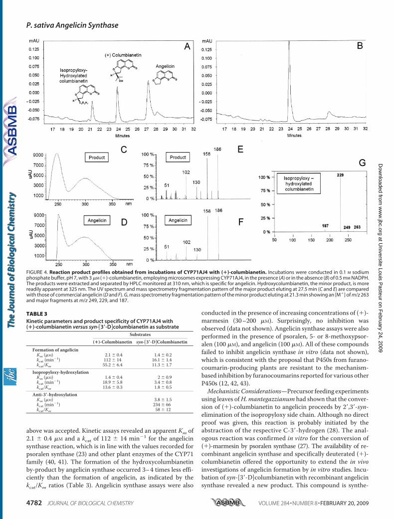
above was accepted. Kinetic assays revealed an apparent Km of2.1 � 0.4 �M and a kcat of 112 � 14 min�1 for the angelicinsynthase reaction, which is in line with the values recorded forpsoralen synthase (23) and other plant enzymes of the CYP71family (40, 41). The formation of the hydroxycolumbianetinby-product by angelicin synthase occurred 3–4 times less effi-ciently than the formation of angelicin, as indicated by thekcat/Km ratios (Table 3). Angelicin synthase assays were also
conducted in the presence of increasing concentrations of (�)-marmesin (30–200 �M). Surprisingly, no inhibition wasobserved (data not shown). Angelicin synthase assays were alsoperformed in the presence of psoralen, 5- or 8-methoxypsor-alen (100 �M), and angelicin (100 �M). All of these compoundsfailed to inhibit angelicin synthase in vitro (data not shown),which is consistent with the proposal that P450s from furano-coumarin-producing plants are resistant to the mechanism-based inhibition by furanocoumarins reported for various otherP450s (12, 42, 43).Mechanistic Considerations—Precursor feeding experiments
using leaves ofH.mantegazzianum had shown that the conver-sion of (�)-columbianetin to angelicin proceeds by 2�,3�-syn-elimination of the isopropyloxy side chain. Although no directproof was given, this reaction is probably initiated by theabstraction of the respective C-3�-hydrogen (28). The anal-ogous reaction was confirmed in vitro for the conversion of(�)-marmesin by psoralen synthase (27). The availability of re-combinant angelicin synthase and specifically deuterated (�)-columbianetin offered the opportunity to extend the in vivoinvestigations of angelicin formation by in vitro studies. Incu-bation of syn-[3�-D]columbianetin with recombinant angelicinsynthase revealed a new product. This compound is synthe-
FIGURE 4. Reaction product profiles obtained from incubations of CYP71AJ4 with (�)-columbianetin. Incubations were conducted in 0.1 M sodiumphosphate buffer, pH 7, with 3 �M (�)-columbianetin, employing microsomes expressing CYP71AJ4, in the presence (A) or in the absence (B) of 0.5 mM NADPH.The products were extracted and separated by HPLC monitored at 310 nm, which is specific for angelicin. Hydroxycolumbianetin, the minor product, is morereadily apparent at 325 nm. The UV spectrum and mass spectrometry fragmentation pattern of the major product eluting at 27.5 min (C and E) are comparedwith those of commercial angelicin (D and F). G, mass spectrometry fragmentation pattern of the minor product eluting at 21.3 min showing an [M�] of m/z 263and major fragments at m/z 249, 229, and 187.
TABLE 3Kinetic parameters and product specificity of CYP71AJ4 with(�)-columbianetin versus syn-3�-Dcolumbianetin as substrate
Substrates(�)-Columbianetin syn-3�-DColumbianetin
Formation of angelicinKm (�M) 2.1 � 0.4 1.4 � 0.2kcat (min�1) 112 � 14 16.1 � 1.4kcat/Km 55.2 � 6.4 11.3 � 1.7
Isopropyloxy-hydroxylationKm (�M) 1.4 � 0.4 2 � 0.9kcat (min�1) 18.9 � 5.8 3.4 � 0.8kcat/Km 13.6 � 0.3 1.8 � 0.5
Anti-3�-hydroxylationKm (�M) 3.8 � 1.5kcat (min�1) 234 � 66kcat/Km 58 � 12
P. sativa Angelicin Synthase
4782 JOURNAL OF BIOLOGICAL CHEMISTRY VOLUME 284 • NUMBER 8 • FEBRUARY 20, 2009
at Universite Louis P
asteur on February 24, 2009
ww
w.jbc.org
Dow
nloaded from

sized at the expense of a relatively large decrease in the forma-tion of angelicin and hydroxycolumbianetin by-product, ascompared with control assays employing natural (�)-columbi-anetin as substrate (Fig. 5A). HPLC and gas chromatography-mass spectrometry (n) analyses unambiguously assigned thenew product as another hydroxylated columbianetin derivativeretaining the deuterium label (Fig. 5B). Roughly 2�g of the newproduct was collected from several incubations for NMR anal-ysis. The LC-SPE-1H NMR spectrum displayed two AB spinsystems comprising four doublet resonances for H-3 (� 6.17,JH-3–H-4 � 9.5 Hz), H-4 (� 7.78, JH-4–H-3 � 9.5 Hz), H-5 (� 7.48,JH-5–H-6 � 8.5Hz), andH-6 (� 6.80, JH-6–H-5 � 8.5Hz) (Fig. 5C),matching the chemical shifts and coupling constants of corre-sponding protons in syn-[3�-D]columbianetin aswell as the twosinglets due to two methyl groups at � 1.22 and 1.21 (notshown). The presence of these proton signals suggests thathydroxylation can only occur at C-2� or C-3�. Hydroxylation atC-2� would, in the spectrum of the deuterated substrate,[3�-D]columbianetin, wipe out the doublet for H-2� at � 4.77(JH-2�–H-3� � 9.8 Hz), and change the broad doublet at � 3.28(JH-3�–H-2� � 9.8 Hz) to a singlet. This was apparently not thecase. Instead, the signal of H-3� disappeared from the spectrumof the new product, indicating that the proton had beenabstracted. Thus, hydroxylation at C-2� can be excluded, andthe deuterium label at C-3� must be retained. Moreover, com-pared with the deuterated substrate (44), which was measuredunder the same conditions as the product (see “ExperimentalProcedures”), the signal ofH-2�was shifted upfield to � 4.36 andcollapsed to a singlet, confirming loss of the adjacent proton atC-3� (Fig. 5C). The unresolved signals between � 3.2 and 3.9
ppm can be ascribed to impurities of the minute product sam-ple. Moreover, hydroxylation at C-2� is highly improbable inlight of the literature (1, 2). Taken together, these results arefully consistent with the new product as syn-[3�-D](anti-3�-hy-droxy)-columbianetin (Fig. 5D).Kinetic studies revealed that the affinities of recombinant
angelicin synthase to natural columbianetin or syn-[3�-D]co-lumbianetin were almost identical (Table 3). However, the con-version rates kcat to angelicin and its derivative hydroxylated inthe isopropyloxy residue observed in assays with naturalcolumbianetin were dramatically affected when the C-3� deu-terium-labeled substrate was used, because the ratio kcat/Kmdropped from 55.2 to 11.3 (Table 3). Furthermore, gas chroma-tography-mass spectrometry analysis of angelicin isolated fromthe CYP71AJ4 assays with syn-[3�-D]columbianetin revealedthe loss of deuterium (data not shown), demonstrating thatsyn-3�D was the site of attack by the iron-oxo center of angeli-cin synthase. In contrast, formation of the new producthydroxylated in anti-orientation at C-3� in these assays wasconsiderably favored, as indicated by a kcat of 234 � 66 min�1
(Table 3).
DISCUSSION
Despite the considerable interest in furanocoumarins astherapeutics or plants’ herbivore defense factors, the biosyn-thesis and genetics of these compounds, particularly the angu-lar furanocoumarins, are still poorly understood (45, 46). Evo-lution of the angular pathway is thought to have developed laterfrom genes of the pathway leading to psoralens and as a conse-quence of herbivore selection pressures (47); such a later devel-
FIGURE 5. Reaction product profiles obtained from incubations of CYP71AJ4 with syn-[3�-D]columbianetin. A, HPLC separation profile of the reactionproduct generated by CYP71AJ4 with syn-[3�-D]columbianetin in the presence (�) or absence (�) of 0.5 mM NADPH. The products were extracted andseparated by HPLC monitored at 325 nm, which is specific for hydroxycolumbianetin. Elution of syn-[3�-D]columbianetin was observed at 24 min, whereas themajor product peak eluting at 18.8 min corresponds to syn-[3�-D](anti-3�-hydroxy)-columbianetin. B, mass spectrometry fragmentation pattern of the productshowing an [M�] of m/z 263 and major fragments at m/z 247, 229, and 205. C, 1H NMR spectrum of the product. The asterisks indicate likely chemical impurities.D, regio- and stereospecific hydroxylation of syn-[3�-D]columbianetin catalyzed by CYP71AJ4.
P. sativa Angelicin Synthase
FEBRUARY 20, 2009 • VOLUME 284 • NUMBER 8 JOURNAL OF BIOLOGICAL CHEMISTRY 4783
at Universite Louis P
asteur on February 24, 2009
ww
w.jbc.org
Dow
nloaded from

opment would predict a close relationship between enzymesinvolved in the two pathways. Following this hypothesis,sequence elements of CYP71AJ1, the first coumarin-specificP450 gene isolated recently from A. majus and encoding psor-alen synthase (23), were used as primers to amplify relatedgenes from plants producing both linear and angular furano-coumarins. Celery (A. graveolens) and parsnip (P. sativa) fulfillthis requirement and belong to the same taxon (Apiaceae), sug-gesting a common codon usage. Three new members of theCYP71AJ subfamily (CYP71AJ2 to -4) were cloned by thisapproach and expressed with a relatively low activity in yeastcells. The marginal degree of expression was also encounteredwith CYP71AJ1 and partially resolved by replacing the N-ter-minalmembrane anchorwith that ofCYP73A1 (23). This, how-ever, turned out not to be necessary for CYP71AJ2 to -4, sincethe activity of the transformants was sufficient for kinetic iden-tification. CYP71AJ2 (celery) and 71AJ3 (parsnip) encode pso-ralen synthases showing substrate affinities to (�)-marmesin or(�)-5-hydroxymarmesin very similar to those reported forCYP71AJ1 (23). It is noteworthy that in these enzymes, SRS4and SRS5 were completely conserved, whereas SRS6 showedsome sequence divergence.CYP71AJ4 (parsnip) was shown to encode angelicin syn-
thase, thus representing a P450 dedicated to angular furano-coumarin biosynthesis. The recombinant enzyme exhibitedstringent substrate selectivity (Km � 2.1 � 0.4 �M) to (�)-co-lumbianetin and yielded a by-product in minor quantity iden-tified as a (�)-columbianetin derivative hydroxylated in the iso-propyloxy side chain. CYP71AJ4 could be cloned from leavesonly after wounding to elicit furanocoumarins (data notshown), which is fully compatible with the proposed role ofangelicin in plant defense against herbivores. Activity assaysconducted with syn-[3�-D]columbianetin (Fig. 5) severelyaffected the kinetics of angelicin formation as well as the prod-uct pattern, shifting the reaction toward the formation of anti-3�-hydroxy-syn-[3�-D]columbianetin. The considerable iso-topic effect (i.e. the drop in kcat/Km) (Table 3) clearly indicatedthat angelicin synthase attacks the substrate by abstracting thesyn-hydrogen from C-3� in the context of the 2�,3�-eliminationreaction.However, the enzyme is also capable of abstracting theanti-hydrogen from C-3�, which is then followed by reboundhydroxylation at C-3�, and this reaction is strongly favored ifdeuterium replaces the syn-3�-hydrogen (Table 3). The datasuggest that the initial abstraction and the elimination reaction(angelicin formation) or rebound hydroxylation (formation ofanti-3�-hydroxycolumbianetin) by angelicin synthase do notoccur concomitantly but, rather, sequentially. This might alsobe the reason for the by-product formation when unlabeled(�)-columbianetin is used as a substrate. Overall, subtle differ-ences become apparent in the catalytic processes catalyzed byangelicin synthase (CYP71AJ4) compared with those catalyzedby psoralen synthase (CYP71AJ1). Psoralen synthase exclu-sively abstracts the hydrogen isotope syn to the C-3 substituentwhen syn-[3�-D]- or anti-[3�-D]marmesin is offered as a sub-strate with a kinetic isotope effect of kH/kD �4 (27), which isconsistent with previous precursor feeding studies (48). Mostimportantly, psoralen is the only product of the reaction.Both CYP71AJ3 (psoralen synthase) and CYP71AJ4 (angeli-
cin synthase) were isolated from P. sativa and constitute thefirst paralogs stricto sensu described for the linear and angularfuranocoumarin pathways. The overall sequence identity of70% suggests that CYP71AJ3 and CYP71AJ4 could have arisenfroma commonancestor of unknown functionality by a processinvolving gene duplication followed by mutations. This is con-sistent with a reduced sequence identity in the SRS elements to40% that fully matches a divergence of substrate specificitybetween the two enzymes. The competitive inhibition of A.majus psoralen synthase by (�)-columbianetin was demon-strated previously and illustrated by in silico docking experi-ments (23), which supported the idea of psoralen synthasestructurally close to angelicin synthase. Alternatively, in thisreport, (�)-marmesin was not accepted as a substrate or com-petitive inhibitor by angelicin synthase. Altogether, these dif-ferences suggest a mode of action for angelicin synthase that isdistinct from that for psoralen synthase, although the reasonsunderlying the discrepancy remain to be determined.The expression of CYP71AJ4-like genes is necessary but not
sufficient for the evolution of the angular furanocoumarinpathway. Indeed, angelicin synthase needs (�)-columbianetinas a substrate and depends on a transferase activity prenylatingcarbon-8 of umbelliferone (Fig. 1). The complementary charac-terization of the umbelliferone 8-prenyltransferase will consti-tute a major step forward to our understanding of the diver-gence between angular and linear furanocoumarin pathways inhigher plants.
Acknowledgments—We thank Prof. R. Caniato (Padova, Italy) for thegifts of (�)-marmesin and 5-OH-marmesin and Prof. W. Boland forthe gift of syn-[3�-D]columbianetin and helpful discussions. We arealso indebted to Prof. J. Hohmann for a reference sample of columbia-nadin. Themass spectroscopy by Dr. G. Laufenberg andN. Zitzer andthe technical assistance of S. Berthet and M. Callier are gratefullyappreciated. We thank Emily Wheeler (Jena) for editorial assistance.
REFERENCES1. Murray, R. D. H., Mendez, J., and Brown, S. A. (1982) The Natural Cou-
marins, John Wiley & Sons, Inc., New York2. Murray, R. D. H. (2002) Prog. Chem. Org. Nat. Prod. 83, 1–6193. Kawase, M., Sakagami, H., Motohashi, N., Hauer, H., Chatterjee, S. S.,
Spengler, G., Vigyikanne, A. V., Molnar, A., and Molnar, J. (2005) In Vivo19, 705–711
4. Pereira, L. E., Villinger, F., Wulff, H., Sankaranarayanan, A., Raman, G.,and Ansari, A. A. (2007) Exp. Biol. Med. 232, 1338–1354
5. Bourgaud, F., Allard, N., Guckert, A., and Forlot, P. (1989)Natural Sourcesof Fucocoumarins, pp. 219–230, John Libbey Eurotext, Ltd., Paris
6. Bourgaud, F., Hehn, A., Larbat, R., Doerper, S., Gontier, E., and Kellner,Matern, U. (2006) Phytochem. Rev. 5, 293–308
7. Beier, R. C., and Oertli, E. H. (1983) Phytochemistry 22, 2595–25978. Baskin, J. M., Ludlow, C. J., Harris, T. M., and Wolf, F. T. (1967) Phyto-
chemistry 6, 1209–12139. Ojala, T., Remes, S., Haansuu, P., Vuorela, H., Hiltunen, R., Haahtela, K.,
and Vuorela, P. (2000) J. Ethnopharmacol. 73, 299–30510. Neal, J. J., and Wu, D. (1994) Pestic. Biochem. Phys. 50, 43–5011. Dall’Acqua, F., Vedaldi, D., and Recher, M. (1978) Photochem. Photobiol.
27, 33–3612. Letteron, P., Descatoir, D., Larrey,M., Tinel, J., Geneve, J., and Pessayre, D.
(1986) J. Pharmacol. Exp. Ther. 238, 685–69213. Scott, J. G. (1996) Insect Biochem. Mol. Biol. 26, 645–64914. Zumwalt, J. G., and Neal, J. J. (1993) Comp. Biochem. Phys. 106, 111–118
P. sativa Angelicin Synthase
4784 JOURNAL OF BIOLOGICAL CHEMISTRY VOLUME 284 • NUMBER 8 • FEBRUARY 20, 2009
at Universite Louis P
asteur on February 24, 2009
ww
w.jbc.org
Dow
nloaded from

15. Berenbaum, M. R., and Zangerl, A. R. (1996) Recent Adv. Phytochem. 30,1–24
16. Ma, R., Cohen, M. B., Berenbaum, M. R., and Schuler, M. A. (1994) Arch.Biochem. Biophys. 310, 332–340
17. Berenbaum, M. R., and Zangerl, A. R. (2006) Ecology 87, 3070–308118. Kai, K., Mizutani, M., Kawamura, N., Yamamoto, R., Tamai, M., Yamagu-
chi, H., Sakata, K., and Shimizu, B. (2008) Plant J. 55, 989–99919. Endler, A., Martens, S., Wellmann, F., and Matern, U. (2008) Plant Mol.
Biol. 67, 335–34620. Dhillon, D. S., and Brown, S. A. (1976) Arch. Biochem. Biophys. 177,
74–8321. Hamerski, D., and Matern, U. (1988) Eur. J. Biochem. 171, 369–37522. Wendorff, H., and Matern, U. (1986) Eur. J. Biochem. 161, 391–39823. Larbat, R., Kellner, S., Specker, S., Hehn, A., Gontier, E., Hans, J., Bour-
gaud, F., and Matern, U. (2007) J. Biol. Chem. 282, 542–55424. Hamerski, D., and Matern, U. (1988) FEBS Lett. 239, 263–26525. Hehmann, M., Lukacin, R., Ekiert, H., and Matern, U. (2004) Eur. J. Bio-
chem. 271, 932–94026. Stanjek, V., Piel, J., and Boland, W. (1999) Phytochemistry 50, 1141–114527. Stanjek, V., Miksch, M., Luer, P., Matern, U., and Boland, W. (1999) An-
gew. Chemie-Int. Ed. 38, 400–40228. Stanjek, V., and Boland, W. (1998) Helv. Chim. Acta 81, 1596–160729. Stemmer, W. P. C., and Morris, S. K. (1992) BioTechniques 13, 214–22030. Pompon, D., Louerat, B., Bronine, A., and Urban, P. (1996) Methods En-
zymol. 272, 51–6431. Gietz, D., St. Jean, A., Woods, R. A., and Schiestl, R. H. (1992) Nucleic
Acids Res. 20, 142532. Bradford, M. M. (1976) Anal. Biochem. 7, 248–254
33. Omura, T., and Sato, R. (1964) J. Biol. Chem. 239, 2370–237834. Liu, R., Sun, Q., Shi, Y., and Kong, L. (2005) J. Chromatogr. A 1076,
127–13235. Wei, Y., and Ito, Y. (2006) J. Chromatogr. A 1115, 112–11736. Ito, Y. (2005) J. Chromatogr. A 1065, 145–16837. Cappeletti, E. M., Innocenti, G., Caniato, R., Filippini, R., and Piovan, A.
(1998) in Biotechnology in Agriculture and Forestry - 41. Medicinal andAromatic Plants (Bajaj, P. S., ed), pp. 238–260, Springer, Heidelberg
38. Zschoke, S., Liu, J. H., Stuppner, H., and Bauer, R. (1998) Phytochem. Anal.9, 283–290
39. Gotoh, O. (1992) J. Biol. Chem. 267, 83–9040. Takahashi, S., Zhao, Y., O’Maille, P. E., Greenhagen, B. T., Noel, J. P.,
Coates, R. M., and Chappell, J. (2005) J. Biol. Chem. 280, 3686–369641. Takahashi, S., Yeo, Y. S., Zhao, Y., O’Maille, P. E., Greenhagen, B. T., Noel,
J. P., Coates, R. M., and Chappell, J. (2007) J. Biol. Chem. 282,31744–31754
42. Koenigs, I. L., and Trager, W. F. (1998) Biochemistry 37, 13184–1319343. Gravot, A., Larbat, R., Hehn, A., Lievre, K., Gontier, E., Goergen, J.-L., and
Bourgaud, F. (2004) Arch. Biochem. Biophys. 422, 71–8044. Stanjek, V. (1998) Studien zur Biosynthese der Furanocumarine, Ph.D.
thesis, Universitat Bonn, Germany45. Stern, R. S. (2007) New Eng. J. Med. 357, 682–69046. Schuler, M. A., and Berenbaum,M. R. (2003) Proc. Natl. Acad. Sci. U. S. A.
100, Suppl. 2, 14593–1459847. Berenbaum, M., and Feeny, P. (1981) Science 212, 927–92948. Stanjek, V., Miksch, M., and Boland, W. (1997) Tetrahedron 53,
17699–17710
P. sativa Angelicin Synthase
FEBRUARY 20, 2009 • VOLUME 284 • NUMBER 8 JOURNAL OF BIOLOGICAL CHEMISTRY 4785
at Universite Louis P
asteur on February 24, 2009
ww
w.jbc.org
Dow
nloaded from