isolation and characterization of new c-terminal substitution … · febs letters 400 (1997)...
TRANSCRIPT
FEBS Letters 400 (1997) 122-126 FEBS 17975
Isolation and characterization of new C-terminal substitution mutations affecting secretion of polygalacturonase in Erwinia carotovora
ssp. carotovora
T. Palomäki, H.T. Saarilahti* Department of Biosciences, Division of Genetics, Biocenter 2, P.O.Box 56, University of Helsinki, FIN-00014 Helsinki, Finland
Received 2 September 1996; revised version received 11 November 1996
Abstract An intact C-terminus was previously shown to be required for stability and secretion of the polygalacturonase (PehA) in Erwinia carotovora ssp. carotovora. Here we have analyzed the effects of amino acid (aa) substitutions generated to five C-terminal positions of PehA. Conservation of two hydrophobic and one non-hydrophobic residue (V372, V374 and N371, respectively) was found to be essential for maintenance of the protein stability. As an exception, one of the mutants (V372G) did not show major effects on protein stability, as determined by immunohlots and enzyme activity assay, yet it prevented the secretion completely. We conclude that the C-terminus of PehA is directly involved in the formation or stabilization of a conformation-sensitive structure needed for recognition of the protein as secreted.
Key words: Gram-negative; Protein secretion; Polygalacturonase; Mutagenesis; Secretion-deficient mutant
1. Introduction
The plant pathogenic bacterium Erwinia carotovora ssp. carotovora (Ecc) causes soft-rot disease in various crop plants by secreting plant cell wall degrading enzymes, such as pectate lyase, pectin lyase, polygalacturonase (Peh), cellulase and pro-tease [1,2]. Production and secretion of these extracellular en-zymes are essential for virulence, since mutants defective either in their production (exp, aep and rex mutations) or secretion (put mutations) are avirulent or exhibit a reduced virulence [3-6]. Pectinases and cellulases are secreted through the general secretion (GSP) [7] or type II secretion pathway [8] which is a central mechanism for targeting extracellular pro-teins in Gram-negative bacteria. Secretion through the Ecc Out pathway requires at least 13 accessory proteins that are considerably conserved with the corresponding components in other bacteria, such as E. chrysanthemi, Klebsiella, Xanthomo-nas and Aeromonas [9]. Secretion is a two-step process where the proteins are first translocated in a signal peptide depend-ent manner across the cytoplasmic membrane by the Sec sys-tem [10,11]. Next, the proteins must fold in their final con-formation in the periplasm before they are routed further across the outer membrane and finally released into the extra-cellular milieu [12-14].
Secreted proteins are supposed to possess another targeting signal in their mature amino acid sequence that is recognized by the components of the secretion apparatus. Regions con-
»Corresponding author. Fax. (358) (9) 70859079. E-mail: [email protected]
taining information needed for secretion have been localized in aerolysin of Aeromonas salmonicida [15] and exotoxin A of Pseudomonas aeruginosa [16,17]. Lu and Lory [17] showed that a segment between amino acids 60 and 120 is needed for translocation across the outer membrane. Interestingly, McVay and Hamood conclude that even the C-terminus of exotoxin A would include similar information [18]. Thus, the protein would contain several, separate targeting signals. Py et al. [13] suggested that information needed for secretion of the cellulase EGZ of E. chrysanthemi could lie in several parts of the protein, yet in this case the separate regions were not independently able to promote secretion, but rather they were thought to form a single targeting signal. Also, since EGZ has to form disulfide bonds in the periplasm before secretion, and the region flanking a bond-forming cysteine shares similarity with the corresponding region in two pectin-ases, this region could contain information relevant for secre-tion [14].
Heterologous proteins can be routed out of the cell via GSP using the targeting information of a secretory protein. Kor-nacker and Pugsley [19] showed that the N-terminal two-thirds of Klebsiella oxytoca pullulanase (Pul) was sufficient to direct secretion of the fused passenger molecule, ß-lactam-ase (Bla). On the other hand, corresponding PehA-Bla hybrid proteins remained cell-bound [6]. Recently, we showed that the C-terminus of PehA contains information related to secre-tion, and e.g. PehA-Bla hybrid proteins can specifically be secreted by the Ecc GSP, provided that no more than the last two aa of the PehA domain were removed from the fusion [20]. To analyze in detail the relevance of the C-terminus of PehA for correct extracellular targeting we have here created defined substitutions to five terminal residues and character-ized the properties of the mutant proteins in Ecc.
2. Materials and methods
2.1. Bacterial strains, plasmids and growth conditions The bacterial strains and plasmids used in the study are listed in
Table 1. E. coli and Ecc cultures were grown in L-medium [21] in a rotary shaker at 37 and 28°C, respectively. Ampicillin (Ap) was added at 150 μg ml-1. For protein analysis and enzymatic assays in Ecc, the cultures were supplemented with 0.4% glycerol and grown to station-ary phase, diluted at 1:100 and grown further for 20 h before collect-ing samples.
2.2. Genetic techniques E. coli cells were made competent and transformed according to
Maniatis et al. [22]. Electroporation of Ecc cells was performed as described previously [20].
2.3. DNA techniques Plasmid DNA was isolated by alkaline lysis according to Birnboim
and Doly [23]. Restriction enzymes, T4 ligase, T4 polynucleotide ki-
0014-5793/97/S17.00 © 1997 Federation of European Biochemical Societies. All rights reserved. P77S0014-5793 (9 6)01369-5
T. Palomäki, Η.Τ. SaarilahtiIFEBS Letters 400 (1997) 122-126 123
nase and alkaline phosphatase were used according to the manufac-turer's instructions (Promega, Madison, WI, USA). DNA fragments were separated by agarose-gel electrophoresis [22]. Site-directed mu-tagenesis of the pehA gene was performed with polymerase chain reaction (PCR) using degenerate oligonucleotides. PCR was per-formed with the following oligonucleotides: no. 9512, TA ACG GAT CCA AGC ATA AAG CCC AGT GCT TTT hybridized to the pehA promoter region at 194-172 nt upstream from the transla-tion start site. no. 949, TA AGG ATC CTA CTT CTT GNC (G/ C)T(A/T/G) GAC GTT C; no. 9414, CA TGG ATC CTA CTT CTT GAN GTT GAC G; no. 9415, CA TGG ATC CTA CTT CTT GAC GTT GNC GTT CT; no. 9518, CA TGG ATC CTA CTT CTT GAC GTT GAC GTT CNT GAT TTG CC; no. 9519, CA TGG ATC CTA CTT CTT GAC GTT GAC GNT CTT GAT TTG CC; hybrid-ized to the 3' end of the coding sequence of pehA and read towards the gene. The following changes in the aa sequence were created: no. 949, N373->Y/H and V374->A/D/G; no. 9414, V374->L/I/F; no. 9415, V372->A/D/G; no. 9518, K370->T/R/M; no. 9519, N371->S/T/I. DNA was amplified for 20 cycles under the following conditions: denaturation at 94°C for 30 s, annealing at 56°C for 30 s, extension at 72°C for 2 min, using Pfu polymerase (Stratagene, La Jolla, CA, USA). Mutated pehA fragments were cloned in pUC18 at the BamHI or Sma\ site, and the plasmids were propagated in R coli followed by transfer to Ecc. DNA sequence analysis was performed from plasmids employing the dideoxy chain-termination method of Sanger et al. [24] using a Sequenase quick-denature plasmid sequenc-ing kit (US Biochemicals, Cleveland, OH). The following oligo-nucleotide, TC CCA GAC TGG AGT GAT ATC, hybridizing in Has pehA gene at 1043-1062 nt downstream from the translation start point, was used as a primer in sequencing.
2.4. Protein analysis SDS-PAGE was performed in 12% polyacrylamide gels according
to Laemmli [25] and transfer of proteins to nitrocellulose filters ac-cording to Burnette [26]. Proteins were detected with a polyclonal rabbit anti-PehA antiserum and an alkaline phosphatase-conjugated second antibody as described [20]. Periplasmic proteins were isolated by osmotic shock treatment as described [27], except that in the final step the periplasmic proteins were recovered in 20 mM MgCl2.
2.5. Enzyme assays Polygalacturonase [28] and ß-lactamase [29] activities were assayed
as described.
3. Results
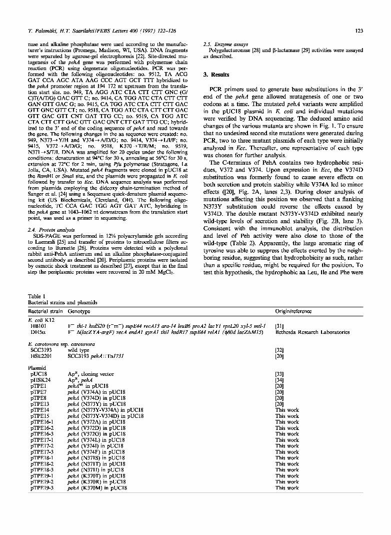
PCR primers used to generate base substitutions in the 3' end of the pehA gene allowed mutagenesis of one or two codons at a time. The mutated pehA variants were amplified in the pUC18 plasmid in E. coli and individual mutations were verified by DNA sequencing. The deduced amino acid changes of the various mutants are shown in Fig. 1. To ensure that no undesired second site mutations were generated during PCR, two to three mutant plasmids of each type were initially analyzed in Ecc. Thereafter, one representative of each type was chosen for further analysis.
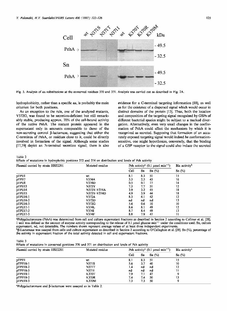
The C-terminus of PehA contains two hydrophobic resi-dues, V372 and V374. Upon expression in Ecc, the V374D substitution was formerly found to cause severe effects on both secretion and protein stability while V374A led to minor effects ([20], Fig. 2A, lanes 2,3). During closer analysis of mutations affecting this position we observed that a flanking N373Y substitution could reverse the effects caused by V374D. The double mutant N373Y-V374D exhibited nearly wild-type levels of secretion and stability (Fig. 2B, lane 3). Consistent with the immunoblot analysis, the distribution and level of Peh activity were also close to those of the wild-type (Table 2). Apparently, the large aromatic ring of tyrosine was able to suppress the effects exerted by the neigh-boring residue, suggesting that hydrophobicity as such, rather than a specific residue, might be required for the position. To test this hypothesis, the hydrophobic aa Leu, He and Phe were
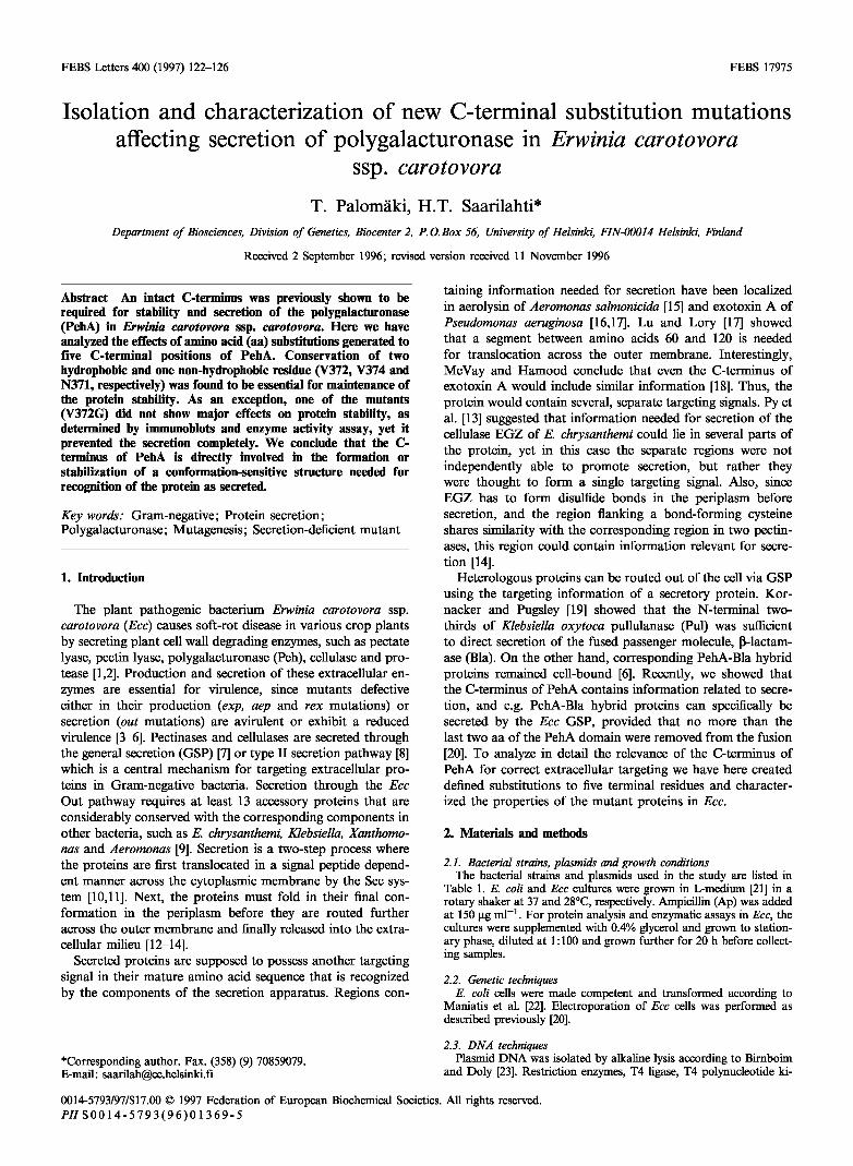
Table 1 Bacterial strains and plasmids
Bacterial strain Genotype Origin/reference
E coli K12 HB101 DH5a
F - thi-1 hsdS20 (r~m_) supE44 recA13 ara-14 leuB6 proA2 lacYl rpsL20 xyl-5 mtl-1 F - AQacZYA-argF) recA endAl gyrAl Ml hsdR17 supE44 relAl (§80d lacZAM15)
[31] Bethesda Research Laboratories
K carotovora ssp. carotovora SCC3193 wild type HSE2201 SCC3193;?e/U::Tn773/
[32] [20]
Plasmid pUC18 ApR, cloning vector pHSK24 ApR, pehA pTPEl pehÄ* in pUC18 pTPE7 pehA (V374A) in pUC18 pTPE8 pehA (V374D) in pUC18 pTPE13 pehA (N373Y) in pUC18 pTPE14 pehA (N373Y-V374A) in pUC18 pTPE15 pehA (N373Y-V374D) in pUC18 pTPE16-l pehA (V372A) in pUC18 pTPE16-2 pehA (V372D) in pUC18 pTPE16-3 pehA (V372G) in pUC18 pTPE17-l pehA (V374L) in pUC18 pTPE17-2 pehA (V374I) in pUC18 pTPE17-3 pehA (V374F) in pUC18 pTPE18-l pehA (N371S) in pUC18 pTPE18-2 pehA (N371T) in pUC18 pTPE18-3 pehA (Ν371Γ) in pUC18 pTPE19-l pehA (K370T) in pUC18 pTPE19-2 pehA (K370R) in pUC18 pTPE19-3 pehA (K370M) in pUC18
[33] [34] [20] [20] [20] [20] This work This work This work This work This work This work This work This work This work This work This work This work This work This work

124 T. Palomäki, Η.Τ. Saarilahti/FEBS Letters 4M (1997) 122-126
sp 26 aa
PehA 376 aa 7 aa1
from wild-type PehA by immunoblot analysis (Fig. 3) and enzyme assays (Table 3).
4. Discussion
Plasm id
pTPEl pTPE7 pTPE8 pTPE13 pTPE14 pTPE15 pTPE16-l pTPE16-2 pTPE16-3 pTPE17-l pTPE17-2 pTPE17-3 pTPE18-l pTPE18-2 pTPE18-3 pTPE19-l pTPE19-2 pTPE19-3
Mutated 370 371 372 373 residues
wt V374A V374D N373Y N373Y-V374A N373Y-V374D V372A V372D V372G V374L V374I V374F N371S N371T N371I K370T K370R K370M
K
T R M
N
S Γ
V
A D G
N
Y Y Y
374 375 3
V K A . D
A D
L I F
Fig. 1. C-terminal mutations of PehA. The substituted residue and its location are shown for the seven last amino acids, sp, signal pep-tide.
substituted for Val-374. In all cases, secretion and stability of the protein were judged unchanged in immunoblot analysis (Fig. 2B), and the level and distribution of Peh activity were very similar to those of the wild-type enzyme (Table 2).
Next, the analysis was extended to position 372. Again, mutants having a V372A or V372D substitution exhibited altered phenotypes, with Asp causing more severe effects than Ala (Fig. 2A). In fact, in immunoblots, the V372A and V372D substitutions closely resembled the respective substitu-tions of V374 (Fig. 2A). This suggests that probably both positions must be occupied by a hydrophobic amino acid. In accordance with this, even the substitution V372G was expected to affect protein stability, as occurred when Gly was substituted for V374 [20]. However, the V372G mutant appeared stable in immunoblot analysis (Fig. 2A) and showed considerable polygalacturonase activity (Table 2). Some of the mutant protein could also be detected in the culture super-natant, yet the level of extracellular V372G did not exceed that of the unspecific leakage observed for the non-secreting control ß-lactamase (Table 2). Further, the mutant protein was localized by osmotic shock treatment to the periplasmic fraction (data not shown).
We also studied the significance of two non-hydrophobic residues, K370 and N371, which are conserved between sev-eral eukaryotic and prokaryotic polygalacturonases [20]. Ser, Thr and He were substituted for the N371, and Thr, Arg and Met for K370 (Fig. 1). More pronounced effects were caused only by the N371T and N371I substitutions, which rendered the PehA entirely unstable (Fig. 3). The different effects caused by the N371S and N371T substitutions are note-worthy, since Ser and Thr are generally regarded as similar in sequence comparisons. Unlike the observed clear require-ment for Asn in position 371, no apparent reason was found for the observed conservation of K370. Three mutants K370T, K370R and K370M showed no significant difference
Previously, we showed that an intact C-terminus is essential for correct targeting of the major secreted enzyme PehA in Ecc, and that alterations within the C-terminus also affect protein conformation [20]. In this study, it is shown that a terminal hydrophobic cluster is required both structurally and for correct targeting of the protein. The cluster consists of two aa, V372 and V374. Non-hydrophobic aa, especially Asp, caused in either position severe effects on protein stability, suggesting that the protein conformation was affected. In most cases, decreased stability paralleled a lowered level of secretion. For the latter position, the mutant phenotype ex-hibited by V374D could be suppressed by a second-site sub-stitution N373Y. Further, all hydrophobic aa substituting for V374 retained the full wild-type characteristics of PehA. The effects of hydrophobic substitutions were not specifically tested for the position 372, but the characteristics of V372A and V372D, as compared to V374A and V374D, suggest that
A
Cell
PehA ^
Sn
PefaA^
B
Cell
IVhA
Sn
lVh \ 1 L- 1 1. \
-49.5
-32.5
-49,5
-32.5
S" ^ ^ A4 ^ ^ ^ kDa
- 49.5
-32.5
- 49,5
-32.5
Fig. 2. (A) Analysis of aa substitutions in the positions 372 and 374. HSE2201 cells harboring the various plasmids were grown to a stationary phase in L-medium supplemented with 0.4% glycerol and Ap 150 μg ml-1. 10 ul aliquots of cell and supernatant fractions were subjected to SDS-PAGE and immunoblot analysis as described in Section 2. Cell refers to cellular fraction and Sn to culture super-natant fraction. (B) Effects of hydrophobic substitutions in positions 373 and 374. Analysis was performed as described in panel A.
T. Palomäki, Η.Τ. SaarilahtiIFEBS Letters 400 (1997) 122-126 125
Cell
PehA ■
Sn
PehA >
* V -v1 - ^ ' -ί1 V V V kDa ^ ¿ Λ ^
«SJg f i ^ ^Wi j l ^ - t f i pS · 3 ^ » s ^
"<ϋ*"
■ 49,5
-32,5
■ 49.5
32,5
Fig. 3. Analysis of aa substitutions at the conserved residues 370 and 371. Analysis was carried out as described in Fig. 2A.
hydrophobicity, rather than a specific aa, is probably the main criterion for both positions.
As an exception to the rule, one of the analyzed mutants, V372G, was found to be secretion-deficient but still remark-ably stable, producing approx. 70% of the cell-bound activity of the native PehA. The mutant protein appeared in the supernatant only in amounts comparable to those of the non-secreting control ß-lactamase, suggesting that either the C-terminus of PehA, or residues close to it, could be directly involved in formation of the signal. Although some studies [17,19] depict an N-terminal secretion signal, there is also
evidence for a C-terminal targeting information [18], as well as for the existence of a dispersed signal which would occur in distinct domains of the protein [13]. Thus, both the location and composition of the targeting signal recognized by GSPs of different bacterial species might be subject to a marked diver-gation. Alternatively, even very small changes in the confor-mation of PehA could affect the mechanism by which it is recognized as secreted. Supposing that formation of an accu-rately exposed targeting signal would indeed be conformation-sensitive, one might hypothesize, conversely, that the binding of a GSP receptor to the signal could also induce the secreted
Table 2 Effects of mutations in hydrophobic positions 372 and 374 on distribution and levels of Peh activity
Plasmid carried by strain HSE2201 Mutated residue Peh activity8 (0.1 umol min - 1)
Cell Sn Sn (%)
Bla activityb
Sn (%)
pTPEl pTPE7 pTPE8 pTPE13 pTPE14 pTPE15 pTPE16-l pTPE16-2 pTPE16-3 pTPE17-l pTPE17-2 pTPE17-3
wt V374A V374D N373Y N373Y-V374A N373Y-V374D V372A V372D V372G V374L V374I V374F
8.1 3.3 0.5 7.3 3.9 4.9 8.3 nd 5.6 8.6 8.7 8.8
8.3 2.5 0.1 7.7 3.2 3.9 6.1 nd 0.6 8.1 8.4 7.9
51 43 17 51 45 44 42 nd 10 49 49 47
15 16 14 12 18 18 12 13 10 12 13 11
"Polygalacturonase (PehA) was determined from cell and culture supernatant fractions as described in Section 2 according to Collmer et al. [28]. 1 unit was defined as the amount of enzyme activity corresponding to the release of 0.1 umol glucose min-1 under the conditions used. Sn, culture supernatant, nd, not detectable. The numbers shown represent average values of at least three independent experiments. bß-Lactamase was assayed from cells and culture supernatant as described in Section 2 according to O'Callaghan et al. [29]. Sn (%), percentage of the activity in supernatant fraction of the total activity detected in cell and supernatant fractions.
Table 3 Effects of mutations in conserved positions 370 and 371 on distribution and levels of Peh activity
Plasmid carried by strain HSE2201 Mutated residue Peh activity8 (0.1 umol min - 1)
Cell Sn Sn (%)
Bla activity"
Sn (%)
pTPEl pTPE18-l pTPE18-2 pTPE18-3 pTPE19-l pTPE19-2 pTPE19-3
wt N371S N371T N371I K370T K370R K370M
8.1 5.6 1.4 nd 7.9 7.4 7.3
8.3 3.7 nd nd 7.1 7.4 7.3
51 40 nd nd 47 50 50
15 10 12 11 9
13 9
"Polygalacturonase and ß-lactamase were assayed as in Table 2.
126 T. Palomäki, Η.Τ. Saarilahti/FEBS Letters 400 (1997) 122-126
protein to undergo a transient change of conformation during translocation. If correct, this might provide at least a partial explanation for the observations that chimeric proteins, in which the conformation of the passenger molecule most hkely cannot be affected by GSP, are often secreted less efficiently through the GSP than the native ones, or not secreted at all [20,30].
In addition to hydrophobic residues, several positions har-boring a non-hydrophobic aa were studied. The substitutions N371T and N371I rendered PehA extremely unstable, and little, if any of the mutant proteins could be detected in im-munoblots. Thus, the structural importance of N371 is in agreement with its conservation within several polygalacturo-nases [20], in contrast to the other conserved residue K370, which appeared dispensable. Because the two extreme ter-minal K375 and K376 are also unimportant [20], all the three terminal lysines seem to lack significance.
Currently, we are performing random mutagenesis over the entire pehA to search for stable non-secreting mutants. Iden-tification of regions accumulating mutations that impair the protein with secretion while leaving other properties un-changed should give us tools for the accurate localization and characterization of the targeting signal of the PehA pro-tein.
Acknowledgements: We thank Dr. P. Heino for critically reading the manuscript. This work was supported by the Academy of Finland.
References
[1] Pérombelon, M.C.M. and Kelman, A. (1980) Annu. Rev. Phyto-pathol. 18, 361-387.
[2] Collmer, A.C. and Keen, N.T. (1986) Annu. Rev. Phytopathol. 24, 383^109.
[3] Pirhonen, M., Saarilahti, H., Karlsson, M.-B. and Palva, E.T. (1991) Mol. Plant-Microbe Interact. 4, 276-283.
[4] Murata, H., McEvoy, J.L., Chatterjee, A., Collmer, A. and Chat-terjee, A.K. (1991) Mol. Plant-Microbe Interact. 4, 239-246.
[5] Jones, S. et al. (1993) EMBO J. 12, 2477-2482. [6] Saarilahti, H.T., Pirhonen, M., Karlsson, M.-B., Flego, D. and
Palva, E.T. (1992) Mol. Gen. Genet. 234, 81-88. [7] Pugsley, A.P. (1993) Microbiol. Rev. 57, 50-108. [8] Salmond, G.P.C. and Reeves, PJ. (1993) Trends Biochem. Sei.
18, 7-12.
[9
[10]
[11
[12]
[13:
[14]
[is:
[16]
[17 [is:
[I?
[20]
[21
[22]
[23:
[24]
[25 [26] [27 [28
[29
[30]
[31
[32]
[33:
[34]
Reeves, P.J., Whitcombe, D., Wharam, S., Gibson, M. and Alli-son, G. (1993) Mol. Microbiol. 8, 443^156. Pugsley, A.P., Poquet, I. and Kornacker, M.G. (19910 Mol. Mi-crobiol. 5, 865-873. Poquet, I., Faucher, D. and Pugsley, A.P. (1993) EMBO J. 12, 271-278. Pugsley, A.P. (1992) Proc. Nati. Acad. Sei. USA 89, 12058-12062. Py, B., Chippaux, M. and Barras, F. (1993) Mol. Microbiol. 7, 785-793. Bortoli-German, I., Brun, E., Py, B., Chippaux, M. and Barras, F. (1994) Mol. Microbiol. 11, 545-553. Wong, K.R. and Buckley, J.T. (1991) J. Biol. Chem. 266, 14451-14456. Hamood, A.N., Olson, J.C., Vincent, T.S. and Iglewski, B.H. (1989) J. Bacteriol. 171, 1817-1824. Lu, H.-M. and Lory, S. (1996) EMBO J. 15, 429-436. McVay, C.S. and Hamood, A.N. (1995) Mol. Gen. Genet. 249, 515-525. Kornacker, M.G. and Pugsley, A.P. (1990) Mol. Microbiol. 4, 1101-1109. Palomäki, T. and Saarilahti, H.T. (1995) Mol. Microbiol. 17, 449-459. Miller, J. (1972) Experiments in Molecular Genetics, Cold Spring Harbor Laboratory Press, Cold Spring Harbor, NY. Maniatis, T., Fritsch, E.F. and Sambrook, J. (1982) Molecular Cloning: A Laboratory Manual, Cold Spring Harbor Laboratory Press, Cold Spring Harbor, NY. Birnboim, H.C. and Doly, J. (1979) Nucl. Acids Res. 7, 1513-1523. Sanger, F., Nieten, S. and Coulson, A.R. (1977) Proc. Nati. Acad. Sei. USA 74, 5463-5467. Laemmli, U.K. (1970) Nature 227, 680-685. Burnette, N.W. (1981) Anal. Biochem. 112, 195-203. Palva, E.T. (1978) J. Bacteriol. 136, 286-294. Collmer, A., Whalen, C.H., Beer, S.V. and Bateman, D.F. (1982) J. Bacteriol. 149, 626-634. O'Callaghan, C.H., Morris, A., Kirby, S.M. and Shingler, A.H. (1972) Antimicrob. Agents Chemother. 1, 283-288. Sauvonnet, N., Poquet, I. and Pugsley, A. (1995) J. Bacteriol. 177, 5238-5246. Boyer, H.W. and Roulland-Dussoix, D. (1969) J. Mol. Biol. 41, 459-472. Pirhonen, M., Heino, P., Helander, I., Harju, P. and Palva, E.T. (1988) Microb. Pathog. 4, 359-367. Yanisch-Perron, C, Vieira, J. and Messing, J. (1985) Gene 33, 103-119. Saarilahti, H.T., Heino, P., Pakkanen, R., Kalkkinen, N., Palva, I. and Palva, E.T. (1990) Mol. Microbiol. 4, 1037-1044.