isolation and characterization of kurstakin and surfactin
TRANSCRIPT

This article has been accepted for publication and undergone full peer review but has not been through the copyediting, typesetting, pagination and proofreading process which may lead to differences between this version and the Version of Record. Please cite this article as doi: 10.1002/jms.4302
This article is protected by copyright. All rights reserved.
Isolation and characterization of kurstakin and surfactin isoforms produced by
Enterobacter cloacae C3 strain
Nawel Jemila,*,
Noomen Hmidet
a, Angeles Manresa
b, Francesc Rabanal
c and Moncef Nasri
a
aLaboratoire de Génie Enzymatique et de Microbiologie, Université de Sfax, Ecole Nationale
d’Ingénieurs de Sfax, B.P. 1173-3038 Sfax, Tunisia.
bSection of Microbiology, Department of Biology, Health and Environment, Faculty of
Pharmacy, University of Barcelona, Joan XXIII s/n, 08028 Barcelona, Spain.
cSection of Organic Chemistry, Department of Inorganic and Organic Chemistry, Faculty of
Chemistry, University of Barcelona, Martí i Franquès, 1, 08028 Barcelona, Spain
* Corresponding author. Tel.: +216 50 498 815; fax: +216 74 275 595.
E-mail address: [email protected]

This article is protected by copyright. All rights reserved.
Abstract
In this work the extraction, structural analysis and identification as well as antimicrobial,
anti-adhesive and anti-biofilm activities of lipopeptides produced by Enterobacter cloacae
C3 strain were studied. A combination of chromatographic and spectroscopic techniques
offers opportunities for a better characterization of the biosurfactant structure. Thin layer
chromatography (TLC) and HPLC for amino acid composition determination are used.
Efficient spectroscopic techniques have been utilized for investigations on the biochemical
structure of biosurfactants, such as Fourier transform infrared (FT-IR) spectroscopy and mass
spectrometry analysis. This is the first work describing the production of different isoforms
belonging to kurstakin and surfactin families by E. cloacae strain. Three kurstakin
homologues differing by the fatty acid chain length from C10 to C12 were detected. The
spectrum of lipopeptides belonging to surfactin family contains various isoforms differing by
the fatty acid chain length as well as the amino acids at positions four and seven. Lipopeptide
C3 extract exhibited important antibacterial activity against Gram-positive and Gram-
negative bacteria, antifungal activity and interesting anti-adhesive and disruptive properties
against biofilm formation by human pathogenic bacterial strains: Salmonella typhimurium,
Klebsiella pneumoniae, Staphylococcus aureus, Bacillus cereus and Candida albicans.
Keywords: Enterobacter cloacae C3; Kurstakin; Surfactin; Structure characterization; Anti-
adhesive property.

This article is protected by copyright. All rights reserved.
Introduction
Biosurfactants are synthesized by a wide variety of microorganisms, mainly by bacteria
and several yeasts.[1]
Lipopeptides are among the most studied bioactive molecules, produced
by multiple bacterial genera such as Bacillus,[2]
Paenibacillus,[3]
Pontibacter,[4]
Achromobacter,[5]
Corynebacterium,[6]
Pseudomonas,[7]
Streptomyces,[8]
Citrobacter and
Enterobacter.[9,10]
Lipopeptides are classified in various families and isoforms according to the peptide
amino acid composition as well as the fatty acid chain length and the type of fatty acid
binding. A common feature is the presence of an acyl chain bound to a cyclic peptide
sequence; the peptide portion could be composed of either anionic or cationic residues with D
or L configuration and might contain non-proteogenic or unusual amino acids. The peptide
portion is non-ribosomally generated; the synthesis is directed by large multi-enzyme
complex called Non-Ribosomal Peptide Synthetase (NRPS).[11]
The most known lipopeptide
families are: surfactin, iturin and fengycin-plipastatin. Surfactin and iturin lipopeptide
compounds are cyclic lipoheptapeptides which contain a β-hydroxy and a β-amino fatty acid
chain, respectively, as lipophilic moieties. Fengycin lipopeptides are cyclic lipodecapeptides
with a β-hydroxy fatty acid chain. In addition to surfactin, iturin and fengycin families,
kurstakin represents a new family of lipopeptides discovered in 2000 produced by Bacillus
thuringiensis and it is considered as a biomarker of this species. Kurstakins were also
detected in other species belonging to Bacillus genus such as B. cereus; they are
lipoheptapeptides displaying antifungal activities.[12]
The first isolated kurstakins did not
contain a β-hydroxy fatty acid and were classified as linear molecules. It has been shown that
they can be found in the form of partially cyclic compounds,[13]
as well as in cyclic
structures,[12]
which places them in a class of non-cationic cyclic lipopeptides.[14]
Cyclic
lipopeptide biosurfactants like surfactin, iturin, bacillomycin, fengycin and kurstakin are

This article is protected by copyright. All rights reserved.
largely produced by species of the genus Bacillus which are Gram-positive bacteria
exhibiting antimicrobial activity.[15,16]
There are few studies describing the production of
these lipopeptide families by Gram-negative bacteria.[9,10]
Usually, Gram-negative bacteria of
the genera Pseudomonas, Klebsiella and Enterobacter produce rhamnolipid and glycolipid
biosurfactants.[17-20]
Different analytical techniques for chemical characterization of lipopeptides have been
applied to elucidate their structure such as infrared spectroscopy (IR), amino acid analysis,
high performance liquid chromatography (HPLC), capillary chromatography coupled to mass
spectrometry (MS), gas chromatography (GC-MS) and UV/Vis spectroscopy,[21]
nuclear
magnetic resonance (NMR) spectroscopy and liquid chromatography-mass spectrometry
(LC-MS).[22]
Furthermore, matrix-assisted laser desorption ionization-time of flight mass
spectrometry (MALDI-TOF-MS) has proven to be very effective in the detection and
identification of lipopeptides. Tandem mass spectrometry is a simple, fast, sensitive method
and the appropriate technique to elucidate complex structures and mixtures on biological
processes. Thousands of reports on applications of MS for microorganism characterization in
research, clinical microbiology, food safety, environmental monitoring, and quality have been
published.[23]
There is a high demand for new antimicrobial agents because of the increased resistance
shown by pathogenic microorganisms against the existing antimicrobial drugs. Several
natural lipopeptides produced by microorganisms have been developed as new therapeutic
products and exploited for biomedical applications thanks to their antibacterial,[24,25]
antifungal,[26]
antiviral[27]
and anti-adhesive properties[4,6,28]
against several pathogenic
microorganisms. In fact, some of the oldest available antibiotics in the market are cyclic
antimicrobial peptides, such as polymyxins, gramicidin and bacitracin.[29,30]

This article is protected by copyright. All rights reserved.
The present work provides an insight into the search of new bioactive molecules from the
Gram-negative bacteria E. cloacae C3. Chemical structure characterization and identification
of different lipopeptide isoforms produced, as well as antimicrobial, anti-adhesive and anti-
biofilm activities were carried out.
Experimental
Bacterial strain and biosurfactants production
The microorganism used in this study was isolated from soil at the area “Nakta” near the
company “British gas”, Sfax City, Tunisia, contaminated by natural-gas condensate, which
comes from the gas of Miskar Asset field. It was identified as Enterobacter cloacae C3 strain
based on the 16S rDNA gene sequence analysis.[31]
It was inoculated into a 250 ml shake
flask containing 25 ml Luria-Bertani broth medium and cultivated at 37 °C with shaking at
200 rpm for 18 h. A 3% (v/v) of inoculum [OD600 nm = 6.7] was transferred into a 2 l shake
flask containing 250 ml of Landy medium[32]
and incubated in an orbital shaker at 30 °C and
150 rpm for 72 h.
Biosurfactants extraction
Biosurfactants recovery was performed as reported in our previous study.[33]
Acid
precipitated biosurfactant (1 g) was subjected to extraction with 45 ml tetrahydrofuran (THF)
solvent four times and the mixture was stirred and centrifuged at 8000 rpm, for 15 min at 4
°C. The recuperated organic phases were combined and concentrated in a rotary vacuum
evaporator (Büchi laborotechnik AG Postfach, Switzerland) at 40 °C.

This article is protected by copyright. All rights reserved.
Chemical characterization and identification of biosurfactants
FT-IR spectra of the crude dried biosurfactants
The functional groups and the chemical bonds present in the crude biosurfactants C3
were determined using Fourier transform infrared spectroscopy (FT-IR) in order to determine
the chemical nature of biosurfactants. FT-IR analysis was performed by using Analect
Instruments fx-6 160 FT-IR spectrometer at a wavenumber range 4000-400 cm-1
.
Thin layer chromatography (TLC) analysis
Biosurfactants extract was tested by TLC on silica gel plates 60 G (Macherel-Nagel,
Düren, Germany) with mobile phase: chloroform/methanol/water (65:25:4). Staining was
carried out with phosphomolybdic acid for the detection of lipids and o- Tolidine specific for
peptide moiety, to detect the spots showing the presence of both fatty acid and peptide
moieties.
Bradford assay for protein quantitation
The protein content of biosurfactants C3 was measured using Bradford Assay Kit
through the microassay procedure as described in our previous study.[34]
Amino acid composition determination
The crude biosurfactants (4 mg) were hydrolyzed in 1 ml 6 M HCl at 110 °C overnight in
a sealed tube. Aliquotes of AABA (L-α-Aminobutyric acid) and NLE (L-Norleucine)
solutions were added as internal standards. Samples were evaporated to dryness and
resuspended in water. The amino acids were then analyzed by HPLC with UV detection,
using the Waters AccQTag pre-column derivatization method.[35]
This article is protected by copyright. All rights reserved.
Characterization of the lipopeptides by mass spectrometry (ESI and MALDI-TOF)
The molecular weight of the lipopeptide molecules was determined by positive/negative-
ion modes electrospray ionization (ESI) analyses (LC/MSD-TOF, Agilent Technologies, Palo
Alto, CA). The capillary voltage was 4 kV and 3.5 kV for the positive/negative-ion modes,
respectively, with nitrogen as the nebulizing and drying gas. Tandem mass spectrometry
(4800 Plus MALDI TOF/TOF, ABSciex, Dublin, CA) was used in the experiment. The full
mass spectrum was acquired in the reflector positive-ion mode for the lipopeptides, using
dihydroxybenzoic acid (DHB) as the matrix. Subsequent fragmentation of the observed ions
was obtained by positive MS2 analysis.
Antimicrobial activities
The antimicrobial activity of C3 lipopeptides was estimated by agar well diffusion
method against selected human pathogens. Antibacterial activity was tested against three
Gram-positive bacteria: Bacillus cereus ATCC 11778, Micrococcus luteus ATCC 4698,
Staphylococcus aureus ATCC 25923 and five Gram-negative bacteria: Escherichia coli
ATCC 25922, Klebsiella pneumoniae ATCC 13883, Salmonella enterica ATCC 27853,
Salmonella typhimurium ATCC 19430 and Enterobacterium sp. Antifungal activity was
tested against Aspergillus niger, Aspergillus flavus and Fusarium oxysporum.
The culture suspension (200 μl) of the tested microorganisms (106
colony-forming units
cfu/ml) of bacteria cells (estimated by absorbance at 600 nm) and 108
spores/ml of fungal
strains (measured by Malassez blade) were spread uniformly using sterile pipette on Luria-
Bertani agar and malt extract agar media, respectively. Then, wells were made using a sterile
well borer and were filled with 100 μl of lipopeptide sample (2 mg/ml concentration).

This article is protected by copyright. All rights reserved.
The zone of growth inhibition was measured in millimeters after incubation for 24 h at
37 °C for bacteria and for 72 h at 30 °C for fungal strains. All the results were represented as
the average of three independent experiments with ≤ 5% deviation.
Anti-adhesion treatment with lipopeptide C3 extract
For surface pre-treatment, the wells of a sterile polystyrene microtiter plate (Costar;
Corning Incorporated, Corning, NY, USA) were filled with 200 µl of C3 lipopeptide extract
at different concentrations ranging from 0.008 to 1 mg/ml, dissolved in PBS composed of
(g/l): NaCl, 8; KCl, 0.2; Na2HPO4, 1.44; KH2PO4, 0.24 (pH 7.2). Microtiter plates were
incubated for 6 h at room temperature (25 °C) and then washed twice with PBS.
For biofilm formation, Klebsiella pneumoniae ATCC 1388, Staphylococcus aureus
ATCC 25923, Salmonella typhimurium ATCC 14028, Bacillus cereus ATCC 11778 and
Candida albicans ATCC 10231 were cultured overnight in Luria Bertani medium (LB).
Cultures were diluted 1/100 in the medium proposed by O´Toole[36]
(g/l): glucose, 2;
casamino acids, 5; KH2PO4, 3; K2HPO4, 7; (NH4)2SO4, 2, MgSO4 7H2O, 0.12. Then, 200 µl
of each dilution were added to the microtiter plate wells and plates were incubated for 20 h at
37 °C. Cultures were discarded and wells were washed three times with distilled water to
remove non-adherent cells, fixed for 15 min with methanol and stained with 125 µl crystal
violet (0.1%, w/v) for 20 min, then washed with water and drying. For quantifying the
microbial adhesion, 200 µl of acetic acid in water (33%, v/v) were added and the absorbance
was determined at 595 nm. Percentages of microbial adhesion inhibition were calculated
using the following formula:
Microbial adhesion inhibition =[1 − (Ac/A0)] × 100
This article is protected by copyright. All rights reserved.
where Ac represents the absorbance of the well with lipopeptides at concentration c and A0
represents the absorbance of the positive control wells (in absence of lipopeptides). Negative
control wells contained only lipopeptides dissolved in PBS. Assays were carried out three
times with ≤ 5% deviation.
Mature biofilm treatment with lipopeptide C3 extract
The wells of a sterile polystyrene microtiter plate were loaded with 200 µl of bacterial
suspension prepared as mentioned above, then the plates were incubated for 20 h at 37 °C.
After incubation, the unattached microbial cells were removed by washing the wells three
times with distilled water. Then, 200 µl of C3 lipopeptides at different concentrations ranging
from 0.008 to 1 mg/ml, were added to each well and the plates were incubated for 6 h at room
temperature (25 °C). The quantification was carried out as in the pre-treatment. All the results
were represented as the average of three independent experiments with ≤ 5% deviation.
Methods of analysis
All data presented are the average of at least three measurements which deviated by not
more than 5%.
Results and discussion
Chemical structure characterization of biosurfactants
Preliminary chemical characterization
The IR spectrum of the crude biosurfactants from E. cloacae C3 strain showed several
strong bands (Figure 1). The peak with the highest absorbance in the spectrum at 1655 cm-1
,
results from the stretching mode of the carbonyl group (C=O) of the amide bond (-CONH-),
also there is a small contribution from carbonyls of the ester bond and carboxyl side chains of

This article is protected by copyright. All rights reserved.
some amino acids such as Glu, Gln, Asp and Asn indicating the presence of peptide groups in
the molecule.[37]
Adjacent to this peak, there is another high intensity peak at 1540 cm-1
resulting from the deformation mode of N–H bonds.[38]
The absorbance peaks at 2959, 2928
and 2850 cm-1
indicate the presence of C–H bonds of the alkyl chains. Another peak at 1405
cm-1
corresponds to C–H bending vibrations, it is common in compounds with alkyl chains.
The ester carbonyl group is detected from the absorbance peaks at 1057, 1254 and 1104 cm−1
.
The strong peak at 3300 cm-1
can be attributed to the presence of carboxyl side chains of
glutamic/aspartic acids (O-H stretching) and –NH bonds of the amide group which overlaps
the stretching in the same region. The observed peaks are similar to those reported by Das et
al.[39]
and Jemil et al.[28]
for lipopeptide biosurfactants. The lipopeptide biosurfactant,
surfactin (Sigma) also yielded a similar IR absorption pattern and absorbed approximately at
the same wavenumber positions.[39]
The characterization of biosurfactant C3 extract by TLC analysis showed many spots at
different levels of migration after spraying with phosphomolybdic acid reagent, yellow color
was revealed after treatment with o- Tolidine which correspond to the presence of peptide
moieties. The appearance of many spots at different levels of migration suggest the presence
of lipopeptide molecules with different polarities.
Amino acid composition determination
The amino acid content of the crude lipopeptides synthesized by E. cloacae C3 strain
was determined and results are presented in Table 1. The pairs Glu/Gln and Asp/Asn cannot
be determined by this technique as hydrolysis of the peptide converts Gln and Asn amino
acids into Glu and Asp, respectively. The sample of the crude lipopeptides C3 has a 36% of
peptide content. Amino acids with high molar ratio are Leu, Glx and Asx with percentages of
13.54%, 12.47% and 11.39%, respectively. The amino acid Leu is present with three or four
This article is protected by copyright. All rights reserved.
residues in surfactin lipopeptide and with four or five residues in pumilacidin lipopeptide.
The amino acids Glu or Gln are present with one or three residues in different lipopeptide
isoforms belonging to different families. Asp is in the composition of surfactin and
pumilacidin with one residue.
Amino acids Gly and Ala are present in lipopeptides with percentages of about 9.0% and
8.0%, respectively. These two amino acids are in the composition of lipopeptides belonging
to kurstakin family with one residue. Val and Thr have molar ratios of 6.6% and 6.0%,
respectively, in lipopeptides C3. The amino acid Val is with one or two residues in surfactin
lipopeptide and Thr is with one residue in kurstakin molecules. Also, Ile and Ser have molar
ratios of 5.5% and 5.3%, respectively. The amino acid Ile is in the composition of
lipopeptides belonging to surfactin family with one residue at most and Ser is in the
composition of kurstakin lipopeptide with one residue. While, the amino acid His with 2.0%
molar ratio, is present with one residue at position 5 in the peptide moiety of kurstakin
isoforms.
Detection of lipopeptides by mass spectrometry analysis
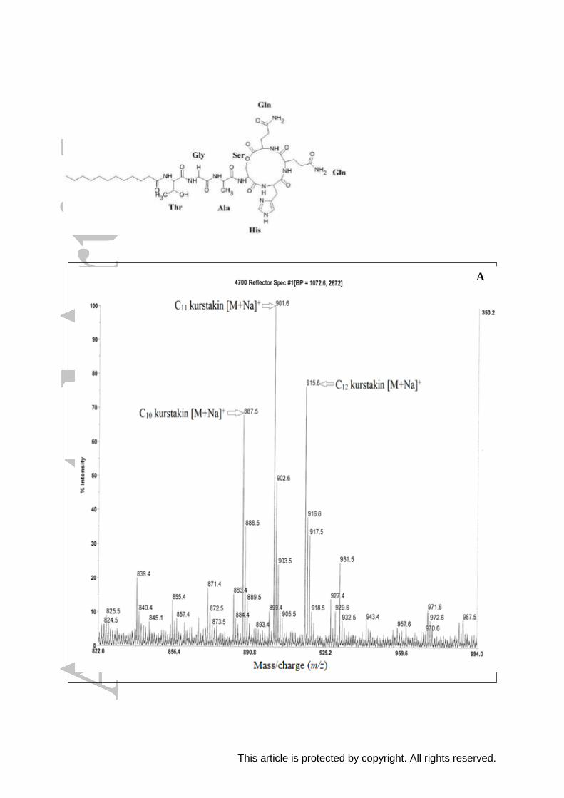
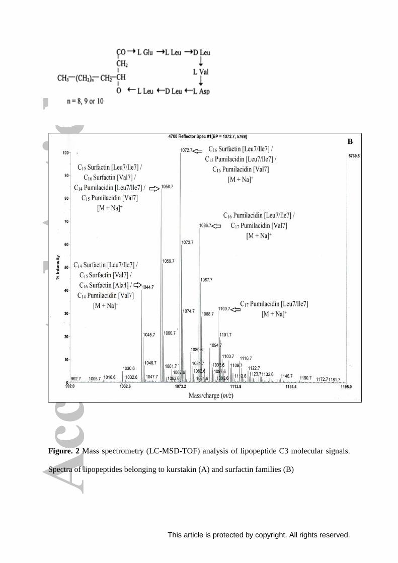
Mass spectrometry analysis of lipopeptide C3 extract reported in Figure 2 shows the
presence of two well-resolved clusters of peaks, the first at m/z values between 887.5 and
915.6 (Figure 2A) and the second within the mass range 1044.7 and 1100.7 Da (Figure 2B).
By comparing the mass (m/z) with the mass values reported for others identified
lipopeptides,[12,34,40]
we can conclude that the first group of peaks (887.5 - 915.6 Da)
corresponds to kurstakin lipopeptide and the second (1044.7 - 1100.7 Da) corresponds to
lipopeptides belonging to surfactin family.

This article is protected by copyright. All rights reserved.
Identification of kurstakin isoforms
Figure 2A shows different peaks corresponding to kurstakin lipopeptide isoforms. The
precursor ion at m/z 887.5 corresponds to a sodium ion adduct of cyclic kurstakin lipopeptide
with a fatty acid chain of 10 carbon atoms. Other lipopeptide homologues detected at m/z
901.6 and 915.6 could be attributed to C11 and C12 kurstakin [M + Na]+, respectively. Each
adduct yields an isotopic distribution of 3 peaks differing by 1 mass unit.
Kurstakin lipopeptide synthesized by E. cloacae C3 strain consist of cyclic
lipoheptapeptides with fatty acid chains from C10 to C12 differing by (m/z of CH2 = 14 Da)
and a peptidic sequence composed of L Thr - Gly - L Ala - L Ser - L His - L Gln - L Gln with
the presence of an amide bond between the fatty acid chain and the first threonine residue and
the presence of a lactone linkage between the serine at position 4 and the C terminus of
glutamine at position 7. Abderrahmani et al.[41]
detected the presence of the three kurstakin
isoforms C11, C12 and C13 in six B. thuringiensis strains. According to Béchet et al.[12]
,
kurstakins were typically identified by the molecular ions at m/z 889, 905, 917 and 933.
Identification and characterization of surfactin and pumilacidin lipopeptides by tandem
mass spectrometry
The precursor ions at m/z 1044.7, 1058.7, 1072.7, 1086.7 and 1100.7 (Figure 2B) were
assigned as the sodium ion adducts of homologous surfactin lipopeptides with 1021.7,
1035.7, 1049.7, 1063.7 and 1077.7 Da mass, respectively. The structure characterization of
surfactin lipopeptides was elucidated by MS2
fragment analysis. The tandem mass
spectrometry analysis was used to carry out the fragmentation of lipopeptides in order to
obtain more precise information on their chemical structure. However, there is more
ambiguity in the fragmentation of the parent ions detected by LC/MSD-TOF analysis and we
This article is protected by copyright. All rights reserved.
obtained two different fragmentation models for each peak corresponding to two different
molecules (Figures 3 and 4).
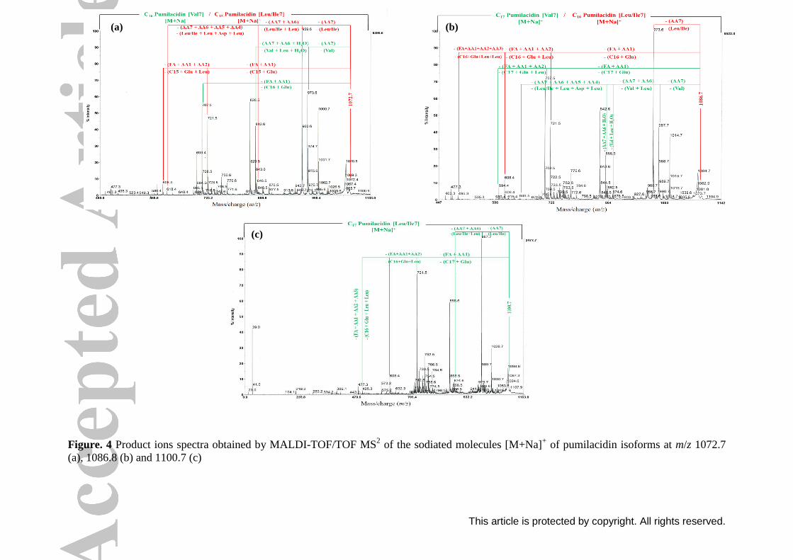
The fragmentation patterns of the peak 1044.7 were illustrated in Figure 3a. The first
fragmentation model resulted in the appearance of product ions from the C-terminal tail of
the aliphatic peptide moiety at m/z 945.6, 832.6 and 717.5 corresponding respectively, to the
losses of Val (−99 Da), Val-Leu (−212 Da) and Val-Leu-Asp (−327 Da) from the parent ion
m/z 1044.7. Therefore, the amino acid at position 7 is Val. An other product ion resulted from
the fragmentation on the side of the fatty acid chain and the N-terminal tail, m/z observed was
693.4 corresponding to the losses of C15 β-hydroxy fatty acid chain-Glu (−351 Da) from the
precursor ion m/z 1044.7. The obtained results indicated that the peak at m/z 1044.7 may
corresponds to surfactin, a cyclic lipopeptide with a fatty acid chain of 15 carbon atoms and
Val residue at position 7. The second fragmentation pattern resulted in the appearance of
product ions from the C-terminal tail of the aliphatic peptide moiety at m/z 931.6, 818.5 and
800.5 corresponding respectively, to the losses of Leu/Ile (−113 Da), Leu/Ile-Leu (−226 Da)
and Leu/Ile-Leu-H2O (−244 Da) from the parent ion m/z 1044.7. Therefore, the amino acid at
position 7 is Leu/Ile. Other product ions resulted from the fragmentation on the side of the
fatty acid chain and the N-terminal tail, m/z observed were 594.4 and 463.3 corresponding
respectively, to the losses of C14 β-hydroxy fatty acid chain-Glu-Leu (−450 Da) and C14 β-
hydroxy fatty acid chain-Glu-Leu-Leu-H2O (−581 Da) from the precursor ion m/z 1044.7.
The obtained results indicated that the peak at m/z 1044.7 may also corresponds to surfactin,
a cyclic lipopeptide with a fatty acid chain of 14 carbon atoms and Leu/Ile residue at position
7. Thus, we can conclude that this lipopeptide may corresponds to C14 surfactin [Leu/Ile7]
and C15 surfactin [Val7].
The same fragmentation sites were observed for the parent ions m/z 1058.7, 1072.7,
1086.7 and 1100.7 (Figures 3 and 4). Results showed that these lipopeptides correspond to
This article is protected by copyright. All rights reserved.
C16 surfactin [Val7] and C15 surfactin [Leu/Ile7], C16 pumilacidin [Val7] and C15 pumilacidin
[Leu/Ile7], C17 pumilacidin [Val7] and C16 pumilacidin [Leu/Ile7], and C17 pumilacidin
[Leu/Ile7], respectively.
The precusor ions at m/z values of 1044.7 and 1058.7 correspond to surfactin isoforms
differing by the acid chain length (m/z of CH2 = 14 Da). Also, the difference between the
sodiated molecules [M + Na]+ at m/z 1058.7 and 1072.7 is 14 Da, these two lipopeptides have
the same acid chain length but differ by the amino acid at position 4, which is valine for
surfactin and leucine for pumilacidin lipopeptide. Based on the fragmentation results, the
precursor ions at m/z values of 1072.7, 1086.7 and 1100.7 were assigned as the sodium ion
adducts of pumilacidin isoforms differing by the acid chain length. According to Pabel et
al.[42]
, the mass peak 1086.9 was assigned to sodiated C17 pumilacidin.
Our results of fragmentation of the parent ions at m/z 1044.7 and 1058.7 resulting in the
sodiated lipopeptides C14 surfactin [Leu/Ile7] and C15 surfactin [Leu/Ile7], respectively, are in
accordance with those demonstrated by Pecci et al.[43]
, Jemil et al.
[34] and Dimkić et al.
[44],
who characterized lipopeptides produced by B. licheniformis V9T14, B. methylotrophicus
DCS1 and Bacillus spp. strains, respectively. Also, according to You et al.[10]
, the
fragmentation of the parent ion at m/z 1058.6 was recognized to be sodium adducts of C15
surfactin. Plaza et al.[45]
reported that the sodiated molecules [M + Na]+ m/z 1044, 1058 and
1072 correspond to C14, C15 and C16 surfactin homologues, respectively, obtained from
lipopeptides produced by B. subtilis KP7 strain. According to Savadogo et al.[46]
, two strains
B. subtilis S6 and B. licheniformis S12 produce biomolecules with m/z related to [M + Na]+
forms of surfactin C14 (m/z 1044) and [M + Na]+ forms of surfactin C15 (m/z 1058). In another
study, Chen et al.[40]
reported that the sodium peaks at m/z 1044 and 1058 are the
characteristic peaks of surfactin molecular weight produced by B. licheniformis MB01.

This article is protected by copyright. All rights reserved.
This is the first work describing kurstakin, surfactin and pumilacidin lipopeptide mixture
production from E. cloacae strain. In fact, the study of You et al.[10]
describes the production
of surfactin homologues from Enterobacter sp. N18 strain. Mandal et al.[9]
reported that the
comprehensive mass spectral (MALDI-TOF-MS and GC-MS) analysis of HPLC purified
antimicrobial lipopeptides obtained from E. cloacae subsp. dissolvens S-11 strain revealed
the occurrence of C17 fengycin B’2, C14 iturin and C15 kurstakin. Whereas, the antimicrobial
lipopeptide obtained from E. homaechei S-5, E. mori S-9, Enterobacter sp. S-4, E. cloacae
subsp. dissolvens S-10 and S-12 is C15 kurstakin.
Antimicrobial activities
The antimicrobial activities of lipopeptide mixture produced by E. cloacae C3 strain
were tested against different Gram-positive, Gram-negative bacteria and fungi strains. The
results showed that lipopeptides C3 exhibited interesting antibacterial and antifungal
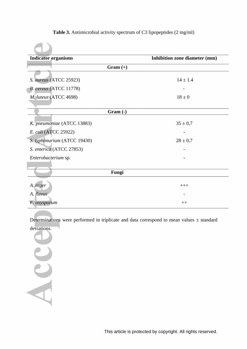
activities (Table 3). K. pneumoniae is the most sensitive strain toward antibacterial activity of
lipopeptides C3 with a maximum zone diameter inhibition of 35 mm, while the lowest
inhibition activity was observed against S. aureus with a zone diameter inhibition of 14 mm.
However, lipopeptides C3 did not exhibit antibacterial activity against B. cereus, E. coli, S.
enterica and Enterobacterium sp. at 2 mg/ml concentration. The inhibitory activity was more
effective against Gram-negative bacteria compared to Gram-positive bacteria with inhibition
zones diameters in the range of 28-35 mm and 14-18 mm, respectively. Our results are in
contrast with those of Ben Ayed et al.[25]
who reported that Gram-positive bacteria are more
sensitive to the inhibitory activity of lipopeptides produced by B. mojavensis A21 strain,
compared to Gram-negative bacteria.
Surfactin lipopeptides are the first and the most well-known member by their
antimicrobial activities. According to Iyer and Sandhya[47]
, the maximum inhibition activity

This article is protected by copyright. All rights reserved.
of the crude surfactin sample (1.5 mg/ml) was observed against Salmonella paratyphi A,
Staphylococcus sp. and E. coli with a zone diameter inhibition of 6 mm. Also,
Sivapathasekaran et al.[48]
showed the antibacterial activity of HPLC purified fractions
containing surfactin. Mandal et al.[9]
reported that lipopeptides belonging to kurstakin, iturin
and fengycin families produced by Citrobacter S-3 and Enterobacter S-11 strains, have an
unusual broad sprectrum antibacterial activity. Lipopeptides which differ in their
composition, follow the same mechanisms such as involving pore formation on bacterial
membrane[49]
or by other non-specific interactions with the membrane[50]
as a result of their
antimicrobial activity. Sotirova et al.[51]
reported that biosurfactants act disturbing the
cytoplasmic membrane, as they have an amphipathic nature that allows its interaction with
phospholipids, altering permeability with consequent cell damage.
Lipopeptides produced by E. cloacae C3 strain showed an interesting antifungal activity
against A. niger and F. oxysporum, but without inhibitory effect against A. flavus. Some other
results have mentioned the antifungal activity of surfactins[52-55]
which would participate to
the preservation of the products against molds. Similar results are reported by Jadhav et al.[20]
who showed that biosurfactants produced by Enterobacter sp. MS16 strain exhibited a
potential antifungal activity and inhibit fungal spores germination. The antimicrobial
activities of lipopeptides C3 may be related to a synergistic effect of both surfactins and
kurstakins. The antimicrobial properties of biosurfactants have been widely reported.
However, the biosurfactants with antimicrobial properties reported till date are produced
mostly by the terrestrial origin microorganisms as a part of defence mechanism to survive in
complex environments.[56]
This article is protected by copyright. All rights reserved.
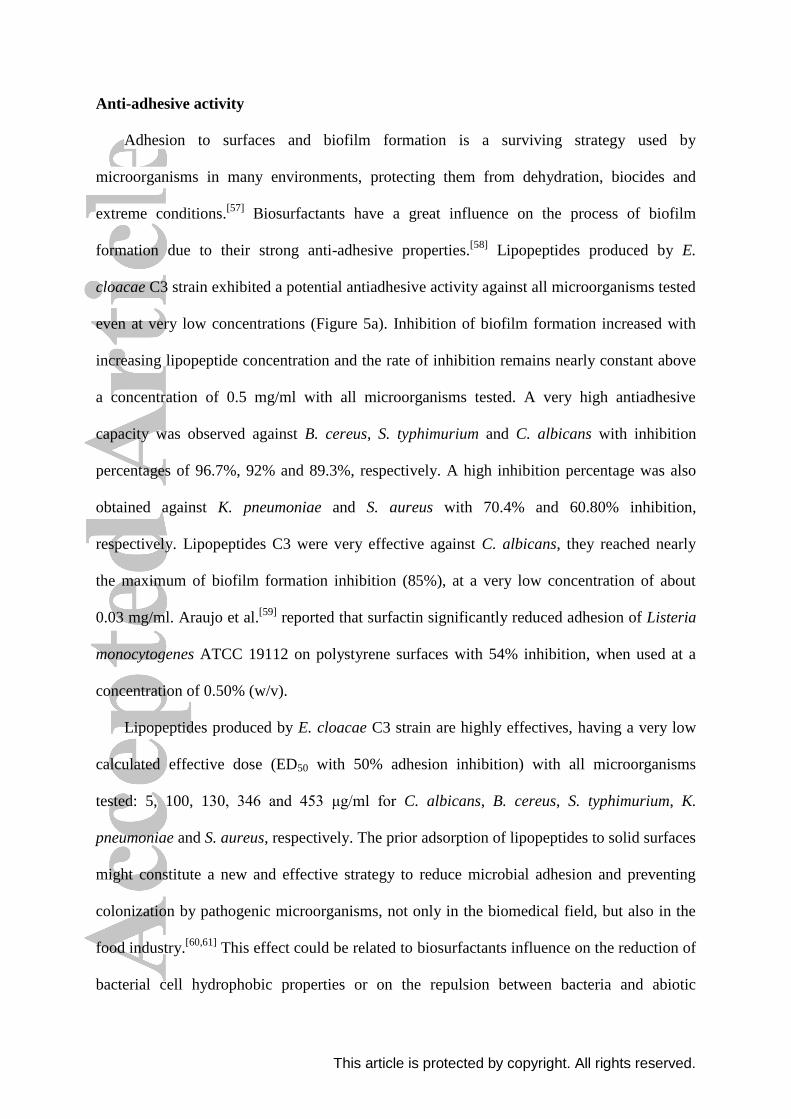
Anti-adhesive activity
Adhesion to surfaces and biofilm formation is a surviving strategy used by
microorganisms in many environments, protecting them from dehydration, biocides and
extreme conditions.[57]
Biosurfactants have a great influence on the process of biofilm
formation due to their strong anti-adhesive properties.[58]
Lipopeptides produced by E.
cloacae C3 strain exhibited a potential antiadhesive activity against all microorganisms tested
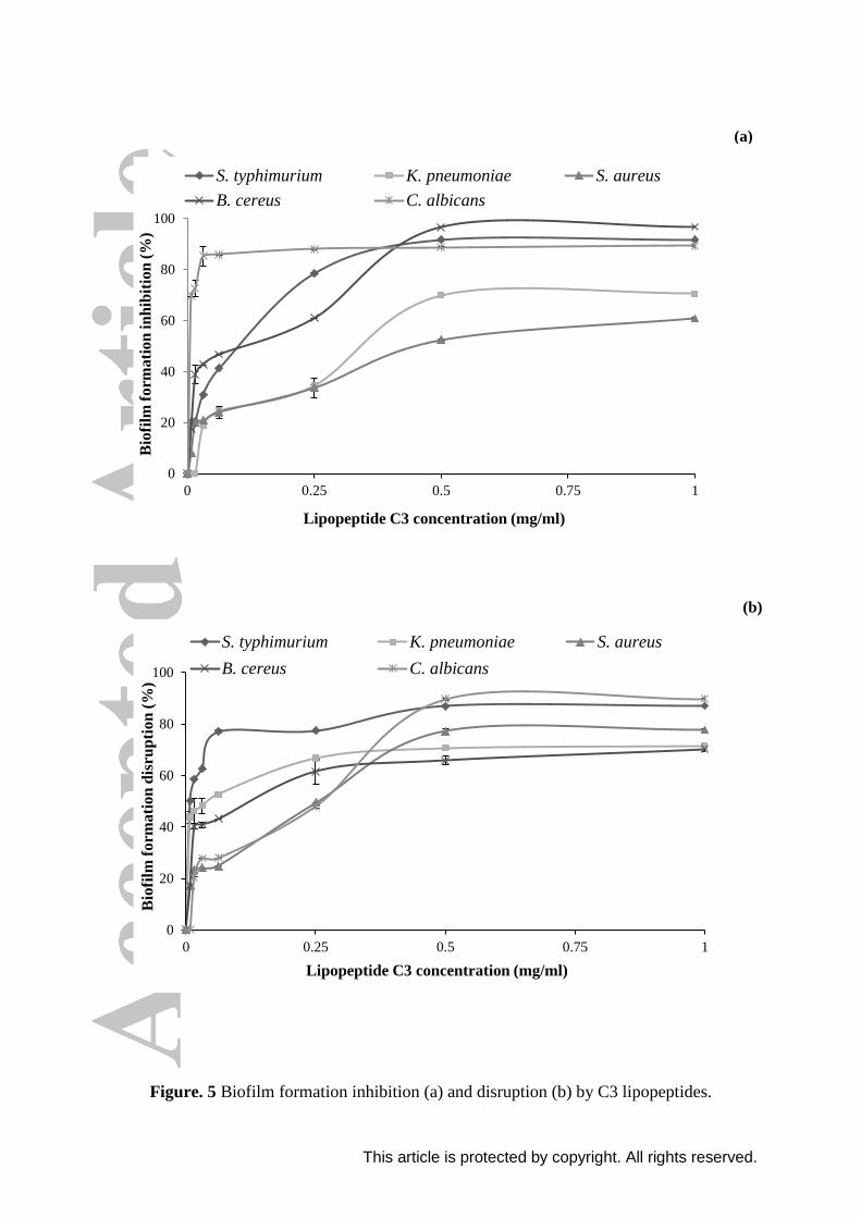
even at very low concentrations (Figure 5a). Inhibition of biofilm formation increased with
increasing lipopeptide concentration and the rate of inhibition remains nearly constant above
a concentration of 0.5 mg/ml with all microorganisms tested. A very high antiadhesive
capacity was observed against B. cereus, S. typhimurium and C. albicans with inhibition
percentages of 96.7%, 92% and 89.3%, respectively. A high inhibition percentage was also
obtained against K. pneumoniae and S. aureus with 70.4% and 60.80% inhibition,
respectively. Lipopeptides C3 were very effective against C. albicans, they reached nearly
the maximum of biofilm formation inhibition (85%), at a very low concentration of about
0.03 mg/ml. Araujo et al.[59]
reported that surfactin significantly reduced adhesion of Listeria
monocytogenes ATCC 19112 on polystyrene surfaces with 54% inhibition, when used at a
concentration of 0.50% (w/v).
Lipopeptides produced by E. cloacae C3 strain are highly effectives, having a very low
calculated effective dose (ED50 with 50% adhesion inhibition) with all microorganisms
tested: 5, 100, 130, 346 and 453 μg/ml for C. albicans, B. cereus, S. typhimurium, K.
pneumoniae and S. aureus, respectively. The prior adsorption of lipopeptides to solid surfaces
might constitute a new and effective strategy to reduce microbial adhesion and preventing
colonization by pathogenic microorganisms, not only in the biomedical field, but also in the
food industry.[60,61]
This effect could be related to biosurfactants influence on the reduction of
bacterial cell hydrophobic properties or on the repulsion between bacteria and abiotic
This article is protected by copyright. All rights reserved.
surfaces.[62]
According to Araujo et al.[59]
, biofilm formation is inhibited by the conditioning
of polystyrene and stainless steel 304 with rhamnolipids and surfactin biosurfactants,
transforming the surfaces hydrophilic or less hydrophobic compared to the control. The
decrease in surface hydrophobicity as a result of conditioning by biosurfactants entails a
decrease in hydrophobic interactions with cell wall of microorganisms and as a result,
adhesion/biofilm formation is reduced.
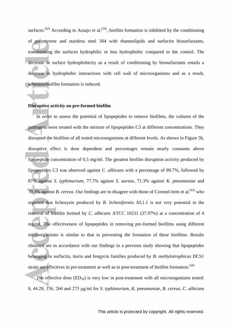
Disruptive activity on pre-formed biofilm
In order to assess the potential of lipopeptides to remove biofilms, the cultures of the
pathogens were treated with the mixture of lipopeptides C3 at different concentrations. They
disrupted the biofilms of all tested microorganisms at different levels. As shown in Figure 5b,
disruptive effect is dose dependent and percentages remain nearly constants above
lipopeptide concentration of 0.5 mg/ml. The greatest biofilm disruption activity produced by
lipopeptides C3 was observed against C. albicans with a percentage of 89.7%, followed by
87% against S. typhimurium, 77.7% against S. aureus, 71.3% against K. pneumoniae and
70.3% against B. cereus. Our findings are in disagree with those of Coronel-león et al.[63]
who
reported that lichenysin produced by B. licheniformis AL1.1 is not very potential in the
removal of biofilm formed by C. albicans ATCC 10231 (37.97%) at a concentration of 4
mg/ml. The effectiveness of lipopeptides in removing pre-formed biofilms using different
microorganisms is similar to that in preventing the formation of these biofilms. Results
obtained are in accordance with our findings in a previous study showing that lipopeptides
belonging to surfactin, iturin and fengycin families produced by B. methylotrophicus DCS1
strain are effectives in pre-treatment as well as in post-treatment of biofilm formation.[28]
The effective dose (ED50) is very low in post-treatment with all microorganisms tested:
8, 44.28, 156, 260 and 273 μg/ml for S. typhimurium, K. pneumoniae, B. cereus, C. albicans
This article is protected by copyright. All rights reserved.
and S. aureus, respectively. Biosurfactants can adsorb at the interface between the attached
biofilm-forming bacteria and the solid surface by orienting polar and nonpolar groups. This
interaction between biosurfactants and the surface alters the surface hydrophobicity, thereby
interfering with microbial adhesion and desorption processes.[64,65]
The results suggest that
lipopeptides produced by E. cloacae C3 strain are potential against all microorganisms tested.
Conclusion
In this study, different cyclic lipopeptides belonging to kurstakin and surfactin families
were detected in E. cloacae C3 strain and their structures were elucidated through tandem
mass spectrometry. Twelve lipopeptide variants belonging to the two different families were
identified; lipopeptide isoforms differ by the fatty acid chain length as well as the amino acid
composition of the peptide cycle. These lipopeptides exhibited an important antimicrobial
activity mainly against K. pneumoniae. In addition, they displayed an excellent anti-adhesive
and disruptive properties against biofilm formation by a variety of bacteria. In conclusion, E.
cloacae C3 strain is a good biocontrol and therapeutic agent for use in combating many
diseases and infections thanks to the antimicrobial and anti-adhesive properties of
lipopeptides produced.
Acknowledgements
This work was funded by «Ministry of Higher Education and Scientific Research», the
Ministerio de Economía y Competitividad. Spain, project CTQ2014-59632-R, and by the IV
Pla de Recerca de Catalunya (Generalitat de Catalunya), grant 2014SGR-534.
This article is protected by copyright. All rights reserved.
References
[1] T.O. Femi-Ola, O.A. Oluwole, T.O. Olowomofe, H. Yakubu. Isolation and screening of
biosurfactant-producing bacteria from soil contaminated with domestic waste water. Br. J.
Env. Sci. 2015, 3, 58.
[2] M.H. Lee, J. Lee, Y.D. Nam, J.S. Lee, M.J. Seo, S.H. Yi. Characterization of
antimicrobial lipopeptides produced by Bacillus sp. LM7 isolated from chungkookjang, a
Korean traditional fermented soybean food. Int. J. Food Microbiol. 2016, 221, 12.
[3] G. Aktuganov, J. Jokela, H. Kivelä, E. Khalikova, A. Melentjeva, N. Galimzianova, L.
Kuzmina, P. Kouvonen, J.P. Himanen, P. Susi, T. Korpela. Isolation and identification of
cyclic lipopeptides from Paenibacillus ehimensis, strain IB-X-b. J. Chromatogr. B. 2014,
973, 9.
[4] S.S. Balan, G.G. Kumar, S. Jayalakshmi. Pontifactin, a new lipopeptide biosurfactant
produced by a marine Pontibacter korlensis strain SBK-47: Purification, characterization and
its biological evaluation. Process Biochem. 2016, 51, 2198.
[5] M.C. Deng, J. Li, Y.H. Hong, X.M. Xu, W.X. Chen, J.P. Yuan, J. Peng, M. Yi, J.H.
Wang. Characterization of a novel biosurfactant produced by marine hydrocarbon-degrading
bacterium Achromobacter sp. HZ01. J. Appl. Microbiol. 2016, 120, 889.
[6] D. Dalili, M. Amini, M.A. Framarzi, M.R. Fazeli, M.R. Khoshayand, N. Samadi. Isolation
and structural characterization of Coryxin, a novel cyclic lipopeptide from Corynebacterium
This article is protected by copyright. All rights reserved.
xerosis NS5 having emulsifying and anti-biofilm activity. Colloids Surf. B. Biointerfaces.
2015, 135, 425.
[7] J.M. Raaijmakers, I. De Brujin, M.J. De Kock. Cyclic lipopeptide production by plant-
associated Pseudomonas spp.: diversity, activity, biosynthesis, and regulation. Mol. Plant
Microbe Interact. 2006, 19, 699.
[8] D. Sharma, S.M. Mandal, R.K. Manhas. Purification and characterization of a novel
lipopeptide from Streptomyces amritsarensis sp. nov. active against methicillin-resistant
Staphylococcus aureus. AMB express. 2014, 4, 50.
[9] S.M. Mandal, S. Sharma, A.K. Pinnaka, A. Kumari, S. Korpole. Isolation and
characterization of diverse antimicrobial lipopeptides produced by Citrobacter and
Enterobacter. BMC Microbiol. 2013, 13, 152.
[10] J. You, S.Z. Yang, B.Z. Mu. Structural characterization of lipopeptides from
Enterobacter sp. strain N18 reveals production of surfactin homologues. Eur. J. Lipid Sci.
Technol. 2015, 117, 890.
[11] V. Leclère, M. Béchet, A. Adam, J.S. Guez, B. Wathelet, M. Ongena, P. Thonart, F.
Gancel, M. Chollet-Imbert, P. Jacques. Mycosubtilin overproduction by Bacillus subtilis
BBG100 enhances the organism's antagonistic and biocontrol activities. Appl. Environ.
Microbiol. 2005, 71, 4577.
[12] M. Béchet, T. Caradec, W. Hussein, A. Abderrahmani, M. Chollet, V. Leclère, T.
Dubois, D. Lereclus, M. Pupin, P. Jacques. Structure, biosynthesis, and properties of

This article is protected by copyright. All rights reserved.
kurstakins, nonribosomal lipopeptides from Bacillus spp.. Appl. Microbiol. Biotechnol. 2012,
95, 593.
[13] Y. Hathout, Y.P. Ho, V. Ryzhov, P. Demirev, C. Fenselau. Kurstakins: a new class of
lipopeptides isolated from Bacillus thuringiensis. J. Nat. Prod. 2000, 63, 1492.
[14] S.A. Cochrane, J.C. Vederas. Lipopeptides from Bacillus and Paenibacillus spp.: a gold
mine of antibiotic candidates. Med. Res. Rev. 2016, 36, 4.
[15] L. Rodrigues, I.M. Banat, J. Teixeira, R. Oliveira. Biosurfactants: potential applications
in medicine. J. Antimicrob. Chemother. 2006, 57, 609.
[16] J.M. Raaijmakers, I. De Bruijn, O. Nybroe, M. Ongena. Natural functions of
lipopeptides from Bacillus and Pseudomonas: more than surfactants and antibiotics. FEMS
Microbiol. Rev. 2010, 34, 1037.
[17] J. Arutchelvi, M. Doble. Characterization of glycolipid biosurfactant from Pseudomonas
aeruginosa CPCL isolated from petroleum-contaminated soil. Lett. Appl. Microbiol. 2010,
51, 75.
[18] P. Bahrali, B.K. Konwar. Production and physico-chemical characterization of a
biosurfactant produced by Pseudomonas aeruginosa OBP1 isolated from petroleum sludge.
Appl. Biochem. Biotechnol. 2011, 164, 1444.

This article is protected by copyright. All rights reserved.
[19] R.M. Jain, K. Mody, A. Mishra, B. Jha. Physicochemical characterization of
biosurfactant and its potential to remove oil from soil and cotton cloth. Carbohydr. Polym.
2012, 89, 1110.
[20] M. Jadhav, A. Kagalkar, S. Jadhav, S. Govindwar. Isolation, characterization, and
antifungal application of a biosurfactant produced by Enterobacter sp. MS16. Eur. J. Lipid
Sci. Technol. 2011, 113, 1347.
[21] T.J.P. Smyth, A. Perfumo, S. McClean, R. Marchant, I.M. Banat. Isolation and analysis
of lipopeptides and high molecular weight biosurfactants, in Handbook of Hydrocarbon and
Lipid Microbiology, (Eds: K.N. Timmis), Springer, Berlin; Heidelberg, 2010, pp. 3687-3704.
[22] S. Son, S.K. Ko, M. Jang, J.W. Kim, G.S. Kim, J.K. Lee, E.S. Jeon, Y. Futamura, I.J.
Ryoo, J.S. Lee, H. Oh, Y.S. Hong, B.Y. Kim, S. Takahashi, H. Osada, J.H. Jang, J.S. Ahn.
New cyclic lipopeptides of the iturin class produced by saltern-derived Bacillus sp.
KCB14S006. Mar. Drugs. 2016, 14, 72.
[23] V. Havlicek, K. Lemr, K.A. Schug. Current trends in microbial diagnostics based on
mass spectrometry. Anal. Chem. 2013, 85, 790.
[24] T. Schneider, A. Müller, H. Miess, H. Gross. Cyclic lipopeptides as antibacterial agents
– Potent antibiotic activity mediated by intriguing mode of actions. Int. J. Med. Microbiol.
2014, 304, 37.
This article is protected by copyright. All rights reserved.
[25] H. Ben Ayed, N. Hmidet, M. Béchet, M. Chollet, G. Chataigné, V. Leclère, P. Jacques,
M. Nasri. Identification and biochemical characteristics of lipopeptides from Bacillus
mojavensis A21. Process Biochem. 2014, 49, 1699.
[26] A. El Arbi, A. Rochex, G. Chataigné, M. Béchet, D. Lecouturier, S. Arnauld, N.
Gharsallah, P. Jacques. The Tunisian oasis ecosystem is a source of antagonistic Bacillus spp.
producing diverse antifungal lipopeptides. Res. Microbiol. 2016, 167, 46.
[27] W. Wu, J. Wang, D. Lin, L. Chen, X. Xie, X. Shen, Q. Yang, Q. Wu, J. Yang, J. He, S.
Liu. Super short membrane-active lipopeptides inhibiting the entry of influenza A virus.
Biochim. Biophys. Acta, Biomembranes. 2015, 1848, 2344.
[28] N. Jemil, H. Ben Ayed, A. Manresa, M. Nasri, N. Hmidet. Antioxidant properties,
antimicrobial and anti-adhesive activities of DCS1 lipopeptides from Bacillus
methylotrophicus DCS1. BMC Microbiol. 2017, 17, 144.
[29] F. Rabanal, Y. Cajal. Recent advances and perspectives in the design and development
of polymyxins. Nat. Prod. Rep. 2017, 34, 886.
[30] F. Rabanal, Y. Cajal. Therapeutic potential of antimicrobial peptides, in New Weapons to
Control Bacterial Growth, (Eds: T. Villa, M. Vinas), Springer, Cham., Switzerland, 2016, pp.
433-451.

This article is protected by copyright. All rights reserved.
[31] N. Jemil, N. Hmidet, H. Ben Ayed, M. Nasri. Physicochemical characterization of
Enterobacter cloacae C3 lipopeptides and their applications in enhancing diesel oil
biodegradation. Process Saf. Environ. Prot. 2018, 117, 399.
[32] M. Landy, G.H. Warren, S.B. Rosenman, L.G. Colio. Bacillomycin: an antibiotic from
Bacillus subtilis active against pathogenic fungi. Proc. Soc. Exp. Biol. Med. 1948, 67, 539.
[33] N. Jemil, H. Ben Ayed, N. Hmidet, M. Nasri. Characterization and properties of
biosurfactants produced by a newly isolated strain Bacillus methylotrophicus DCS1 and their
applications in enhancing solubility of hydrocarbon. World J. Microbiol. Biotechnol. 2016,
32, 175.
[34] N. Jemil, A. Manresa, F. Rabanal, H. Ben Ayed, N. Hmidet, M. Nasri. Structural
characterization and identification of cyclic lipopeptides produced by Bacillus
methylotrophicus DCS1 strain. J. Chromatogr. B. 2017, 1060, 374.
[35] S.A. Cohen, D.P. Michaud. Synthesis of a fluorescent derivatizing reagent, 6
aminoquinolyl-N-hydroxysuccinimidyl carbamate, and its application for the analysis of
hydrolysate amino acids via high-performance liquid chromatography. Anal. Biochem. 1993,
211, 279.
[36] G.A. O’Toole. Microtiter dish biofilm formation assay. J. Vis. Exp. 2011, 47, 2437.
[37] A.F. de Faria, D.S. Teodoro-Martinez, G.N.O. Barbosa, B.G. Vaz, Í.S. Silva, J.S. Garcia,
M.R. Tótola, M.N. Eberlin, M. Grossman, O.L. Alves, L.R. Durrant. Production and
This article is protected by copyright. All rights reserved.
structural characterization of surfactin (C14/Leu7) produced by Bacillus subtilis isolate LSFM-
05 grown on raw glycerol from the biodiesel industry. Process Biochem. 2011, 46, 1951.
[38] F. Rivardo, R.J. Turner, G. Allegrone, H. Ceri, M.G. Martinotti. Anti-adhesion activity
of two biosurfactants produced by Bacillus spp. prevents biofilm formation of human
bacterial pathogens. Appl. Microbiol. Biotechnol. 2009, 83, 541.
[39] P. Das, S. Mukherjee, R. Sen. Antimicrobial potential of a lipopeptide biosurfactant
derived from a marine Bacillus circulans. J. Appl. Microbiol. 2008, 104, 1675.
[40] Y. Chen, S.A. Liu, H. Mou, Y. Ma, M. Li, X. Hu. Characterization of lipopeptide
biosurfactants produced by Bacillus licheniformis MB01 from marine sediments. Front.
Microbiol. 2017, 8, 871.
[41] A. Abderrahmani, A. Tapi, F. Nateche, M. Chollet, V. Leclère, B. Whatelet, H. Hacene,
P. Jacques. Bioinformatics and molecular approaches to detect NRPS genes involved in the
biosynthesis of kurstakin from Bacillus thuringiensis. Appl. Microbiol. Biotechnol. 2011, 92,
571.
[42] C.T. Pabel, J. Vater, C. Wilde, P. Franke, J. Hofemeister, B. Adler, G. Bringmann, J.
Hacker, U. Hentschel. Antimicrobial activities and matrix-assisted laser desorption/ionization
mass spectrometry of Bacillus isolates from the marine sponge Aplysina aerophoba. Mar.
Biotechnol. 2003, 5, 424.
This article is protected by copyright. All rights reserved.
[43] Y. Pecci, F. Rivardo, M.G. Martinotti, G. Allegrone. LC/ESI-MS/MS characterization of
lipopeptide biosurfactants produced by Bacillus licheniformis V9T14 strain. J. Mass
Spectrom. 2010, 45, 772.
[44] I. Dimkić, S. Stanković, M. Nišavić, M. Petković, P. Ristivojević, D. Fira, T. Berić. The
profile and antimicrobial activity of Bacillus lipopeptide extracts of five potential biocontrol
strains. Front. Microbiol. 2017, 8, 925.
[45] G. Płaza, J. Chojniak, K. Rudnicka, K. Paraszkiewicz, P. Bernat. Detection of
biosurfactants in Bacillus species: genes and products identification. J. Appl. Microbiol.
2015, 119, 1023.
[46] A. Savadogo, A. Tapi, M. Chollet, B. Wathelet, A.S. Traoré, P. Jacques. Identification of
surfactin producing strains in Soumbala and Bikalga fermented condiments using polymerase
chain reaction and matrix assisted laser desorption/ionization-mass spectrometry methods.
Int. J. Food microbiol. 2011, 151, 299.
[47] P. Iyer, P. Sandhya. Antimicrobial activity of lipopeptide biosurfactant-surfactin. World
J. Pharm. Pharm. Sci. 2015, 4, 1642.
[48] C. Sivapathasekaran, S. Mukherjee, R. Sen. Matrix assisted laser desorption ionization-
time of flight mass spectral analysis of marine lipopeptides with potential therapeutic
implications. Int. J. Pept. Res. Ther. 2010, 16, 79.
This article is protected by copyright. All rights reserved.
[49] M. Deleu, M. Paquot, T. Nylander. Fengycin interaction with lipid monolayers at the air-
aqueous interface-implications for the effect of fengycin on biological membranes. J. Colloid
Interface Sci. 2005, 283, 358.
[50] R. Bessalle, A. Kapitkovsky, A. Gorea, I. Shalit, M. Fridkin. All-D-magainin: chirality,
antimicrobial activity and proteolytic resistance. FEBS Lett. 1990, 274, 151.
[51] A.V. Sotirova, D.I. Spasova, D.N. Galabova, E. Karpenko, A. Shulga. Rhamnolipid-
biosurfactant permeabilizing effects on Gram-positive and Gram-negative bacterial strains.
Curr. Microbiol. 2008, 56, 639.
[52] X. Huang, Y. Wang, Y. Cui, X. Hua. Optimization of antifungal effect of surfactin and
iturin to Penicillium notatum in syrup of peach by RSM. Int. J. Pept. Res. Ther. 2010, 16, 63.
[53] P.A. McCarron, R.F. Donnelly, W. Marouf, D.E. Calvert. Anti-adherent and antifungal
activities of surfactant-coated poly(ethylcyanoacrylate) nanoparticles. Int. J. Pharm. 2007,
340, 182.
[54] K. Nagórska, M. Bikowski, M. Obuchowski. Multicellular behaviour and production of
a wide variety of toxic substances support usage of Bacillus subtilis as a powerful biocontrol
agent. Acta Biochim. Pol. 2007, 54, 495.
[55] J. Vater. Lipopeptides, an attractive class of microbial surfactants. Prog. Colloid
Polymer Sci. 1986, 72, 12.

This article is protected by copyright. All rights reserved.
[56] N.H. Youssef, K.E. Dunacn, D.P. Nagle, K.N. Savage, R.M. Knapp, M.J. McInerney.
Comparison of methods to detect biosurfactant production by diverse microorganisms. J.
Microbiol. Methods. 2004, 56, 339.
[57] W.M. Dunne. Bacterial adhesion: seen any good biofilms lately?. Clin. Microbiol. Rev.
2002, 15, 155.
[58] A. Singh, J.D. Van Hamme, O.P. Ward. Surfactants in microbiology and biotechnology:
Part 2. Application aspects. Biotechnol. Adv. 2007, 25, 99.
[59] L.V. Araujo, C.R. Guimarães, R.L.S. Marquita, V.M.J. Santiago, M.P. Souza, M.
Nitschke, D.M.G. Freire. Rhamnolipid and surfactin: Anti-adhesion/antibiofilm and
antimicrobial effects. Food control. 2016, 63, 171.
[60] L.R. Rodrigues, I.M. Banat, J.A. Teixeira, R. Oliveira. Strategies for the prevention of
microbial biofilm formation on silicone rubber voice prostheses. J. Biomed. Mater. Res. Part
B: Appl. Biomater. 2007, 81, 358.
[61] M.E. Falagas, G.C. Makris. Probiotic bacteria and biosurfactants for nosocomial
infection control: a hypothesis. J. Hosp. Infect. 2009, 71, 301.
[62] M. Zezzi do Valle Gomez, M. Nitschke. Evaluation of rhamnolipid and surfactin to
reduce the adhesion and remove biofilms of individual and mixed cultures of food pathogenic
bacteria. Food Control. 2012, 25, 441.

This article is protected by copyright. All rights reserved.
[63] J. Coronel-León, A.M. Marqués, J. Bastida, A. Manresa. Optimizing the production of
the biosurfactant lichenysin and its application in biofilm control. J. Appl. Microbiol. 2015,
150, 99.
[64] T.R. Neu. Significance of bacterial surface-active compounds in interaction of bacteria
with interfaces. Microbiol. Rev. 1996, 60, 151.
[65] E.J. Gudiña, J.A. Teixeira, L.R. Rodrigues. Isolation and functional characterization of a
biosurfactant produced by Lactobacillus paracasei. Colloids Surf. B. Biointerfaces. 2010, 76,
298.

This article is protected by copyright. All rights reserved.
Table 1. Total amino acids in the hydrolyzed crude lipopeptides C3
Amino acid Molar ratio (%)
Leu 13.54
Asx 12.47
Glx 11.39
Gly 8.91
Ala 8.11
Val 6.64
Thr 6.03
Ile 5.50
Ser 5.29
Pro 3.22
Tyr 3.02
His 2.01
Phe 3.42
Arg 3.15

This article is protected by copyright. All rights reserved.
Table 2. Different lipopeptide isoforms identified by mass spectrometry
Family [M + Na]+ ion Nature of lipopeptide isoforms
Kurstakin
Surfactin
887.5
901.6
915.6
1044.6
1058.6
1072.6
1086.6
1100.7
C10 Kurstakin
C11 Kurstakin
C12 Kurstakin
C14 Surfactin [Leu7/Ile7] and C15
Surfactin [Val7]
C15 Surfactin [Leu7/Ile7] and C16
Surfactin [Val7]
C15 Pumilacidin [Leu7/Ile7] and C16
Pumilacidin [Val7]
C16 Pumilacidin [Leu7/Ile7] and C17
Pumilacidin [Val7]
C17 Pumilacidin [Leu7/Ile7]
This article is protected by copyright. All rights reserved.
Table 3. Antimicrobial activity spectrum of C3 lipopeptides (2 mg/ml)
Indicator organisms Inhibition zone diameter (mm)
Gram (+)
S. aureus (ATCC 25923)
B. cereus (ATCC 11778)
M. luteus (ATCC 4698)
14 ± 1.4
-
18 ± 0
Gram (-)
K. pneumoniae (ATCC 13883)
E. coli (ATCC 25922)
S. typhimurium (ATCC 19430)
S. enterica (ATCC 27853)
Enterobacterium sp.
35 ± 0,7
-
28 ± 0,7
-
-
Fungi
A. niger
A. flavus
F. oxysporum
+++
-
++
Determinations were performed in triplicate and data correspond to mean values ± standard
deviations.

This article is protected by copyright. All rights reserved.
Figure. 1 Fourier transforms infrared spectrum of biosurfactants synthesized by E. cloacae
C3 strain
This article is protected by copyright. All rights reserved.
A
This article is protected by copyright. All rights reserved.
Figure. 2 Mass spectrometry (LC-MSD-TOF) analysis of lipopeptide C3 molecular signals.
Spectra of lipopeptides belonging to kurstakin (A) and surfactin families (B)
B

This article is protected by copyright. All rights reserved.
Figure. 3 Product ions spectra obtained by MALDI-TOF/TOF MS2 of the sodiated molecules
[M+Na]+ of surfactin isoforms at m/z 1044.7 (a) and 1058.7 (b)
(a)
(b)
This article is protected by copyright. All rights reserved.
Figure. 4 Product ions spectra obtained by MALDI-TOF/TOF MS2 of the sodiated molecules [M+Na]
+ of pumilacidin isoforms at m/z 1072.7
(a), 1086.8 (b) and 1100.7 (c)
(a) (b)
(c)
This article is protected by copyright. All rights reserved.
Figure. 5 Biofilm formation inhibition (a) and disruption (b) by C3 lipopeptides.
0
20
40
60
80
100
0 0.25 0.5 0.75 1
Bio
film
fo
rma
tio
n in
hib
itio
n (
%)
Lipopeptide C3 concentration (mg/ml)
S. typhimurium K. pneumoniae S. aureus
B. cereus C. albicans
0
20
40
60
80
100
0 0.25 0.5 0.75 1
Bio
film
fo
rm
ati
on
dis
rup
tio
n (
%)
Lipopeptide C3 concentration (mg/ml)
S. typhimurium K. pneumoniae S. aureus
B. cereus C. albicans
(a)
(b)