isolation and characterization of extracellular vesicles
TRANSCRIPT

NanoscaleAdvances
REVIEW
Ope
n A
cces
s A
rtic
le. P
ublis
hed
on 1
6 Fe
brua
ry 2
021.
Dow
nloa
ded
on 3
/13/
2022
3:1
5:53
PM
. T
his
artic
le is
lice
nsed
und
er a
Cre
ativ
e C
omm
ons
Attr
ibut
ion
3.0
Unp
orte
d L
icen
ce.
View Article OnlineView Journal | View Issue
Isolation and cha
University of British Columbia, Faculty of P
1Z3, Canada. E-mail: [email protected]
Cite this:Nanoscale Adv., 2021, 3, 1830
Received 14th August 2020Accepted 13th February 2021
DOI: 10.1039/d0na00676a
rsc.li/nanoscale-advances
1830 | Nanoscale Adv., 2021, 3, 1830
racterization of extracellularvesicles for clinical applications in cancer – time forstandardization?
Nikki Salmond and Karla C. Williams *
Extracellular vesicles (EVs) are nanometer sized lipid enclosed particles released by all cell types into the
extracellular space and biological fluids in vivo, and into cell culture media in vitro. An important
physiological role of EVs is cell–cell communication. EVs interact with, and deliver, their contents to
recipient cells in a functional capacity; this makes EVs desirable vehicles for the delivery of therapeutic
cargoes. In addition, as EVs contain proteins, lipids, glycans, and nucleic acids that reflect their cell of
origin, their potential utility in disease diagnosis and prognostication is of great interest. The number of
published studies analyzing EVs and their contents in the pre-clinical and clinical setting is rapidly
expanding. However, there is little standardization as to what techniques should be used to isolate, purify
and characterize EVs. Here we provide a comprehensive literature review encompassing the use of EVs
as diagnostic and prognostic biomarkers in cancer. We also detail their use as therapeutic delivery
vehicles to treat cancer in pre-clinical and clinical settings and assess the EV isolation and
characterization strategies currently being employed. Our report details diverse isolation strategies which
are often dependent upon multiple factors such as biofluid type, sample volume, and desired purity of
EVs. As isolation strategies vary greatly between studies, thorough EV characterization would be of great
importance. However, to date, EV characterization in pre-clinical and clinical studies is not consistently
or routinely adhered to. Standardization of EV characterization so that all studies image EVs, quantitate
protein concentration, identify the presence of EV protein markers and contaminants, and measure EV
particle size and concentration is suggested. Additionally, the use of RNase, DNase and protease EV
membrane protection control experiments is recommended to ensure that the cargo being investigated
is truly EV associated. Overall, diverse methodology for EV isolation is advantageous as it can support
different sample types and volumes. Nevertheless, EV characterization is crucial and should be
performed in a rigorous manor.
Introduction to extracellular vesiclesEV biogenesis
Extracellular vesicles (EVs) are lipid membrane enclosed nano-sized particles released into the extracellular environment andbiological uids by virtually every cell line and cell type. Theterm EV is used to encompass a large family of vesiclesincluding, exosomes, microvesicles, oncosomes and apoptoticbodies.1 Exosome (�30–150 nm) biogenesis occurs within thelate endosome through inward membrane budding and ssionwhich generates intraluminal vesicles and creates a structureclassically described as the multivesicular body (MVB). Fusionof the MVB at the plasma membrane releases intraluminalvesicles into the extracellular environment as exosomes.1–3
Microvesicle (�50–1000 nm), oncosome (�1–10 mm), andapoptotic body (�1–5 mm) biogenesis occurs via direct
harmaceutical Sciences, Vancouver, V6T
a
–1852
membrane budding and ssion at the plasma membrane.1,4
Oncosome biogenesis has been associated with cancer cells,5–7
and apoptotic bodies are released by cells undergoing apoptosis(Fig. 1).8,9
EV content and function
EVs contain ribonucleic acid (RNA),10–13 deoxyribonucleic acid(DNA),14,15 proteins,6,16 lipids,17–20 metabolites21,22 andglycans23–25 from the cell of origin. Although originally thoughtto be trash containers for cellular waste removal,26 work over thelast decade has highlighted the diverse and important functionsof EVs in cell–cell communication, maintaining homeostasis,and in multiple pathological conditions including cancer.27,28
Aer release into the extracellular space, or into biologicaluids (i.e. blood, urine, saliva, breast milk),29–31 EVs are taken upby recipient cells and deliver their functional protein andnucleic acid contents to alter the recipient cell phenotype.27 Thisexchange of information between cells not only occurs locally
© 2021 The Author(s). Published by the Royal Society of Chemistry

Fig. 1 Schematic of EV family and biogenesis. Exosome biogenesistakes place within the endosomal system whereby the inward buddingof the endosomal membrane forms intraluminal vesicles within thelate endosomes (LE). The LE contains cargo from multiple vesiculartrafficking routes (EE: early endosome; TGN: trans-Golgi network).The resultant multivesicular body (MVB) fuses with the plasmamembrane to release intraluminal vesicles into the extracellular envi-ronment as exosomes. Microvesicles, oncosomes and apoptoticbodies all shed directly from the plasma membrane. Collectively thisfamily of vesicles is termed extracellular vesicles.
Review Nanoscale Advances
Ope
n A
cces
s A
rtic
le. P
ublis
hed
on 1
6 Fe
brua
ry 2
021.
Dow
nloa
ded
on 3
/13/
2022
3:1
5:53
PM
. T
his
artic
le is
lice
nsed
und
er a
Cre
ativ
e C
omm
ons
Attr
ibut
ion
3.0
Unp
orte
d L
icen
ce.
View Article Online
between neighboring cells, but also with cells at distant sites ofthe body aer transport in biological uids such as blood.32–36
EVs have a well-established role in physiological processes suchas coordinating the immune response,37 coagulation38 andangiogenesis.39 Just as EVs are effective in delivering cargo torecipient cells to support healthy homeostasis, EVs fromdiseased cells also deliver their contents to recipient cellspromoting multifarious phenotypic changes locally andregionally at distant organs. EVs have been found to haveimportant roles in neurodegenerative,40 cardiovascular,41 andinammatory42 disease states amongst many.
EV function in cancer
A rapidly expanding eld of EV research is that of EVs in cancerbiology. EVs released by cancer cells have demonstrated roles inall stages of cancer progression and metastasis. However, as thefocus of this review is on EV developments in the clinical settingwe will only briey describe some highlights on EV functions incancer and direct readers to comprehensive reviews by othersdetailing EV-based cell-to-cell communication,28,43,44 immunemodulation,45–48 drug resistance,49 and the pre-metastatic nichein cancer.33
The rst report suggesting a role for EVs in neoplastic cellfunction was published in 1981.50 Following this, some of therst discoveries on EV function in cancer identied immuno-modulatory roles for EVs. In 1998, a pivotal study by Zitvogel, L.et al. isolated tumor antigen-presenting exosomes fromdendritic cells pulsed with tumor antigens and demonstratedtheir immunostimulatory effect which decreased tumor growth,and in some instances even resulted in tumor eradication.51
Furthermore, it was shown that tumor-derived EVs transfertumor antigens to dendritic cells and stimulate antitumoreffects.52 These early studies introduced the eld to the possi-bility that EVs could be used in a therapeutic capacity for cancer
© 2021 The Author(s). Published by the Royal Society of Chemistry
treatment. Whilst the immunostimulatory properties of EVs canincrease tumor recognition, studies focused on cancer cellderived EVs have also detailed potent immunosuppression of T-cells,53,54 Natural Killer cells,55 promotion of myeloid suppressorcell differentiation56 and pro-tumorigenic differentiation ofmacrophages.57 Taken together, all these studies highlight thecomplex and diverse actions of EVs.
Cancer cell derived EVs not only modulate the immunesystem but also support the transfer of EV nucleic acid andprotein contents between neoplastic cells to promote tumori-genic phenotypes. For example, it has been observed thatmutant Epidermal Growth Factor Receptor (EGFR) protein canbe transferred to neighboring cells via cancer cell derived EVsand promote a tumorigenic phenotype in recipient cells.6
Additionally, EVs released by glioblastoma cells deliver proteinand RNA cargoes to recipient cells to promote angiogenesis andtumorigenesis.12 Cancer cell EVs support not only local cell-to-cell communication, but also act on cells at distant sites. Thiswas elegantly shown by Zomer, et al. (2015) using intravital invivo imaging to show the transfer of cre recombinase containingEVs from tumor cells to less malignant recipient cre reportercells both locally and systemically. Not only was cre transferredin a functional capacity, but less malignant recipient cellsstarted to display increased migratory and metastatic pheno-types demonstrating EV mediated transfer of tumorigenicphenotypes between different cell populations.32 Additionally,melanoma derived EVs educate bone marrow progenitor cellstowards a pro-metastatic phenotype, and promote vascularleakiness at pre-metastatic sites, thus, playing a role in pre-metastatic niche formation.36 Pancreatic cancer cell derivedEVs have been found to promote brotic pre-metastatic envi-ronment formation in the liver through education of kupffercells and recruitment of macrophages.35 Interestingly, EVpriming of the metastatic niche is thought to depend uponspecic integrin expression on cancer cell derived EVs; integrinsappear to impart a tropism of EVs to specic extracellularmatrices within organs and can be predictive of metastaticsite.35 These studies are a few of many which highlight thefunctional role of cancer derived EVs and support the notion ofa unique molecular prole which could potentially be used fordisease detection, monitoring and prognosis.
EVs as non-invasive biomarkers in cancer
Growing interest in the use of EVs for biomedical research hasled to an explosion of EV-based publications aiming to developliquid biopsies for cancer. EVs act as windows of informationabout the cell from which they derived in their nucleic acid,protein and lipid signatures. In cancer, cells oen exhibitunique nucleic acid/protein/lipid proles that should be re-ected in the EVs that those cells release. For example, pro-teomic analysis identied epithelial mesenchymal transition(EMT) associated proteins in EVs released by metastatic bladdercancer cells but not non-metastatic bladder cancer cells.58
Additionally, alterations and mutations in cancer cell DNA canbe detected in the EVs released by cancer cells.14,15,59 Thissuggests that interrogation of EV content can be exploited for
Nanoscale Adv., 2021, 3, 1830–1852 | 1831
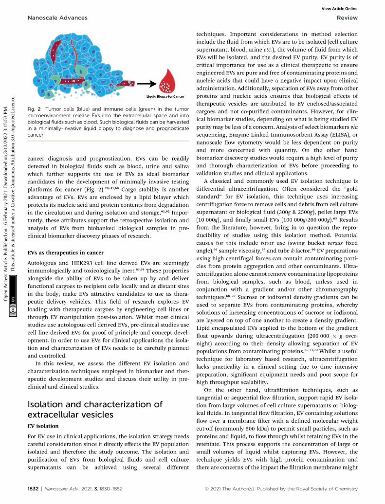
Fig. 2 Tumor cells (blue) and immune cells (green) in the tumormicroenvironment release EVs into the extracellular space and intobiological fluids such as blood. Such biological fluids can be harvestedin a minimally-invasive liquid biopsy to diagnose and prognosticatecancer.
Nanoscale Advances Review
Ope
n A
cces
s A
rtic
le. P
ublis
hed
on 1
6 Fe
brua
ry 2
021.
Dow
nloa
ded
on 3
/13/
2022
3:1
5:53
PM
. T
his
artic
le is
lice
nsed
und
er a
Cre
ativ
e C
omm
ons
Attr
ibut
ion
3.0
Unp
orte
d L
icen
ce.
View Article Online
cancer diagnosis and prognostication. EVs can be readilydetected in biological uids such as blood, urine and salivawhich further supports the use of EVs as ideal biomarkercandidates in the development of minimally invasive testingplatforms for cancer (Fig. 2).29–31,60 Cargo stability is anotheradvantage of EVs. EVs are enclosed by a lipid bilayer whichprotects its nucleic acid and protein contents from degradationin the circulation and during isolation and storage.61,62 Impor-tantly, these attributes support the retrospective isolation andanalysis of EVs from biobanked biological samples in pre-clinical biomarker discovery phases of research.
EVs as therapeutics in cancer
Autologous and HEK293 cell line derived EVs are seeminglyimmunologically and toxicologically inert.63,64 These propertiesalongside the ability of EVs to be taken up by and deliverfunctional cargoes to recipient cells locally and at distant sitesin the body, make EVs attractive candidates to use as thera-peutic delivery vehicles. This eld of research explores EVloading with therapeutic cargoes by engineering cell lines orthrough EV manipulation post-isolation. Whilst most clinicalstudies use autologous cell derived EVs, pre-clinical studies usecell line derived EVs for proof of principle and concept devel-opment. In order to use EVs for clinical applications the isola-tion and characterization of EVs needs to be carefully plannedand controlled.
In this review, we assess the different EV isolation andcharacterization techniques employed in biomarker and ther-apeutic development studies and discuss their utility in pre-clinical and clinical studies.
Isolation and characterization ofextracellular vesiclesEV isolation
For EV use in clinical applications, the isolation strategy needscareful consideration since it directly effects the EV populationisolated and therefore the study outcome. The isolation andpurication of EVs from biological uids and cell culturesupernatants can be achieved using several different
1832 | Nanoscale Adv., 2021, 3, 1830–1852
techniques. Important considerations in method selectioninclude the uid from which EVs are to be isolated (cell culturesupernatant, blood, urine etc.), the volume of uid from whichEVs will be isolated, and the desired EV purity. EV purity is ofcritical importance for use as a clinical therapeutic to ensureengineered EVs are pure and free of contaminating proteins andnucleic acids that could have a negative impact upon clinicaladministration. Additionally, separation of EVs away from otherproteins and nucleic acids ensures that biological effects oftherapeutic vesicles are attributed to EV enclosed/associatedcargoes and not co-puried contaminants. However, for clin-ical biomarker studies, depending on what is being studied EVpurity may be less of a concern. Analysis of select biomarkers viasequencing, Enzyme Linked Immunosorbent Assay (ELISA), ornanoscale ow cytometry would be less dependent on purityand more concerned with quantity. On the other handbiomarker discovery studies would require a high level of purityand thorough characterization of EVs before proceeding tovalidation studies and clinical applications.
A classical and commonly used EV isolation technique isdifferential ultracentrifugation. Oen considered the “goldstandard” for EV isolation, this technique uses increasingcentrifugation force to remove cells and debris from cell culturesupernatant or biological uid (300g & 2500g), pellet large EVs(10 000g), and nally small EVs (100 000g/200 000g).65 Resultsfrom the literature, however, bring in to question the repro-ducibility of studies using this isolation method. Potentialcauses for this include rotor use (swing bucket versus xedangle),66 sample viscosity,67 and tube k-factor.66 EV preparationsusing high centrifugal forces can contain contaminating parti-cles from protein aggregation and other contaminants. Ultra-centrifugation alone cannot remove contaminating lipoproteinsfrom biological samples, such as blood, unless used inconjunction with a gradient and/or other chromatographytechniques.68–70 Sucrose or iodixonal density gradients can beused to separate EVs from contaminating proteins, wherebysolutions of increasing concentrations of sucrose or iodixonalare layered on top of one another to create a density gradient.Lipid encapsulated EVs applied to the bottom of the gradientoat upwards during ultracentrifugation (200 000 � g over-night) according to their density allowing separation of EVpopulations from contaminating proteins.65,71,72 Whilst a usefultechnique for laboratory based research, ultracentrifugationlacks practicality in a clinical setting due to time intensivepreparation, signicant equipment needs and poor scope forhigh throughput scalability.
On the other hand, ultraltration techniques, such astangential or sequential ow ltration, support rapid EV isola-tion from large volumes of cell culture supernatants or biolog-ical uids. In tangential ow ltration, EV containing solutionsow over a membrane lter with a dened molecular weightcut-off (commonly 500 kDa) to permit small particles, such asproteins and liquid, to ow through whilst retaining EVs in theretentate. This process supports the concentration of large orsmall volumes of liquid whilst capturing EVs. However, thetechnique yields EVs with high protein contamination andthere are concerns of the impact the ltration membrane might
© 2021 The Author(s). Published by the Royal Society of Chemistry

Review Nanoscale Advances
Ope
n A
cces
s A
rtic
le. P
ublis
hed
on 1
6 Fe
brua
ry 2
021.
Dow
nloa
ded
on 3
/13/
2022
3:1
5:53
PM
. T
his
artic
le is
lice
nsed
und
er a
Cre
ativ
e C
omm
ons
Attr
ibut
ion
3.0
Unp
orte
d L
icen
ce.
View Article Online
have upon EV integrity. Where high purity is required, tangen-tial ow ltration would need to be used in conjunction witha second technique to improve purity, such as size exclusionchromatography.73–76 Alternatively, sequential ltration usesa combination of three ltration steps to isolate EVs devoid ofcontaminating proteins. First, dead end ltration is used toremove cells and debris. This is followed by tangential owltration to concentrate the sample and retain EVs as describedabove. Finally ltration occurs through a track edgedmembrane of increasing pore sizes (50–200 nm) to isolate andfractionate EVs by size.76
Size exclusion chromatography is an efficient chromatog-raphy technique that separates particles based on size. This hasbeen adapted to separate and purify EVs from proteins incomplex biological samples. When biological uids such asblood plasma/serum is applied to a sepharose size exclusioncolumn, differential exclusion causes EVs to elute rst andseparately from proteins which get trapped in the resin poresand only begin to elute in later fractions.77 However, sizeexclusion chromatography technology cannot efficiently sepa-rate EVs from lipoproteins of similar size when used to purifyEVs from plasma or serum. For complete lipoprotein removala mixture of isolation and purication methods would need tobe carried out including density gradient ultracentrifugationfollowed by size exclusion chromatography.68 Other chroma-tography techniques that have been developed for EV purica-tion include affinity purication and ion exchangechromatography. Membrane affinity purication methods suchas exoEasy spin columns can isolate EVs from biologicalsamples, however purity may be sub-optimal in comparison tosize exclusion chromatography.78 Affinity based methods havealso been optimized using the calcium sensitive, phosphati-dylserine binding protein Tim4. EVs bound to Tim4 cansubsequently be simply released by addition of calcium chela-tors.79 Other immuno-affinity capture agents can includeheparin, tetraspanins and Epithelial Cell Adhesion Molecule(EpCAM).80–82 However the disadvantage of immuno-affinitycapture is that only select populations of EVs are puried andEVs can be difficult to remove from the substrate without veryharsh conditions such as low pH. A nal chromatographymethod that is proving to be efficient for the isolation of EVsfrom cell culture supernatants in a scalable and efficientmanner is anion exchange chromatography.83,84 Negativelycharged EVs bind to positively charged columns and EVs arethen eluted from the column using increasing concentrations ofsalt. Using anion exchange chromatography EVs can be isolatedfrom 1 liter of cell culture conditioned media within 2 hourswith minimal user input.83 This approach to EV isolationdemonstrates scalability and rapid isolation suggesting it mayhave promise in supporting the use of EVs as therapeutics.
A popular method for isolation of EVs from clinical biolog-ical samples is by precipitation using commercially availablereagents. The precipitation of EVs using polyethylene glycol(PEG),85 or commercially available reagents such as exoquick86
allows EVs to be pelleted by centrifugation at lower speeds,removing the need for time consuming and equipmentdependent ultracentrifugation. EVs can be captured from small
© 2021 The Author(s). Published by the Royal Society of Chemistry
volumes of biological uids, or larger volumes of pre-concentrated biological uids/cell culture supernatants.Although very user friendly and amenable for use with a largenumber of biological samples, some reports suggest that EVpurity aer precipitation can be low as precipitation can alsopellet proteins and lipoproteins.86,87 A second purication stepaer precipitation may be necessary to isolate a pure prepara-tion of EVs. Additionally, precipitation reagents remaining inEV preparations can affect recipient cell viability EV and bio-logical activity.88,89
Finally microuidic chips are an emerging technology usefulfor the capture and analysis of EVs from small volumes ofclinical samples and show promise for liquid biopsy diagnosisof disease. Microuidic devices have been engineered forimmuno-capture using tumor specic antigens or othermarkers of interest. For example, Human Epidermal GrowthFactor Receptor (HER2) and Prostate Specic Antigen (PSA)positive tumor derived EVs have been captured on chipsemploying nanoshearing uid ow. Captured EVs can then bequantied by a colorimetric reaction.90 Others have isolated andthen eluted EVs for downstream analysis. For instance, EGFRwild type or EGFRvIII EVs can be isolated and quantied fromglioblastoma patient plasma. Additionally EVs have been elutedfrom a chip and used for further in-depth RNA sequencing ofEGFRvIII EVs.91 An alternative chip device approach usedEpCAM aptamers to capture EVs, and electro-oxidation of metalnanoparticles to detect specic epitopes present upon capturedEVs (specically EpCAM and Prostate Specic MembraneAntigen (PSMA)). Oxidation of metal particles provides anelectro chemical peak that can be used as a read out for quan-tication of captured EVs.92 Microuidic chips have also beendesigned with specic size thresholds to capture tumor derivedmicrovesicles. The EVs travel through the microuidic chip andare eluted from different ports (dependent on size) for furtherdownstream processing.93 Table 1 summarizes the pros andcons of current popular EV isolation techniques.
EV characterization
Equally as important as selecting a method for EV isolation andpurication is the characterization of EVs before use in down-stream assays. There is an array of different methods that can beused to validate EV size, concentration, purity, and biomarkerpresence. Western blotting is a standard method used wherebyprobing for different EV markers in conjunction witha biomarker of interest can be carried out. EV markers that canbe used include CD63, CD81, CD9, ALIX, TSG101, Flotillin andAnnexins amongst many others.72 These markers do notdifferentiate between different EV subtypes but do conrm thepresence of EVs. Western blots can, to some extent, also beuseful in the analysis of EV purity. Western blotting forcontaminant proteins such as cell organelle specic proteinsand albumin which should be absent from EV preparations, canbe useful to determine the extent of protein contamination inthe EV preparation.72 To analyze the concentration and size ofEVs there are several technologies available such as nano-particle tracking analysis (NTA) and tunable resistive pulse
Nanoscale Adv., 2021, 3, 1830–1852 | 1833
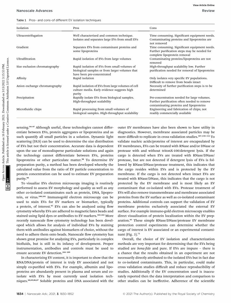
Table 1 Pros- and cons-of different EV isolation techniques
Isolation Pros Cons
Ultracentrifugation Well characterized and common technique.Isolates and separates large EVs from small EVs
Time consuming. Signicant equipment needs.Contaminating proteins and lipoproteins arenot removed
Gradient Separates EVs from contaminant proteins andsome lipoproteins
Time consuming. Signicant equipment needs.Further purication steps may be needed forcomplete lipoprotein removal
Ultraltration Rapid isolation of EVs from large volumes Contaminating proteins/lipoproteins are notremoved
Size exclusion chromatography Rapid isolation of EVs from small volumes ofbiological samples or from larger volumes thathave been pre-concentrated
High-throughput scalability low. Furtherpurication needed for removal of lipoproteins
Affinity Rapid isolation Only isolates very specic EV populations.Difficult to remove from beads intact
Anion exchange chromatography Rapid isolation of EVs from large volumes of cellculture media. Early evidence suggests highpurity
Necessity of further purication steps is to bedetermined
Precipitation Rapidly isolate EVs from biological samples.High-throughput scalability
Pre-concentration needed for large volumes.Further purication oen needed to removecontaminating proteins and lipoproteins
Microuidic chips Rapid processing from small volumes ofbiological samples. High-throughput scalability
Engineering and fabrication of chips; notreadily commercially available
Nanoscale Advances Review
Ope
n A
cces
s A
rtic
le. P
ublis
hed
on 1
6 Fe
brua
ry 2
021.
Dow
nloa
ded
on 3
/13/
2022
3:1
5:53
PM
. T
his
artic
le is
lice
nsed
und
er a
Cre
ativ
e C
omm
ons
Attr
ibut
ion
3.0
Unp
orte
d L
icen
ce.
View Article Online
sensing.94–97 Although useful, these technologies cannot differ-entiate between EVs, protein aggregates or lipoproteins and assuch quantify all small particles in a solution. Dynamic lightscattering (DLS) can be used to determine the size distributionof EVs but not their concentration. Accurate data is dependentupon the use of monodisperse particulate solutions and againthe technology cannot differentiate between EVs, proteinslipoproteins or other particulate matter.98 To determine EVpreparation purity, a method has been developed whereby thecalculated value from the ratio of EV particle concentration toprotein concentration can be used to estimate EV preparationpurity.99
To visualize EVs electron microscopy imaging is routinelyperformed to assess EV morphology and quality as well as anyother co-isolated contaminates such as protein, DNA, lipopro-tein, or virus.100,101 Immunogold electron microscopy can beused to stain EVs for EV markers or biomarker, typicallya protein, of interest.65 EVs can also be analyzed using owcytometry whereby EVs are adhered to magnetic/latex beads andstained using lipid dyes or antibodies to EV markers.102,103 Morerecently nanoscale ow cytometry technology has been devel-oped which allows for analysis of individual EVs by stainingthem with antibodies against biomarkers of choice, without theneed to adhere them onto beads. Nanoscale ow cytometry hasshown great promise for analyzing EVs, particularly in complexbiouids, but is still in its infancy of development. Properinstrumentation, antibodies and controls must be used toensure accurate EV detection.104–109
In characterizing EV content, it is important to show that theRNA/DNA/protein of interest is truly EV associated and notsimply co-puried with EVs. For example, albumin and lipo-proteins are abundantly present in plasma and serum and co-isolate with EVs by most currently used isolation tech-niques.68,69,86,87 Soluble proteins and DNA associated with the
1834 | Nanoscale Adv., 2021, 3, 1830–1852
outer EV membranes have also been shown to have utility indiagnostics. However, membrane associated particles may bemore difficult to replicate in cross validation studies.107,110–112 Tovalidate nucleic acids/proteins of interest are encapsulated byEV membranes, EVs can be treated with RNase and/or DNase orprotease with and without tritonX-100/detergent lysis. If thecargo is detected when EVs are treated with RNase/DNase/protease, but are not detected if detergent lysis of EVs is fol-lowed by RNase/DNase/protease treatment, this indicates thatthe cargo resides within EVs and is protected by the EVmembrane. If the cargo is not detected when intact EVs aretreated with RNase/DNase, this indicates that the cargo is notprotected by the EV membrane and is most likely an EVcontaminant that co-isolated with EVs. Protease treatment ofEVs will also remove transmembrane andmembrane associatedproteins from the EV surface as well as co-isolated contaminantproteins. Additional controls can support the validation of EVmembrane proteins exclusively associated the external EVsurface. For example immuno-gold electronmicroscopy enablesdirect visualization of protein localization within the EV prep-aration.65 These simple RNase/DNase/protease EV membraneprotection control experiments can determine whether thecargo of interest is EV associated or an experimental contami-nant (Fig. 3).72
Overall, the choice of EV isolation and characterizationmethods are very important for determining that the EVs beingstudied are bona-de and pure. If EVs are impure – there isa chance that the results obtained in an experiment are notnecessarily directly attributed to the isolated EVs but in fact dueto co-isolated contaminants. This, in particular, could makecross validation studies difficult and reduce reproducibility ofstudies. Additionally if the EV concentration used is inaccu-rately reported then the data interpretation and comparison toother studies can be ineffective. Adherence of the scientic
© 2021 The Author(s). Published by the Royal Society of Chemistry
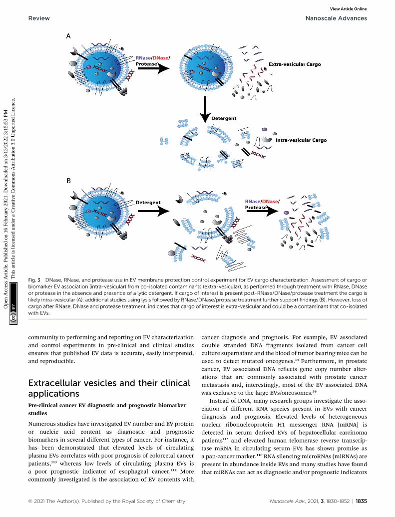
Fig. 3 DNase, RNase, and protease use in EV membrane protection control experiment for EV cargo characterization. Assessment of cargo orbiomarker EV association (intra-vesicular) from co-isolated contaminants (extra-vesicular), as performed through treatment with RNase, DNaseor protease in the absence and presence of a lytic detergent. If cargo of interest is present post-RNase/DNase/protease treatment the cargo islikely intra-vesicular (A); additional studies using lysis followed by RNase/DNase/protease treatment further support findings (B). However, loss ofcargo after RNase, DNase and protease treatment, indicates that cargo of interest is extra-vesicular and could be a contaminant that co-isolatedwith EVs.
Review Nanoscale Advances
Ope
n A
cces
s A
rtic
le. P
ublis
hed
on 1
6 Fe
brua
ry 2
021.
Dow
nloa
ded
on 3
/13/
2022
3:1
5:53
PM
. T
his
artic
le is
lice
nsed
und
er a
Cre
ativ
e C
omm
ons
Attr
ibut
ion
3.0
Unp
orte
d L
icen
ce.
View Article Online
community to performing and reporting on EV characterizationand control experiments in pre-clinical and clinical studiesensures that published EV data is accurate, easily interpreted,and reproducible.
Extracellular vesicles and their clinicalapplicationsPre-clinical cancer EV diagnostic and prognostic biomarkerstudies
Numerous studies have investigated EV number and EV proteinor nucleic acid content as diagnostic and prognosticbiomarkers in several different types of cancer. For instance, ithas been demonstrated that elevated levels of circulatingplasma EVs correlates with poor prognosis of colorectal cancerpatients,113 whereas low levels of circulating plasma EVs isa poor prognostic indicator of esophageal cancer.114 Morecommonly investigated is the association of EV contents with
© 2021 The Author(s). Published by the Royal Society of Chemistry
cancer diagnosis and prognosis. For example, EV associateddouble stranded DNA fragments isolated from cancer cellculture supernatant and the blood of tumor bearingmice can beused to detect mutated oncogenes.14 Furthermore, in prostatecancer, EV associated DNA reects gene copy number alter-ations that are commonly associated with prostate cancermetastasis and, interestingly, most of the EV associated DNAwas exclusive to the large EVs/oncosomes.59
Instead of DNA, many research groups investigate the asso-ciation of different RNA species present in EVs with cancerdiagnosis and prognosis. Elevated levels of heterogeneousnuclear ribonucleoprotein H1 messenger RNA (mRNA) isdetected in serum derived EVs of hepatocellular carcinomapatients115 and elevated human telomerase reverse transcrip-tase mRNA in circulating serum EVs has shown promise asa pan-cancer marker.116 RNA silencing microRNAs (miRNAs) arepresent in abundance inside EVs and many studies have foundthat miRNAs can act as diagnostic and/or prognostic indicators
Nanoscale Adv., 2021, 3, 1830–1852 | 1835
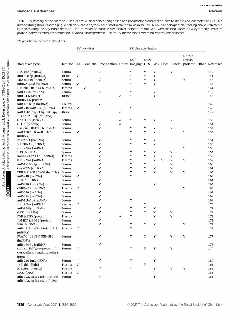
Table 2 Summary of the methods used in pre-clinical cancer diagnostic and prognostic biomarker studies to isolate and characterize EVs. UC:ultracentrifugation. EM/imaging: electronmicroscopy/any other method used to visualize EVs. NTA/DLS: nanoparticle tracking analysis/dynamiclight scattering (or any other method used to measure particle size and/or concentration). WB: western blot. Flow: flow cytometry. Protein:protein concentration determination. RNase/DNase/protease: use of EV membrane protection control experiments
EV pre-clinical cancer biomarkers
Biomarker (type) Biouid
EV isolation EV characterization
ReferenceUC Gradient Precipitation OtherEM/imaging
NTA/DLS WB Flow Protein
RNase/DNase/protease Other
HOTTIP (lncRNA) Serum 3 Y Y Y Y 140miR-30c-5p (miRNA) Urine 3 Y Y Y 141LINC02418 (lncRNA) Serum 3 Y Y Y Y 142miRNA-320d (miRNA) Serum 3 Y Y Y 143Hsa-circ-0065149 (circRNA) Plasma 3 144miR-1246 (miRNA) Serum 3 Y Y 145miR-21 & MMP1(miRNA & protein)
Urine 3 Y Y 146
miR-181b-5p (miRNA) Ascites 3 147miR-21& miR-92a (miRNA) Plasma 3 Y 148miR-19b1-5p, 21-5p, 136-5p,139-5p, 210-3p (miRNAs)
Urine 3 149
GNAQ-6:1 (lncRNA) Serum 3 Y Y Y Y 150GPC-1 (protein) Serum 3 3 151Hsa-circ-0004771 (circRNA) Serum 3 Y Y Y Y 152miR-150-5p & miR-99b-5p(miRNA)
Serum 3 Y Y Y Y 153
Pcsk2-2:1 (lncRNA) Serum 3 Y Y Y 1545 lncRNAs (lncRNA) Serum 3 Y Y Y 1554 miRNAs (miRNA) Serum 3 Y 156H19 (lncRNA) Serum 3 Y Y Y Y 157lncSLC2A12-10:1 (lncRNA) Plasma 3 Y Y Y Y 1588 miRNAs (miRNA) Plasma 3 Y Y Y Y Y 159miR-1910p-3p (miRNA) Serum 3 Y Y Y 160Circ-PNN (circRNA) Serum 3 Y Y Y Y 161TBILA & AGAP2-AS1 (lncRNA) Serum 3 Y Y Y Y Y 162miR-210 (miRNA) Serum 3 Y 163HULC (lncRNA) Serum 3 164miR-320d (miRNA) Serum 3 Y 165CEBPA-AS1 (lncRNA) Plasma 3 Y 166miR-378 (miRNA) Serum 3 167miR-874 (miRNA) Serum 3 168miR-10b-5p (miRNA) Serum 3 Y 1698 miRNAs (miRNA) Ascites 3 Y 170miR-17-5p (miRNA) Serum 3 Y Y Y 171GAS5 (lncRNA) Serum 3 Y Y Y Y 172FGB & FGG (protein) Plasma 3 Y Y Y 173*c-MET & PDL1 (protein) Serum 3 174H19 (lncRNA) Serum 3 Y Y Y Y 175miR-4525, miR-451a& miR-21(miRNA)
Plasma 3 Y 176
PCAT-1, UBC1 & SNHG16(lncRNA)
Serum 3 Y Y Y Y Y 177
miR-454-3p (miRNA) Serum 3 178alpha-2-HS-(glycoprotein) &extracellular matrix protein 1(protein)
Serum 3 Y Y Y Y 179
miR-210 (microRNA) Serum 3 Y Y 18016 lipids (lipid) Plasma 3 Y 181PTENP1 (lncRNA) Plasma 3 Y Y Y Y 182KRAS (DNA) Plasma 3 183miR-122, miR-125b, miR-145,miR-192, miR-194, miR-29a,
Serum 3 Y Y 184
1836 | Nanoscale Adv., 2021, 3, 1830–1852 © 2021 The Author(s). Published by the Royal Society of Chemistry
Nanoscale Advances Review
Ope
n A
cces
s A
rtic
le. P
ublis
hed
on 1
6 Fe
brua
ry 2
021.
Dow
nloa
ded
on 3
/13/
2022
3:1
5:53
PM
. T
his
artic
le is
lice
nsed
und
er a
Cre
ativ
e C
omm
ons
Attr
ibut
ion
3.0
Unp
orte
d L
icen
ce.
View Article Online
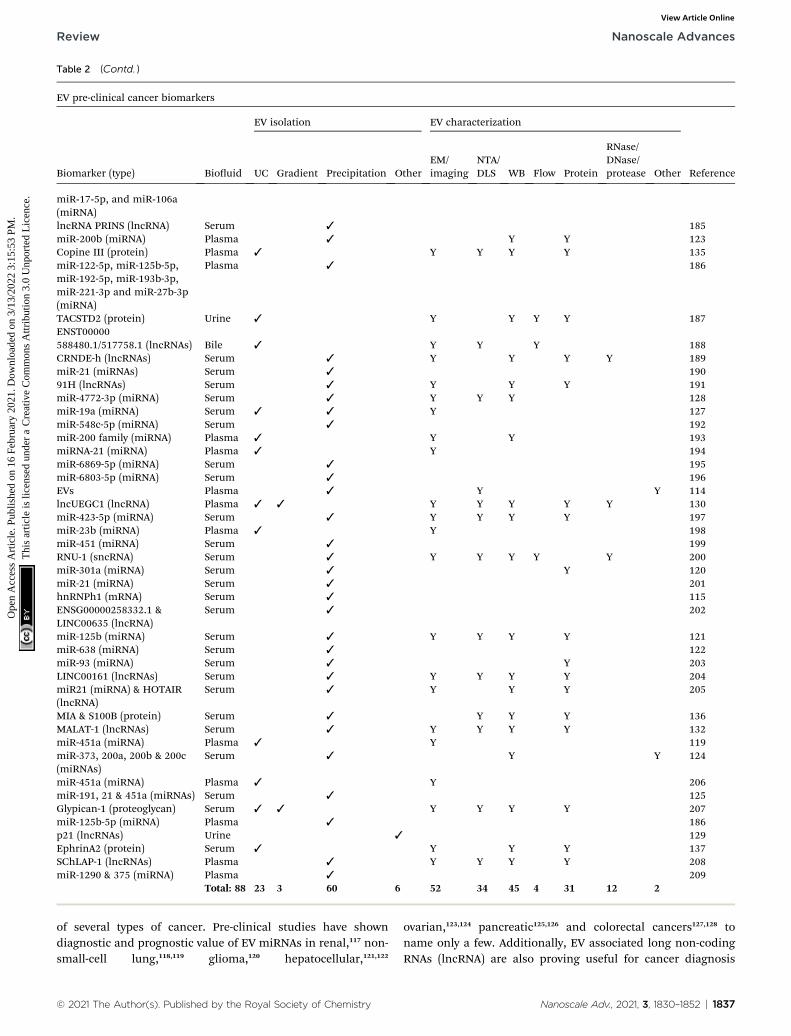
Table 2 (Contd. )
EV pre-clinical cancer biomarkers
Biomarker (type) Biouid
EV isolation EV characterization
ReferenceUC Gradient Precipitation OtherEM/imaging
NTA/DLS WB Flow Protein
RNase/DNase/protease Other
miR-17-5p, and miR-106a(miRNA)lncRNA PRINS (lncRNA) Serum 3 185miR-200b (miRNA) Plasma 3 Y Y 123Copine III (protein) Plasma 3 Y Y Y Y 135miR-122-5p, miR-125b-5p,miR-192-5p, miR-193b-3p,miR-221-3p and miR-27b-3p(miRNA)
Plasma 3 186
TACSTD2 (protein) Urine 3 Y Y Y Y 187ENST00000588480.1/517758.1 (lncRNAs) Bile 3 Y Y Y 188CRNDE-h (lncRNAs) Serum 3 Y Y Y Y 189miR-21 (miRNAs) Serum 3 19091H (lncRNAs) Serum 3 Y Y Y 191miR-4772-3p (miRNA) Serum 3 Y Y Y 128miR-19a (miRNA) Serum 3 3 Y 127miR-548c-5p (miRNA) Serum 3 192miR-200 family (miRNA) Plasma 3 Y Y 193miRNA-21 (miRNA) Plasma 3 Y 194miR-6869-5p (miRNA) Serum 3 195miR-6803-5p (miRNA) Serum 3 196EVs Plasma 3 Y Y 114lncUEGC1 (lncRNA) Plasma 3 3 Y Y Y Y Y 130miR-423-5p (miRNA) Serum 3 Y Y Y Y 197miR-23b (miRNA) Plasma 3 Y 198miR-451 (miRNA) Serum 3 199RNU-1 (sncRNA) Serum 3 Y Y Y Y Y 200miR-301a (miRNA) Serum 3 Y 120miR-21 (miRNA) Serum 3 201hnRNPh1 (mRNA) Serum 3 115ENSG00000258332.1 &LINC00635 (lncRNA)
Serum 3 202
miR-125b (miRNA) Serum 3 Y Y Y Y 121miR-638 (miRNA) Serum 3 122miR-93 (miRNA) Serum 3 Y 203LINC00161 (lncRNAs) Serum 3 Y Y Y Y 204miR21 (miRNA) & HOTAIR(lncRNA)
Serum 3 Y Y Y 205
MIA & S100B (protein) Serum 3 Y Y Y 136MALAT-1 (lncRNAs) Serum 3 Y Y Y Y 132miR-451a (miRNA) Plasma 3 Y 119miR-373, 200a, 200b & 200c(miRNAs)
Serum 3 Y Y 124
miR-451a (miRNA) Plasma 3 Y 206miR-191, 21 & 451a (miRNAs) Serum 3 125Glypican-1 (proteoglycan) Serum 3 3 Y Y Y Y 207miR-125b-5p (miRNA) Plasma 3 186p21 (lncRNAs) Urine 3 129EphrinA2 (protein) Serum 3 Y Y Y 137SChLAP-1 (lncRNAs) Plasma 3 Y Y Y Y 208miR-1290 & 375 (miRNA) Plasma 3 209
Total: 88 23 3 60 6 52 34 45 4 31 12 2
Review Nanoscale Advances
Ope
n A
cces
s A
rtic
le. P
ublis
hed
on 1
6 Fe
brua
ry 2
021.
Dow
nloa
ded
on 3
/13/
2022
3:1
5:53
PM
. T
his
artic
le is
lice
nsed
und
er a
Cre
ativ
e C
omm
ons
Attr
ibut
ion
3.0
Unp
orte
d L
icen
ce.
View Article Online
of several types of cancer. Pre-clinical studies have showndiagnostic and prognostic value of EV miRNAs in renal,117 non-small-cell lung,118,119 glioma,120 hepatocellular,121,122
© 2021 The Author(s). Published by the Royal Society of Chemistry
ovarian,123,124 pancreatic125,126 and colorectal cancers127,128 toname only a few. Additionally, EV associated long non-codingRNAs (lncRNA) are also proving useful for cancer diagnosis
Nanoscale Adv., 2021, 3, 1830–1852 | 1837
Nanoscale Advances Review
Ope
n A
cces
s A
rtic
le. P
ublis
hed
on 1
6 Fe
brua
ry 2
021.
Dow
nloa
ded
on 3
/13/
2022
3:1
5:53
PM
. T
his
artic
le is
lice
nsed
und
er a
Cre
ativ
e C
omm
ons
Attr
ibut
ion
3.0
Unp
orte
d L
icen
ce.
View Article Online
and prognostication in pre-clinical studies. EV associatedlncRNA-p21 is elevated in the urine of prostate cancer patientsand can distinguish between benign disease and cancer.129
Plasma EV associated lncRNA UEGC1 shows promise as a diag-nostic for early stage gastric cancer,130 and serum derived EV-associated HOTTIP may be useful as a diagnostic and prog-nostic indicator of gastric cancer.131 Finally, lncRNA MALAT1 iselevated in serum EVs from non-small-cell lung cancerpatients.132 The less common family of circular RNAs isbecoming increasingly recognized to be associated with EVsand potentially useful as a cancer biomarker.133 In fact, thepresence of circular RNA-PDE8A is associated with pancreaticcancer diagnosis and progression.134
Finally, proteins either encapsulated by or associated withEV membranes can also be used for biomarker discovery. Forexample, circulating EV associated Copine III has been found tohave diagnostic and prognostic signicance for colorectalcancer.135 Melanoma biomarkers MIA and S100B are found onserum EVs and are predictive of diagnosis and prognosis ofmelanoma.136 Ascites derived EV associated E-cadherin hasdiagnostic utility in ovarian cancer and serum EV associatedEphrinA2 may aid diagnosis of prostate cancer.137,138
There is a large number of research studies publishedregarding the analysis of EVs as biomarkers for cancer. Toassess the EV isolation and characterization methods employedby the large number of EV studies, we selected studies based ona previous meta-analysis study that looked at the clinicalsignicance of EVs in cancer.139 This study narrowed down theavailable selection of cancer EV biomarker literature between2010–September 2018. The authors did a literature search usingthe key words ‘exosome and cancer and diagnosis or prognosis’.The authors then reviewed all literature and included onlystudies that involved EVs in cancer patient biouids with atleast 10 patients and matched controls being used. Thebiomarker had to have clinical signicance and reporting ofspecicity and sensitivity for diagnostic markers, and con-dence interval reported for prognostic markers. The literaturesearch was narrowed down to include 60 studies. We furthernarrowed these studies down to only include studies that usedat least 20 patient samples and matched controls. We then usedthe identical search terms and found that since September 2018
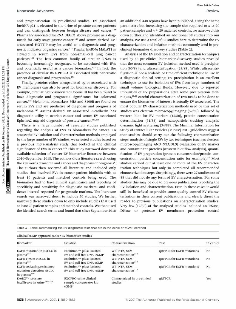
Table 3 Table summarizing the EV diagnostic tests that are in the clinic
Clinical/cGMP approved cancer EV biomarker studies
Biomarker Isolation Chara
EGFR mutation in NSCLC inplasma217
Exolution™ plus: isolatedEV and cell free DNA. cGMP
WB, Nchara
EGFR T790M NSCLC inplasma212
Exolution™ plus: isolatedEV and cell free DNA cGMP
WB, Nchara
EGFR activating/resistancemutation detection NSCLCin plasma210
Exolution™ plus: isolatedEV and cell free DNA. cGMP
WB, Nchara
ExoDX™ prostateintelliscore in urine213–215
EXOPRO urine clinicalsample concentrator kit.cGMP
Charastudie
1838 | Nanoscale Adv., 2021, 3, 1830–1852
an additional 448 reports have been published. Using the sameparameters but increasing the sample size required to n $ 20patient samples and n$ 20 matched controls, we narrowed thisdown further and identied an additional 38 studies into ouranalysis. We use a total of 88 studies here to determine the EVcharacterization and isolation methods commonly used in pre-clinical biomarker discovery studies (Table 2).
Analysis of the EV isolation and characterization techniquesused by 88 pre-clinical biomarker discovery studies revealedthat the most common EV isolation method used is precipita-tion (60/88) and ultracentrifugation (23/88). Whilst ultracentri-fugation is not a scalable or time efficient technique to use ina diagnostic clinical setting, EV precipitation is an excellenttechnique to use for isolation of EVs from large numbers ofsmall volume biological uids. However, due to reportedimpurities of EV preparations aer some precipitation tech-niques,86,87 careful characterization of EVs needs to be done toensure the biomarker of interest is actually EV associated. Themost popular EV characterization methods used by this set ofstudies was electron microscopy/imaging (52/88), followed bywestern blot for EV markers (45/88), protein concentrationdetermination (31/88) and nanoparticle tracking analysis/dynamic light scattering (34/88). The Minimal Information forStudy of Extracellular Vesicles (MISEV) 2018 guidelines suggestthat studies should carry out the following characterizationsteps: analysis of single EVs by two techniques (such as electronmicroscopy/imaging AND NTA/DLS) evaluation of EV markerand contaminant proteins (western blot/ow analysis), quanti-cation of EV preparation (protein concentration/protein con-centration : particle concentration ratio for example).72 Moststudies carried out at least one or more of the EV character-ization techniques but only 18 completed all recommendedcharacterization steps. Surprisingly, there were 27 studies out of88 that did not do any form of EV characterization. For somestudies this may be due to previous publications reporting theEV isolation and characterization. Even in these cases it wouldstill be benecial to provide some quality control EV charac-terization in their current publications and clearly direct thereader to previous publications on characterization studies.Very few (12/88) of the analyzed studies included an RNase,DNase or protease EV membrane protection control
or cGMP certified
cterization Test In clinic?
TA, SEMcterization218
qRTPCR for EGFRmutations No
TA, SEMcterization218
qRTPCR for EGFRmutations No
TA, SEMcterization218
qRTPCR for EGFRmutations No
cterized in pre-clinicals
qRTPCR Yes
© 2021 The Author(s). Published by the Royal Society of Chemistry

Review Nanoscale Advances
Ope
n A
cces
s A
rtic
le. P
ublis
hed
on 1
6 Fe
brua
ry 2
021.
Dow
nloa
ded
on 3
/13/
2022
3:1
5:53
PM
. T
his
artic
le is
lice
nsed
und
er a
Cre
ativ
e C
omm
ons
Attr
ibut
ion
3.0
Unp
orte
d L
icen
ce.
View Article Online
experiments to determine whether the EV associated cargo/biomarker was in fact EV enclosed/associated and not a co-isolated contaminant. During the discovery phase ofbiomarker development, this is an important control experi-ment to include to present clear data that could be used to moveforward to a clinical setting.
Clinical translation of EV diagnostic and prognosticbiomarker studies in cancer
While many studies have investigated new cancer biomarkers inpre-clinical settings, there are only a handful of biomarker teststhat have been current good manufacturing practice (cGMP)approved and only one has been approved for use by clinicians(Table 3). Exosome Diagnostics, Inc. has developed workows to
Table 4 EV isolation and characterization methods used in pre-clinidifferential centrifugation/ultracentrifugation. UF: ultrafiltration. GradienEM/imaging: electron microscopy/any other method used to visualize EVany other method used to measure particle size and/or concentration). Wdetermination. RNase/DNase/protease: use of EV membrane protection
EVs as therapeutic delivery vehicles
Cargo (target/drug)
EV isolation EV
DC/UC UFGradient/SC Precipitation SEC
EMima
Small molecule(doxorubicin)
3 3 3 Y
Small molecule(doxorubicin)
3
Small molecule(Piclitaxel)
3 3 Y
Small molecule(piclitaxel)
3 3 Y
Oncolytic virus andpiclitaxel
3
siRNA (Kras) 3 3 Y
siRNA (Rad51, Rad52) 3 YsiRNA (PLK-1) 3
miRNA & siRNA(Let7, VEGF)
3 3 Y
miRNA (mir-134) 3 3 YmiRNA (mir-31,mir-451a)
3 Y
gRNA (reporter) 3 3 Y
DNA (Cas9/gRNA –RUNX2 & CTNNB1)
3 Y
DNA (Cas9/gRNA –PARP1)
3 3 Y
Protein & gRNA(Cas9/gRNARNP – HIV LTR)
3
Protein/mRNA(p53/Cas9gRNA RNP)
3 3 Y
Protein (Bax/srlkB/cre) 3 3
Protein (ww-cre) 3 3
Total: 18 12 5 4 6 3 12
© 2021 The Author(s). Published by the Royal Society of Chemistry
isolate cell free DNA and EV associated DNA/mRNA fromplasma using a cGMP clinically certied process Exolution™Plus. The DNA and EVs are then subject to lysis and quantitativeRT-PCR (qRTPCR) analysis of EGFRmutations. This has clinicalutility for lung cancer patients to direct patient treatmentstrategies.210–212 The same company has developed an additionalcGMP and clinically approved technology – ExoPro urine clin-ical sample concentrator kit – to isolate EVs from urine byultraltration centrifugation and to extract EV associated RNA.QRTPCR was used to analyze the expression of a set of genes(PCA3, SPDEF, ERG) and found to have utility as non-invasivetest to risk stratify men with suspected prostate cancer andimprove the identication of individuals with clinically signi-cant disease.213–215
cal studies generating EVs as therapeutic delivery vehicles. DC/UC:t/SC: gradient/sucrose cushion. SEC: size exclusion chromatography.s. NTA/DLS: nanoparticle tracking analysis/dynamic light scattering (orB: western blot. Flow: flow cytometry. Protein: protein concentrationcontrol experiments. N/A: not applicable
characterization
Reference/ging
NTA/DLS WB Flow Protein
DNase/RNase/protease Detergent
Y N/A 219
Y Y Y N/A 220
Y Y Y N/A 221
Y Y Y N/A 222
Y N/A 223
Y Y Y RNase,proteinase K
Y 224
Y Y Y 225Y 226
Y Y RNase 227
Y Y RNase 228Y Y Y Y RNase Y 240
Y Y Y RNase,proteinase K
Y 231
Y Y Y DNase,proteinase K
233
Y Y Y DNase 232
Y Y 234
Y Y 238
Y Y Y 237Y Y Y Proteinase K Y 23916 13 2 14 8 4
Nanoscale Adv., 2021, 3, 1830–1852 | 1839

Nanoscale Advances Review
Ope
n A
cces
s A
rtic
le. P
ublis
hed
on 1
6 Fe
brua
ry 2
021.
Dow
nloa
ded
on 3
/13/
2022
3:1
5:53
PM
. T
his
artic
le is
lice
nsed
und
er a
Cre
ativ
e C
omm
ons
Attr
ibut
ion
3.0
Unp
orte
d L
icen
ce.
View Article Online
Why are there so many pre-clinical studies identifying newcancer biomarkers and so few are getting to clinic? There areseveral reasons why a bottle neck exists. These include lack ofimplementation of robust methodologies that comply withgood laboratory practice (GLP) and cGMP protocols that are alsoapproved for use in the clinic.216 Currently, Exosome Diagnos-tics, Inc. uses their cGMP and clinical laboratory approvedtechnologies to isolate EVs from prostate cancer patient urineand non-small cell lung carcinoma patient plasma. In order toreduce the bottle neck, there is a need for the development ofa simple EV isolation protocol that is cGMP approved and canbe used widely in a clinical setting to support the analysis ofmultiple clinical samples in a high through put manner. Whilstreagents have been developed that are commonly used in pre-clinical biomarker discovery studies and are also GLP andcGMP compliant, such as exoquick (EV precipitation reagent),there is conicting information in the literature regarding theefficiency of precipitation in isolating pure EV preparationsfrom biological samples.78,86–88
Pre-clinical development of EV therapeutic delivery vehiclesfor cancer
Many research groups are developing therapeutic EVs that canbe used as delivery vehicles of small molecules (RNA, DNA,proteins and pharmacological agents) for cancer treatment(Table 4). EVs loaded with small molecules can deliver chemo-therapeutics such as paclitaxel and doxorubicin to cancer cellsand reduce off target effects on non-specic organs.219–222 Cargoloading of small molecules can be achieved through manualloading of EVs post-EV isolation by incubation, electroporationor sonication. It has also been demonstrated that cells can beengineered to load oncolytic virus into EVs. EVs loaded withoncolytic virus and paclitaxel increased antitumor effects bothin vitro and in vivo as compared to paclitaxel loaded EVsalone.223 For these systems, EV contaminating proteins must beassessed in addition to an assessment of efficient removal ofexcess drug; this is essential to allow accurate control of dosageand drug delivery.
EVs can be loaded with small interfering RNA (siRNA) andmiRNA to manipulate gene expression in cancer cells andreduce tumorigenic phenotypes. For example, siRNA tokRASG12D has been packaged into foreskin broblast-derivedEVs by electroporation. Intraperitoneal injection of kRASG12D
siRNA loaded EVs resulted in regression of not only the primarypancreatic tumor, but also the metastatic tumors.224 Loaded EVsdelivering siRNA against Rad51 (ref. 225) and PLK1 (ref. 226) tocancer cells in vitro reversed cancer phenotypes. In anotherexample, EVs were both targeted to nucleoilin (which is over-expressed on breast cancer cell membranes) and loaded withVEGF siRNA and Let-7 miRNA to initiate anti-tumor activity invitro and in vivo.227 Rather than electroporation, another studyused engineered cell lines to overexpress miRNAs of interestwhich passively load into EVs during biogenesis.228
An alternative way to manipulate cancer cell gene expressionis to edit the genome directly using CRISPR/Cas9.229,230 Thistechnology has the potential to treat any disease with a genetic
1840 | Nanoscale Adv., 2021, 3, 1830–1852
basis including cancer, however, it requires an appropriatevehicle for its delivery to diseased cells. Guide RNA can beloaded into EVs using simple over-expression and delivered ina functional capacity to recipient reporter cells in vitro.231 Othermethods have loaded DNA plasmids encoding both the Cas9protein and the guide RNA into isolated EVs by electro-poration232 or lipofectamine hybridosome formation233 todeliver gene editing machinery into cancer cells and mesen-chymal stem cells, respectively. Alternatively, Cas9 proteinforms a ribonuclease protein complex (RNP) with guide RNA,this RNP can be loaded into EVs by rapalog induced dimeriza-tion of two fusion proteins: membrane associated mCherrypicker-DMRA and Cas9-DMRC. Co-expression of vesicularstomatitis virus G protein (VSVg) induces budding of a uniqueform of EVs called ‘Gesicles’. Rapalog induced dimerization ofDMRA and DMRC localizes Cas9-gRNA RNP to themembrane inthe vicinity of EV biogenesis ensuring its incorporation into thespecialized Gesicles.234 Alternative dimerisation systems havealso been designed and used to load Cas9-gRNA into EVs.235,236
Another heterodimerisation system uses light to load proteincargo into EVs. The two fusion proteins, CD9-CIBN and cargo-Cry2, dimerize in the presence of blue light. Dimerization ofcargo protein to membrane localized tetraspanin CD9, leads toincorporation of cargo protein into EVs during biogenesis. Thissystem can deliver proteins such as tumor suppressor Bax, Srlkband Cre to recipient cells in vitro and in vivo.237 Another studyused Arrestin domain containing protein 1 (ARRDC1) whichlocalizes to the plasma membrane (cytosolic side) and recruitsESCRT machinery to initiate ARRDC1-mediated microvesiclebudding (ARMMs). Fusion of tumor suppressor p53 withARRDC1 led to incorporation of functional ARRDC1-p53 intoARRMS.238 Finally, tagging of cargo proteins such as Cre witha WW-tag drives the proteins ubiquitination and localization toendosomal membranes where WW-tagged proteins are incor-porated into intraluminal vesicles during exosome biogenesis.Exosome loaded WW-Cre was functionally delivered in vivo.239
The various approaches that can be used to load therapeuticcargoes into EVs both pre- and post-isolation are summarized ina diagram in Fig. 4.
Diverse types of cargo have been loaded into EVs and thetechniques/strategies used vary from study to study. Table 4details the cargo loading approaches used in pre-clinical ther-apeutic EV studies and the EV characterization techniquesperformed. The majority of studies used differential ultracen-trifugation to isolate EVs (12/18). The second most commontechnique used to isolate EVs was EV precipitation reagentssuch as exoquick or PEG, or in one case immunoprecipitation(6/18). In some studies that used precipitation, a second EVpurication procedure was carried out. The majority of studiesthat did not use a second purication technique aer precipi-tation included RNase/DNase/protease EV membrane protec-tion control experiments. Less than half of the studiescharacterized isolated EVs with imaging, NTA/DLS, proteinquantication AND western blot or ow cytometry for EVmarkers (8/18), and 4/18 used only one or two of the MISEVrecommended EV characterization techniques. Over half of theeligible reviewed studies carried out, RNase/DNase/protease EV
© 2021 The Author(s). Published by the Royal Society of Chemistry
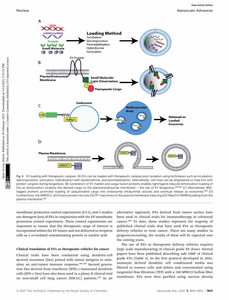
Fig. 4 EV loading with therapeutic cargoes. (A) EVs can be loaded with therapeutic cargoes post-isolation using techniques such as incubation,electroporation, sonication, hybridization with lipofectamine, and permeabilization. Alternatively, cell lines can be engineered to load EVs withprotein cargoes during biogenesis. (B) Generation of EV marker and cargo fusion proteins enables light/ligand induced dimerization loading ofEVs as dimerization localizes the desired cargo to the plasma/endosomal membrane – the site of EV biogenesis.234,237 (C) Alternatively WW-tagged proteins promotes loading of ubiquitinated cargo into endosomal intraluminal vesicles and eventual release as exosomes.239 (D)Furthermore, the ARRDC1-p53 fusion protein recruits ESCRTmachinery to the plasmamembrane inducing p53 filled EV ARMMbudding from theplasma membrane.238
Review Nanoscale Advances
Ope
n A
cces
s A
rtic
le. P
ublis
hed
on 1
6 Fe
brua
ry 2
021.
Dow
nloa
ded
on 3
/13/
2022
3:1
5:53
PM
. T
his
artic
le is
lice
nsed
und
er a
Cre
ativ
e C
omm
ons
Attr
ibut
ion
3.0
Unp
orte
d L
icen
ce.
View Article Online
membrane protection control experiments (8/13), and 4 studiesuse detergent lysis of EVs in conjunction with the EVmembraneprotection control experiment. These control experiments areimportant to ensure that the therapeutic cargo of interest isincorporated within the EV lumen and not delivered to recipientcells as a co-isolated contaminating protein or nucleic acid.
Clinical translation of EVs as therapeutic vehicles for cancer
Clinical trials have been conducted using dendritic-cellderived exosomes (Dex) pulsed with tumor antigens to stim-ulate an anti-tumor immune response.241,242 Second genera-tion Dex derived from interferon (IFN)-g-maturated dendriticcells (IFN-g-Dex) have also been used in a phase II clinical trialon non-small cell lung cancer (NSCLC) patients.243 As an
© 2021 The Author(s). Published by the Royal Society of Chemistry
alternative approach, EVs derived from tumor ascites havebeen used in clinical trials for immunotherapy in colorectalcancer.244 To date, these studies represent the majority ofpublished clinical trials that have used EVs as therapeuticdelivery vehicles to treat cancer. There are many studies inprogress/recruiting, the results of these will be expected overthe coming years.
The use of EVs as therapeutic delivery vehicles requireslarge scale manufacturing of clinical grade EV doses. Severalpapers have been published describing safe GMP of clinicalgrade EVs (Table 5). In the rst protocol developed in 2002,monocyte derived dendritic cell conditioned media wasltered to remove cells and debris and concentrated usingtangential ow ltration (TFF) with a 500 MWCO hollow bermembrane. EVs were then puried using sucrose density
Nanoscale Adv., 2021, 3, 1830–1852 | 1841
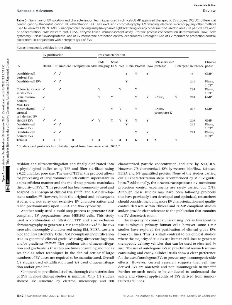
Table 5 Summary of EV isolation and characterization techniques used in clinical/cGMP approved therapeutic EV studies. DC/UC: differentialcentrifugation/ultracentrifugation. UF: ultrafiltration. SEC: size exclusion chromatography. EM/imaging: electron microscopy/any other methodused to visualize EVs. NTA/DLS: nanoparticle tracking analysis/dynamic light scattering (or any other method used to measure particle size and/or concentration). WB: western blot. ELISA: enzyme linked immunosorbent assay. Protein: protein concentration determination. Flow: flowcytometry. RNase/DNase/protease: use of EV membrane protection control experiments. Detergent: use of EV membrane protection controlexperiment in conjunction with detergent lysis of EVs
EVs as therapeutic vehicles in the clinic
EV
EV purication EV characterization
ReferenceClinicalphaseDC/UC UF Gradient Precipitation SEC
EM/imaging
NTA/DLS WB ELISA Protein Flow
DNase/RNase/protease Detergent
Dendritic cellderived EVs
3 3 Y Y Y 75 GMPa
Dendritic cell EVs 3 3 Y 241 Phase,1 CTa
Colorectal cancerascites EVs
3 3 Y Y Y 244 Phase,1 CT
Bone marrowderivedMSC EVs
3 Y Y Y Y RNase, Y 248 GMP
Mesenchymalstromalcell derived EV
3 Y Y Y RNase,proteinase K
247 GMP
Hek293 EVs 3 3 3 Y Y Y Y Y Y 246 GMPDendritic cellderived EVs
3 3 Y 242 Phase,1 CTa
Dendritic cellderived EVs
3 3 Y Y 243 Phase,2 CTa
Total: 8 4 5 5 0 1 3 3 3 4 6 4 2 1
a Studies used protocols formulated/adapted from Lamparski et al., 2002.75
Nanoscale Advances Review
Ope
n A
cces
s A
rtic
le. P
ublis
hed
on 1
6 Fe
brua
ry 2
021.
Dow
nloa
ded
on 3
/13/
2022
3:1
5:53
PM
. T
his
artic
le is
lice
nsed
und
er a
Cre
ativ
e C
omm
ons
Attr
ibut
ion
3.0
Unp
orte
d L
icen
ce.
View Article Online
cushion and ultracentrifugation and nally dialtrated intoa physiological buffer using TFF and lter sterilized usinga 0.22 mm lter pore size. The use of TFF in the protocol allowsfor processing of large volumes of cell culture supernatant ina time efficient manner and the multi-step process maximizesthe purity of EVs.75 This protocol has been commonly used andadapted in subsequent clinical trials241–243 and GMP develop-ment studies.245 However, both the original and subsequentstudies did not carry out extensive EV characterization andrelied predominantly upon ELISA and ow cytometry.
Another study used a multi-step process to generate GMPcompliant EV preparations from HEK293 cells. This studyused a combination of ltration, TFF and size exclusionchromatography to generate GMP compliant EVs.246 The EVswere also thoroughly characterized using EM, ELISA, westernblot and ow cytometry. Other GMP compliant EV puricationstudies generated clinical grade EVs using ultracentrifugationand/or gradients.244,247,248 The problem with ultracentrifuga-tion and gradients is that they are time consuming and not asscalable as other techniques in the clinical setting if largenumbers of EV doses are required to be manufactured. Overall5/8 studies used ultraltration and 8/8 used ultracentrifuga-tion and/or gradient.
Compared to pre-clinical studies, thorough characterizationof EVs in most clinical studies is minimal. Only 3/8 studiesshowed EV structure by electron microscopy and 3/8
1842 | Nanoscale Adv., 2021, 3, 1830–1852
characterized particle concentration and size by NTA/DLS.However, 7/8 characterized EVs by western blot/ow, 4/8 usedELISA and 6/8 quantied protein. None of the studies carriedout all characterization steps recommended by MISEV guide-lines.72 Additionally, the RNase/DNase/protease EV membraneprotection control experiments are rarely carried out (2/8).Although these studies may have been following protocolsthat have previously been developed and optimized, researchersshould consider includingmore EV characterization and qualitycontrol datasets within clinical and cGMP compliant studiesand/or provide clear reference to the publication that containsthe EV characterization.
The majority of clinical studies using EVs as therapeuticsuse autologous primary human cells however some GMPstudies have explored the purication of clinical grade EVsfrom cell lines. This is a stark contrast to pre-clinical studieswhere the majority of studies use human cell lines to generatetherapeutic delivery vehicles that can be used in vitro and invivo. The use of autologous EVs in pre-clinical research is timeconsuming and costly. Clinical trials show a clear preferencefor the use of autologous EVs to prevent any immunogenic sideeffects. However, current research suggests that cell linederived EVs are non-toxic and non-immunogenic in vivo.63,64
Further research needs to be conducted to understand thesafety and clinical applicability of EVs derived from immor-talized cell lines.
© 2021 The Author(s). Published by the Royal Society of Chemistry
Review Nanoscale Advances
Ope
n A
cces
s A
rtic
le. P
ublis
hed
on 1
6 Fe
brua
ry 2
021.
Dow
nloa
ded
on 3
/13/
2022
3:1
5:53
PM
. T
his
artic
le is
lice
nsed
und
er a
Cre
ativ
e C
omm
ons
Attr
ibut
ion
3.0
Unp
orte
d L
icen
ce.
View Article Online
The need for standardization ofextracellular vesicle isolation andcharacterization protocols for clinicalapplications
Our review of the literature investigates the most common EVisolation and characterization techniques that are used by pre-clinical and clinical studies. The review has uncovered impor-tant points regarding what we should be doing as a scienticcommunity when isolating and characterizing our EVs.
Identication of the optimal EV isolation protocol for pre-clinical studies is essential and dependent upon the uid andvolume from which EVs are being isolated. We conclude that itis possible to use any isolation technique deemed necessary(most frequently used was ultracentrifugation and precipita-tion) as long as characterization steps are employed: electronmicroscopy (or alternative), western blot/ow for EV markersand contaminants, EV preparation quantitation (proteinconcentration quantication for example) and nanoparticletracking analysis (or alternative technology). In addition, thenumber of studies employing RNase/DNase/protease EVmembrane protection control experiments were surprisinglylow. Performing these simple experiments, would providestrong evidence that the biomarker being investigated or thetherapeutic cargo loaded is in fact EV-associated/enclosed.Thorough EV characterization and EV membrane protectioncontrol experiments are important for the identication of bonade EV cargoes; particularly given that other particles in bio-logical uids likely reside in EV preparations. These otherparticles may also have biomarker utility as has been shown forthe extracellular particle termed exomere.249,250 Exomeres lacka lipid bilayer and likely arise through a distinct and differentbiogenesis pathway from EVs. Exomeres have a physiologicalrole and have demonstrated potential as biomarkers forcancer.249,250 It is likely that all extracellular particles (such asEVs and exomeres) have potential to support the developmentof new diagnostic and prognostic testing. However, thoroughcharacterization of the particle that one is working with willimprove our understanding of the cargoes that they carry andthe biological functions that they may have, enhance ourunderstanding of small particles, and support studyreproducibility.
For EV isolation in a clinical setting more cGMP approvedtechnologies for small scale and large scale EV isolation need tobe developed. Large scale isolation of EV therapeutic deliveryvehicles in the clinic usemulti-stepmethods to isolate pure EVs.However most methods include an ultracentrifugation and/orgradient step which are not easily scaled up and thereforethesemethodsmay not be suitable for large scale isolation of EVtherapeutics in the clinic. Ultraltration technologies such asTFF can concentrate and isolate EVs at a large scale, buta second purication step is necessary. Therefore, other EVpurication strategies that can isolate EVs in a large scale,quickly with minimal hands on input need to be developed. EVcharacterization in clinical studies oen relies upon past
© 2021 The Author(s). Published by the Royal Society of Chemistry
publications using the same or similar isolation strategy.Improved reporting on past study EV characterization andinclusion of EV quality control and characterization would bebenecial in clinical studies.
Conclusion
Is there a need for EV purication and characterization stan-dardization? One could argue that there is not a need forstandardization of EV purication protocols for pre-clinical andclinical studies, as long as puried EVs are thoroughly charac-terized and controlled. There is plenty of room for technologydevelopment in terms of EV isolation and purication forbiomarker analysis and for therapeutic delivery vehicle devel-opment. MISEV recommend that EVs are characterized on thesingle molecule level using two techniques (electronmicroscopy/other imaging techniques and by a particle analysistechnology such as NTA/DLS), analyzed for EV markers andabsence of contaminants (western blot/ow cytometry), andthat the EV preparation should be quantitated (proteinconcentration or protein concentration to particle numberratio).72 Currently a large proportion of pre-clinical and clinicalstudies still do not fully characterize the isolated EVs. EVcharacterization should be standardized and become routinewithin the eld. Additionally, the use of the RNase/DNase/protease EV membrane protection control experiment shouldalso become a new routine standard control experimentincluded in all EV biomarker and therapeutic loading studies.Without these simple controls, there is no way the reader canknow if the cargo/biomarker of interest reported by the study isactually EV associated or is rather a co-isolated contaminant.
Author contributions
NS and KCW conceptualized review. NS draed the manuscript.NS and KCW reviewed and edited the manuscript. KCWgenerated manuscript gures.
Conflicts of interest
There are no conicts of interest to declare
Acknowledgements
This work was supported by the Department of DefenseCongressionally Directed Medical Research Programs, BreastCancer Research Program (W81XWH-16-1-0525) and theNatural Sciences and Engineering Research Council of Canada(NSERC) Discovery Grants program. K. C. W. holds a Tier 2Canada Research Chair and a Michael Smith Foundation forHealth Research Scholar award.
References
1 G. van Niel, G. D'Angelo and G. Raposo, Shedding light onthe cell biology of extracellular vesicles, Nat. Rev. Mol. CellBiol., 2018, 19, 213–228, DOI: 10.1038/nrm.2017.125.
Nanoscale Adv., 2021, 3, 1830–1852 | 1843
Nanoscale Advances Review
Ope
n A
cces
s A
rtic
le. P
ublis
hed
on 1
6 Fe
brua
ry 2
021.
Dow
nloa
ded
on 3
/13/
2022
3:1
5:53
PM
. T
his
artic
le is
lice
nsed
und
er a
Cre
ativ
e C
omm
ons
Attr
ibut
ion
3.0
Unp
orte
d L
icen
ce.
View Article Online
2 B. T. Pan, K. Teng, C. Wu, M. Adam and R. M. Johnstone,Electron microscopic evidence for externalization of thetransferrin receptor in vesicular form in sheepreticulocytes, J. Cell Biol., 1985, 101, 942–948, DOI:10.1083/jcb.101.3.942.
3 C. Harding, J. Heuser and P. Stahl, Receptor-mediatedendocytosis of transferrin and recycling of the transferrinreceptor in rat reticulocytes, J. Cell Biol., 1983, 97, 329–339, DOI: 10.1083/jcb.97.2.329.
4 C. Tricarico, J. Clancy and C. D'Souza-Schorey, Biology andbiogenesis of shed microvesicles, Small GTPases, 2017, 8,220–232, DOI: 10.1080/21541248.2016.1215283.
5 D. Di Vizio, et al., Large oncosomes in human prostatecancer tissues and in the circulation of mice withmetastatic disease, Am. J. Pathol., 2012, 181, 1573–1584,DOI: 10.1016/j.ajpath.2012.07.030.
6 K. Al-Nedawi, et al., Intercellular transfer of the oncogenicreceptor EGFRvIII by microvesicles derived from tumourcells, Nat. Cell Biol., 2008, 10, 619–624, DOI: 10.1038/ncb1725.
7 T. H. Lee, et al., Microvesicles as mediators of intercellularcommunication in cancer–the emerging science of cellular'debris', Semin. Immunopathol., 2011, 33, 455–467, DOI:10.1007/s00281-011-0250-3.
8 G. K. Atkin-Smith, et al., A novel mechanism of generatingextracellular vesicles during apoptosis via a beads-on-a-string membrane structure, Nat. Commun., 2015, 6, 7439,DOI: 10.1038/ncomms8439.
9 M. Hristov, W. Erl, S. Linder and P. C. Weber, Apoptoticbodies from endothelial cells enhance the number andinitiate the differentiation of human endothelialprogenitor cells in vitro, Blood, 2004, 104, 2761–2766, DOI:10.1182/blood-2003-10-3614.
10 H. Valadi, et al., Exosome-mediated transfer of mRNAs andmicroRNAs is a novel mechanism of genetic exchangebetween cells, Nat. Cell Biol., 2007, 9, 654–659, DOI:10.1038/ncb1596.
11 E. N. Nolte-'t Hoen, et al., Deep sequencing of RNA fromimmune cell-derived vesicles uncovers the selectiveincorporation of small non-coding RNA biotypes withpotential regulatory functions, Nucleic Acids Res., 2012, 40,9272–9285, DOI: 10.1093/nar/gks658.
12 J. Skog, et al., Glioblastoma microvesicles transport RNAand proteins that promote tumour growth and providediagnostic biomarkers, Nat. Cell Biol., 2008, 10, 1470–1476, DOI: 10.1038/ncb1800.
13 J. Ratajczak, et al., Embryonic stem cell-derivedmicrovesicles reprogram hematopoietic progenitors:evidence for horizontal transfer of mRNA and proteindelivery, Leukemia, 2006, 20, 847–856, DOI: 10.1038/sj.leu.2404132.
14 B. K. Thakur, et al., Double-stranded DNA in exosomes:a novel biomarker in cancer detection, Cell Res., 2014, 24,766–769, DOI: 10.1038/cr.2014.44.
15 C. Kahlert, et al., Identication of double-stranded genomicDNA spanning all chromosomes with mutated KRAS andp53 DNA in the serum exosomes of patients with
1844 | Nanoscale Adv., 2021, 3, 1830–1852
pancreatic cancer, J. Biol. Chem., 2014, 289, 3869–3875,DOI: 10.1074/jbc.C113.532267.
16 C. Thery, et al., Proteomic analysis of dendritic cell-derivedexosomes: a secreted subcellular compartment distinctfrom apoptotic vesicles, J. Immunol., 2001, 166, 7309–7318, DOI: 10.4049/jimmunol.166.12.7309.
17 J. F. Brouwers, et al., Distinct lipid compositions of twotypes of human prostasomes, Proteomics, 2013, 13, 1660–1666, DOI: 10.1002/pmic.201200348.
18 R. A. Haraszti, et al., High-resolution proteomic andlipidomic analysis of exosomes and microvesicles fromdifferent cell sources, J. Extracell. Vesicles, 2016, 5, 32570,DOI: 10.3402/jev.v5.32570.
19 K. Trajkovic, et al., Ceramide triggers budding of exosomevesicles into multivesicular endosomes, Science, 2008,319, 1244–1247, DOI: 10.1126/science.1153124.
20 A. Llorente, et al., Molecular lipidomics of exosomesreleased by PC-3 prostate cancer cells, Biochim. Biophys.Acta, 2013, 1831, 1302–1309, DOI: 10.1016/j.bbalip.2013.04.011.
21 X. Luo, M. An, K. C. Cuneo, D. M. Lubman and L. Li, High-Performance Chemical Isotope Labeling LiquidChromatography Mass Spectrometry for ExosomeMetabolomics, Anal. Chem., 2018, 90, 8314–8319, DOI:10.1021/acs.analchem.8b01726.
22 M. Clos-Garcia, et al., Metabolic alterations in urineextracellular vesicles are associated to prostate cancerpathogenesis and progression, J. Extracell. Vesicles, 2018,7, 1470442, DOI: 10.1080/20013078.2018.1470442.
23 C. Williams, et al., Assessing the role of surface glycans ofextracellular vesicles on cellular uptake, Sci. Rep., 2019, 9,11920, DOI: 10.1038/s41598-019-48499-1.
24 C. Williams, et al., Glycosylation of extracellular vesicles:current knowledge, tools and clinical perspectives, J.Extracell. Vesicles, 2018, 7, 1442985, DOI: 10.1080/20013078.2018.1442985.
25 Y. Liang, et al., Complex N-linked glycans serve asa determinant for exosome/microvesicle cargorecruitment, J. Biol. Chem., 2014, 289, 32526–32537, DOI:10.1074/jbc.M114.606269.
26 R. M. Johnstone, M. Adam, J. R. Hammond, L. Orr andC. Turbide, Vesicle formation during reticulocytematuration. Association of plasma membrane activitieswith released vesicles (exosomes), J. Biol. Chem., 1987,262, 9412–9420.
27 M. Mathieu, L. Martin-Jaular, G. Lavieu and C. Thery,Specicities of secretion and uptake of exosomes andother extracellular vesicles for cell-to-cell communication,Nat. Cell Biol., 2019, 21, 9–17, DOI: 10.1038/s41556-018-0250-9.
28 J. Maia, S. Caja, M. C. StranoMoraes, N. Couto and B. Costa-Silva, Exosome-Based Cell-Cell Communication in theTumor Microenvironment, Front. Cell Dev. Biol., 2018, 6,18, DOI: 10.3389/fcell.2018.00018.
29 M. P. Caby, D. Lankar, C. Vincendeau-Scherrer, G. Raposoand C. Bonnerot, Exosomal-like vesicles are present in
© 2021 The Author(s). Published by the Royal Society of Chemistry
Review Nanoscale Advances
Ope
n A
cces
s A
rtic
le. P
ublis
hed
on 1
6 Fe
brua
ry 2
021.
Dow
nloa
ded
on 3
/13/
2022
3:1
5:53
PM
. T
his
artic
le is
lice
nsed
und
er a
Cre
ativ
e C
omm
ons
Attr
ibut
ion
3.0
Unp
orte
d L
icen
ce.
View Article Online
human blood plasma, Int. Immunol., 2005, 17, 879–887,DOI: 10.1093/intimm/dxh267.
30 C. Lasser, et al., Human saliva, plasma and breast milkexosomes contain RNA: uptake by macrophages, J. Transl.Med., 2011, 9, 9, DOI: 10.1186/1479-5876-9-9.
31 T. Pisitkun, R. F. Shen and M. A. Knepper, Identicationand proteomic proling of exosomes in human urine,Proc. Natl. Acad. Sci. U. S. A., 2004, 101, 13368–13373,DOI: 10.1073/pnas.0403453101.
32 A. Zomer, et al., In Vivo imaging reveals extracellularvesicle-mediated phenocopying of metastatic behavior,Cell, 2015, 161, 1046–1057, DOI: 10.1016/j.cell.2015.04.042.
33 I. Wortzel, S. Dror, C. M. Kenic and D. Lyden, Exosome-Mediated Metastasis: Communication from a Distance,Dev. Cell, 2019, 49, 347–360, DOI: 10.1016/j.devcel.2019.04.011.
34 A. Hoshino, et al., Tumour exosome integrins determineorganotropic metastasis, Nature, 2015, 527, 329–335, DOI:10.1038/nature15756.
35 B. Costa-Silva, et al., Pancreatic cancer exosomes initiatepre-metastatic niche formation in the liver, Nat. Cell Biol.,2015, 17, 816–826, DOI: 10.1038/ncb3169.
36 H. Peinado, et al., Melanoma exosomes educate bonemarrow progenitor cells toward a pro-metastaticphenotype through MET, Nat. Med., 2012, 18, 883–891,DOI: 10.1038/nm.2753.
37 G. Raposo, et al., B lymphocytes secrete antigen-presentingvesicles, J. Exp. Med., 1996, 183, 1161–1172, DOI: 10.1084/jem.183.3.1161.
38 I. Del Conde, C. N. Shrimpton, P. Thiagarajan andJ. A. Lopez, Tissue-factor-bearing microvesicles arise fromlipid ras and fuse with activated platelets to initiatecoagulation, Blood, 2005, 106, 1604–1611, DOI: 10.1182/blood-2004-03-1095.
39 A. Brill, O. Dashevsky, J. Rivo, Y. Gozal and D. Varon,Platelet-derived microparticles induce angiogenesis andstimulate post-ischemic revascularization, Cardiovasc.Res., 2005, 67, 30–38, DOI: 10.1016/j.cardiores.2005.04.007.
40 J. Howitt and A. F. Hill, Exosomes in the Pathology ofNeurodegenerative Diseases, J. Biol. Chem., 2016, 291,26589–26597, DOI: 10.1074/jbc.R116.757955.
41 A. Ibrahim and E. Marban, Exosomes: FundamentalBiology and Roles in Cardiovascular Physiology, Annu.Rev. Physiol., 2016, 78, 67–83, DOI: 10.1146/annurev-physiol-021115-104929.
42 B. D. Chan, et al., Exosomes in Inammation andInammatory Disease, Proteomics, 2019, 19, e1800149,DOI: 10.1002/pmic.201800149.
43 A. Becker, et al., Extracellular Vesicles in Cancer: Cell-to-Cell Mediators of Metastasis, Cancer Cell, 2016, 30, 836–848, DOI: 10.1016/j.ccell.2016.10.009.
44 C. Ciardiello, et al., Focus on Extracellular Vesicles: NewFrontiers of Cell-to-Cell Communication in Cancer, Int. J.Mol. Sci., 2016, 17, 175, DOI: 10.3390/ijms17020175.
45 F. Fanini and M. Fabbri, Cancer-derived exosomicmicroRNAs shape the immune system within the tumor
© 2021 The Author(s). Published by the Royal Society of Chemistry
microenvironment: State of the art, Semin. Cell Dev. Biol.,2017, 67, 23–28, DOI: 10.1016/j.semcdb.2016.12.004.
46 C. Sheehan and C. D’Souza-Schorey, Tumor-derivedextracellular vesicles: molecular parcels that enableregulation of the immune response in cancer, J. Cell Sci.,2019, 132(20), DOI: 10.1242/jcs.235085.
47 P. D. Robbins and A. E. Morelli, Regulation of immuneresponses by extracellular vesicles, Nat. Rev. Immunol.,2014, 14, 195–208, DOI: 10.1038/nri3622.
48 D. W. Greening, S. K. Gopal, R. Xu, R. J. Simpson andW. Chen, Exosomes and their roles in immune regulationand cancer, Semin. Cell Dev. Biol., 2015, 40, 72–81, DOI:10.1016/j.semcdb.2015.02.009.
49 N. M. Namee and L. O'Driscoll, Extracellular vesicles andanti-cancer drug resistance, Biochim. Biophys. Acta, Rev.Cancer, 2018, 1870, 123–136, DOI: 10.1016/j.bbcan.2018.07.003.
50 E. G. Trams, C. J. Lauter, N. Salem Jr and U. Heine,Exfoliation of membrane ecto-enzymes in the form ofmicro-vesicles, Biochim. Biophys. Acta, 1981, 645, 63–70,DOI: 10.1016/0005-2736(81)90512-5.
51 L. Zitvogel, et al., Eradication of established murine tumorsusing a novel cell-free vaccine: dendritic cell-derivedexosomes, Nat. Med., 1998, 4, 594–600, DOI: 10.1038/nm0598-594.
52 J. Wolfers, et al., Tumor-derived exosomes are a source ofshared tumor rejection antigens for CTL cross-priming,Nat. Med., 2001, 7, 297–303, DOI: 10.1038/85438.
53 A. J. Abusamra, et al., Tumor exosomes expressing Fasligand mediate CD8+ T-cell apoptosis, Blood Cells, Mol.,Dis., 2005, 35, 169–173, DOI: 10.1016/j.bcmd.2005.07.001.
54 A. Clayton, S. Al-Taei, J. Webber, M. D. Mason and Z. Tabi,Cancer exosomes express CD39 and CD73, which suppressT cells through adenosine production, J. Immunol., 2011,187, 676–683, DOI: 10.4049/jimmunol.1003884.
55 C. Liu, et al., Murine mammary carcinoma exosomespromote tumor growth by suppression of NK cellfunction, J. Immunol., 2006, 176, 1375–1385, DOI: 10.4049/jimmunol.176.3.1375.
56 X. Xiang, et al., Induction of myeloid-derived suppressorcells by tumor exosomes, Int. J. Cancer, 2009, 124, 2621–2633, DOI: 10.1002/ijc.24249.
57 T. Cooks, et al., Mutant p53 cancers reprogrammacrophages to tumor supporting macrophages viaexosomal miR-1246, Nat. Commun., 2018, 9, 771, DOI:10.1038/s41467-018-03224-w.
58 D. K. Jeppesen, et al., Quantitative proteomics offractionated membrane and lumen exosome proteinsfrom isogenic metastatic and nonmetastatic bladdercancer cells reveal differential expression of EMT factors,Proteomics, 2014, 14, 699–712, DOI: 10.1002/pmic.201300452.
59 T. Vagner, et al., Large extracellular vesicles carry most ofthe tumour DNA circulating in prostate cancer patientplasma, J. Extracell. Vesicles, 2018, 7, 1505403, DOI:10.1080/20013078.2018.1505403.
Nanoscale Adv., 2021, 3, 1830–1852 | 1845
Nanoscale Advances Review
Ope
n A
cces
s A
rtic
le. P
ublis
hed
on 1
6 Fe
brua
ry 2
021.
Dow
nloa
ded
on 3
/13/
2022
3:1
5:53
PM
. T
his
artic
le is
lice
nsed
und
er a
Cre
ativ
e C
omm
ons
Attr
ibut
ion
3.0
Unp
orte
d L
icen
ce.
View Article Online
60 K. B. Johnsen, J. M. Gudbergsson, T. L. Andresen andJ. B. Simonsen, What is the blood concentration ofextracellular vesicles? Implications for the use ofextracellular vesicles as blood-borne biomarkers ofcancer, Biochim. Biophys. Acta, Rev. Cancer, 2019, 1871,109–116, DOI: 10.1016/j.bbcan.2018.11.006.
61 Q. Ge, et al., miRNA in plasma exosome is stable underdifferent storage conditions, Molecules, 2014, 19, 1568–1575, DOI: 10.3390/molecules19021568.
62 Y. Cheng, Q. Zeng, Q. Han and W. Xia, Effect of pH,temperature and freezing-thawing on quantity changesand cellular uptake of exosomes, Protein Cell, 2019, 10,295–299, DOI: 10.1007/s13238-018-0529-4.
63 X. Zhu, et al., Comprehensive toxicity and immunogenicitystudies reveal minimal effects in mice following sustaineddosing of extracellular vesicles derived from HEK293Tcells, J. Extracell. Vesicles, 2017, 6, 1324730, DOI: 10.1080/20013078.2017.1324730.
64 A. F. Saleh, et al., Extracellular vesicles induce minimalhepatotoxicity and immunogenicity, Nanoscale, 2019, 11,6990–7001, DOI: 10.1039/c8nr08720b.
65 C. Thery, S. Amigorena, G. Raposo and A. Clayton, Isolationand characterization of exosomes from cell culturesupernatants and biological uids, Curr. Protoc. Cell Biol.,2006, 3(3.22), DOI: 10.1002/0471143030.cb0322s30.
66 A. Cvjetkovic, J. Lotvall and C. Lasser, The inuence of rotortype and centrifugation time on the yield and purity ofextracellular vesicles, J. Extracell. Vesicles, 2014, 3, DOI:10.3402/jev.v3.23111.
67 F. Momen-Heravi, et al., Impact of biouid viscosity on sizeand sedimentation efficiency of the isolated microvesicles,Front. Physiol., 2012, 3, 162, DOI: 10.3389/fphys.2012.00162.
68 N. Karimi, et al., Detailed analysis of the plasmaextracellular vesicle proteome aer separation fromlipoproteins, Cell. Mol. Life Sci., 2018, 75, 2873–2886, DOI:10.1007/s00018-018-2773-4.
69 B. W. Sodar, et al., Low-density lipoprotein mimics bloodplasma-derived exosomes and microvesicles duringisolation and detection, Sci. Rep., 2016, 6, 24316, DOI:10.1038/srep24316.
70 Y. Yuana, J. Levels, A. Grootemaat, A. Sturk andR. Nieuwland, Co-isolation of extracellular vesicles andhigh-density lipoproteins using density gradientultracentrifugation, J. Extracell. Vesicles, 2014, 3, DOI:10.3402/jev.v3.23262.
71 K. Li, D. K. Wong, K. Y. Hong and R. L. Raffai, Cushioned-Density Gradient Ultracentrifugation (C-DGUC): A Renedand High Performance Method for the Isolation,Characterization, and Use of Exosomes, Methods Mol.Biol., 2018, 1740, 69–83, DOI: 10.1007/978-1-4939-7652-2_7.
72 C. Thery, et al., Minimal information for studies ofextracellular vesicles 2018 (MISEV2018): a positionstatement of the International Society for ExtracellularVesicles and update of the MISEV2014 guidelines, J.Extracell. Vesicles, 2018, 7, 1535750, DOI: 10.1080/20013078.2018.1535750.
1846 | Nanoscale Adv., 2021, 3, 1830–1852
73 R. A. Haraszti, et al., Exosomes Produced from 3D Culturesof MSCs by Tangential Flow Filtration Show Higher Yieldand Improved Activity, Mol. Ther., 2018, 26, 2838–2847,DOI: 10.1016/j.ymthe.2018.09.015.
74 G. Corso, et al., Reproducible and scalable purication ofextracellular vesicles using combined bind-elute and sizeexclusion chromatography, Sci. Rep., 2017, 7, 11561, DOI:10.1038/s41598-017-10646-x.
75 H. G. Lamparski, et al., Production and characterization ofclinical grade exosomes derived from dendritic cells, J.Immunol. Methods, 2002, 270, 211–226, DOI: 10.1016/s0022-1759(02)00330-7.
76 M. L. Heinemann and J. Vykoukal, Sequential Filtration: AGentle Method for the Isolation of FunctionalExtracellular Vesicles, Methods Mol. Biol., 2017, 1660, 33–41, DOI: 10.1007/978-1-4939-7253-1_4.
77 A. N. Boing, et al., Single-step isolation of extracellularvesicles by size-exclusion chromatography, J. Extracell.Vesicles, 2014, 3, DOI: 10.3402/jev.v3.23430.
78 R. Stranska, et al., Comparison of membrane affinity-basedmethod with size-exclusion chromatography for isolation ofexosome-like vesicles from human plasma, J. Transl. Med.,2018, 16, 1, DOI: 10.1186/s12967-017-1374-6.
79 W. Nakai, et al., A novel affinity-based method for theisolation of highly puried extracellular vesicles, Sci. Rep.,2016, 6, 33935, DOI: 10.1038/srep33935.
80 L. Balaj, et al., Heparin affinity purication of extracellularvesicles, Sci. Rep., 2015, 5, 10266, DOI: 10.1038/srep10266.
81 B. J. Tauro, et al., Comparison of ultracentrifugation,density gradient separation, and immunoaffinity capturemethods for isolating human colon cancer cell lineLIM1863-derived exosomes, Methods, 2012, 56, 293–304,DOI: 10.1016/j.ymeth.2012.01.002.
82 M. P. Oksvold, A. Neurauter and K. W. Pedersen, Magneticbead-based isolation of exosomes,Methods Mol. Biol., 2015,1218, 465–481, DOI: 10.1007/978-1-4939-1538-5_27.
83 N. Heath, et al., Rapid isolation and enrichment ofextracellular vesicle preparations using anion exchangechromatography, Sci. Rep., 2018, 8, 5730, DOI: 10.1038/s41598-018-24163-y.
84 M. Kosanovic, B. Milutinovic, S. Goc, N. Mitic andM. Jankovic, Ion-exchange chromatography purication ofextracellular vesicles, Biotechniques, 2017, 63, 65–71, DOI:10.2144/000114575.
85 A. K. Ludwig, et al., Precipitation with polyethylene glycolfollowed by washing and pelleting by ultracentrifugationenriches extracellular vesicles from tissue culturesupernatants in small and large scales, J. Extracell.Vesicles, 2018, 7, 1528109, DOI: 10.1080/20013078.2018.1528109.
86 M. Ding, et al., Comparison of commercial exosomeisolation kits for circulating exosomal microRNAproling, Anal. Bioanal. Chem., 2018, 410, 3805–3814,DOI: 10.1007/s00216-018-1052-4.
87 M. Macias, et al., Comparison of six commercial serumexosome isolation methods suitable for clinical
© 2021 The Author(s). Published by the Royal Society of Chemistry

Review Nanoscale Advances
Ope
n A
cces
s A
rtic
le. P
ublis
hed
on 1
6 Fe
brua
ry 2
021.
Dow
nloa
ded
on 3
/13/
2022
3:1
5:53
PM
. T
his
artic
le is
lice
nsed
und
er a
Cre
ativ
e C
omm
ons
Attr
ibut
ion
3.0
Unp
orte
d L
icen
ce.
View Article Online
laboratories. Effect in cytokine analysis, Clin. Chem. Lab.Med., 2019, 57, 1539–1545, DOI: 10.1515/cclm-2018-1297.
88 A. Gamez-Valero, et al., Size-Exclusion Chromatography-based isolation minimally alters Extracellular Vesicles'characteristics compared to precipitating agents, Sci. Rep.,2016, 6, 33641, DOI: 10.1038/srep33641.
89 L. Paolini, et al., Residual matrix from different separationtechniques impacts exosome biological activity, Sci. Rep.,2016, 6, 23550, DOI: 10.1038/srep23550.
90 R. Vaidyanathan, et al., Detecting exosomes specically:a multiplexed device based on alternating currentelectrohydrodynamic induced nanoshearing, Anal. Chem.,2014, 86, 11125–11132, DOI: 10.1021/ac502082b.
91 E. Reategui, et al., Engineered nanointerfaces formicrouidic isolation and molecular proling of tumor-specic extracellular vesicles, Nat. Commun., 2018, 9, 175,DOI: 10.1038/s41467-017-02261-1.
92 Y. G. Zhou, et al., Interrogating CirculatingMicrosomes andExosomes Using Metal Nanoparticles, Small, 2016, 12, 727–732, DOI: 10.1002/smll.201502365.
93 S. M. Santana, M. A. Antonyak, R. A. Cerione and B. J. Kirby,Microuidic isolation of cancer-cell-derived microvesiclesfrom hetergeneous extracellular shed vesicle populations,Biomed. Microdevices, 2014, 16, 869–877, DOI: 10.1007/s10544-014-9891-z.
94 R. A. Dragovic, et al., Sizing and phenotyping of cellularvesicles using Nanoparticle Tracking Analysis,Nanomedicine, 2011, 7, 780–788, DOI: 10.1016/j.nano.2011.04.003.
95 J. de Vrij, et al., Quantication of nanosized extracellularmembrane vesicles with scanning ion occlusion sensing,Nanomedicine, 2013, 8, 1443–1458, DOI: 10.2217/nnm.12.173.
96 J. C. Akers, et al., Comparative Analysis of Technologies forQuantifying Extracellular Vesicles (EVs) in ClinicalCerebrospinal Fluids (CSF), PLoS One, 2016, 11, e0149866,DOI: 10.1371/journal.pone.0149866.
97 S. L. Maas, M. L. Broekman and J. de Vrij, Tunable ResistivePulse Sensing for the Characterization of ExtracellularVesicles, Methods Mol. Biol., 2017, 1545, 21–33, DOI:10.1007/978-1-4939-6728-5_2.
98 R. Szatanek, et al., The Methods of Choice for ExtracellularVesicles (EVs) Characterization, Int. J. Mol. Sci., 2017, 18(6),1153, DOI: 10.3390/ijms18061153.
99 J. Webber and A. Clayton, How pure are your vesicles?, J.Extracell. Vesicles, 2013, 2, DOI: 10.3402/jev.v2i0.19861.
100 Y. Yuana, et al., Cryo-electron microscopy of extracellularvesicles in fresh plasma, J. Extracell. Vesicles, 2013, 2,DOI: 10.3402/jev.v2i0.21494.
101 L. G. Rikkert, R. Nieuwland, L. Terstappen andF. A. W. Coumans, Quality of extracellular vesicle imagesby transmission electron microscopy is operator andprotocol dependent, J. Extracell. Vesicles, 2019, 8, 1555419,DOI: 10.1080/20013078.2018.1555419.
102 A. Clayton, et al., Analysis of antigen presenting cell derivedexosomes, based on immuno-magnetic isolation and ow
© 2021 The Author(s). Published by the Royal Society of Chemistry
cytometry, J. Immunol. Methods, 2001, 247, 163–174, DOI:10.1016/s0022-1759(00)00321-5.
103 H. Suarez, et al., A bead-assisted ow cytometry method forthe semi-quantitative analysis of Extracellular Vesicles, Sci.Rep., 2017, 7, 11271, DOI: 10.1038/s41598-017-11249-2.
104 A. Morales-Kastresana, et al., High-delity detection andsorting of nanoscale vesicles in viral disease and cancer,J. Extracell. Vesicles, 2019, 8, 1597603, DOI: 10.1080/20013078.2019.1597603.
105 J. P. Nolan and J. C. Jones, Detection of platelet vesicles byow cytometry, Platelets, 2017, 28, 256–262, DOI: 10.1080/09537104.2017.1280602.
106 G. C. t. Brittain, et al., A Novel Semiconductor-Based FlowCytometer with Enhanced Light-Scatter Sensitivity for theAnalysis of Biological Nanoparticles, Sci. Rep., 2019, 9,16039, DOI: 10.1038/s41598-019-52366-4.
107 N. Salmond, K. Khanna, G. R. Owen and K. C. Williams,Nanoscale ow cytometry for immunophenotyping andquantitating extracellular vesicles in blood plasma,Nanoscale, 2021, 13, 2012–2025, DOI: 10.1039/d0nr05525e.
108 A. Morales-Kastresana, et al., Labeling ExtracellularVesicles for Nanoscale Flow Cytometry, Sci. Rep., 2017, 7,1878, DOI: 10.1038/s41598-017-01731-2.
109 J. A. Welsh, et al., MIFlowCyt-EV: a framework forstandardized reporting of extracellular vesicle owcytometry experiments, J. Extracell. Vesicles, 2020, 9,1713526, DOI: 10.1080/20013078.2020.1713526.
110 J. C. Gross, V. Chaudhary, K. Bartscherer and M. Boutros,Active Wnt proteins are secreted on exosomes, Nat. CellBiol., 2012, 14, 1036–1045, DOI: 10.1038/ncb2574.
111 E. Lazaro-Ibanez, et al., DNA analysis of low- and high-density fractions denes heterogeneous subpopulationsof small extracellular vesicles based on their DNA cargoand topology, J. Extracell. Vesicles, 2019, 8, 1656993, DOI:10.1080/20013078.2019.1656993.
112 E. I. Buzas, E. A. Toth, B. W. Sodar and K. E. Szabo-Taylor,Molecular interactions at the surface of extracellularvesicles, Semin. Immunopathol., 2018, 40, 453–464, DOI:10.1007/s00281-018-0682-0.
113 J. Silva, et al., Analysis of exosome release and its prognosticvalue in human colorectal cancer, Genes ChromosomesCancer, 2012, 51, 409–418, DOI: 10.1002/gcc.21926.
114 Y. Matsumoto, et al., Quantication of plasma exosome isa potential prognostic marker for esophageal squamouscell carcinoma, Oncol. Rep., 2016, 36, 2535–2543, DOI:10.3892/or.2016.5066.
115 H. Xu, X. Dong, Y. Chen and X. Wang, Serum exosomalhnRNPH1 mRNA as a novel marker for hepatocellularcarcinoma, Clin. Chem. Lab. Med., 2018, 56, 479–484, DOI:10.1515/cclm-2017-0327.
116 H. Goldvaser, et al., Characterisation of blood-derivedexosomal hTERT mRNA secretion in cancer patients:a potential pan-cancer marker, Br. J. Cancer, 2017, 117,353–357, DOI: 10.1038/bjc.2017.166.
117 N. Fujii, et al., Extracellular miR-224 as a prognostic markerfor clear cell renal cell carcinoma, Oncotarget, 2017, 8,109877–109888, DOI: 10.18632/oncotarget.22436.
Nanoscale Adv., 2021, 3, 1830–1852 | 1847

Nanoscale Advances Review
Ope
n A
cces
s A
rtic
le. P
ublis
hed
on 1
6 Fe
brua
ry 2
021.
Dow
nloa
ded
on 3
/13/
2022
3:1
5:53
PM
. T
his
artic
le is
lice
nsed
und
er a
Cre
ativ
e C
omm
ons
Attr
ibut
ion
3.0
Unp
orte
d L
icen
ce.
View Article Online
118 Q. Liu, et al., Circulating exosomal microRNAs asprognostic biomarkers for non-small-cell lung cancer,Oncotarget, 2017, 8, 13048–13058, DOI: 10.18632/oncotarget.14369.
119 R. Kanaoka, et al., Usefulness of Plasma ExosomalMicroRNA-451a as a Noninvasive Biomarker for EarlyPrediction of Recurrence and Prognosis of Non-Small CellLung Cancer, Oncology, 2018, 94, 311–323, DOI: 10.1159/000487006.
120 F. Lan, et al., Serum exosomal miR-301a as a potentialdiagnostic and prognostic biomarker for human glioma,Cell. Oncol., 2018, 41, 25–33, DOI: 10.1007/s13402-017-0355-3.
121 W. Liu, et al., Serum exosomal miR-125b is a novelprognostic marker for hepatocellular carcinoma,OncoTargets Ther., 2017, 10, 3843–3851, DOI: 10.2147/OTT.S140062.
122 M. Shi, et al., Decreased levels of serum exosomal miR-638predict poor prognosis in hepatocellular carcinoma, J. Cell.Biochem., 2018, 119, 4711–4716, DOI: 10.1002/jcb.26650.
123 C. Pan, et al., Exosomal microRNAs as tumor markers inepithelial ovarian cancer, Mol. Oncol., 2018, 12, 1935–1948, DOI: 10.1002/1878-0261.12371.
124 X. Meng, et al., Diagnostic and prognostic relevance ofcirculating exosomal miR-373, miR-200a, miR-200b andmiR-200c in patients with epithelial ovarian cancer,Oncotarget, 2016, 7, 16923–16935, DOI: 10.18632/oncotarget.7850.
125 T. Goto, et al., An elevated expression of serum exosomalmicroRNA-191, - 21, -451a of pancreatic neoplasm isconsidered to be efficient diagnostic marker, BMC Cancer,2018, 18, 116, DOI: 10.1186/s12885-018-4006-5.
126 R. Que, G. Ding, J. Chen and L. Cao, Analysis of serumexosomal microRNAs and clinicopathologic features ofpatients with pancreatic adenocarcinoma, World J. Surg.Oncol., 2013, 11, 219, DOI: 10.1186/1477-7819-11-219.
127 T. Matsumura, et al., Exosomal microRNA in serum isa novel biomarker of recurrence in human colorectalcancer, Br. J. Cancer, 2015, 113, 275–281, DOI: 10.1038/bjc.2015.201.
128 C. Liu, et al., Serum exosomal miR-4772-3p is a predictor oftumor recurrence in stage II and III colon cancer,Oncotarget, 2016, 7, 76250–76260, DOI: 10.18632/oncotarget.12841.
129 M. Isin, et al., Exosomal lncRNA-p21 levels may help todistinguish prostate cancer from benign disease, Front.Genet., 2015, 6, 168, DOI: 10.3389/fgene.2015.00168.
130 L. Y. Lin, et al., Tumor-originated exosomal lncUEGC1 asa circulating biomarker for early-stage gastric cancer, Mol.Canc., 2018, 17, 84, DOI: 10.1186/s12943-018-0834-9.
131 R. Zhao, et al., Exosomal long noncoding RNA HOTTIP aspotential novel diagnostic and prognostic biomarker testfor gastric cancer, Mol. Canc., 2018, 17, 68, DOI: 10.1186/s12943-018-0817-x.
132 R. Zhang, et al., Serum long non coding RNA MALAT-1protected by exosomes is up-regulated and promotes cellproliferation and migration in non-small cell lung cancer,
1848 | Nanoscale Adv., 2021, 3, 1830–1852
Biochem. Biophys. Res. Commun., 2017, 490, 406–414, DOI:10.1016/j.bbrc.2017.06.055.
133 Y. Li, et al., Circular RNA is enriched and stable inexosomes: a promising biomarker for cancer diagnosis,Cell Res., 2015, 25, 981–984, DOI: 10.1038/cr.2015.82.
134 Z. Li, et al., Tumor-released exosomal circular RNA PDE8Apromotes invasive growth via the miR-338/MACC1/METpathway in pancreatic cancer, Canc. Lett., 2018, 432, 237–250, DOI: 10.1016/j.canlet.2018.04.035.
135 B. Sun, et al., Circulating exosomal CPNE3 as a diagnosticand prognostic biomarker for colorectal cancer, J. Cell.Physiol., 2019, 234, 1416–1425, DOI: 10.1002/jcp.26936.
136 E. Alegre, et al., Circulating melanoma exosomes asdiagnostic and prognosis biomarkers, Clin. Chim. Acta,2016, 454, 28–32, DOI: 10.1016/j.cca.2015.12.031.
137 S. Li, et al., Exosomal ephrinA2 derived from serum asa potential biomarker for prostate cancer, J. Cancer, 2018,9, 2659–2665, DOI: 10.7150/jca.25201.
138 M. K. S. Tang, et al., Soluble E-cadherin promotes tumorangiogenesis and localizes to exosome surface, Nat.Commun., 2018, 9, 2270, DOI: 10.1038/s41467-018-04695-7.
139 C. H. Wong and Y. C. Chen, Clinical signicance ofexosomes as potential biomarkers in cancer, World J ClinCases, 2019, 7, 171–190, DOI: 10.12998/wjcc.v7.i2.171.
140 F. Oehme, et al., Low level of exosomal long non-codingRNA HOTTIP is a prognostic biomarker in colorectalcancer, RNA Biol., 2019, 16, 1339–1345, DOI: 10.1080/15476286.2019.1637697.
141 S. Song, et al., Urinary exosome miR-30c-5p as a biomarkerof clear cell renal cell carcinoma that inhibits progressionby targeting HSPA5, J. Cell. Mol. Med., 2019, 23, 6755–6765, DOI: 10.1111/jcmm.14553.
142 Y. Zhao, et al., Long noncoding RNA LINC02418 regulatesMELK expression by acting as a ceRNA and may serve asa diagnostic marker for colorectal cancer, Cell Death Dis.,2019, 10, 568, DOI: 10.1038/s41419-019-1804-x.
143 Y. Tang, et al., Tumor-derived exosomal miRNA-320d asa biomarker for metastatic colorectal cancer, J. Clin. Lab.Anal., 2019, 33, e23004, DOI: 10.1002/jcla.23004.
144 Y. Shao, et al., Hsa_circ_0065149 is an Indicator for EarlyGastric Cancer Screening and Prognosis Prediction,Pathol. Oncol. Res., 2020, 26, 1475–1482, DOI: 10.1007/s12253-019-00716-y.
145 Y. Shi, et al., Exosomal miR-1246 in serum as a potentialbiomarker for early diagnosis of gastric cancer, Int. J.Clin. Oncol., 2020, 25, 89–99, DOI: 10.1007/s10147-019-01532-9.
146 W. Ando, et al., Novel breast cancer screening: combinedexpression of miR-21 and MMP-1 in urinary exosomesdetects 95% of breast cancer without metastasis, Sci. Rep.,2019, 9, 13595, DOI: 10.1038/s41598-019-50084-5.
147 J. Yun, et al., Exosomal miR-181b-5p Downregulation inAscites Serves as a Potential Diagnostic Biomarker forGastric Cancer-associated Malignant Ascites, J. Gastric.Cancer, 2019, 19, 301–314, DOI: 10.5230/jgc.2019.19.e27.
148 N. Soeda, et al., Plasma exosome-encapsulated microRNA-21 and microRNA-92a are promising biomarkers for the
© 2021 The Author(s). Published by the Royal Society of Chemistry
Review Nanoscale Advances
Ope
n A
cces
s A
rtic
le. P
ublis
hed
on 1
6 Fe
brua
ry 2
021.
Dow
nloa
ded
on 3
/13/
2022
3:1
5:53
PM
. T
his
artic
le is
lice
nsed
und
er a
Cre
ativ
e C
omm
ons
Attr
ibut
ion
3.0
Unp
orte
d L
icen
ce.
View Article Online
prediction of peritoneal recurrence in patients with gastriccancer, Oncol Lett, 2019, 18, 4467–4480, DOI: 10.3892/ol.2019.10807.
149 G. Gullu Amuran, et al., Urinary micro-RNA expressionsand protein concentrations may differentiate bladdercancer patients from healthy controls, Int. Urol. Nephrol.,2020, 52, 461–468, DOI: 10.1007/s11255-019-02328-6.
150 S. Li, et al., Exosomal long noncoding RNA lnc-GNAQ-6:1may serve as a diagnostic marker for gastric cancer, Clin.Chim. Acta, 2020, 501, 252–257, DOI: 10.1016/j.cca.2019.10.047.
151 E. Buscail, et al., High Clinical Value of Liquid Biopsy toDetect Circulating Tumor Cells and Tumor Exosomes inPancreatic Ductal Adenocarcinoma Patients Eligible forUp-Front Surgery, Cancers, 2019, 11, DOI: 10.3390/cancers11111656.
152 B. Pan, et al., Identication of Serum Exosomal hsa-circ-0004771 as a Novel Diagnostic Biomarker of ColorectalCancer, Front. Genet., 2019, 10, 1096, DOI: 10.3389/fgene.2019.01096.
153 Y. J. Zhao, et al., Circulating Exosomal miR-150-5p andmiR-99b-5p as Diagnostic Biomarkers for ColorectalCancer, Front. Oncol., 2019, 9, 1129, DOI: 10.3389/fonc.2019.01129.
154 C. Cai, et al., Serum Exosomal Long Noncoding RNA pcsk2-2:1 As A Potential Novel Diagnostic Biomarker For GastricCancer, OncoTargets Ther., 2019, 12, 10035–10041, DOI:10.2147/OTT.S229033.
155 Y. Zhang, et al., Identication of an exosomal long non-coding RNAs panel for predicting recurrence risk inpatients with colorectal cancer, Aging, 2020, 12, 6067–6088, DOI: 10.18632/aging.103006.
156 S. Tang, et al., Combination of Four Serum ExosomalMiRNAs as Novel Diagnostic Biomarkers for Early-StageGastric Cancer, Front. Genet., 2020, 11, 237, DOI: 10.3389/fgene.2020.00237.
157 G. Zhong, et al., Determination of Serum Exosomal H19 asa Noninvasive Biomarker for Breast Cancer Diagnosis,OncoTargets Ther., 2020, 13, 2563–2571, DOI: 10.2147/OTT.S243601.
158 P. Zheng, et al., Plasma Exosomal Long Noncoding RNAlnc-SLC2A12-10:1 as a Novel Diagnostic Biomarker forGastric Cancer, OncoTargets Ther., 2020, 13, 4009–4018,DOI: 10.2147/OTT.S253600.
159 M. Liang, et al., A Panel of Plasma Exosomal miRNAs asPotential Biomarkers for Differential Diagnosis of ThyroidNodules, Front. Genet., 2020, 11, 449, DOI: 10.3389/fgene.2020.00449.
160 B. Wang, et al., Exosomal miR-1910-3p promotesproliferation, metastasis, and autophagy of breast cancercells by targeting MTMR3 and activating the NF-kappaBsignaling pathway, Canc. Lett., 2020, 489, 87–99, DOI:10.1016/j.canlet.2020.05.038.
161 Y. Xie, et al., RNA-Seq Proling of Serum Exosomal CircularRNAs Reveals Circ-PNN as a Potential Biomarker forHuman Colorectal Cancer, Front. Oncol., 2020, 10, 982,DOI: 10.3389/fonc.2020.00982.
© 2021 The Author(s). Published by the Royal Society of Chemistry
162 Y. Tao, et al., Exploration of Serum Exosomal LncRNATBILA and AGAP2-AS1 as Promising Biomarkers forDiagnosis of Non-Small Cell Lung Cancer, Int J Biol Sci,2020, 16, 471–482, DOI: 10.7150/ijbs.39123.
163 F. Lan, X. Yue and T. Xia, Exosomal microRNA-210 isa potentially non-invasive biomarker for the diagnosisand prognosis of glioma, Oncol Lett, 2020, 19, 1967–1974,DOI: 10.3892/ol.2020.11249.
164 K. Takahashi, et al., Circulating extracellular vesicle-encapsulated HULC is a potential biomarker for humanpancreatic cancer, Cancer Sci., 2020, 111, 98–111, DOI:10.1111/cas.14232.
165 W. Li, et al., Downregulation of serum exosomal miR-320dpredicts poor prognosis in hepatocellular carcinoma, J.Clin. Lab. Anal., 2020, 34, e23239, DOI: 10.1002/jcla.23239.
166 H. Y. Piao, S. Guo, Y. Wang and J. Zhang, Exosomal LongNon-Coding RNA CEBPA-AS1 Inhibits Tumor Apoptosisand Functions as a Non-Invasive Biomarker for Diagnosisof Gastric Cancer, OncoTargets Ther., 2020, 13, 1365–1374,DOI: 10.2147/OTT.S238706.
167 Y. Zhang and H. Xu, Serum exosomal miR-378 upregulationis associated with poor prognosis in non-small-cell lungcancer patients, J. Clin. Lab. Anal., 2020, 34, e23237, DOI:10.1002/jcla.23237.
168 N. Zhang, et al., Reduced serum exosomal miR-874expression predicts poor prognosis in colorectal cancer,Eur Rev Med Pharmacol Sci, 2020, 24, 664–672, DOI:10.26355/eurrev_202001_20043.
169 H. J. Cho, et al., Serum Exosomal MicroRNA, miR-10b-5p,as a Potential Diagnostic Biomarker for Early-StageHepatocellular Carcinoma, J Clin Med, 2020, 9, DOI:10.3390/jcm9010281.
170 B. Roman-Canal, et al., EV-Associated miRNAs fromPeritoneal Lavage are a Source of Biomarkers inEndometrial Cancer, Cancers, 2019, 11, 839, DOI: 10.3390/cancers11060839.
171 Y. Zhang, Y. Zhang, Y. Yin and S. Li, Detection of circulatingexosomal miR-17-5p serves as a novel non-invasivediagnostic marker for non-small cell lung cancer patients,Pathol., Res. Pract., 2019, 215, 152466, DOI: 10.1016/j.prp.2019.152466.
172 C. Li, et al., Tumor-derived exosomal lncRNA GAS5 asa biomarker for early-stage non-small-cell lung cancerdiagnosis, J. Cell. Physiol., 2019, 234, 20721–20727, DOI:10.1002/jcp.28678.
173 M. Kuang, et al., FGB and FGG derived from plasmaexosomes as potential biomarkers to distinguish benignfrom malignant pulmonary nodules, Clin. Exp. Med.,2019, 19, 557–564, DOI: 10.1007/s10238-019-00581-8.
174 A. Lux, C. Kahlert, R. Grutzmann and C. Pilarsky, c-Met andPD-L1 on Circulating Exosomes as Diagnostic andPrognostic Markers for Pancreatic Cancer, Int. J. Mol. Sci.,2019, 20, 3305, DOI: 10.3390/ijms20133305.
175 J. Wang, K. Yang, W. Yuan and Z. Gao, Determination ofSerum Exosomal H19 as a Noninvasive Biomarker forBladder Cancer Diagnosis and Prognosis, Med. Sci.Monit., 2018, 24, 9307–9316, DOI: 10.12659/MSM.912018.
Nanoscale Adv., 2021, 3, 1830–1852 | 1849

Nanoscale Advances Review
Ope
n A
cces
s A
rtic
le. P
ublis
hed
on 1
6 Fe
brua
ry 2
021.
Dow
nloa
ded
on 3
/13/
2022
3:1
5:53
PM
. T
his
artic
le is
lice
nsed
und
er a
Cre
ativ
e C
omm
ons
Attr
ibut
ion
3.0
Unp
orte
d L
icen
ce.
View Article Online
176 S. Kawamura, et al., Exosome-encapsulated microRNA-4525, microRNA-451a and microRNA-21 in portal veinblood is a high-sensitive liquid biomarker for theselection of high-risk pancreatic ductal adenocarcinomapatients, J Hepatobiliary Pancreat Sci, 2019, 26, 63–72,DOI: 10.1002/jhbp.601.
177 S. Zhang, et al., Evaluation of serum exosomal LncRNA-based biomarker panel for diagnosis and recurrenceprediction of bladder cancer, J. Cell. Mol. Med., 2019, 23,1396–1405, DOI: 10.1111/jcmm.14042.
178 N. Shao, et al., miR-454-3p Is an Exosomal Biomarker andFunctions as a Tumor Suppressor in Glioma, Mol. CancerTher., 2019, 18, 459–469, DOI: 10.1158/1535-7163.MCT-18-0725.
179 L. Niu, et al., Tumor-derived exosomal proteins asdiagnostic biomarkers in non-small cell lung cancer,Cancer Sci., 2019, 110, 433–442, DOI: 10.1111/cas.13862.
180 X. Wang, et al., Serum exosomal miR-210 as a potentialbiomarker for clear cell renal cell carcinoma, J. Cell.Biochem., 2018, DOI: 10.1002/jcb.27347.
181 T. W. M. Fan, et al., Exosomal lipids for classifying earlyand late stage non-small cell lung cancer, Anal. Chim.Acta, 2018, 1037, 256–264, DOI: 10.1016/j.aca.2018.02.051.
182 R. Zheng, et al., Exosome-transmitted long non-coding RNAPTENP1 suppresses bladder cancer progression, Mol.Canc., 2018, 17, 143, DOI: 10.1186/s12943-018-0880-3.
183 V. Bernard, et al., Circulating Nucleic Acids Are AssociatedWith Outcomes of Patients With Pancreatic Cancer,Gastroenterology, 2019, 156, 108–118, DOI: 10.1053/j.gastro.2018.09.022.
184 X. Xue, Y. Zhao, X. Wang, L. Qin and R. Hu, Developmentand validation of serum exosomal microRNAs asdiagnostic and prognostic biomarkers for hepatocellularcarcinoma, J. Cell. Biochem., 2019, 120, 135–142, DOI:10.1002/jcb.27165.
185 L. Sedlarikova, et al., Circulating exosomal long noncodingRNA PRINS-First ndings in monoclonal gammopathies,Hematol Oncol, 2018, 36, 786–791, DOI: 10.1002/hon.2554.
186 X. Zhou, et al., Plasma miRNAs in diagnosis and prognosisof pancreatic cancer: A miRNA expression analysis, Gene,2018, 673, 181–193, DOI: 10.1016/j.gene.2018.06.037.
187 C. L. Chen, et al., Comparative and targeted proteomicanalyses of urinary microparticles from bladder cancerand hernia patients, J. Proteome Res., 2012, 11, 5611–5629,DOI: 10.1021/pr3008732.
188 X. Ge, et al., The diagnostic/prognostic potential andmolecular functions of long non-coding RNAs in theexosomes derived from the bile of humancholangiocarcinoma, Oncotarget, 2017, 8, 69995–70005,DOI: 10.18632/oncotarget.19547.
189 T. Liu, et al., Exosomal long noncoding RNA CRNDE-h asa novel serum-based biomarker for diagnosis andprognosis of colorectal cancer, Oncotarget, 2016, 7, 85551–85563, DOI: 10.18632/oncotarget.13465.
190 R. Uratani, et al., Diagnostic Potential of Cell-Free andExosomal MicroRNAs in the Identication of Patients
1850 | Nanoscale Adv., 2021, 3, 1830–1852
with High-Risk Colorectal Adenomas, PLoS One, 2016, 11,e0160722, DOI: 10.1371/journal.pone.0160722.
191 T. Gao, et al., Exosomal lncRNA 91H is associated with poordevelopment in colorectal cancer by modifying HNRNPKexpression, Canc. Cell Int., 2018, 18, 11, DOI: 10.1186/s12935-018-0506-2.
192 Z. Y. Peng, R. H. Gu and B. Yan, Downregulation ofexosome-encapsulated miR-548c-5p is associated withpoor prognosis in colorectal cancer, J. Cell. Biochem.,2018, DOI: 10.1002/jcb.27291.
193 S. Santasusagna, et al., Prognostic Impact of miR-200Family Members in Plasma and Exosomes from Tumor-Draining versus Peripheral Veins of Colon CancerPatients, Oncology, 2018, 95, 309–318, DOI: 10.1159/000490726.
194 M. Tsukamoto, H. Iinuma, T. Yagi, K. Matsuda andY. Hashiguchi, Circulating Exosomal MicroRNA-21 asa Biomarker in Each Tumor Stage of Colorectal Cancer,Oncology, 2017, 92, 360–370, DOI: 10.1159/000463387.
195 S. Yan, et al., MicroRNA-6869-5p acts as a tumor suppressorvia targeting TLR4/NF-kappaB signaling pathway incolorectal cancer, J. Cell. Physiol., 2018, 233, 6660–6668,DOI: 10.1002/jcp.26316.
196 S. Yan, et al., Exosomal miR-6803-5p as potential diagnosticand prognostic marker in colorectal cancer, J. Cell.Biochem., 2018, 119, 4113–4119, DOI: 10.1002/jcb.26609.
197 H. Yang, et al., Exosomal miR-423-5p targets SUFU topromote cancer growth and metastasis and serves asa novel marker for gastric cancer, Mol. Carcinog., 2018,57, 1223–1236, DOI: 10.1002/mc.22838.
198 Y. Kumata, et al., Exosomeencapsulated microRNA23b asa minimally invasive liquid biomarker for the predictionof recurrence and prognosis of gastric cancer patients ineach tumor stage, Oncol. Rep., 2018, 40, 319–330, DOI:10.3892/or.2018.6418.
199 F. Liu, Z. Bu, F. Zhao and D. Xiao, Increased T-helper 17 celldifferentiation mediated by exosome-mediated microRNA-451 redistribution in gastric cancer inltrated T cells,Cancer Sci., 2018, 109, 65–73, DOI: 10.1111/cas.13429.
200 L. Manterola, et al., A small noncoding RNA signaturefound in exosomes of GBM patient serum as a diagnostictool, Neuro Oncol., 2014, 16, 520–527, DOI: 10.1093/neuonc/not218.
201 W. Liu, S. Chen and B. Liu, Diagnostic and prognosticvalues of serum exosomal microRNA-21 in children withhepatoblastoma: a Chinese population-based study,Pediatr. Surg. Int., 2016, 32, 1059–1065, DOI: 10.1007/s00383-016-3960-8.
202 H. Xu, Y. Chen, X. Dong and X. Wang, Serum ExosomalLong Noncoding RNAs ENSG00000258332.1 andLINC00635 for the Diagnosis and Prognosis ofHepatocellular Carcinoma, Cancer Epidemiol. Biomark.Prev., 2018, 27, 710–716, DOI: 10.1158/1055-9965.EPI-17-0770.
203 X. Xue, X. Wang, Y. Zhao, R. Hu and L. Qin, Exosomal miR-93 promotes proliferation and invasion in hepatocellularcarcinoma by directly inhibiting TIMP2/TP53INP1/
© 2021 The Author(s). Published by the Royal Society of Chemistry
Review Nanoscale Advances
Ope
n A
cces
s A
rtic
le. P
ublis
hed
on 1
6 Fe
brua
ry 2
021.
Dow
nloa
ded
on 3
/13/
2022
3:1
5:53
PM
. T
his
artic
le is
lice
nsed
und
er a
Cre
ativ
e C
omm
ons
Attr
ibut
ion
3.0
Unp
orte
d L
icen
ce.
View Article Online
CDKN1A, Biochem. Biophys. Res. Commun., 2018, 502, 515–521, DOI: 10.1016/j.bbrc.2018.05.208.
204 L. Sun, et al., Serum and exosome long non coding RNAs aspotential biomarkers for hepatocellular carcinoma, J.Cancer, 2018, 9, 2631–2639, DOI: 10.7150/jca.24978.
205 J. Wang, et al., Combined detection of serum exosomalmiR-21 and HOTAIR as diagnostic and prognosticbiomarkers for laryngeal squamous cell carcinoma, Med.Oncol., 2014, 31, 148, DOI: 10.1007/s12032-014-0148-8.
206 K. Takahasi, et al., Usefulness of exosome-encapsulatedmicroRNA-451a as a minimally invasive biomarker forprediction of recurrence and prognosis in pancreaticductal adenocarcinoma, J Hepatobiliary Pancreat Sci, 2018,25, 155–161, DOI: 10.1002/jhbp.524.
207 S. A. Melo, et al., Glypican-1 identies cancer exosomes anddetects early pancreatic cancer, Nature, 2015, 523, 177–182,DOI: 10.1038/nature14581.
208 Y. H. Wang, et al., Tumor-Derived Exosomal LongNoncoding RNAs as Promising Diagnostic Biomarkers forProstate Cancer, Cell. Physiol. Biochem., 2018, 46, 532–545, DOI: 10.1159/000488620.
209 X. Huang, et al., Exosomal miR-1290 and miR-375 asprognostic markers in castration-resistant prostatecancer, Eur. Urol., 2015, 67, 33–41, DOI: 10.1016/j.eururo.2014.07.035.
210 E. Castellanos-Rizaldos, et al., Exosome-based detection ofactivating and resistance EGFR mutations from plasma ofnon-small cell lung cancer patients, Oncotarget, 2019, 10,2911–2920, DOI: 10.18632/oncotarget.26885.
211 A. K. Krug, et al., Improved EGFR mutation detection usingcombined exosomal RNA and circulating tumor DNA inNSCLC patient plasma, Ann. Oncol., 2018, 29, 700–706,DOI: 10.1093/annonc/mdx765.
212 E. Castellanos-Rizaldos, et al., Exosome-Based Detection ofEGFR T790M in Plasma from Non-Small Cell Lung CancerPatients, Clin. Cancer Res., 2018, 24, 2944–2950, DOI:10.1158/1078-0432.CCR-17-3369.
213 M. J. Donovan, et al., A molecular signature of PCA3 andERG exosomal RNA from non-DRE urine is predictive ofinitial prostate biopsy result, Prostate Cancer ProstaticDis., 2015, 18, 370–375, DOI: 10.1038/pcan.2015.40.
214 J. McKiernan, et al., A Prospective Adaptive Utility Trial toValidate Performance of a Novel Urine Exosome GeneExpression Assay to Predict High-grade Prostate Cancer inPatients with Prostate-specic Antigen 2-10 ng ml�1 atInitial Biopsy, Eur. Urol., 2018, 74, 731–738, DOI: 10.1016/j.eururo.2018.08.019.
215 J. McKiernan, et al., A Novel Urine Exosome GeneExpression Assay to Predict High-grade Prostate Cancer atInitial Biopsy, JAMA Oncol, 2016, 2, 882–889, DOI:10.1001/jamaoncol.2016.0097.
216 A. Yekula, et al., From laboratory to clinic: Translation ofextracellular vesicle based cancer biomarkers, Methods,2020, 177, 58–66, DOI: 10.1016/j.ymeth.2020.02.003.
217 A. K. Krug, et al., Improved EGFR mutation detection usingcombined exosomal RNA and circulating tumor DNA in
© 2021 The Author(s). Published by the Royal Society of Chemistry
NSCLC patient plasma, Ann. Oncol., 2018, 29, 2143, DOI:10.1093/annonc/mdy261.
218 L. Mohrmann, et al., Liquid Biopsies Using PlasmaExosomal Nucleic Acids and Plasma Cell-Free DNACompared with Clinical Outcomes of Patients withAdvanced Cancers, Clin. Cancer Res., 2018, 24, 181–188,DOI: 10.1158/1078-0432.CCR-17-2007.
219 Y. Tian, et al., A doxorubicin delivery platform usingengineered natural membrane vesicle exosomes fortargeted tumor therapy, Biomaterials, 2014, 35, 2383–2390, DOI: 10.1016/j.biomaterials.2013.11.083.
220 C. Schindler, et al., Exosomal delivery of doxorubicinenables rapid cell entry and enhanced in vitro potency,PLoS One, 2019, 14, e0214545, DOI: 10.1371/journal.pone.0214545.
221 H. Saari, et al., Microvesicle- and exosome-mediated drugdelivery enhances the cytotoxicity of Paclitaxel inautologous prostate cancer cells, J. Controlled Release,2015, 220, 727–737, DOI: 10.1016/j.jconrel.2015.09.031.
222 M. S. Kim, et al., Development of exosome-encapsulatedpaclitaxel to overcome MDR in cancer cells,Nanomedicine, 2016, 12, 655–664, DOI: 10.1016/j.nano.2015.10.012.
223 M. Garofalo, et al., Antitumor effect of oncolytic virus andpaclitaxel encapsulated in extracellular vesicles for lungcancer treatment, J. Controlled Release, 2018, 283, 223–234, DOI: 10.1016/j.jconrel.2018.05.015.
224 S. Kamerkar, et al., Exosomes facilitate therapeutictargeting of oncogenic KRAS in pancreatic cancer, Nature,2017, 546, 498–503, DOI: 10.1038/nature22341.
225 T. A. Shtam, et al., Exosomes are natural carriers ofexogenous siRNA to human cells in vitro, Cell Commun.Signaling, 2013, 11, 88, DOI: 10.1186/1478-811X-11-88.
226 K. A. Greco, et al., PLK-1 Silencing in Bladder Cancer bysiRNA Delivered With Exosomes, Urology, 2016, 91, 241–247, DOI: 10.1016/j.urology.2016.01.028.
227 Y. Wang, et al., Nucleolin-targeted Extracellular Vesicles asa Versatile Platform for Biologics Delivery to Breast Cancer,Theranostics, 2017, 7, 1360–1372, DOI: 10.7150/thno.16532.
228 K. O'Brien, et al., miR-134 in extracellular vesicles reducestriple-negative breast cancer aggression and increasesdrug sensitivity, Oncotarget, 2015, 6, 32774–32789, DOI:10.18632/oncotarget.5192.
229 L. Cong, et al., Multiplex genome engineering usingCRISPR/Cas systems, Science, 2013, 339, 819–823, DOI:10.1126/science.1231143.
230 P. Mali, et al., RNA-guided human genome engineering viaCas9, Science, 2013, 339, 823–826, DOI: 10.1126/science.1232033.
231 O. G. de Jong, et al., A CRISPR-Cas9-based reporter systemfor single-cell detection of extracellular vesicle-mediatedfunctional transfer of RNA, Nat. Commun., 2020, 11, 1113,DOI: 10.1038/s41467-020-14977-8.
232 S. M. Kim, et al., Cancer-derived exosomes as a deliveryplatform of CRISPR/Cas9 confer cancer cell tropism-dependent targeting, J. Controlled Release, 2017, 266, 8–16, DOI: 10.1016/j.jconrel.2017.09.013.
Nanoscale Adv., 2021, 3, 1830–1852 | 1851

Nanoscale Advances Review
Ope
n A
cces
s A
rtic
le. P
ublis
hed
on 1
6 Fe
brua
ry 2
021.
Dow
nloa
ded
on 3
/13/
2022
3:1
5:53
PM
. T
his
artic
le is
lice
nsed
und
er a
Cre
ativ
e C
omm
ons
Attr
ibut
ion
3.0
Unp
orte
d L
icen
ce.
View Article Online
233 Y. Lin, et al., Exosome-Liposome Hybrid NanoparticlesDeliver CRISPR/Cas9 System in MSCs, Adv Sci, 2018, 5,1700611, DOI: 10.1002/advs.201700611.
234 L. A. Campbell, et al., Gesicle-Mediated Delivery of CRISPR/Cas9 Ribonucleoprotein Complex for Inactivating the HIVProvirus, Mol. Ther., 2019, 27, 151–163, DOI: 10.1016/j.ymthe.2018.10.002.
235 Y. Ye, et al., An engineered exosome for deliveringsgRNA:Cas9 ribonucleoprotein complex and genomeediting in recipient cells, Biomater. Sci., 2020, 8, 2966–2976, DOI: 10.1039/d0bm00427h.
236 P. Gee, et al., Extracellular nanovesicles for packaging ofCRISPR-Cas9 protein and sgRNA to induce therapeuticexon skipping, Nat Commun, 2020, 11, 1334, DOI:10.1038/s41467-020-14957-y.
237 N. Yim, et al., Exosome engineering for efficientintracellular delivery of soluble proteins using opticallyreversible protein-protein interaction module, Nat.Commun., 2016, 7, 12277, DOI: 10.1038/ncomms12277.
238 Q. Wang, et al., ARMMs as a versatile platform forintracellular delivery of macromolecules, Nat. Commun.,2018, 9, 960, DOI: 10.1038/s41467-018-03390-x.
239 U. Sterzenbach, et al., Engineered Exosomes as Vehicles forBiologically Active Proteins, Mol. Ther., 2017, 25, 1269–1278, DOI: 10.1016/j.ymthe.2017.03.030.
240 M. A. C. Pomatto, et al., Improved Loading of Plasma-Derived Extracellular Vesicles to Encapsulate AntitumormiRNAs, Mol. Ther.–Methods Clin. Dev., 2019, 13, 133–144,DOI: 10.1016/j.omtm.2019.01.001.
241 M. A. Morse, et al., A phase I study of dexosomeimmunotherapy in patients with advanced non-small celllung cancer, J. Transl. Med., 2005, 3, 9, DOI: 10.1186/1479-5876-3-9.
242 B. Escudier, et al., Vaccination of metastatic melanomapatients with autologous dendritic cell (DC) derived-
1852 | Nanoscale Adv., 2021, 3, 1830–1852
exosomes: results of therst phase I clinical trial, J.Transl. Med., 2005, 3, 10, DOI: 10.1186/1479-5876-3-10.
243 B. Besse, et al., Dendritic cell-derived exosomes asmaintenance immunotherapy aer rst linechemotherapy in NSCLC, Oncoimmunology, 2016, 5,e1071008, DOI: 10.1080/2162402X.2015.1071008.
244 S. Dai, et al., Phase I clinical trial of autologous ascites-derived exosomes combined with GM-CSF for colorectalcancer, Mol. Ther., 2008, 16, 782–790, DOI: 10.1038/mt.2008.1.
245 H. Navabi, et al., Preparation of human ovarian cancerascites-derived exosomes for a clinical trial, Blood Cells,Mol., Dis., 2005, 35, 149–152, DOI: 10.1016/j.bcmd.2005.06.008.
246 D. C. Watson, et al., Scalable, cGMP-compatiblepurication of extracellular vesicles carrying bioactivehuman heterodimeric IL-15/lactadherin complexes, J.Extracell. Vesicles, 2018, 7, 1442088, DOI: 10.1080/20013078.2018.1442088.
247 K. Pachler, et al., A Good Manufacturing Practice-gradestandard protocol for exclusively human mesenchymalstromal cell-derived extracellular vesicles, Cytotherapy,2017, 19, 458–472, DOI: 10.1016/j.jcyt.2017.01.001.
248 M. Mendt, et al., Generation and testing of clinical-gradeexosomes for pancreatic cancer, JCI Insight, 2018, 3,e99263, DOI: 10.1172/jci.insight.99263.
249 H. Zhang, et al., Identication of distinct nanoparticles andsubsets of extracellular vesicles by asymmetric ow eld-ow fractionation, Nat. Cell Biol., 2018, 20, 332–343, DOI:10.1038/s41556-018-0040-4.
250 Q. Zhang, et al., Transfer of Functional Cargo in Exomeres,Cell reports, 2019, 27, 940–954, DOI: 10.1016/j.celrep.2019.01.009.
© 2021 The Author(s). Published by the Royal Society of Chemistry