isolated fetal pyelectasis and chromosomal abnormalities
TRANSCRIPT
American Journal of Obstetrics and Gynecology (2005) 193, 732-8
www.ajog.org
Isolated fetal pyelectasis and chromosomal abnormalities
Claudio Coco, MD,a,b Philippe Jeanty, MD, PhDa
Department of Ultrasound, Women’s Health Alliance, Nashville, TN a; Maternal Fetal Medicine Center, ArtemisaGroups, Rome, Italyb
Received for publication July 29, 2003; revised January 19, 2005; accepted February 9, 2005
KEY WORDSAneuploidy
TrisomyLow-risk populationPyelectasisDilated renal pelvis
Soft markersPrenatal diagnosisFetal ultrasound
Objective: The primary objective of this study was to determine if isolated pyelectasis is a risk
factor for trisomy 21.Study design: Twelve thousand, six hundred and seventy-two unselected singleton fetuses wereexamined by prenatal ultrasound during the second trimester at a single institution. The
sensitivity, specificity, positive predictive value, negative predictive value, and likelihood ratio ofpyelectasis (either isolated or in association with other soft markers/structural anomalies) todetect trisomy 21 were calculated.
Results: Pyelectasis (anteroposterior pelvic diameter R4 mm) was detected in 2.9% (366/12,672)of the fetuses. Among these, 83.3% (305/366) were isolated, and 16.7% (61/366) were associatedwith other markers/structural anomalies. The prevalence of trisomy 21 was 0.087% (11/12,672)
and, among these fetuses, 2 (18.1%) had pyelectasis, 1 isolated, and 1 associated with othermarkers/structural anomalies. The presence of isolated pyelectasis had 9.09% sensitivity, 97.6%specificity, 0.33% positive predictive value, and 99.9% negative predictive value to detect fetuseswith trisomy 21. The likelihood ratio of trisomy 21 in this group of fetuses was 3.79 (95% CI
0.582–24.616). Among fetuses with pyelectasis and other associated markers/structural anom-alies, the sensitivity, specificity, positive predictive value, negative predictive value, and likelihoodratio for trisomy 21 were 9.09%, 99.5%, 1.64%, 99.9%, and 19.2 (95% CI 2.91–126.44).
Conclusion: In the absence of other findings, isolated pyelectasis is not a justification for theperformance of an amniocentesis.� 2005 Elsevier Inc. All rights reserved.
Pyelectasis, which is a dilatation of the renal pelvisvisible with ultrasound, is an anatomic variant thatrarely has pathologic significance for fetal and postnatalrenal function. Generally, the renal pelvis is collapsedand, thus, undetectable with ultrasound.
The renal pelvis diameter may be affected by maternalhydration,1-3 although this view is not shared by all
Reprints not available from the authors. Address correspondence to
Claudio Coco, MD, Artemisa Groups, Viale Liegi 45, 00198 Rome,
Italy.
E-mail: [email protected]
0002-9378/$ - see front matter � 2005 Elsevier Inc. All rights reserved.
doi:10.1016/j.ajog.2005.02.074
investigators.4,5 Persutte et al6 reported that repeatedmeasurements of the renal pelvis performed withina period of 2 hours present high variability, and thatthis observation was independent from the maternalhydration status. A possible genetic predisposition topyelectasis in consecutive pregnancies has been proposedby Degani et al.7
In 1990, Benacerraf et al8 were first to suggest anassociation between pyelectasis and aneuploidy. Theyestimated a 3.3% risk for Down syndrome when pye-lectasis was present. In this study, we investigated theprevalence and possible association of pyelectasis with
Coco and Jeanty 733
aneuploidy in an unselected population of patients. Wedetermined whether the finding of fetal pyelectasis in thesecond trimester justified alteration in patient manage-ment, in particular, whether or not patients should besubjected to a karyotype.
Various criteria have been used to define fetal renalpyelectasis. For example, Benacerraf et al defined pye-lectasis as a renal pelvis anteroposterior diameter greateror equal to 4 mm in fetuses between 15 and 20 weeks; ananteroposterior renal pelvis diameter greater than orequal to 5 mm in fetuses between 20 and 30 weeks; and arenal pelvis anteroposterior diameter greater or equal to7 mm in fetuses between 30 and 40 weeks. Other authorsused a cutoff of 4 mm to define pyelectasis.7,9-12 In ourstudy, the criterion used to select fetuses with pyelectasiswas an anteroposterior renal pelvis diameter of 4 mm ormore.
Material and methods
In contrast to most published series, our patients camefrom a homogeneous base low-risk population from ourpractice, which serves the needs of a group of about 30obstetricians. Patients referred by physicians outside ourultrasound practice were not included in the studybecause they were more likely to have been referredfor a suspected anomaly and, therefore, may not haverepresented a low-risk population.
From the 16,272 midgestation patients referred to ourcenter from January 1998 to December 2002, 12,672patients between 16 and 23 gestational weeks wereincluded in the study. The other patients were removedbecause their initial examinations do not fall within the16 to 23 weeks frame. None of the patients had a firsttrimester aneuploidy screening.
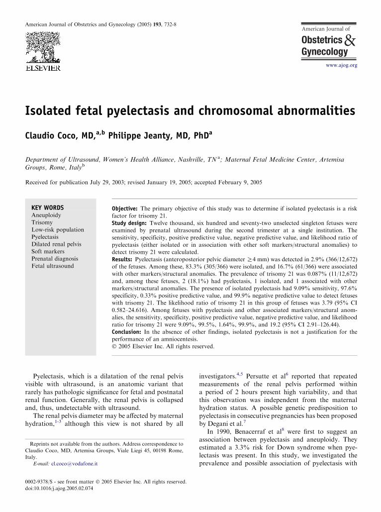
Figure 1 Measurement of the renal pelvis in the anteropos-terior dimension in a fetus with bilateral pyelectasis of 4.6 mm.
All patients underwent a thorough ultrasound exam-ination which, aside from the AIUM-ACR guidelines,sought as many soft markers as possible, includingnuchal thickening, pyelectasis, echogenic intracardiacfoci, brachymesophalangia of the fifth digit, as well as asimian crease, whenever possible, for these more subtlemarkers were only looked at for a few seconds and, ifnot seen, simply passed over. In only a small percentageof fetuses did we identify this sign in accord withprevious work.13
Hands, feet, and limb segments were included.Outflowtracts, return of pulmonary veins to the left atrium, and,when possible, both arches were also part of the routine.
Examinations were performed using either AcusonXP 128 (Siemens Medical Systems, Mountain View,Calif), Sequoia (Siemens Medical Systems), or Voluson730 (General Electric Medical Systems, Kretztechnik,Zipf, Austria) scanners.
The results only include data from 2D examinations,not from 3D.
The aim of this study was to determine if isolatedpyelectasis is a risk factor for trisomy 21. Because of thecompounding risk in the presence of other markers, weanalyzed the population with isolated pyelectasis and thepopulation with other markers separately. There is ageneral consensus that pyelectasis is only a marker fortrisomy 21 and not for other aneuploidy, and thus, ouranalysis only considered cases of trisomy 21. We alsoreported the total cases of aneuploidies recognized in thestudy period and the presence of pyelectasis in these cases.
The sensitivity, specificity, positive predictive value,negative predictive value, and likelihood ratios with95% CIs of pyelectasis to detect trisomy 21 werecalculated. Separate analyses were conducted for fetuseswith isolated pyelectasis, and for those with pyelectasisand other associated markers/structural anomalies. Thefollowing markers/structural anomalies were included:choroid plexus cysts, echogenic heart focus, 2-vesselcord, nuchal thickening, heart defects, diaphragmatichernia, esophageal atresia, omphalocele, facial cleft,micrognathia, myelomeningocele, growth restriction,shortening of the limbs, radial aplasia, (for long boneswe routinely measured humerus and femur, and welooked at ulna-radius and tibia-fibula, but only mea-sured them if they appeared abnormal), overlappingfingers, talipes, rocker bottom feet, clinodactyly, andbrachymesophalangia of the fifth digit.
Likelihood ratios were also calculated by the for-mula: Likelihood ratioC = sensitivity /(1� Specificity);likelihood ratio � = (1-Sensitivity)/Specificity.
The likelihood ratio expresses the odds that pyelec-tasis occurs in fetuses with Down syndrome versus theodds that pyelectasis occurs in fetuses without Downsyndrome (Figure 1).
Follow-up information in all the cases was obtainedby amniocentesis, infant postnatal reports, or by
734 Coco and Jeanty
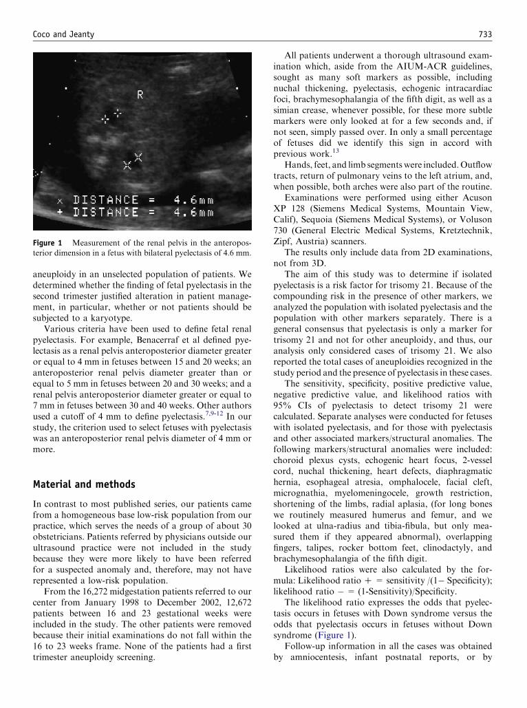
Figure 2 The distribution of pyelectasis with and without trisomy 21 in the patients examined.
contacting the referring physician or pediatrician. Ifthese options failed, the mother was interviewed. Thefollow-up was obtained in every case of pyelectasis.
Results
The mean maternal age was 27.2 G (standard deviation5.5), ranging from 15 to 42 years old. The prevalence ofpyelectasis in our population was 2.9% (366/12,672). In57% (208/366) of cases, the anteroposterior pelvis diam-eter measured 4 to 5 mm, in 23% (85/366) the renal pelvismeasured 5 to 6 mm, in 13% (48/366) the dilatation was 6to 7 mm, in 5% (19/366) it was 7 to 8 mm, and in 1% (4/366) the renal pelvis measured 8 mm, 1 fetus had 12 mmdilatation, and 1 had 16 mm dilatation.
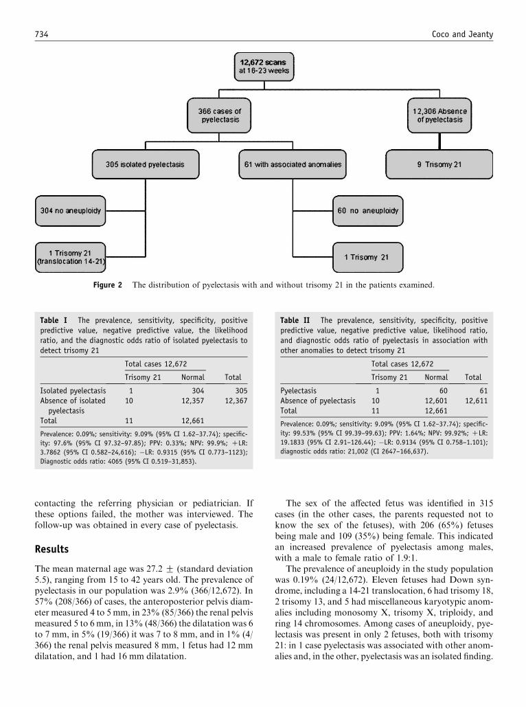
Table I The prevalence, sensitivity, specificity, positivepredictive value, negative predictive value, the likelihoodratio, and the diagnostic odds ratio of isolated pyelectasis todetect trisomy 21
Total cases 12,672
Trisomy 21 Normal Total
Isolated pyelectasis 1 304 305Absence of isolatedpyelectasis
10 12,357 12,367
Total 11 12,661
Prevalence: 0.09%; sensitivity: 9.09% (95% CI 1.62–37.74); specific-
ity: 97.6% (95% CI 97.32–97.85); PPV: 0.33%; NPV: 99.9%; CLR:
3.7862 (95% CI 0.582–24,616); �LR: 0.9315 (95% CI 0.773–1123);
Diagnostic odds ratio: 4065 (95% CI 0.519–31,853).
The sex of the affected fetus was identified in 315cases (in the other cases, the parents requested not toknow the sex of the fetuses), with 206 (65%) fetusesbeing male and 109 (35%) being female. This indicatedan increased prevalence of pyelectasis among males,with a male to female ratio of 1.9:1.
The prevalence of aneuploidy in the study populationwas 0.19% (24/12,672). Eleven fetuses had Down syn-drome, including a 14-21 translocation, 6 had trisomy 18,2 trisomy 13, and 5 had miscellaneous karyotypic anom-alies including monosomy X, trisomy X, triploidy, andring 14 chromosomes. Among cases of aneuploidy, pye-lectasis was present in only 2 fetuses, both with trisomy21: in 1 case pyelectasis was associated with other anom-alies and, in the other, pyelectasis was an isolated finding.
Table II The prevalence, sensitivity, specificity, positivepredictive value, negative predictive value, likelihood ratio,and diagnostic odds ratio of pyelectasis in association withother anomalies to detect trisomy 21
Total cases 12,672
Trisomy 21 Normal Total
Pyelectasis 1 60 61Absence of pyelectasis 10 12,601 12,611Total 11 12,661
Prevalence: 0.09%; sensitivity: 9.09% (95% CI 1.62–37.74); specific-
ity: 99.53% (95% CI 99.39–99.63); PPV: 1.64%; NPV: 99.92%; CLR:
19.1833 (95% CI 2.91–126.44); �LR: 0.9134 (95% CI 0.758–1.101);
diagnostic odds ratio: 21,002 (CI 2647–166,637).
Coco and Jeanty 735
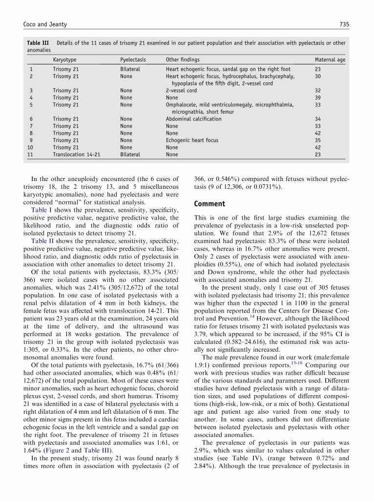
Table III Details of the 11 cases of trisomy 21 examined in our patient population and their association with pyelectasis or otheranomalies
Karyotype Pyelectasis Other findings Maternal age
1 Trisomy 21 Bilateral Heart echogenic focus, sandal gap on the right foot 232 Trisomy 21 None Heart echogenic focus, hydrocephalus, brachycephaly,
hypoplasia of the fifth digit, 2-vessel cord30
3 Trisomy 21 None 2-vessel cord 324 Trisomy 21 None None 395 Trisomy 21 None Omphalocele, mild ventriculomegaly, microphthalmia,
micrognathia, short femur33
6 Trisomy 21 None Abdominal calcification 347 Trisomy 21 None None 338 Trisomy 21 None None 429 Trisomy 21 None Echogenic heart focus 3510 Trisomy 21 None None 4211 Translocation 14-21 Bilateral None 23
In the other aneuploidy encountered (the 6 cases oftrisomy 18, the 2 trisomy 13, and 5 miscellaneouskaryotypic anomalies), none had pyelectasis and wereconsidered ‘‘normal’’ for statistical analysis.
Table I shows the prevalence, sensitivity, specificity,positive predictive value, negative predictive value, thelikelihood ratio, and the diagnostic odds ratio ofisolated pyelectasis to detect trisomy 21.
Table II shows the prevalence, sensitivity, specificity,positive predictive value, negative predictive value, like-lihood ratio, and diagnostic odds ratio of pyelectasis inassociation with other anomalies to detect trisomy 21.
Of the total patients with pyelectasis, 83.3% (305/366) were isolated cases with no other associatedanomalies, which was 2.41% (305/12,672) of the totalpopulation. In one case of isolated pyelectasis with arenal pelvis dilatation of 4 mm in both kidneys, thefemale fetus was affected with translocation 14-21. Thispatient was 23 years old at the examination, 24 years oldat the time of delivery, and the ultrasound wasperformed at 18 weeks gestation. The prevalence oftrisomy 21 in the group with isolated pyelectasis was1:305, or 0.33%. In the other patients, no other chro-mosomal anomalies were found.
Of the total patients with pyelectasis, 16.7% (61/366)had other associated anomalies, which was 0.48% (61/12,672) of the total population. Most of these cases wereminor anomalies, such as heart echogenic focus, choroidplexus cyst, 2-vessel cords, and short humerus. Trisomy21 was identified in a case of bilateral pyelectasis with aright dilatation of 4 mm and left dilatation of 6 mm. Theother minor signs present in this fetus included a cardiacechogenic focus in the left ventricle and a sandal gap onthe right foot. The prevalence of trisomy 21 in fetuseswith pyelectasis and associated anomalies was 1:61, or1.64% (Figure 2 and Table III).
In the present study, trisomy 21 was found nearly 8times more often in association with pyelectasis (2 of
366, or 0.546%) compared with fetuses without pyelec-tasis (9 of 12,306, or 0.0731%).
Comment
This is one of the first large studies examining theprevalence of pyelectasis in a low-risk unselected pop-ulation. We found that 2.9% of the 12,672 fetusesexamined had pyelectasis: 83.3% of these were isolatedcases, whereas in 16.7% other anomalies were present.Only 2 cases of pyelectasis were associated with aneu-ploidies (0.55%), one of which had isolated pyelectasisand Down syndrome, while the other had pyelectasiswith associated anomalies and trisomy 21.
In the present study, only 1 case out of 305 fetuseswith isolated pyelectasis had trisomy 21; this prevalencewas higher than the expected 1 in 1100 in the generalpopulation reported from the Centers for Disease Con-trol and Prevention.14 However, although the likelihoodratio for fetuses trisomy 21 with isolated pyelectasis was3.79, which appeared to be increased, if the 95% CI iscalculated (0.582–24.616), the estimated risk was actu-ally not significantly increased.
The male prevalence found in our work (male:female1.9:1) confirmed previous reports.15-18 Comparing ourwork with previous studies was rather difficult becauseof the various standards and parameters used. Differentstudies have defined pyelectasis with a range of dilata-tion sizes, and used populations of different composi-tions (high-risk, low-risk, or a mix of both). Gestationalage and patient age also varied from one study toanother. In some cases, authors did not differentiatebetween isolated pyelectasis and pyelectasis with otherassociated anomalies.
The prevalence of pyelectasis in our patients was2.9%, which was similar to values calculated in otherstudies (see Table IV), (range between 0.72% and2.84%). Although the true prevalence of pyelectasis in

736 Coco and Jeanty
Table IV Comparison of several studies on pylelectasis
Author
Prevalenceofpyelectasis Population
Gestationalage
Pyelec.
Cut Off N %
Benacerraf (8) 2.84% 7400 O16 R4 mm between 15-20 weeks 210 2.84%R5 mm between 20-30 weeksR7 mm between 30-40 weeks
Corteville (12) 2.10% 5944 O14 R4 mm before 33 weeks 127 2.14%R7 mm after 33 weeks
Nicolaides (18) NA Selected 15-38 R5 mm 258 d
Wickstrom (10) 0.80% 11340 17-39 R4 mm 82 0.72%Wickstrom (25) 0.72% 7481 R15 R4 mm !33 weeks 121 1.62%
R7 mm O33 weeksOuzounian et al. (28) d d R4 mm 84* dChudleigh et al. (19) 0.73% 101600 16-26 R5 mm 737 0.73%
Havutcu et al. (16) 1.25% 25586 18-24 R5 mm 320 1.25%Persutte et al. (15) 5.50% 5529 16-38 R4 mm 306 5.53%
Nyberg (23) NA Selected 14-20 R3 mm 186Rotmensch (29) NA Selected 14-28 R4 mm between 15-20 weeks
R5 mm between 20-30 weeksSairam (30) 2.34%Current study 2.90% 12672 16-23 R4 mm 366 2.89%
*No distintion there were among isolated and not isolated pyelectasis.y The distinction was made only for major anomaly: 12 had concomitant anomaly: 2 T21, 1 recombinant 8 Syndrome, 3 multiple congenital anomaly,
1 IUGR, 5 fetal or infant losses.z Analisys of 155 fetuses with Down syndrome.x Report of 187 cases of trisomy 21.
the general population was difficult to ascertain, thisstudy provided evidence that is as close to the baseline aspossible because none of the patients were preselected orreferred from other centers. A summary of the previousstudy is provided in Table IV.
Chudleigh19 used the percentage of pyelectasis to thenumber of births, not among the total ultrasound exam,and different parameter inclusions (anteroposterior di-ameter of the renal pelvis of 5 mm up to and including10 mm, while we used 4 mm).
Though there was variability in the prevalence ofpyelectasis in past studies (Table IV), there was a relativeuniformity of result for the prevalence of trisomy 21among fetuses with pyelectasis. Because of the smallprevalence of trisomy 21, 1 or 2 cases could have stronglyaffected the statistical analysis in most studies.
In our study the prevalence of trisomy 21 in isolatedpyelectasis was 1:305; Chudleigh19 reported similarresults (1:217, and 1:324 in women under 36 years) forisolated pyelectasis, but suggested a higher incidence forpyelectasis in association with other anomalies. In thestudy of Corteville,12 among 4 cases of trisomy 21, 3
were in association with ultrasound abnormalities and,in the remaining case of trisomy 21, the abnormality wassuspected. Previous studies did not find any aneuploidyfor isolated fetal pyelectasis.16 Others20,21 concludedthat the presence of pyelectasis increased the risk ofaneuploidy only if it was in association with otheranomalies, such as nuchal fold thickening and a shorthumerus.
Although Persutte et al15 did not specify if his caseswere associated with other structural anomalies or othersoft signs, he found a high incidence of chromosomalanomalies (2 trisomy 21 and 1 recombinant 8 syndrome)in his 306 cases and, in addition, a high incidence ofconcomitant anomalies (9 cases). In the study ofNicolaides et al,18 the suggested risk for isolated pye-lectasis was an increased risk for all aneuploidy.
Different likehood ratios were reported in anotherstudy. In his ‘‘age adjusted ultrasound risk assessment’’(AAURA) for Down syndrome, Nyberg22 used a like-lihood ratio for renal pyelectasis of 1.6, and in his otherstudy,23 an isolated pyelectasis within 14 and 20 weekshad a likelihood ratio of 1.5 (95% CI 0.6–3.6) as an
Coco and Jeanty 737
Table IV (Continued)
PyelectasisNon isolatedpyelectasis Isolated pyelectasis Pyelec. whit other anomalies
N % N % Normal Anormal Normal Anormal
203* 7 Trisomy 21
d d d d d 1 Trisomy 21 6 (3 Trisomy 21; 3 Other)
163 63.18% 95 36.82% 158 1 Trisomy 21 65 30 Trisomies 21, 13, 184 other other
d d d d 79 3 Trisomy 21 d dAll isolated 119 1 Trisomy 21
1 mosaic 46, XY/XYYd 1 Trisomy 21
651 88.33% 98 13.30% 648 3 Trisomy 21 89 3 Trisomy 211 Trisomy 131 Trisomy 82 45, X02 Other
301 94.06% 19 5.94% 301 None 19 None294y 96.08% d d 294y None dy 2 Trisomy 21
1 recombinant 8 Syndromez 5 Trisomy 21 z 16 trisomy 21d None x 13 Trisomy 21
305 83.33% 61 16.67% 304 1 Traslocation 14-21 60 1 Trisomy 21
* No distintion there were among isolated and not isolated pyelectasis.y The distinction was made only for major anomaly: 12 had concomitant anomaly: 2 T21, 1 recombinant 8 Syndrome, 3 multiple congenital anomaly,
1 IUGR, 5 fetal or infant losses.z Analisys of 155 fetuses with Down syndrome.x Report of 187 cases of trisomy 21.
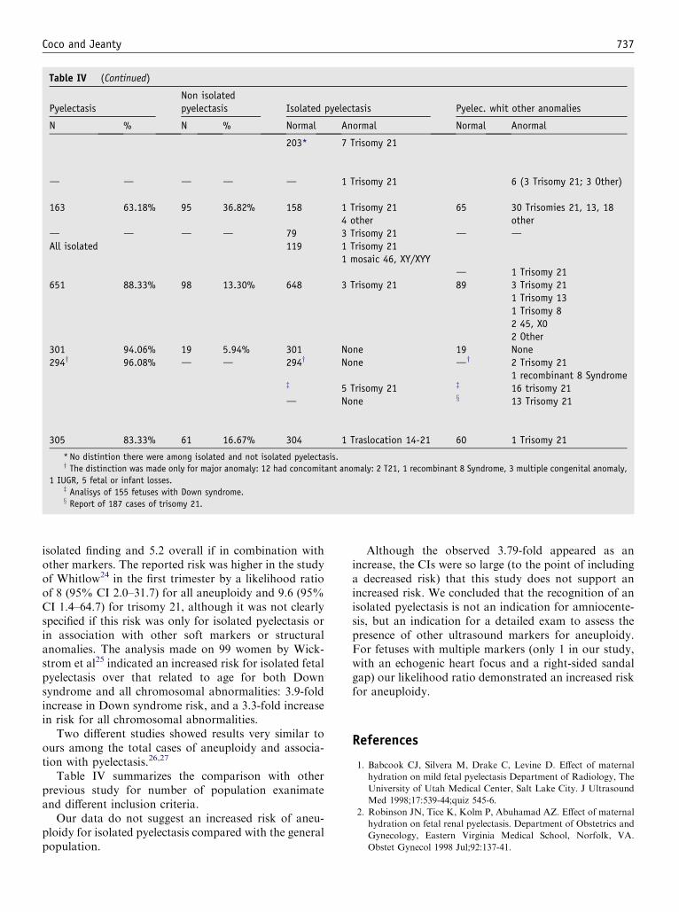
isolated finding and 5.2 overall if in combination withother markers. The reported risk was higher in the studyof Whitlow24 in the first trimester by a likelihood ratioof 8 (95% CI 2.0–31.7) for all aneuploidy and 9.6 (95%CI 1.4–64.7) for trisomy 21, although it was not clearlyspecified if this risk was only for isolated pyelectasis orin association with other soft markers or structuralanomalies. The analysis made on 99 women by Wick-strom et al25 indicated an increased risk for isolated fetalpyelectasis over that related to age for both Downsyndrome and all chromosomal abnormalities: 3.9-foldincrease in Down syndrome risk, and a 3.3-fold increasein risk for all chromosomal abnormalities.
Two different studies showed results very similar toours among the total cases of aneuploidy and associa-tion with pyelectasis.26,27
Table IV summarizes the comparison with otherprevious study for number of population exanimateand different inclusion criteria.
Our data do not suggest an increased risk of aneu-ploidy for isolated pyelectasis compared with the generalpopulation.
Although the observed 3.79-fold appeared as anincrease, the CIs were so large (to the point of includinga decreased risk) that this study does not support anincreased risk. We concluded that the recognition of anisolated pyelectasis is not an indication for amniocente-sis, but an indication for a detailed exam to assess thepresence of other ultrasound markers for aneuploidy.For fetuses with multiple markers (only 1 in our study,with an echogenic heart focus and a right-sided sandalgap) our likelihood ratio demonstrated an increased riskfor aneuploidy.
References
1. Babcook CJ, Silvera M, Drake C, Levine D. Effect of maternal
hydration on mild fetal pyelectasis Department of Radiology, The
University of Utah Medical Center, Salt Lake City. J Ultrasound
Med 1998;17:539-44;quiz 545-6.
2. Robinson JN, Tice K, Kolm P, Abuhamad AZ. Effect of maternal
hydration on fetal renal pyelectasis. Department of Obstetrics and
Gynecology, Eastern Virginia Medical School, Norfolk, VA.
Obstet Gynecol 1998 Jul;92:137-41.

738 Coco and Jeanty
3. Graif M, Kessler A, Hart S, Daitzchman M, Mashiach S, Boichis
H, et al. Renal pyelectasis in pregnancy: correlative evaluation of
fetal and maternal collecting systems. Department of Diagnostic
Imaging, Chaim Sheba Medical Center, Tel Hashomer, Israel. Am
J Obstet Gynecol 1992;167:1304-6.
4. Hoddick WK, Filly RA, Mahony BS, Callen PW. Minimal fetal
renal pyelectasis. J Ultrasound Med 1985;4:85-9.
5. Allen KS, Arger PH, Mennuti M, Coleman BG, Mintz MC,
Fishman M. Effects of maternal hydration on fetal renal pyelec-
tasis. Radiology 1987;163:807-9.
6. Persutte WH, Hussey M, Chyu J, Hobbins JC. Striking findings
concerning the variability in the measurement of the fetal renal
collecting system. Ultrasound Obstet Gynecol 2000;15:186-90.
7. Degani S, Leibovitz Z, Shapiro I, Gonen R, Ohel G. Department
of Obstetrics and Gynecology, Bnai-Zion Medical Center, Haifa,
Israel. Fetal pyelectasis in consecutive pregnancies: a possible
genetic predisposition. Ultrasound Obstet Gynecol 1997;10:19-21.
8. Benacerraf BR, Mandell J, Estroff JA, Harlow BL, Frigoletto FD
Jr. Fetal pyelectasis: a possible association with Down syndrome.
Department of Obstetrics and Gynecology, Brigham & Women’s
Hospital, Boston, Massachusetts. Obstet Gynecol 1990;76:58-60.
9. Gotoh H, Masuzaki H, Fukuda H, Yoshimura S, Ishimaru T.
Detection and assessment of pyelectasis in the fetus: relationship to
postnatal renal function Department of Obstetrics and Gynecol-
ogy, Nagasaki University School of Medicine, Japan. Obstet
Gynecol 1998;92:226-31.
10. Wickstrom E, Maizels M, Sabbagha RE, Tamura RK, Cohen LC,
Pergament E. Isolated fetal pyelectasis: assessment of risk for
postnatal uropathy and Down syndrome. Ultrasound Obstet
Gynecol 1996;8:236-40.
11. Adra AM, Mejides AA, Dennaoui MS, Beydoun SN. Fetal
pyelectasis: is it always ‘‘physiologic?’’ Department of Obstetrics
and Gynecology, University of Miami School of Medicine,
Florida. Am J Obstet Gynecol 1995;173:1263-6.
12. Corteville JE, Dicke JM, Crane JP. Fetal pyelectasis and Down
syndrome: is genetic amniocentesis warranted? Obstet Gynecol
1992;79:770-2.
13. Jeanty P. Prenatal detection of simian crease. J Ultrasound Med
1990;9:131-6.
14. Centers for Disease Control and Prevention. Down syndrome
prevalence at birthdUnited States, 1983-1990. Accessed August
26, 1994. Available at: http://www.cdc.gov/mmwr/preview/
mmwrhtml/00032401.htm.
15. Persutte WH, Koyle M, Lenke RR, Klas J, Ryan C, Hobbins JC.
Mild pyelectasis ascertained with prenatal ultrasonography is
pediatrically significant. Ultrasound Obstet Gynecol 1997;10:12-8.
16. Havutcu AE, Nikolopoulos G, Adinkra P, Lamont RF. The
association between fetal pyelectasis on second trimester ultra-
sound scan and aneuploidy among 25,586 low risk unselected
women. Prenat Diagn 2002;22:1201-6.
17. Petrikovsky BM, Nardi DA, Rodis JF, Hoegsberg B. Elevated
maternal serum alpha-fetoprotein and mild fetal uropathy. Obstet
Gynecol 1991;78:262-4.
18. Nicolaides KH, Cheng HH, Abbas A, Snijders RJ, Gosden C. Fetal
renal defects: associated malformations and chromosomal defects.
Harris Birthright Research Centre for Fetal Medicine, King’s
College Hospital, London, UK. Fetal Diagn Ther 1992;7:1-11.
19. Chudleigh PM, Chitty LS, Pembrey M, Campbell S. The associ-
ation of aneuploidy and mild fetal pyelectasis in an unselected
population: the results of a multicenter study. Ultrasound Obstet
Gynecol 2001;17:197-202.
20. Vintzileos AM, Egan JF. Adjusting the risk for trisomy 21 on the
basis of second-trimester ultrasonography. Am J Obstet Gynecol
1995;172:837-44.
21. Vintzileos AM, Campbell WA, Guzman ER, Smulian JC, McLean
DA, Ananth CV. Second-trimester ultrasound markers for detec-
tion of trisomy 21: which markers are best? Obstet Gynecol
1997;89:941-4.
22. Nyberg DA, Luthy DA, Resta RG, Nyberg BC, Williams MA.
Age-adjusted ultrasound risk assessment for fetal Down’s syn-
drome during the second trimester: description of the method
and analysis of 142 cases. Ultrasound Obstet Gynecol 1998;
12:8-14.
23. Nyberg DA, Souter VL, El-Bastawissi A, Young S, Luthhardt F,
Luthy DA. Isolated sonographic markers for detection of fetal
Down syndrome in the second trimester of pregnancy. J Ultra-
sound Med 2001;20:1053-63.
24. Whitlow BJ, Lazanakis ML, Kadir RA, Chatzipapas I, Econo-
mides DL. The significance of choroid plexus cysts, echogenic
heart foci and renal pyelectasis in the first trimester. Ultrasound
Obstet Gynecol 1998;12:385-90.
25. Wickstrom EA, Thangavelu M, Parilla BV, Tamura RK, Sabba-
gha RE. A prospective study of the association between isolated
fetal pyelectasis and chromosomal abnormality. Obstet Gynecol
1996;88:379-82.
26. Salihu HM, Boos R, Schmidt W. Antenatally detectable markers
for the diagnosis of autosomally trisomic fetuses in at-risk preg-
nancies. Am J Perinatol 1997;14:257-61.
27. Vergani P, Locatelli A, Piccoli MG, Ceruti P, Mariani E, Pezzullo
JC, et al. Best second trimester sonographic markers for the
detection of trisomy 21. J Ultrasound Med 1999;18:469-73.
28. Ouzounian JG, Castro MA, Fresquez M, al-Sulyman OM, Kovacs
BW. Prognostic significance of antenatally detected fetal pyelecta-
sis. Ultrasound Obstet Gynecol 1996;7:424-8.
29. Rotmensch S, Liberati M, Bronshtein M, Schoenfeld-Dimaio M,
Shalev J, Ben-Rafael Z, et al. Prenatal sonographic findings in
187 fetuses with Down syndrome. Prenat Diagn 1997;17:1001-9.
30. Sairam S, Al-Habib A, Sasson S, Thilaganathan B. Natural history
of fetal hydronephrosis diagnosed on mid-trimester ultrasound.
Ultrasound Obstet Gynecol 2001;17:191-6.