isolated adrenocorticotropic hormone deficiency and
TRANSCRIPT

CASE REPORT Open Access
Isolated adrenocorticotropic hormonedeficiency and thyroiditis associated withnivolumab therapy in a patient withadvanced lung adenocarcinoma: a casereport and review of the literatureNobumasa Ohara1*, Michi Kobayashi1,2, Kazumasa Ohashi3, Ryo Ito3, Yohei Ikeda4, Gen Kawaguchi4,Yuichiro Yoneoka5, Go Hasegawa6 and Toshinori Takada3
Abstract
Introduction: Immune checkpoint inhibitors are a promising class of anticancer drugs. The clinical benefits affordedby immune checkpoint inhibitors can be accompanied by immune-related adverse events that affect multiple organs,and endocrine immune-related adverse events include thyroiditis and hypophysitis. Hypophysitis is less frequent andhas a less severe clinical presentation in patients treated with other immune checkpoint inhibitors, such as nivolumab,pembrolizumab, and atezolizumab, than in those treated with ipilimumab. However, studies have described isolatedadrenocorticotropic hormone deficiency cases associated with nivolumab, pembrolizumab, and atezolizumab therapy,most of which occurred during the course of immune checkpoint inhibitor therapy. We report a rare case of patientwith isolated adrenocorticotropic hormone deficiency that occurred after nivolumab therapy.
Case presentation: A 69-year-old Japanese woman with advanced lung adenocarcinoma developed painless thyroiditiswith transient elevations of serum thyroid hormones during 3months of cancer treatment with nivolumab and beganthyroid hormone replacement therapy for subsequent primary hypothyroidism. Four months after nivolumab therapywas discontinued, she developed isolated adrenocorticotropic hormone deficiency; corticosteroid replacement therapyrelieved her secondary adrenal insufficiency symptoms, such as anorexia and fatigue. Human leukocyte antigen typingrevealed the presence of DRB1*04:05-DQB1*04:01-DQA1*03:03 and DRB1*09:01-DQB1*03:03-DQA1*03:02 haplotypes,which increase susceptibility to autoimmune polyendocrine syndrome associated with thyroid and pituitary disorders inthe Japanese population.
Conclusions: Our patient developed thyroiditis during cancer treatment with nivolumab and subsequently exhibitedisolated adrenocorticotropic hormone deficiency 4months after discontinuing the drug. Administration of nivolumab incombination with a genetic predisposition to polyglandular autoimmunity probably caused both the thyroiditis andhypophysitis, resulting in primary hypothyroidism and isolated adrenocorticotropic hormone deficiency, respectively, inour patient. The present case highlights the need for physicians to be aware that endocrine immune-related adverseevents, including hypophysitis, can occur more than several months after discontinuing a drug.
Keywords: Human leukocyte antigen, Hydrocortisone, Isolated adrenocorticotropic hormone deficiency, Lungadenocarcinoma, Nivolumab, Thyroiditis
* Correspondence: [email protected] of Endocrinology and Metabolism, Uonuma Institute ofCommunity Medicine, Niigata University Medical and Dental Hospital, 4132Urasa, Minamiuonuma, Niigata 949-7302, JapanFull list of author information is available at the end of the article
© The Author(s). 2019 Open Access This article is distributed under the terms of the Creative Commons Attribution 4.0International License (http://creativecommons.org/licenses/by/4.0/), which permits unrestricted use, distribution, andreproduction in any medium, provided you give appropriate credit to the original author(s) and the source, provide a link tothe Creative Commons license, and indicate if changes were made. The Creative Commons Public Domain Dedication waiver(http://creativecommons.org/publicdomain/zero/1.0/) applies to the data made available in this article, unless otherwise stated.
Ohara et al. Journal of Medical Case Reports (2019) 13:88 https://doi.org/10.1186/s13256-019-2002-2

IntroductionImmune checkpoint inhibitors (ICIs) are a promisingnew class of anticancer drugs that reactivate cytotoxicT cells which, in turn, destroy tumor cells [1]. BecauseICIs generate dysimmune toxicities (autoimmunity) bycreating an imbalance within the immune system, theclinical benefits afforded by ICIs can be accompaniedby immune-related adverse events (IRAEs) that affectmultiple organs, mainly the skin, gut, liver, lung, andendocrine tissues [2]. Common endocrine IRAEs in-clude thyroiditis and hypophysitis, whereas uncom-mon IRAEs include adrenalitis and type 1 diabetesmellitus [3].Compared with those treated with ipilimumab, an
anti-cytotoxic T lymphocyte antigen 4 monoclonal anti-body, hypophysitis is less frequent and has a less severeclinical presentation in patients treated with other ICIs,such as nivolumab and pembrolizumab, which areanti-programmed cell death protein 1 (PD-1) monoclo-nal antibodies, and atezolizumab, an anti-programmedcell death ligand 1 (PD-L1) monoclonal antibody [4–6].However, studies have described isolated adrenocortico-tropic hormone deficiency (IAD) cases associated withnivolumab, pembrolizumab, and atezolizumab therapy[7–22], most of which have occurred during the courseof ICI therapy.IAD is a pituitary disorder characterized by secondary
adrenal insufficiency (AI) with low or absent cortisolproduction but normal secretion of pituitary hormonesother than adrenocorticotropic hormone (ACTH) [23].Patients with IAD typically present with anorexia, fa-tigue, and general weakness, but corticosteroid replace-ment therapy is effective for managing this disorder.
We report a case of a patient with advanced lung adeno-carcinoma (LAC) who developed thyroiditis during nivo-lumab therapy and subsequently exhibited IAD 4monthsafter the discontinuation of nivolumab. We also reviewpreviously reported cases of nivolumab-related thyroiditisand IAD.
Case presentationA 69-year-old Japanese woman who had been undergo-ing cancer treatment for advanced LAC was admittedto our hospital in January 2018 because of anorexia, fa-tigue, and general weakness. The patient had a mater-nal family history of esophageal cancer. The patient hadbeen a housewife since her 20s, had never smoked ciga-rettes, and did not have a drinking habit. The patient’smedical history was unremarkable until June 2016,when an abnormal x-ray shadow was found in her rightlung. A computed tomographic (CT) scan revealed atumor (3.1 cm) in the upper lobe of her right lung(Fig. 1a), right hilar and mediastinal lymph node swell-ings, and liver tumors. A transbronchoscopic biopsyfrom the lung tumor revealed LAC with vascular invasion(Fig. 2). IHC revealed no anaplastic lymphoma kinase re-arrangement, and a genetic analysis of the cancer cells de-tected no epidermal growth factor receptor mutation.Whole-body technetium-99m methylene diphosphonatescintigraphy revealed multiple lesions at the thoracic andlumbar vertebrae, sternum, ilium, and right ischial bones.Brain magnetic resonance imaging (MRI) revealed tumorsin the left temporal lobe and right cerebellar hemisphere(Fig. 1b). As a result, the patient was diagnosed with LACwith distant metastases to the brain, liver, and bones(cT2aN2M1b, stage IV) [24].
ba
Fig. 1 Radiological findings (July 2016). a Chest computed tomographic scan showing a tumor (3.1 cm) in the upper lobe of the right lung (arrow). bT1-weighted transverse magnetic resonance imaging of the brain showing a tumor in the left temporal lobe (arrow) and a tumor in the rightcerebellar hemisphere (short arrow)
Ohara et al. Journal of Medical Case Reports (2019) 13:88 Page 2 of 10

The patient underwent stereotactic radiation surgery(total, 22 Gy) for her metastatic brain tumors in July 2016.Thereafter, she received four courses of chemotherapy withintravenous cisplatin, pemetrexed, and bevacizumab fromJuly 2016 to October 2016; this treatment regimen effect-ively controlled her advanced LAC with a Response Evalu-ation Criteria in Solid Tumors (RECIST) classification ofpartial response [25]. The patient subsequently receivednine courses of maintenance chemotherapy with intraven-ous pemetrexed and bevacizumab from November 2016 toApril 2017. CT scans performed in May 2017 revealed noprogression of the primary LAC or metastatic brain andbone lesions, but they showed evidence of progression ofthe metastatic liver tumors.Subsequently, the patient began second-line
chemotherapy with intravenous nivolumab (133 mg[3 mg/kg] every 2 weeks) in May 2017 (Fig. 3). Thy-roid function was routinely monitored in July 2017after five courses of nivolumab therapy, and sheshowed high levels of serum free thyroxine (FT4,
1.91 ng/dl) and low levels of thyroid-stimulating hor-mone (TSH, 0.04 μIU/ml). The patient had no symp-toms of thyrotoxicosis or exophthalmos but hadmild and soft struma without any pain or fever.Ultrasonography revealed rough and mildly lowechogenicity in a slightly enlarged thyroid glandwithout a tumor (Fig. 4a, b), and technetium-99mpertechnetate thyroid scintigraphy revealed a lowthyroid uptake of 0.1% (reference range, 0.5–4%) inthe entire thyroid gland (Fig. 4c). The patient hadnegative test results for TSH-binding inhibitory im-munoglobulin (TBII), thyroglobulin autoantibody(TgAb), and thyroid peroxidase autoantibody(TPOAb). On the basis of these findings, she was di-agnosed with painless thyroiditis induced by nivolu-mab and was closely followed without medication.The patient exhibited primary hypothyroidism (FT4,0.66 ng/dl; TSH, 11.41 μIU/ml) in September 2017and initiated thyroid hormone replacement therapywith oral levothyroxine (50 μg/day).
a b
c d
Fig. 2 Histopathological findings of biopsy specimen from the left lung tumor (July 2016). a, b Microscopic examination showing bronchialmucosal infiltration by poorly differentiated lung adenocarcinoma (a), and vascular invasion is seen (arrows) (b) (H&E staining). c, d The cytoplasmof the tumor cells was immunohistochemically positive for surfactant protein A (c), and the tumor cell nuclei were positive for thyroidtranscription factor 1 (d). Ly Lymph duct, V Vein
Ohara et al. Journal of Medical Case Reports (2019) 13:88 Page 3 of 10

Because the CT scans performed after the sixth cycleof nivolumab revealed enlargements of the metastaticliver tumors, nivolumab therapy was discontinued inAugust 2017. The patient received three courses ofthird-line chemotherapy with docetaxel and ramuciru-mab beginning in September 2017, which effectivelycontrolled her LAC (RECIST classification of partial re-sponse), but this treatment was terminated in October2017 because of side effects, such as joint pain and
rhabdomyolysis. The patient did not consent to continueanticancer drug therapy after considering the potentialbenefits and side effects, and she chose to receive bestsupportive care.In November 2017, the patient’s body weight, blood
pressure, and pulse rate were 45 kg, 121/67 mmHg, and76 beats per minute, respectively. She had normal levelsof serum electrolytes (sodium 140 mEq/L, potassium4.3 mEq/L, and chloride 106 mEq/L). However, the
Fig. 3 Clinical course of the patient before the onset of isolated adrenocorticotropic hormone deficiency. Bmab Bevacizumab, CDDP Cisplatin,DTX Docetaxel, FT4 Free thyroxine, IAD Isolated adrenocorticotropic hormone deficiency, PEM Pemetrexed, Rmab Ramucirumab, TSHThyroid-stimulating hormone
b ca
Fig. 4 Thyroid gland imaging findings (July 2017). a, b Ultrasonography of the thyroid gland showing rough and mildly low echogenicity in aslightly enlarged thyroid gland without a tumor (a). Color Doppler images revealed no increased blood flow in the thyroid gland (b). cTechnetium-99m pertechnetate thyroid scintigraphy showing a low thyroid uptake (0.1%, reference range 0.5–4%) in the entire thyroid glandwithout a hot spot
Ohara et al. Journal of Medical Case Reports (2019) 13:88 Page 4 of 10

patient developed acute anorexia, fatigue, and generalweakness in December 2017 and was admitted to theDepartment of Respiratory Medicine at our hospital inJanuary 2018.On admission, the patient had clear consciousness
and did not complain of headache, abdominal pain,diarrhea, or joint and muscle pain. Her height, bodyweight, body temperature, blood pressure, and pulserate were 153 cm, 38 kg, 36.9 °C, 90/54 mmHg, and 73beats per minute, respectively. She had mild and softgoiter without pain. No heart murmur, chest rales, rash,vitiligo, skin pigmentation, or peripheral edema was de-tected. No paralysis, cerebellar ataxia, pyramidal orextrapyramidal tract symptoms, visual disturbance,hearing loss, dysarthria, or epileptic seizures werefound. A blood analysis revealed hyponatremia (serumsodium 124 mEq/L); asymptomatic hypoglycemia (fast-ing plasma glucose 65 mg/dl); and low levels of immu-noreactive insulin (< 0.2 μU/ml), ACTH (2.6 pg/ml),and cortisol (< 0.2 μg/dl) (Table 1). Because AI was sus-pected, oral levothyroxine was discontinued on day 2 ofadmission because hormone replacement therapy withthyroid hormone alone can exaggerate AI symptomswhen hypothyroidism and AI coexist. The patient wasreferred to the Department of Endocrinology and Me-tabolism on day 5 of admission.A rapid cosyntropin stimulation test suggested sec-
ondary AI (Table 2). Dynamic tests assessing the se-cretion of pituitary hormones showed the normalrelease of growth hormone (GH), TSH, and prolactin;age-appropriate release of luteinizing hormone andfollicle-stimulating hormone; but no ACTH releasefollowing a corticotropin-releasing hormone load(Table 3). A GH-releasing peptide 2 loading test alsoshowed no ACTH release, whereas GH release wassufficient (Table 4). These findings were indicative ofIAD. A brain MRI study revealed slight atrophy of theanterior pituitary with a pituitary height of 2.2 mm(Fig. 5).The patient had negative test results for anti-pituitary cell
antibody, TgAb, TPOAb, and TBII as well as otherorgan-specific autoantibodies, including glutamic acid de-carboxylase autoantibody, insulin autoantibody, gastric
Table 1 Laboratory findings at admission (January 2018)
Hematology
Red blood cells 379 × 104/μl (386–492)
Hemoglobin 11.3 g/dl (11.6–14.8)
Hematocrit 33.7% (35.1–44.4)
White blood cells 3600/μl (3300–8600)
Platelets 20.9 × 10⁴/μl (15.8–34.8)
Blood chemistry
Fasting plasma glucose 65 mg/dl (70–109)
Immunoreactive insulin < 0.2 μU/ml (2.2–12.4)
Glycated hemoglobin 5.2% (4.6–6.2)
Total cholesterol 161 mg/dl (142–248)
Triglycerides 62 mg/dl (30–117)
Total protein 6.2 g/dl (6.6–8.1)
Albumin 3.7 g/dl (4.1–5.1)
Aspartate aminotransferase 31 IU/L (13–30)
Alanine aminotransferase 12 IU/L (7–23)
Creatine phosphokinase 59 IU/L (41–153)
Urea nitrogen 11.4 mg/dl (8.0–18.4)
Creatinine 0.66 mg/dl (0.46–0.79)
Uric acid 2.5 mg/dl (2.6–5.5)
Sodium 124mEq/L (137–147)
Potassium 4.8 mEq/L (3.5–4.7)
Chloride 90 mEq/L (98–108)
Calcium 8.7 mg/dl (8.8–10.1)
C-reactive protein 0.16 mg/dl (0–0.14)
Immunoglobulin G4 35 mg/dl (5–117)
Carcinoembryonic antigen 208.0 ng/ml (0–4.76)
Sialyl-Lewis X 26.6 U/ml (0–37.9)
Cytokeratin 19 fragments 1.6 ng/ml (0–2.1)
Squamous cell carcinoma antigen 1.1 ng/ml (0–1.5)
Plasma osmolality 254 mOsm/L (275–290)
Arginine vasopressin 1.0 pg/ml
Thyroid-stimulating hormone 4.84 μIU/ml (0.50–5.00)
Free thyroxine 1.44 ng/dl (0.90–1.70)
Free triiodothyronine 3.22 pg/ml (2.30–4.00)
Adrenocorticotropic hormone 2.6 pg/ml (7.2–63.3)
Serum cortisol < 0.2 μg/dl (4.5–21.1)
Dehydroepiandrosterone sulfate < 20 ng/ml (120–1330)
Plasma renin activity 0.2 ng/ml/h (0.2–2.3)
Aldosterone 8.8 ng/dl (3.0–15.9)
The reference range for each parameter is shown in parenthesesBlood samples were taken in the morning (10 a.m.) with the patient in supineposition. The patient was taking thyroid hormone replacement therapy withoral levothyroxine (50 μg/day) for primary hypothyroidism
Table 2 Endocrinological investigation: rapid cosyntropinstimulation test in January 2018 (day 6 after admission)
Time (min)
0 30 60
Cortisol (μg/dl) < 0.2 1.0 1.4
Aldosterone 5.7 14.1 16.5
Blood samples were taken at 0 (just before) and at 30 and 60 min aftersynthetic adrenocorticotropic hormone 1–24 (cosyntropin hydroxide 0.25 mg)was intravenously administered in the morning (9 a.m.). The patient had a lowplasma adrenocorticotropic hormone level (2.3 pg/ml) and low plasma reninactivity (< 0.2 ng/ml/h) just before cosyntropin administration
Ohara et al. Journal of Medical Case Reports (2019) 13:88 Page 5 of 10

parietal cell autoantibody, intrinsic factor autoantibody, ad-renocortical autoantibody, antinuclear antibody, Sjögren’ssyndrome A and B antibodies, anti-citrullinated peptideantibody, and rheumatoid factor.Human leukocyte antigen (HLA) typing revealed the
presence of A*08:01/12:02, B*48:01/52:01, and C*08:01/12:02 class I genes and DRB1*04:05/09:01, DQB1*03:03/04:01, DQA1*03:02/03:03, and DPB1*04:02/14:01 class IIgenes.The patient began corticosteroid replacement therapy
with oral hydrocortisone (15mg/day) for AI secondary toIAD on the afternoon of day 8 of admission. Subsequently,she resumed oral levothyroxine (50 μg/day) for primaryhypothyroidism on day 9 of admission.The patient experienced improvements in anorexia,
fatigue, and general weakness and became ambulatorywithin days. Her hyponatremia was corrected within 1week, and her hypoglycemia was resolved. Laboratory
data obtained on day 21 of admission showed normallevels of serum sodium (140 mEq/L), potassium (4.3mEq/L), chloride (106 mEq/L), FT4 (1.60 ng/dl), TSH(3.28 μIU/ml), and fasting plasma glucose (78 mg/dl).The patient was discharged on day 25 of admission.The subsequent clinical course of the patient was
mostly uneventful for 6 months after discharge. As herLAC progressed, her ability to perform activities of dailyliving decreased, and in November 2018, she was trans-ferred to a local hospital to receive terminal care.
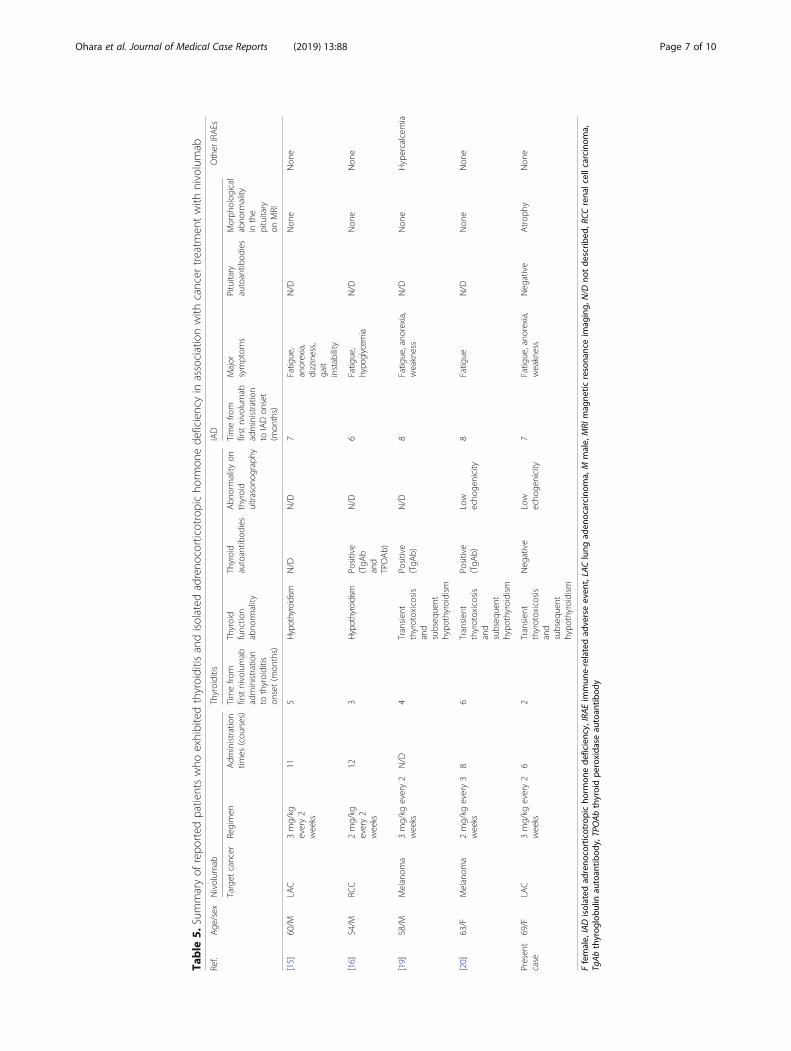
DiscussionOur patient with advanced LAC developed painlessthyroiditis associated with a transient elevation ofserum thyroid hormones (thyrotoxicosis) during 3months of second-line chemotherapy with nivolumaband began thyroid hormone replacement therapy forsubsequent primary hypothyroidism. Four months afternivolumab therapy was discontinued, the patient devel-oped IAD. Other than reports regarding nivolumab,there have been few reports on thyroid or pituitary dys-function associated with the anticancer agents that ourpatient took, including bevacizumab and ramucirumab(Fig. 3) [26–29]. External brain radiotherapy can pro-duce pituitary dysfunction that is usually associatedwith GH deficiency, but it seldom causes IAD [30].These findings suggest that our patient exhibited bothICI-related thyroiditis and IAD induced by nivolumab.Table 5 summarizes the characteristics of reported pa-
tients who exhibited nivolumab-related thyroiditis andIAD [15, 16, 19, 20]. The cases describe both male and fe-male adults with melanoma, LAC, or renal cell carcinoma.They developed thyroiditis that resulted in hypothyroidismand subsequently developed IAD.Thyroiditis is the most frequent IRAE associated with
endocrine organs, regardless of the different ICIs [3, 6].Thyroiditis usually occurs within a few months of initiat-ing ICI therapy with or without verifiable circulatingthyroid autoantibodies, such as TgAb and TPOAb. Com-mon clinical manifestations of ICI-related thyroiditis in-clude painless thyroiditis characterized by a transientthyrotoxicosis and subsequent occurrence of persistentprimary hypothyroidism [31, 32]. In our patient, physicalfindings, thyroid gland image findings (Fig. 4), and theclinical course (Fig. 3) indicated that she had a typicalform of ICI-related thyroiditis.Studies on patients with IAD associated with nivolu-
mab have shown that the latency from initial nivolumabadministration to IAD onset ranged approximatelyfrom 4 to 8 months, with a mean period of roughly 6months [7–20], which appears to be longer than that ofipilimumab-related hypophysitis (approximately 2months)[5]. Most cases of nivolumab-related IAD occurred duringthe course of or within 1 month of the discontinuation of
Table 3 Endocrinological investigation: CRH/GRF/TRH/LHRHstimulation test in January 2018 (day 7 after admission)
Time (min)
0 15 30 60 90 120
Adrenocorticotropichormone (pg/ml)
2.1 3.5 4.0 3.4 2.9 2.2
Cortisol (μg/dl) < 0.2 < 0.2 < 0.2 < 0.2 < 0.2 < 0.2
Thyroid-stimulatinghormone (μIU/ml)
5.47 22.86 33.95 28.66 23.64 17.65
Growth hormone(ng/ml)
7.67 19.96 34.68 38.76 37.53 21.17
Prolactin (ng/ml) 22.6 75.6 94.4 84.6 61.2 44.6
Luteinizing hormone(mIU/ml)
10.4 21.8 30.9 45.3 47.9 43.1
Follicle-stimulating hormone(mIU/ml)
32.8 36.2 33.6 36.6 45.6 45.5
The following synthetic hypothalamic hormones were intravenouslyadministered in the morning (9 a.m.): human corticotropin-releasing hormone(CRH; 100 μg), growth hormone-releasing factor (GRF; 100 μg), thyrotropin-releasing hormone (TRH; 500 μg), and luteinizing hormone-releasing hormone(LHRH; 100 μg)The hormone replacement therapy with oral levothyroxine (50 μg/day) forprimary hypothyroidism was discontinued on day 2 after admission, and thepatient’s serum levels of free thyroxine (1.26 ng/dl) and free triiodothyronine(2.77 pg/ml) were measured just before the drug administration
Table 4 Endocrinological investigation: GHRP-2 stimulation testin January 2018 (day 8 after admission)
Time (min)
0 15 30 45 60
Adrenocorticotropichormone (pg/ml)
1.9 2.3 1.9 1.9 3.2
Cortisol (μg/dl) < 0.2 < 0.2 < 0.2 < 0.2 < 0.2
Growth hormone(ng/ml)
3.73 18.02 29.63 32.98 26.21
Growth hormone-releasing peptide (GHRP)-2 (100 μg) was intravenouslyadministered in the morning (9 a.m.)
Ohara et al. Journal of Medical Case Reports (2019) 13:88 Page 6 of 10
Table
5.Summaryof
repo
rted
patientswho
exhibitedthyroiditis
andisolated
adreno
corticotropicho
rmon
ede
ficiencyin
associationwith
cancer
treatm
entwith
nivolumab
Ref.
Age
/sex
Nivolum
abThyroiditis
IAD
Other
IRAEs
Target
cancer
Regimen
Adm
inistration
times
(cou
rses)
Timefro
mfirstnivolumab
administration
tothyroiditis
onset(m
onths)
Thyroid
functio
nabno
rmality
Thyroid
autoantib
odies
Abn
ormality
onthyroid
ultrason
ograph
y
Timefro
mfirstnivolumab
administration
toIADon
set
(mon
ths)
Major
symptom
sPituitary
autoantib
odies
Morph
olog
ical
abno
rmality
inthe
pituitary
onMRI
[15]
60/M
LAC
3mg/kg
every2
weeks
115
Hypothyroidism
N/D
N/D
7Fatig
ue,
anorexia,
dizziness,
gait
instability
N/D
Non
eNon
e
[16]
54/M
RCC
2mg/kg
every2
weeks
123
Hypothyroidism
Positive
(TgA
band
TPOAb)
N/D
6Fatig
ue,
hypoglycem
iaN/D
Non
eNon
e
[19]
58/M
Melanom
a3mg/kg
every2
weeks
N/D
4Transien
tthyrotoxicosis
and
subseq
uent
hypo
thyroidism
Positive
(TgA
b)N/D
8Fatig
ue,ano
rexia,
weakness
N/D
Non
eHypercalcem
ia
[20]
63/F
Melanom
a2mg/kg
every3
weeks
86
Transien
tthyrotoxicosis
and
subseq
uent
hypo
thyroidism
Positive
(TgA
b)Low
echo
genicity
8Fatig
ueN/D
Non
eNon
e
Presen
tcase
69/F
LAC
3mg/kg
every2
weeks
62
Transien
tthyrotoxicosis
and
subseq
uent
hypo
thyroidism
Neg
ative
Low
echo
genicity
7Fatig
ue,ano
rexia,
weakness
Neg
ative
Atrop
hyNon
e
Ffemale,
IADisolated
adreno
corticotropicho
rmon
ede
ficiency,IRAEim
mun
e-relatedad
verseeven
t,LAClung
aden
ocarcino
ma,M
male,
MRI
mag
netic
resona
nceim
aging,
N/D
notde
scrib
ed,R
CCrena
lcellcarcino
ma,
TgAbthyrog
lobu
linau
toan
tibod
y,TPOAbthyroidpe
roxida
seau
toan
tibod
y
Ohara et al. Journal of Medical Case Reports (2019) 13:88 Page 7 of 10

nivolumab therapy [7–19], but one patient with melanomawho completed 6months of nivolumab therapy 2monthsprior developed IAD during subsequent ipilimumab ther-apy [20]. Similarly, our patient with LAC developed IAD 4months after the discontinuation of 3months of nivolumabtherapy. Our patient’s case highlights the need for physi-cians to be aware that endocrine IRAEs, including hypo-physitis, can occur more than several months afterdiscontinuing a drug.Patients with ipilimumab-related hypophysitis usually
exhibit pituitary enlargement as radiological evidence ofpituitary inflammation and pituitary dysfunction withmultiple deficits in anterior pituitary hormones [4, 5, 33].By contrast, many studies of nivolumab-related IAD havereported no morphological pituitary abnormalities [8–17,19, 20], whereas two studies described patients whoshowed mild or slight but definitely reversible pituitary en-largement [7, 18]. However, there was a reported case of apatient with atezolizumab-related IAD who showed anter-ior pituitary atrophy [22]. Similarly, our patient exhibitedmild anterior pituitary atrophy (Fig. 5).Autoimmune polyendocrine syndromes (APS) are a
heterogeneous group of endocrine and nonendocrineorgan-specific autoimmune disorders [34]. For example,autoimmune thyroid diseases can be associated with otherendocrine disorders as part of APS type 3. Genetic factors,including HLA class II genes, play a role in the develop-ment of APS. Our patient with ICI-related thyroiditis andIAD had the HLA-DRB1*04:05-DQB1*04:01-DQA1*03:03and DRB1*09:01-DQB1*03:03-DQA1*03:02 haplotypes,which increase susceptibility to APS type 3 associated withpituitary disorders in the Japanese population [35]. Thesefindings suggest that administration of nivolumab in com-bination with a genetic predisposition to polyglandularautoimmunity caused thyroiditis and hypophysitis in
association with APS type 3, resulting in primaryhypothyroidism and IAD, respectively, in our patient.
ConclusionsWe describe a patient with advanced LAC who devel-oped thyroiditis during 3 months of nivolumab ther-apy and subsequently exhibited IAD 4 months afterthe discontinuation of nivolumab. The combination ofnivolumab administration and genetic factors thatmanifested a susceptibility to polyglandular auto-immunity likely caused her ICI-related thyroiditis andhypophysitis. Thus, physicians should be aware thatICI-related endocrinopathies, including hypophysitis,may occur more than several months after discontinu-ation of the drug.
AbbreviationsACTH: Adrenocorticotropic hormone; AI: Adrenal insufficiency;APS: Autoimmune polyendocrine syndrome; Bmab: Bevacizumab;CDDP: Cisplatin; CRH: Corticotropin-releasing hormone; CT: Computedtomography; DTX: Docetaxel; F: Female; FT4: Free thyroxine; GH: Growthhormone; GHRP: Growth hormone-releasing peptide; GRF: Growthhormone-releasing factor; HLA: Human leukocyte antigen; IAD: Isolatedadrenocorticotropic hormone deficiency; ICI: Immune checkpointinhibitor; IRAE: Immune-related adverse event; LAC: Lungadenocarcinoma; LHRH: Luteinizing hormone-releasing hormone;Ly: Lymph duct; M: Male; MRI: Magnetic resonance imaging; N/D: Notdescribed; PD-1: Anti-programmed cell death protein-1; PD-L1:Anti-programmed cell death ligand-1; PEM: Pemetrexed; RCC: Renal cellcarcinoma; RECIST: Response Evaluation Criteria in Solid Tumors;Rmab: Ramucirumab; TBII: Thyroid-stimulating hormone-binding inhibi-tory immunoglobulin; TgAb: Thyroglobulin autoantibody; TPOAb: Thyroidperoxidase autoantibody; TRH: Thyrotropin-releasing hormone;TSH: Thyroid-stimulating hormone; V: Vein
AcknowledgementsThe authors thank the clinical laboratory technicians of Uonuma Institute ofCommunity Medicine, Niigata University Medical and Dental Hospital, fortheir valuable technical support.
ba c
Fig. 5 Magnetic resonance imaging of the pituitary gland (January 2018). a Sagittal T1-weighted plain magnetic resonance imaging (MRI) studyshowing normal high-intensity signals in the posterior lobe of the pituitary. b, c Gadolinium-enhanced MRI scans (b, sagittal plane; c, coronalplane) showing homogeneous enhancement of the normal hypophyseal stalk and mild atrophy of the anterior lobe of the pituitary gland. Thewidth, length, and height of the pituitary gland are 16.2, 9.8, and 2.2 mm, respectively
Ohara et al. Journal of Medical Case Reports (2019) 13:88 Page 8 of 10
FundingNo sources of funding are declared for this study.
Availability of data and materialsAll data generated or analyzed during this study are included in thispublished article.
Authors’ contributionsNO, MK, KO, RI, YI, and GH contributed to patient management. NO was amajor contributor to the writing of the manuscript. GK, YY, and TT criticallyreviewed the manuscript. All authors read and approved the finalmanuscript.
Ethics approval and consent to participateThis study was performed in accordance with the Declaration of Helsinki.
Consent for publicationWritten informed consent was obtained from the patient for publication ofthis case report and any accompanying images. A copy of the writtenconsent is available for review by the Editor-in-Chief of this journal.
Competing interestsThe authors declare that they have no competing interests.
Publisher’s NoteSpringer Nature remains neutral with regard to jurisdictional claims inpublished maps and institutional affiliations.
Author details1Department of Endocrinology and Metabolism, Uonuma Institute ofCommunity Medicine, Niigata University Medical and Dental Hospital, 4132Urasa, Minamiuonuma, Niigata 949-7302, Japan. 2Department of Diabetes,Endocrinology and Metabolism, Center Hospital of the National Center forGlobal Health and Medicine, Tokyo, Japan. 3Department of RespiratoryMedicine, Uonuma Institute of Community Medicine, Niigata UniversityMedical and Dental Hospital, Niigata, Japan. 4Department of Radiology,Uonuma Institute of Community Medicine, Niigata University Medical andDental Hospital, Niigata, Japan. 5Department of Neurosurgery, UonumaInstitute of Community Medicine, Niigata University Medical and DentalHospital, Niigata, Japan. 6Department of Pathology, Uonuma Institute ofCommunity Medicine, Niigata University Medical and Dental Hospital,Niigata, Japan.
Received: 6 June 2018 Accepted: 29 January 2019
References1. Pennock GK, Chow LQ. The evolving role of immune checkpoint inhibitors
in cancer treatment. Oncologist. 2015;20:812–22.2. Michot JM, Bigenwald C, Champiat S, Collins M, Carbonnel F, Postel-Vinay S,
Berdelou A, Varga A, Bahleda R, Hollebecque A, Massard C, Fuerea A, RibragV, Gazzah A, Armand JP, Amellal N, Angevin E, Noel N, Boutros C, Mateus C,Robert C, Soria JC, Marabelle A, Lambotte O. Immune-related adverseevents with immune checkpoint blockade: a comprehensive review. Eur JCancer. 2016;54:139–48.
3. Barroso-Sousa R, Barry WT, Garrido-Castro AC, Hodi FS, Min L, Krop IE,Tolaney SM. Incidence of endocrine dysfunction following the use ofdifferent immune checkpoint inhibitor regimens: a systematic review andmeta-analysis. JAMA Oncol. 2018;4:173–82.
4. Araujo PB, Coelho MC, Arruda M, Gadelha MR, Neto LV. Ipilimumab-inducedhypophysitis: review of the literature. J Endocrinol Investig. 2015;38:1159–66.
5. Faje A. Immunotherapy and hypophysitis: clinical presentation, treatment,and biologic insights. Pituitary. 2016;19:82–92.
6. Shang YH, Zhang Y, Li JH, Li P, Zhang X. Risk of endocrine adverse events incancer patients treated with PD-1 inhibitors: a systematic review and meta-analysis. Immunotherapy. 2017;9:261–72.
7. Okano Y, Satoh T, Horiguchi K, Toyoda M, Osaki A, Matsumoto S, Tomaru T,Nakajima Y, Ishii S, Ozawa A, Shibusawa N, Shimada T, Higuchi T,Chikamatsu K, Yamada M. Nivolumab-induced hypophysitis in a patientwith advanced malignant melanoma. Endocr J. 2016;63:905–12.
8. Fujimura T, Kambayashi Y, Furudate S, Kakizaki A, Hidaka T, Haga T,Hashimoto A, Morimoto R, Aiba S. Isolated adrenocorticotropic hormonedeficiency possibly caused by nivolumab in a metastatic melanoma patient.J Dermatol. 2017;44:e13–4.
9. Narahira A, Yanagi T, Cho KY, Nakamura A, Miyoshi H, Hata H, Imafuku K,Kitamura S, Shimizu H. Isolated adrenocorticotropic hormone deficiencyassociated with nivolumab therapy. J Dermatol. 2017;44:e70.
10. Ishikawa M, Oashi K. Case of hypophysitis caused by nivolumab. J Dermatol.2017;44:109–10.
11. Kitajima K, Ashida K, Wada N, Suetsugu R, Takeichi Y, Sakamoto S, Uchi H,Matsushima T, Shiratsuchi M, Ohnaka K, Furue M, Nomura M. Isolated ACTHdeficiency probably induced by autoimmune-related mechanism evokedwith nivolumab. Jpn J Clin Oncol. 2017;47:463–6.
12. Cho KY, Miyoshi H, Nakamura A, Kurita T, Atsumi T. Hyponatremia can be apowerful predictor of the development of isolated ACTH deficiencyassociated with nivolumab treatment [letter]. Endocr J. 2017;64:235–6.
13. Takaya K, Sonoda M, Fuchigami A, Hiyoshi T. Isolated adrenocorticotropichormone deficiency caused by nivolumab in a patient with metastatic lungcancer. Intern Med. 2017;56:2463–9.
14. Seki T, Yasuda A, Oki M, Kitajima N, Takagi A, Nakajima N, Miyajima A,Fukagawa M. Secondary adrenal insufficiency following nivolumab therapyin a patient with metastatic renal cell carcinoma. Tokai J Exp Clin Med.2017;42:115–20.
15. Kastrisiou M, Kostadima FL, Kefas A, Zarkavelis G, Kapodistrias N, Ntouvelis E,Petrakis D, Papadaki A, Vassou A, Pentheroudakis G. Nivolumab-inducedhypothyroidism and selective pituitary insufficiency in a patient with lungadenocarcinoma: a case report and review of the literature. ESMO Open.2017;2:e000217.
16. Zeng MF, Chen L, Ye HY, Gong W, Zhou LN, Li YM, Zhao XL. Primaryhypothyroidism and isolated ACTH deficiency induced by nivolumabtherapy: case report and review. Medicine (Baltimore). 2017;96:e8426.
17. Marchand L, Paulus V, Fabien N, Pérol M, Thivolet C, Vouillarmet J, SaintignyP. Nivolumab-induced acute diabetes mellitus and hypophysitis in a patientwith advanced pulmonary pleomorphic carcinoma with a prolonged tumorresponse. J Thorac Oncol. 2017;12:e182–4.
18. Ohara N, Ohashi K, Fujisaki T, Oda C, Ikeda Y, Yoneoka Y, Hashimoto T, HasegawaG, Suzuki K, Takada T. Isolated adrenocorticotropin deficiency due to nivolumab-induced hypophysitis in a patient with advanced lung adenocarcinoma: a casereport and literature review. Intern Med. 2018;57:527–35.
19. Takebayashi K, Ujiie A, Kubo M, Furukawa S, Yamauchi M, Shinozaki H,Suzuki T, Naruse R, Hara K, Tsuchiya T, Inukai T. Isolated adrenocorticotropichormone deficiency and severe hypercalcemia after destructive thyroiditisin a patient on nivolumab therapy with a malignant melanoma. J Clin MedRes. 2018;10:358–62.
20. Ariyasu H, Inaba H, Ota T, Yamaoka H, Furukawa Y, Iwakura H, Doi N,Yamamoto Y, Akamizu T. Thyrotoxicosis and adrenocortical hormonedeficiency during immune-checkpoint inhibitor treatment for malignantmelanoma. In Vivo. 2018;32:345–51.
21. Ariyasu R, Horiike A, Yoshizawa T, Dotsu Y, Koyama J, Saiki M, Sonoda T,Nishikawa S, Kitazono S, Yanagitani N, Nishio M. Adrenal insufficiency related toanti-programmed death-1 therapy. Anticancer Res. 2017;37:4229–32.
22. Kanie K, Iguchi G, Bando H, Fujita Y, Odake Y, Yoshida K, Matsumoto R,Fukuoka H, Ogawa W, Takahashi Y. Two cases of atezolizumab-inducedhypophysitis. J Endocr Soc. 2017;2:91–5.
23. Andrioli M, Pecori Giraldi F, Cavagnini F. Isolated corticotrophin deficiency.Pituitary. 2006;9:289–95.
24. Goldstraw P, Ball D, Jett JR, Le Chevalier T, Lim E, Nicholson AG, ShepherdFA. Non-small-cell lung cancer. Lancet. 2011;378:1727–40.
25. Eisenhauer EA, Therasse P, Bogaerts J, Schwartz LH, Sargent D, Ford R,Dancey J, Arbuck S, Gwyther S, Mooney M, Rubinstein L, Shankar L, Dodd L,Kaplan R, Lacombe D, Verweij J. New response evaluation criteria in solidtumours: revised RECIST guideline (version 1.1). Eur J Cancer. 2009;45:228–4.
26. Chaker L, Bianco AC, Jonklaas J, Peeters RP. Hypothyroidism. Lancet. 2017;390:1550–62.
27. Higham CE, Johannsson G, Shalet SM. Hypopituitarism. Lancet. 2016;388:2403–15.
28. Lauro S, Onesti CE, Righini R, Marchetti P. The use of bevacizumab in non-small cell lung cancer: an update. Anticancer Res. 2014;34:1537–45.
29. Arrieta O, Zatarain-Barrón ZL, Cardona AF, Carmona A, Lopez-Mejia M.Ramucirumab in the treatment of non-small cell lung cancer. Expert OpinDrug Saf. 2017;16:637–44.
Ohara et al. Journal of Medical Case Reports (2019) 13:88 Page 9 of 10

30. Kyriakakis N, Lynch J, Orme SM, Gerrard G, Hatfield P, Loughrey C, Short SC,Murray RD. Pituitary dysfunction following cranial radiotherapy for adult-onset nonpituitary brain tumours. Clin Endocrinol. 2016;84:372–9.
31. O’Malley G, Lee HJ, Parekh S, Galsky MD, Smith CB, Friedlander P,Yanagisawa RT, Gallagher EJ. Rapid evolution of thyroid dysfunction inpatients treated with nivolumab. Endocr Pract. 2017;23:1223–31.
32. Maekura T, Naito M, Tahara M, Ikegami N, Kimura Y, Sonobe S,Kobayashi T, Tsuji T, Minomo S, Tamiya A, Atagi S. Predictive factors ofnivolumab-induced hypothyroidism in patients with non-small cell lungcancer. In Vivo. 2017;31:1035–9.
33. Falorni A, Minarelli V, Bartoloni E, Alunno A, Gerli R. Diagnosis andclassification of autoimmune hypophysitis. Autoimmun Rev. 2014;13:412–6.
34. Cutolo M. Autoimmune polyendocrine syndromes. Autoimmun Rev. 2014;13:85–9.
35. Hashimoto K, Maruyama H, Nishiyama M, Asaba K, Ikeda Y, Takao T, IwasakiY, Kumon Y, Suehiro T, Tanimoto N, Mizobuchi M, Nakamura T.Susceptibility alleles and haplotypes of human leukocyte antigen DRB1,DQA1, and DQB1 in autoimmune polyglandular syndrome type III inJapanese population. Horm Res. 2005;64:253–60.
Ohara et al. Journal of Medical Case Reports (2019) 13:88 Page 10 of 10