iron acquisition mechanisms and their role in the
TRANSCRIPT

REVIEWpublished: 06 November 2017
doi: 10.3389/fcimb.2017.00460
Frontiers in Cellular and Infection Microbiology | www.frontiersin.org 1 November 2017 | Volume 7 | Article 460
Edited by:
Pierre Cornelis,
Vrije Universiteit Brussel, Belgium
Reviewed by:
Vittorio Venturi,
International Centre for Genetic
Engineering and Biotechnology, India
Jonathan Mark Warawa,
University of Louisville, United States
*Correspondence:
Mark S. Thomas
Received: 18 August 2017
Accepted: 18 October 2017
Published: 06 November 2017
Citation:
Butt AT and Thomas MS (2017) Iron
Acquisition Mechanisms and Their
Role in the Virulence of Burkholderia
Species.
Front. Cell. Infect. Microbiol. 7:460.
doi: 10.3389/fcimb.2017.00460
Iron Acquisition Mechanisms andTheir Role in the Virulence ofBurkholderia SpeciesAaron T. Butt and Mark S. Thomas*
Department of Infection, Immunity and Cardiovascular Disease, Faculty of Medicine, Dentistry and Health, University of
Sheffield, Sheffield, United Kingdom
Burkholderia is a genus within the β-Proteobacteriaceae that contains at least 90 validly
named species which can be found in a diverse range of environments. A number of
pathogenic species occur within the genus. These include Burkholderia cenocepacia
and Burkholderia multivorans, opportunistic pathogens that can infect the lungs of
patients with cystic fibrosis, and are members of the Burkholderia cepacia complex
(Bcc). Burkholderia pseudomallei is also an opportunistic pathogen, but in contrast to
Bcc species it causes the tropical human disease melioidosis, while its close relative
Burkholderia mallei is the causative agent of glanders in horses. For these pathogens to
survive within a host and cause disease they must be able to acquire iron. This chemical
element is essential for nearly all living organisms due to its important role in many
enzymes and metabolic processes. In the mammalian host, the amount of accessible
free iron is negligible due to the low solubility of the metal ion in its higher oxidation state
and the tight binding of this element by host proteins such as ferritin and lactoferrin.
As with other pathogenic bacteria, Burkholderia species have evolved an array of iron
acquisition mechanisms with which to capture iron from the host environment. These
mechanisms include the production and utilization of siderophores and the possession
of a haem uptake system. Here, we summarize the knownmechanisms of iron acquisition
in pathogenic Burkholderia species and discuss the evidence for their importance in the
context of virulence and the establishment of infection in the host. We have also carried
out an extensive bioinformatic analysis to identify which siderophores are produced by
each Burkholderia species that is pathogenic to humans.
Keywords: Burkholderia, iron, siderophores, haem uptake, cystic fibrosis, melioidosis
THE ROLE OF IRON IN BACTERIA AND SOURCES OF IRONWITHIN THE HOST
Iron mainly occurs in either of two oxidation states in biological systems, Fe2+ and Fe3+ (alsoreferred to as Fe(II) and Fe(III) or ferrous and ferric, respectively), the latter being the oxidizedform that also prevails in the earth’s crust, whereas the former is favored by low pH and low oxygenconcentrations (Sanchez et al., 2017). It is the ability of iron to be interconverted between these twostates that is the basis of many redox reactions that occur in cells (Andrews et al., 2003). For almostall species of bacteria, iron is essential as it is an important component of many proteins. It mayoccur as part of the haem cofactor, as in cytochromes and haem-type catalases, or as an iron-sulfur

Butt and Thomas Role of Iron Acquisition in the Virulence of Burkholderia
center, as in ferredoxins, rubredoxins, nitrogenase, sulfitereductase, and other iron-sulfur proteins, or as mono- ordinuclear non-haem iron that occurs in Fe-dependent superoxidedismutase and in class Ia ribonucleotide reductases, respectively(Caza and Kronstad, 2013). However, despite the relatively highiron content within humans and animals, it is not freely availabledue to sequestration by proteins that include hemoglobin,transferrin, lactoferrin, and ferritin, and the fact that it is largelypresent in the intracellular compartment (Skaar, 2010). Thispresents a problem to pathogenic microbes that demands thepossession of high affinity iron capturing systems if they are tocope with the otherwise bacteriostatic environment. The readeris referred to the following reviews for a more comprehensivediscussion of this subject (Nairz et al., 2010; Skaar, 2010; Parrowet al., 2013; Runyen-Janecky, 2013).
THE GENUS BURKHOLDERIA
Burkholderia is a genus within the β-Proteobacteriaceae thatcontains at least 90 validly named species but will almost certainlyinclude many more (Depoorter et al., 2016). Members of thegenus are diverse and may be found as free-living species withinsoil or water, or in association with other hosts, includingplants, fungi, animals and humans (Smith et al., 1995; Parkeand Gurian-Sherman, 2001). They contain large genomes in therange of 7–9Mb that are typically organized into two or threechromosomes. Based on 16S rRNA sequences, two major cladesaccount for almost all of the currently described species (theendosymbionts B. rhizoxinica and B. endofungorum being thetwo exceptions; Figure 1). One clade consists of a large groupof environmental and plant-associated species referred to asthe Burkholderia xenovorans group, together with the deeperbranching species B. caryophylli, B. soli, and B. symbiotica. Theother clade consists of two large groups [the B. glathei groupand the B. cepacia complex (or Bcc)] and two smaller groups(the Burkholderia pseudomallei group and a plant pathogenicgroup consisting of Burkholderia gladioli, B. glumae, and B.plantarii) (Depoorter et al., 2016). More recently, most of thenon-pathogenic species (i.e., the B. xenovorans and B. glatheigroups along with B. caryophylli, B. soli, and B. symbiotica) havebeen transferred to the new genus Paraburkholderia (Sawanaet al., 2014; Oren and Garrity, 2015) and subsequently the B.glathei group has been transferred to the new genus Caballeronia(Dobritsa and Samadpour, 2016; Figure 1), although it is notclear whether the new classification schemes will be acceptedby the scientific community (Depoorter et al., 2016). For thepurposes of this review we will refer to all species as belongingto the genus Burkholderia (whichever classification scheme isadopted, the pathogenic species will remain within the genusBurkholderia). Of the pathogenic species, members of the Bccand B. pseudomallei group can cause life-threatening infectionsin humans, and this feature will be discussed in this reviewin the context of their iron acquisition mechanisms. Thephytopathogen B. gladioli also causes opportunistic infections inhumans, but as little is known concerning its iron acquisitionmechanisms it will not be discussed in detail.
BCC INFECTIONS AND CYSTIC FIBROSIS
The Bcc constitute a group of at least 20 closely related specieswithin the genus (Table 1). Members of the Bcc are well-knownfor causing infections in the lungs of cystic fibrosis (CF) patients,although they are also associated with infections of patientswith chronic granulomatous disease and in individuals who arecompromised for other reasons (Song et al., 2011). Althoughalmost all Bcc species have been recovered from CF patientsputum, the most prevalent species are B. cenocepacia andB. multivorans, and consequently they have been the subjectof most studies on potential virulence mechanisms (Reik et al.,2005; Drevinek and Mahenthiralingam, 2010; Zlosnik et al.,2015). Infections with Bcc have variable outcomes and mayinclude transient or chronic asymptomatic infections, or theymay cause a rapid decline in lung function which in some casesis accompanied by bacteraemia leading to death of the patient(“Cepacia syndrome;” Isles et al., 1984; Mahenthiralingam et al.,2001; Courtney et al., 2004; Jones et al., 2004). Infections withBcc species are extremely difficult to eradicate due to their highlevel of intrinsic resistance tomany antibiotics and biocides (Roseet al., 2009; Rhodes and Schweizer, 2016) [Note: prior to 2005,bacteria described as B. cepacia (and as Pseudomonas cepaciaprior to the proposal of the genus Burkholderia in 1992) largelyincluded members of related species within the Bcc that were notrecognized as such at the time (Yabuuchi et al., 1992; Lipuma,2005). This needs to be borne in mind when considering theresults from some of the earlier investigations described below,particularly those in which a large number of “P. cepacia” or “B.cepacia” isolates were analyzed].
Cystic fibrosis (CF) is the most common autosomal recessivedisorder among Caucasians. It is caused by mutations to the geneencoding the CF transmembrane conductor regulator (CFTR),which primarily functions as a gated chloride ion transporter butalso regulates other apical membrane ion transporters (Davieset al., 2007). This defect leads to a more viscous lung mucusthat impairs the action of the mucociliary escalator (Matsuiet al., 1998, 2005; Boucher, 2007). Moreover, the high viscosityof the mucosal secretions may hinder the access of secretedcationic antimicrobial peptides from submucosal glands to theepithelial surface and may also restrict migration of neutrophils(see Doring et al., 2011; Tang et al., 2014 for reviews). Additionaleffects of a defective CFTR are also likely to be at play infacilitating pathogen survival in the airway surface liquid (ASL)of the CF lung (reviewed in Doring and Gulbins, 2009; Tanget al., 2014; Elborn, 2016), including increased abundance ofamino acids (Barth and Pitt, 1996; Thomas et al., 2000), loweredpH (Song et al., 2006; Yoon et al., 2006), increased neutrophil-mediated oxidative stress (Kolpen et al., 2010), inflammation(Perez et al., 2007), hypoxic regions (Worlitzsch et al., 2002), andan altered iron status (see below). These phenomena conspireto make CF patients particularly susceptible to infection from avariety of bacterial, viral and fungal pathogens (Harrison, 2007).
Despite extensive research, the key virulence determinantsof Bcc members that lead to establishment of an infection,persistence and morbidity in CF patients still remain to beestablished. Potential virulence determinants associated with the
Frontiers in Cellular and Infection Microbiology | www.frontiersin.org 2 November 2017 | Volume 7 | Article 460

Butt and Thomas Role of Iron Acquisition in the Virulence of Burkholderia
TABLE 1 | Siderophore biosynthesis in human pathogenic members of the genus Burkholderiaa.
Species Ornibactinb Malleobactinc Cepaciachelind Pyocheline Cepabactinf
BCCg
B. ambifaria + – +h – ?
B. metallica + – + – ?
B. multivorans + – +i – ?
B. pseudomultivorans + – +j – ?
B. pyrrocinia + – + – ?
B. stagnalis + – + – ?
B. ubonensis + – + – ?
Bcc ATCC 31433k + – + – ?
B. anthina + – – + ?
B. cenocepacia + – – + –
B. cepacia + – –l +m
+
B. lata + – – + ?
B. paludis –n – – + ?
B. seminalis + – – + ?
B. stabiliso + – – + ?
B. arborisp ? ? – ? ?
B. contaminans + – – – ?
B. diffusa + – – – ?
B. dolosa + – – – ?
B. latens + – – – ?
B. territorii + – – –q ?
B. vietnamiensis + – – –r –
PSEUDOMALLEI GROUP
B. pseudomallei – + – + ?
B. mallei – + – – ?
OTHERS
B. gladioli – – – – ?
aThe potential of the listed species to produce each siderophore was deduced from a bioinformatic analysis of genome sequences using genes known to encode the biosynthesis of
each siderophore as a search query (except for cepabactin where the biosynthetic genes remain to be identified). In some cases, the production (or not) of a siderophore by a specific
strain has been demonstrated (see below and main text for details).bOrnibactin production has been confirmed in B. ambifaria, B. cenocepacia, B. cepacia, and B. vietnamiensis (Stephan et al., 1993; Meyer et al., 1995; Barelmann et al., 1996; Darling
et al., 1998; Agnoli et al., 2006).cPresumed to be malleobactin E for B. pseudomallei and B. mallei based on the known structure of the B. thailandensis siderophore (Franke et al., 2015).dCepaciachelin production has only been confirmed in B. ambifaria (Meyer et al., 1989).eB. cenocepacia, B. cepacia, B. paludis, and B. pseudomallei have been demonstrated to produce pyochelin whereas B. vietnamiensis isolates do not (Meyer et al., 1995; Darling et al.,
1998; Alice et al., 2006; Kvitko et al., 2012; Ong et al., 2016).fCepabactin has been identified in culture supernatants from two B. cepacia environmental strains (ATCC 25416 and ATCC17759) but not in environmental or clinical isolates of
B. vietnamiensis [including the type strain TVV75 (LMG 10929)] (Meyer et al., 1989, 1995). It was not detected in culture supernatants of B. cenocepacia clinical isolates K56-2 and
715j (Darling et al., 1998).gMember species of the Bcc are as listed in Depoorter et al. (2016) with the addition of B. paludis (Ong et al., 2016) and a potential new member Bcc ATCC 31433 (Loveridge et al.,
2017).hB. ambifaria PHP7 (LMG 11351) and the type strain AMMD (LMG 19182) have been shown to produce cepaciachelin, whereas strains MC40-6, MEX-5 and IOP40-10 do not possess
the required genes (this study; Barelmann et al., 1996; Esmaeel et al., 2016).iAlthough, several B. multivorans strains encode the capacity to produce cepaciachelin, some (including ATCC 17616 and the type strain LMG 13010) do not (this study; Esmaeel et al.,
2016).jCepaciachelin gene cluster is present in B. pseudomultivorans strain MSMB368 but not in other strains currently in the database.kBcc ATCC 31433 is closely related to B. ubonensis, but possibly constitutes a separate species (Loveridge et al., 2017).lB. cepacia LK29 has the genetic capacity to produce cepaciachelin but other B. cepacia strains for which genome sequences are available do not (this study; Esmaeel et al., 2016).mB. cepacia ATCC 25416 (the type strain) and ATCC 17759 produce pyochelin whereas strain GG4 does not encode the capacity to produce this siderophore (Meyer et al., 1995;
Deng et al., 2016; Esmaeel et al., 2016).nB. paludis encodes the ferric ornibactin transport system but not the biosynthetic apparatus. The fact that it retains OrbE may suggest recycling of the siderophore.oAt the time of writing, three B. stabilis complete genome sequences had been deposited in the database. The type strain (ATCC BAA-67) and FERMP-21014 contain the pyochelin
biosynthesis and utilization genes on chromosome 2, whereas strain LA20W lacks these genes and carries the cepaciachelin gene cluster.pSiderophore status is unknown due to unavailability of genome sequence information.qB. territorii A63 contains the pyochelin gene cluster. Other strains, including the type strain, do not have it.rPyochelin was not detected in culture supernatants of clinical and environmental isolates of B. vietnamiensis (Meyer et al., 1995).
Frontiers in Cellular and Infection Microbiology | www.frontiersin.org 3 November 2017 | Volume 7 | Article 460

Butt and Thomas Role of Iron Acquisition in the Virulence of Burkholderia
FIGURE 1 | The major groups of Burkholderia species. Groups are based on the 16S rRNA-based phylogenetic tree (see, for example, Depoorter et al., 2016).
Species most commonly associated with infections in humans are shown in red font (for a full list of the Bcc species see Table 1). Alternative classification schemes
involving the proposed new genera Paraburkholderia and Caballeronia are also indicated (see text for details). Recently, it has been proposed that B. rhizoxinica
should be transferred to a new, as yet unnamed genus (Beukes et al., 2017).
Bcc, including their ability to survive intracellularly (includingwithin macrophages) (Lamothe and Valvano, 2008; Vergunstet al., 2010; Valvano, 2015; Mesureur et al., 2017), havebeen the subject of several comprehensive reviews and arenot discussed here (Drevinek and Mahenthiralingam, 2010;Loutet and Valvano, 2010; Sousa et al., 2011). One of theproblems in identifying the pathogenic mechanisms is that thereare few candidate virulence determinants that are associatedwith all member species of the Bcc, and indeed even amongdifferent strains within the same species some these determinantsmay not be conserved. However, one trait that does appearto be required for virulence in Bcc species is the abilityto acquire iron from iron depleted environments such aswithin the human host. A large number of studies have beencarried out on the virulence strategies of the most commonlyisolated bacterial pathogen from CF patients, P. aeruginosa,particularly in relation to its ability to colonize the CF lungand the role that iron acquisition mechanisms may play inthis process. This knowledge may inform our understandingof the conditions prevailing within the CF lung and the ironacquisition mechanisms that are important for establishmentof an infection by Bcc species. Where relevant, pertinent data
obtained from studies on P. aeruginosa will be discussed in thisreview.
THE ROLE OF IRON ACQUISITIONMECHANISMS IN BCC INFECTIONS
Iron Availability in the CF LungIn considering the potential role of iron acquisition systems in theCF lung it is worth reviewing what we know considering the ironcontent of CF sputum and its bioavailability. Based on the knowniron limiting environment of the ASL of the healthy lung, whereiron is sequestered by lactoferrin, transferrin, and ferritin, it waslong assumed that the CF lung also generated an iron-deficientenvironment (Drevinek et al., 2008). Indeed, the results of someinvestigations into the regulation of iron acquisition genes in themajor CF pathogen, Pseudomonas aeruginosa, appeared to lendsupport to this contention (see below). However, measurementsof the iron content of the CF lung have led to a reappraisal of thisenvironment. It is now clear that the lungs of CF patients have, onaverage, a higher iron content than that of a healthy individual.For example, it was shown that the abundance of the iron storage
Frontiers in Cellular and Infection Microbiology | www.frontiersin.org 4 November 2017 | Volume 7 | Article 460
Butt and Thomas Role of Iron Acquisition in the Virulence of Burkholderia
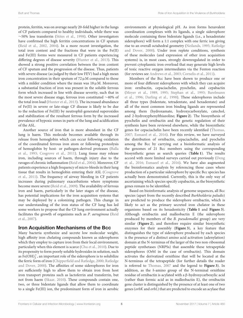
protein, ferritin, was on average nearly 20-fold higher in the lungsof CF patients compared to healthy individuals, while there was∼50% less transferrin (Stites et al., 1998). Other investigatorshave confirmed the high ferritin concentrations in CF sputum(Reid et al., 2002, 2004). In a more recent investigation, thetotal iron content and the fractions that were in the Fe(II)and Fe(III) forms were measured in CF patients experiencingdiffering degrees of disease severity (Hunter et al., 2013). Thisshowed a strong positive correlation between the iron contentof CF sputum and the progression of the disease. Thus, patientswith severe disease (as judged by their low FEV) had a high meaniron concentration in their sputum of 72µM compared to thosewith a milder condition where the mean was 18µM. Moreover,a substantial fraction of iron was present in the soluble ferrousform which increased in line with disease severity, such that inthe most severe disease stage, ferrous iron constituted ∼40% ofthe total iron load (Hunter et al., 2013). The increased abundanceof Fe(II) in severe or late-stage CF disease is likely to be dueto the reduction of Fe(III) by neutrophil-generated superoxidesand stabilization of the resultant ferrous form by the increasedprevalence of hypoxic zones in parts of the lung and acidificationof the ASL.
Another source of iron that is more abundant in the CFlung is haem. This molecule becomes available through itsrelease from hemoglobin which can occur following oxidationof the coordinated ferrous iron atom or following proteolysisof hemoglobin by host- or pathogen-derived proteases (Ballaet al., 1993; Cosgrove et al., 2011). Lung tissue may releaseiron, including sources of haem, through injury due to theravages of chronic inflammation (Reid et al., 2004). Moreover, CFpatients experience a high frequency ofmicro-bleeds in their lungtissue that results in hemoglobin entering their ASL (Cosgroveet al., 2011). The frequency of airway bleeding in CF patientsincreases during pulmonary exacerbations where symptomsbecome more severe (Reid et al., 2009). The availability of ferrousiron and haem, particularly in the later stages of the disease,has potential implications for the iron acquisition systems thatmay be deployed by a colonizing pathogen. This change inour understanding of the iron status of the CF lung has ledsome workers to propose that the CF lung environment actuallyfacilitates the growth of organisms such as P. aeruginosa (Reidet al., 2007).
Iron Acquisition Mechanisms of the BccMany bacteria synthesize and secrete low molecular weight,high affinity iron chelating compounds known as siderophoreswhich they employ to capture iron from their local environment,particularly when this element is scarce (Chu et al., 2010). Due toits propensity to form poorly soluble hydroxides in solution, suchas Fe(OH)+2 , an important role of the siderophore is to solubilizethe ferric form of iron (Chipperfield and Ratledge, 2000; Ratledgeand Dover, 2000). The affinities of some siderophores for ironare sufficiently high to allow them to obtain iron from hostiron transport proteins such as lactoferrin and transferrin, butnot from haem (Skaar, 2010). These compounds contain one,two, or three bidentate ligands that allow them to coordinateto a single Fe(III) ion, the predominant form of iron in aerobic
environments at physiological pH. As iron forms hexavalentcoordination complexes with its ligands, a single siderophoremolecule containing three bidentate ligands (i.e., a hexadentatesiderophore) will form a 1:1 complex with one ferric ion givingrise to an overall octahedral geometry (Neilands, 1995; Ratledgeand Dover, 2000). Under iron replete conditions, synthesisof these molecules (and expression of other iron acquisitionsystems) is, in most cases, strongly downregulated in order toprevent cytoplasmic iron overload that may generate high levelsof toxic reactive oxygen intermediates via the Fenton reaction(for reviews see Andrews et al., 2003; Cornelis et al., 2011).
Members of the Bcc have been shown to produce one ormore of four different siderophores with which they can acquireiron: ornibactin, cepaciachelin, pyochelin, and cepabactin(Meyer et al., 1989, 1995; Stephan et al., 1993; Barelmannet al., 1996; Darling et al., 1998). These siderophores includeall three types (bidentate, tetradentate, and hexadentate) andall of the most common iron binding ligands are representedamong them (hydroxamate, hydroxycarboxylate, catechol,and 2-hydroxyphenylthiazoline; Figure 2). The biosynthesis ofpyochelin and ornibactin and the genetic regulation of theirsynthesis have been reviewed elsewhere, while the biosyntheticgenes for cepaciachelin have been recently identified (Thomas,2007; Esmaeel et al., 2016). For this review, we have surveyedthe distribution of ornibactin, cepaciachelin, and pyochelinamong the Bcc by carrying out a bioinformatic analysis ofthe genomes of 21 Bcc members using the correspondingbiosynthetic genes as search queries (Table 1). The resultsaccord with more limited surveys carried out previously (Denget al., 2016; Esmaeel et al., 2016). We have also augmentedthe bioinformatics analysis by referencing those cases whereproduction of a particular siderophore by specific Bcc species hasactually been demonstrated. Currently, this is the only way ofascertaining which species specify cepabactin, as the biosyntheticgenes remain to be identified.
Based on bioinformatic analysis of genome sequences, all Bccspecies (apart from the recently described Burkholderia paludis)are predicted to produce the siderophore ornibactin, which islikely to act as the primary secreted iron chelator in theseorganisms based on its hexadenticity (Table 1 and Figure 3).Although ornibactin and malleobactin E (the siderophoreproduced by members of the B. pseudomallei group) are verysimilar (Figure 2), and therefore require similar biosyntheticenzymes for their assembly (Figure 3), a key feature thatdistinguishes the type of siderophore produced by each speciesis the presence of a distinct amino acid activation (adenylation)domain at the N-terminus of the larger of the two non-ribosomalpeptide synthetases (NRPSs) that assemble these tetrapeptidesiderophores (OrbI in the case of ornibactin). This domainactivates the derivatized ornithine that will be located at theN-terminus of the tetrapeptide (for further details the readeris referred to Thomas, 2007 and the legend to Figure 3). Inaddition, as the δ-amino group of the N-terminal ornithineresidue of ornibactin is acylated with a β-hydroxycarboxylic acid(rather than formic acid as in malleobactin E), the ornibactingene cluster is distinguished by the presence of at least one of twogenes (orbK and orbL) that are predicted to encode an acylase that
Frontiers in Cellular and Infection Microbiology | www.frontiersin.org 5 November 2017 | Volume 7 | Article 460

Butt and Thomas Role of Iron Acquisition in the Virulence of Burkholderia
FIGURE 2 | Structure of siderophores produced by Burkholderia species. (A) Ornibactins contain an N-terminal ornithine that is acylated with a C4, C6, or C8
β-hydroxycarboxylic acid on the δ-amino nitrogen atom, giving rise to ornibactin-C4, -C6, or -C8. The δ-amino nitrogen atom is also hydroxylated. The other three
amino acids in the tetrapeptide are D-hydroxyaspartate, L-serine and the C-terminal ornithine that is formylated and hydroxylated on the δ-amino nitrogen atom and
the carboxyl group is conjugated to putrescine. As with the malleobactins, they contain two bidentate β-hydroxycarboxylate ligands and a single bidentate
hydroxamate ligand. (B) Malleobactin E, the siderophore-active malleobactin congener of B. thailandensis. (C) The siderophore-active malleobactin congener of B.
xenovorans, tentatively referred to here as “malleobactin X.” (D) Cepaciachelin contains two 2,3-DHBA groups that form amide linkages with the two amino groups of
lysine, which in turn is conjugated to a molecule of putrescine (1,4-diaminobutane) on its α-carboxyl group. (E) Pyochelin contains two less commonly occurring
bidentate iron-chelating groups (2-hydroxyphenyl thiazoline and N-methylthiazolidine-4-carboxylate). (F) Cepabactin, a cyclic hydroxamate bidentate siderophore.
Chemical groups that distinguish the ornibactins and malleobactins are indicated in red circles or ellipses.
catalyses this condensation reaction (Figure 3). Whereas orbLis always present, orbK may contain an internal deletion (as inB. ubonensis) or be absent from the cluster altogether (as in B.vietnamiensis). Although, the single reported B. paludis strainis an environmental isolate, it should be noted that in a surveyof “B. cepacia” CF isolates carried out prior to the taxonomicreorganization of B. cepacia into separate Bcc species, two clinicalstrains were found not to produce detectable levels of ornibactin(Darling et al., 1998). It is not clear to which Bcc member speciesthey belong or whether they are indeed members of the Bcc.One possibility is that these strains were capable of producingornibactin prior to infection but this ability was lost throughmutation during prolonged carriage as has been observed withrespect to production of the major siderophore pyoverdine bysome P. aeruginosa strains isolated from chronically infected CFpatients (De Vos et al., 2001; Smith et al., 2006; Andersen et al.,2015).
In addition, most Bcc species produce one or more secondarysiderophores that are likely to have lower affinity for ironthan ornibactin. The gene clusters specifying the biosynthesisand utilization of two of these siderophores, cepaciachelinand pyochelin, are shown in Figures 4A,B. Based on ourbioinformatics survey, at least 7 species of Bcc, including B.cenocepacia and B. lata, produce pyochelin as the secondary
siderophore, a feature associated with some Pseudomonas species(Cornelis and Matthijs, 2002), whereas in 8 other species,including some strains of B. ambifaria and B. multivorans, thesecondary siderophore is cepaciachelin (Table 1). We have notidentified a species possessing the genetic information requiredto produce both cepaciachelin and pyochelin. Both compoundsare tetradentate siderophores, although the former belongs tothe 2-hydroxyphenylthiazoline family whereas cepaciachelin isa bis-catecholate siderophore (Barelmann et al., 1996; Thomas,2007; Inahashi et al., 2017). Some species do not appearto produce either of these two compounds as a secondarysiderophore (Table 1). Another siderophore, the bidentatecyclic hydroxamate, cepabactin, has been detected in culturesupernatants of some environmental B. cepacia strains inaddition to ornibactin and pyochelin (see Table 1; Meyer et al.,1989, 1995). The ability of some clinical Bcc isolates of unknowntaxonomic status to produce cepabactin has also been observed(Darling et al., 1998). Currently, it is not possible to infer frombioinformatics how widespread the synthesis or utilization of thissiderophore is likely to be among the Bcc, although its productionhas not been observed in B. cenocepacia and B. vietnamiensisstrains (Table 1).
The uptake of ferric-siderophore complexes by Gram-negative bacteria such as the Burkholderia requires an outer
Frontiers in Cellular and Infection Microbiology | www.frontiersin.org 6 November 2017 | Volume 7 | Article 460

Butt and Thomas Role of Iron Acquisition in the Virulence of Burkholderia
FIGURE 3 | Organization of ornibactin, malleobactin, and phymabactin biosynthesis and utilization genes in pathogenic Burkholderia and related species. Genes are
represented by block arrows and are color coded as indicated in the figure [the precise role of the MbtH-like OrbH/MbaG/PhmF proteins is unknown but they are
assumed to be required for biosynthesis of the siderophore based on the requirement for other MbtH-like proteins for NRPS-mediated biosynthesis of some peptides;
(Wolpert et al., 2007; Baltz, 2011)]. The numbering of each gene cluster corresponds to the numbering system for the species listed at the bottom of the figure. The
font color used for each species name corresponds to the siderophore produced as follows: black, ornibactin; red, malleobactins; green, phymabactin.
NRPSpredictor2 (Rottig et al., 2011) was used to predict the siderophore product based on the substrates accepted by the four adenylation domains present in
OrbI/OrbJ, MbaA/MbaB, and PhmA/PhmB for each species. For systems known to specify ornibactin, the first and last (N- and C-terminal) adenylation domains of
the OrbI-OrbJ NRPS pair are both predicted to accept leucine with highest probability, reflecting the presence of N5-3-hydroxyacyl-N5-hydroxyornithine and
N5-formyl-N5-hydroxyornithine, respectively, at these positions in the tetrapeptide product. For malleobactin, the predicted specificity of the N-terminal adenylation
domain changes to β-hydroxytyrosine although the accepted substrate is N5-formyl-N5-hydroxyornithine. In some species, such as B. thailandensis, the N-terminal
N5-formyl-N5-hydroxyornithine is formylated on the N2-amino group upon formation of the tetrapeptide to generate malleobactin E (Franke et al., 2015), whereas in
B. xenovorans the N-terminal N5-formyl-N5-hydroxyornithine does not appear to undergo such a tailoring reaction (Vargas-Straube et al., 2016) and so we tentatively
refer to this siderophore as “malleobactin X.” The N- and C-terminal adenylation domains of PhmA-PhmB are predicted to accept aspartate and cysteine, respectively,
but the structure of the product, phymabactin, is unknown, although it is predicted to have siderophore activity based on its genomic context (Esmaeel et al., 2016). In
all cases, the second and third adenylation domains are predicted to accept aspartate and serine, respectively, which correspond to β-hydroxy-D-aspartate and
L-serine in the final product. The nomenclature proposed for each gene is shown below the gene clusters, and the gene designations are color coded as follows:
ornibactin, black (Agnoli et al., 2006); the two systems for malleobactin, red (upper, Alice et al., 2006; lower, Franke et al., 2013); the two systems for phymabactin,
green (upper, Esmaeel et al., 2016; lower, this study). Genes indicated by a single letter have the same prefix as the gene name at the extreme left. Dashes indicate
the absence of a gene. Question mark indicates where a gene name has not been proposed. The initial annotation of the phymabactin gene cluster (Esmaeel et al.,
2016) did not include the third and last genes in the cluster or the fact that a TBDR gene occurs in other species bearing this gene cluster (upper annotation in green
font). Therefore, we have proposed an alternative nomenclature based on the ornibactin gene cluster (lower annotation in green font). Scale bar refers to gene lengths
and not intergenic regions, which in some cases have been exaggerated to permit alignment of each gene cluster. Species marked with an asterisk belong to a
subclade within the B. xenovorans group and have been reassigned to the new genus Paraburkholderia (Sawana et al., 2014). Collimonas is a genus within the
Oxalobacteraceae, a family belonging to the order Burkholderiales. Member species of the Bcc are enclosed in a box (B. arboris is not listed as its genome sequence
is not currently available). Gene loci are shown in Supplementary Table 1.
Frontiers in Cellular and Infection Microbiology | www.frontiersin.org 7 November 2017 | Volume 7 | Article 460

Butt and Thomas Role of Iron Acquisition in the Virulence of Burkholderia
FIGURE 4 | Organization of the cepaciachelin and pyochelin biosynthesis and
utilization genes and the genes for the haem uptake system in Burkholderia
species. (A) The cepaciachelin gene cluster in Burkholderia species encodes
enzymes for the synthesis of the precursor 2,3-dihydroxybenzoic acid (DHBA)
from chorismate and its assembly into the siderophore. Genes encoding
possible cepaciachelin transport proteins are also present, including the
TonB-dependent receptor, CpcG, and an MFS transporter, CpcH. Most of the
genes have been previously annotated (Esmaeel et al., 2016) but the
annotation has been extended here (genes labeled in red font) and includes
components of a cytoplasmic membrane ABC transporter which may be
involved in uptake of ferric cepaciachelin (CpcF, -I, and -J). EstA is homologous
to the cytoplasmic enterobactin and salmochelin esterases Fes and IroD that
are required for removal of iron from ferric-enterobactins following their uptake.
However, it contains a putative Sec-dependent signal peptide and so may be
periplasmically located like the C. jejuni enterobactin esterase, Cee (Zeng et al.,
2013). This may suggest that the putative cepaciachelin receptor (CpcG) and
the cytoplasmic membrane transporter also recognizes ferric-enterobactin.
With the exception of B. ambifaria and B. pseudomultivorans the
cepaciachelin gene cluster includes a gene encoding a DAHP synthase which
catalyses the first step in the shikimate pathway that leads to the biosynthesis
of chorismate from erythrose-4-phosphate and PEP. Note that in Esmaeel
et al. (2016) cphA-cphC should be annotated as cpcA-cpcC, as shown here
(V. Leclere, personal communication). -, gene not assigned a four letter name.
Gene loci are shown in Supplementary Table 2. (B) The pyochelin gene cluster.
Genes annotated with a single letter are designated with the prefix pch.
Products of the biosynthetic genes generate the precursor salicylic acid from
chorismate (PchAB), activate it (PchD) and assemble it into pyochelin along
with two molecules of cysteine (PchCEFG). The pchHI and fptABCX genes
encode membrane proteins, of which two (FptA and FptX) are involved in the
transport of exogenous ferric-pyochelin across the outer and inner
membranes, respectively. fptBC and pchHI appear not to be essential for
export of pyochelin nor for uptake of iron-bound pyochelin (see Youard et al.,
2011 for a review). Gene loci are shown in Supplementary Table 3.
(C) Organization of the Burkholderia haem uptake genes, bhuRSTUV (Shalom
et al., 2007; Thomas, 2007), also referred to as hmuRSTUV, huvA-hmuSTUV
or omr-hmuSTUV (Yuhara et al., 2008; Kvitko et al., 2012; Tyrrell et al., 2015).
Note that in B. stagnalis a VOC family protein is encoded between bhuU and
bhuV, and in B. gladioli the bhu genes are organized into two operons present
on separate chromosomes. Gene loci of representative species are given in
Supplementary Table 4.
membrane receptor, a 75–85 kDa polypeptide that folds into aβ-barrel containing a central plug domain. Binding of a ferric-siderophore complex to the external face of the receptor triggersa conformational change in the gated receptor that allows accessof the complex to the periplasmic space. The energy required
for this process is derived from the proton motive force throughthe action of the TonB system, a complex of three differentcytoplasmic membrane-anchored protein subunits: TonB, ExbB,and ExbD (Noinaj et al., 2010; Celia et al., 2016). For thisreason, ferric-siderophore receptors are referred to as TonB-dependent receptors (TBDRs) or TonB-dependent transporters(TBDTs). As an example, OrbA is the TBDR for ferric-ornibactin(Figure 5A). Once the ferric-siderophore complex has enteredthe periplasmic space, the ferric ion is transported across thecytoplasmic membrane, either in complex with the siderophoreor following release from the siderophore (depending on thesystem). The cytoplasmic membrane transporters are often ATP-binding cassette (ABC) transporters that consist of a periplasmicbinding protein, an intrinsic membrane protein (the permease)and an ATPase located on the cytoplasmic face of the permease(Krewulak and Vogel, 2008). This type of system operates forthe uptake of ferric ornibactin (Orb-B, -C, and -D) and possiblyalso for cepaciachelin (CpcF, -I, and -J) in the Bcc, as well as forthe import of ferric malleobactin in B. pseudomallei and relatedbacteria (Figures 5A,B; Agnoli et al., 2006). In the case of ferric-pyochelin, a single subunit permease, FptX, appears to serve asthe cytoplasmic membrane transporter (Figure 5C; Cuiv et al.,2004; Cunrath et al., 2015). For ferric-siderophore complexes thatenter the cytoplasm, the iron is removed from the siderophorethrough its reduction to Fe(II), which is presumed to occurfor ornibactin (Agnoli et al., 2006), or through modification orhydrolysis of the siderophore (Brickman and McIntosh, 1992;Hannauer et al., 2010).
Like many other bacterial species, Burkholderia spp.encode additional TBDRs that may allow them to utilizesiderophores that are produced by other bacteria and fungi(“xenosiderophores”), although the potential importance ofthese compounds for pathogenicity is only likely to be realizedin the context of a polymicrobial infection. Based on ananalysis of its translated genome, B. cenocepacia is predictedto encode at least 20 TBDRs, many of which are likely to beinvolved in utilization of xenosiderophores (our unpublishedresults). At present, little is known concerning the nature ofthe xenosiderophores that can be utilized by the Burkholderia.However, the presence of the ornibactin transport genes in B.paludis, but not the biosynthetic genes, strongly suggests thatornibactin is likely to be utilized as a xenosiderophore by thisspecies.
Members of the Bcc also specify iron acquisition systemsthat are not siderophore-dependent. For example, it has beenshown that B. cenocepacia can utilize haem as an iron source(Whitby et al., 2006; Mathew et al., 2014; Tyrrell et al., 2015).B. cenocepacia and B. multivorans contain a cluster of genes(bhuRSTUV) that are predicted to be required for uptake of thismolecule (Figure 4C; Thomas, 2007; Yuhara et al., 2008). [Notethat there is currently a lack of consistency regarding the geneticnomenclature for this system in the Burkholderia (see below andlegend to Figure 4).] Our bioinformatic survey shows that thebhu cluster is present on chromosome 2 in nearly all Bcc speciesfor which whole genome sequence information is available(Supplementary Table 4). The exception is B. vietnamiensiswhichcompletely lacks the bhuRSTUV gene cluster. Although, there
Frontiers in Cellular and Infection Microbiology | www.frontiersin.org 8 November 2017 | Volume 7 | Article 460

Butt and Thomas Role of Iron Acquisition in the Virulence of Burkholderia
FIGURE 5 | Proposed iron uptake pathways in the Burkholderia. (A) The ornibactin/malleobactin uptake system. Ferric-ornibactin is recognized by the OrbA/MbaD
TBDR and is translocated into the periplasmic space through a conformational change in the plug domain of the TBDR that requires energy transduction by the TonB
complex (TonB-ExbB-ExbD). The iron-siderophore complex is then transported across the cytoplasmic membrane by a periplasmic binding protein-dependent ABC
transporter (OrbBCD/MbaLIJ). Oncethe ferric-siderophore complex has been internalized, iron is released from ornibactin through its reduction to the ferrous form by
OrbF/MbaK. (B) Ferric-cepaciachelin is proposed to require the CpcG TBDR. Genes neighboring cpcG encode a periplasmic binding protein-dependent ABC
transporter (CpcFHI) that may be involved in transport of the bis-catecholate complex across the cytoplasmic membrane. (C) Ferric-pyochelin uptake requires the
FptA TBDR and the single subunit cytoplasmic membrane transporter, FptX. (D) Uptake of haem via the Bhu system. Haem uptake is proposed to follow an
analogous pathway to that of ornibactin/malleobactin and cepaciachelin. Cytoplasmic haem is bound by the BhuS protein which is proposed to play a role in haem
trafficking and haemostasis. If required, iron can be released form haem by haem oxygenases (not shown). The FtrABCD system is not shown. OM, outer membrane;
CM, cytoplasmic membrane.
have not been any major studies carried out to investigate therole of this system in haem acquisition in the Bcc, this functionhas been established for the bhuRSTUV system in B. pseudomallei(see below), and so one can confidently infer its role in haemuptake in the Bcc. Haem appears to be an important sourceof iron for P. aeruginosa during colonization of the CF lung,and therefore one might expect members of the Bcc to takeadvantage of this nutrient (Konings et al., 2013). B. cenocepaciacan also obtain iron from ferritin in a protease-dependent process(Whitby et al., 2006; Mathew et al., 2014; Tyrrell et al., 2015).Interestingly, although iron is present in the ferric form whensequestered by ferritin, siderophores are not required for uptakeof ferritin-derived iron in B. pseudomallei (Kvitko et al., 2012).
Ferrous iron is very soluble and can passage across theouter membrane of Gram-negative bacteria through porins inan energy-independent process. However, it requires specifictransporters for translocation across the cytoplasmic membranein all bacteria. The Feo system is one such ferrous iron-specificuptake system that is present in many Gram-positive and Gram-negative bacteria (Lau et al., 2016). This system consists of acytoplasmic membrane transport protein (FeoB) and (in mostcases) a cytoplasmic component of unknown function (FeoA). Insome cases, a third component (FeoC) is also present. Ferrousiron is likely to be an important source of this essential nutrientfor bacteria colonizing the CF lung based on its increasedabundance in the ASL of more severely afflicted CF patients.Accordingly, it has been shown that the Feo system of P.aeruginosa is upregulated during colonization of the lungs ofevery individual in a cohort of 23 infected CF patients (Konings
et al., 2013). For this reason, we conducted a survey of humanpathogenic Burkholderia species for the presence of feoA andfeoB. However, only a small a minority of B. multivorans and B.pseudomultivorans strains (not the type strains), as well as thesingle currently identified B. paludis strain (MSh1), were foundto encode a FeoB-like protein, although these proteins lacked aregion of∼100 amino acids that is present in P. aeruginosa FeoB.There were no matches when FeoA was used as the query in aBLASTP search of the Bcc and B. pseudomallei. We concludethat the vast majority of pathogenic Burkholderia species lack thisparticular ferrous iron uptake system.
B. cenocepacia encodes a siderophore-independentmechanism for iron assimilation that is very similar to theFtrABCD systems reported in Bordetella and Brucella (Brickmanand Armstrong, 2012; Elhassanny et al., 2013; Mathew et al.,2014). These systems share similarities with components ofthe Escherichia coli EfeUOB system that serves to importferrous iron under aerobic conditions at low pH (Cao et al.,2007). Accordingly, in Bordetella spp. and Brucella abortus,ferrous iron is efficiently transported into these bacteria bythe FtrABCD system over the pH range 6.0–7.5, in a processwhere the Fe(II) ion is oxidized to the ferric form using theFtrB cupredoxin component before translocation across thecytoplasmic membrane. In contrast, the B. cenocepacia FtrABCDsystem appears to utilize ferric iron as the substrate. Giventhe absence of a Feo system in B. cenocepacia, this begs thequestion as to the mechanism by which this species can uptakeferrous iron. B. multivorans also possesses an FtrABCD system,but in contrast to that of B. cenocepacia it does utilize ferrous
Frontiers in Cellular and Infection Microbiology | www.frontiersin.org 9 November 2017 | Volume 7 | Article 460

Butt and Thomas Role of Iron Acquisition in the Virulence of Burkholderia
iron as the substrate (S.C. Andrews, unpublished results).Therefore, it is possible that this systemmay play a role in ferrousiron acquisition in the CF lung for at least some Bcc species,particularly given the abundance of ferrous iron in combinationwith the relatively low pH of CF ASL.
Although, the substantial proportion of iron that is in theferrous form and the increased abundance of haem in theASLmay implicate the siderophore-independent iron acquisitionmechanisms of the Bcc in establishing CF lung infections, as yet,the role of these systems in the context of CF have not beeninvestigated in any detail (see below).
Experimental Evidence for the Role of IronAcquisition Systems in the Virulence of theBccIn Vivo Studies
The earliest study on the role of iron acquisition mechanismsin the virulence of members of the Bcc was carried out priorto the discovery of the primary siderophore, ornibactin, in thisgroup of bacteria (Sokol, 1986). Here, it was observed that froma collection of 43 “P. cepacia” CF isolates, ∼50% produceddetectable levels of pyochelin during iron limited growth invitro (Pch+ phenotype). However, 86% of the Pch+ strains wereassociated with infections which had led to the death of thepatient or were responsible for severe infections, whereas only41% of Pch− strains were associated with such outcomes. Thus,while the ability to colonize a CF patient could not be linkedto the ability to produce this siderophore, there was a linkbetween pyochelin production and the morbidity/mortality ofdisease in CF patients. Similarly, while addition of exogenouspyochelin to two pyochelin-negative Bcc strains did not increasebacterial numbers or bacterial persistence in infected rat lungs,it did increase the severity of infection as assessed by lungpathology (Sokol and Woods, 1988). It was proposed thatthe observed enhancement of lung damage brought about byexogenous addition of pyochelin to these strains was mostlikely due to increased dissemination of the bacteria throughoutthe lungs. However, it is not clear from these studies whetherit was the role of pyochelin in iron acquisition that wasresponsible for the more severe outcome or some other effectof the siderophore. In this regard, it is known that apartfrom binding iron and other metals (Cuppels et al., 1987;Visca et al., 1992; Baysse et al., 2000; Braud et al., 2009)pyochelin possesses an inherent chemical reactivity that maycontribute to disease severity. For example, it can promote thedegradation of organotin derivatives (Sun et al., 2006), andperhaps pertinently, it can catalyse the generation of ROS suchas hydroxyl radicals that result in tissue damage (Coffman et al.,1990; Britigan et al., 1994, 1997; Adler et al., 2012). The latterproperty is proposed to contribute to the known antibioticactivity of this siderophore (Adler et al., 2012; Ong et al., 2016).It should be noted that one important Bcc pathogen of CFpatients, B. multivorans, does not produce pyochelin (Table 1)and the clonally related B. cenocepacia CF epidemic strainsK56-2 and J2315 produce very little of this siderophore (seebelow).
A direct genotypic-phenotypic link between iron acquisitionand the virulence of B. cenocepacia was observed during aninvestigation of the virulence potential of ornibactin deficientmutants in rodent models of both chronic and acute respiratoryinfection (Sokol et al., 1999). In this study, mutants derivedfrom the highly transmissible epidemic B. cenocepacia strain,K56-2, which contained an insertionally inactivated pvdAgene that is required for ornibactin synthesis (Figure 3), weregenerated by transposon mutagenesis (pvdA::Tn5-OT182) andallelic replacement (pvdA::tp). Both mutants were significantlyattenuated in these models. Thus, in a rat lung chronic infectionmodel, the number of pvdA::Tn5-OT182 bacterial cells recoveredfrom the lung was 4 logs lower than that of the wild type K56-2 strain at 28 days post infection. Furthermore, the pvdA::tpstrain could not be recovered from the lungs after the samelength of time, suggesting the infection had been cleared.The degree of pathology, as determined by the amount ofinflammatory cell infiltration and exudate in the lungs, wasalso significantly reduced in the K56pvdA::tp strain comparedto that of K56-2. Aerosol administration of K56-2 and thepvdA::Tn5-OT182 mutant into neutropenic mice as an acuterespiratory infection model revealed that whereas the wild typestrain was able to persist in the lung 7 days post infection,the pvdA mutant was cleared from most of the mice after 3days. These experiments suggested the importance of ornibactin-mediated iron acquisition by the bacteria for initial colonization,persistence and resulting pathological changes within the host.However, given the difference in the iron content of the lungs ofa healthy individual compared to those of a CF patient, it is notclear to what extent the conclusions from this study, which didnot involve CF mice or rats, can be extrapolated to the situationin the CF lung.
While the data indicated an important role for ornibactinin lung colonization in these models, the K56-2 strain (andother members of the highly transmissible ET12 epidemiclineage) produces very low amounts of the siderophore pyochelincompared to other B. cenocepacia strains due to a frameshiftmutation in pchF (Darling et al., 1998; Holden et al., 2009)(Based on perusal of the genome sequence, we presume pyochelinbiosynthesis in these strains occurs through independentinitiation of translation from an internal in-frame GUG codonlocated upstream of the frameshift site that results in productionof PchF as two separate components). Thus, the role of pyochelinin the lung infection model could not be established using ET12strains. However, given that ET12 strains cause life-threateninginfections in CF patients, this would again appear to rule outan important role for pyochelin in colonization of the CF lungby B. cenocepacia. Later work using a B. cenocepacia strain(Pc715jorbA::tp) that produced normal amounts of pyochelin butwas unable to utilize ferric-ornibactin due to disruption of thegene encoding the ferric-ornibactin TBDR, OrbA, revealed thatit was cleared from rat lungs much more quickly than the WTstrain (Visser et al., 2004). A ferric-pyochelin receptor mutant(Pc715jfptA::tp) persisted with the same efficiency as that of theWT. These data suggested that while pyochelin may have a rolein the severity of infection, it is unable to compensate for theloss of a functional ornibactin utilization system. Therefore, it is
Frontiers in Cellular and Infection Microbiology | www.frontiersin.org 10 November 2017 | Volume 7 | Article 460

Butt and Thomas Role of Iron Acquisition in the Virulence of Burkholderia
ornibactin which appears to be important in order to establish aninfection in this system. This may be explained by the presumedlower affinity of pyochelin than ornibactin for iron (Cox andGraham, 1979; Visca et al., 1993).
The importance of the ornibactin system for the virulence ofB. cenocepacia has also been assessed in other infection models,including invertebrates and plants. Both a K56-2 orbA mutantand a K56-2 pvdAmutant were attenuated in the Caenorhabditiselegans and Galleria mellonella invertebrate models. The pvdAmutant was also slightly attenuated in the plant alfalfa model(Uehlinger et al., 2009). An orbJ mutant of the B. cenocepacia CFstrain, H111, that is also deficient in the production of ornibactin,was also attenuated in the G. mellonella system (Mathew et al.,2014). Consistent with the virulence of the K56-2 strain in therat lung chronic infection model, an H111 1pchAB pyochelindeficient mutant was still virulent in G. mellonella.
Using a modified signature-tagged mutagenesis (STM)procedure to identify genes required for survival in therat chronic lung infection model, one of the attenuated B.cenocepacia K56-2 mutants which could not survive for 10 daysin this model contained a transposon inserted just upstream of anORF that encoded a haem TBDR-like protein (Hunt et al., 2004).The authors of this study indicated that the gene was the first in acluster of genes associated with haem uptake that were locatedon chromosome 2, and the encoded protein was very similarto the 79 kDa RS03722 gene product of Ralstonia solanacearumstrain GMI1000 (now reannotated as RSp0244). As RSp0244 ishighly similar to BhuR we conclude that the plasposon insertionexerted polar effects on expression of the bhuRSTUV operonthat impaired or abolished haem uptake. In contrast, genesinvolved in the biosynthesis and transport of ornibactin andpyochelin were not implicated in this study (Hunt et al., 2004).These observations would suggest that haem acquisition, andnot siderophore-mediated iron acquisition, is an essential traitfor persistence in the rat lung. Notwithstanding the differenttime courses of the chronic lung infection models, it is not clearwhy the ornibactin system should be implicated in some studies(Sokol et al., 1999; Visser et al., 2004) but not in the STM study,particularly as the ornibactin gene cluster presents a large targetfor plasposon-mediated disruption. One possible explanation isthe selection procedure employed to construct the B. cenocepaciatransposon mutant library. Here, mutants were selected on amineral salts based medium in order to exclude auxotrophs.A K56-2 mutant in which the plasposon has disrupted a generequired for ornibactin synthesis or utilization would effectivelybe unable to obtain iron in a siderophore-dependent manner.Wehave observed that B. cenocepacia siderophore deficient mutantsgrow more slowly that the parental wild type strain on mineralsalts medium (see for example Asghar et al., 2011), and it ispossible that such mutants were omitted from the library of Huntet al. This is not a complete explanation, however, as the haemuptake deficient mutants in the STM study can still produceornibactin and so they might be expected to retain virulencebased on other studies. This may suggest that a combination ofboth iron acquisition systems (haem- and ornibactin-mediated)is required for efficient colonization and persistence in the ratlung model.
Finally, the B. cenocepacia FtrABCD system was alsoinvestigated for a potential role in virulence in the Galleria waxmoth model. Whereas deletion of the ftr system in isolation didnot result in reduced virulence, when deleted in a strain thatwas unable to biosynthesise ornibactin and pyochelin, themutantwas more attenuated in comparison to an ornibactin-negativestrain. This observation suggests that while ornibactin is themoreimportant iron acquisition system for virulence in this model,the FtrABCD system can play a role in iron acquisition duringinfection in the absence of siderophores (Mathew et al., 2014).
In Vitro Studies
Changes in the environmental iron concentration or availabilitycan also trigger adaptive changes in expression of virulence traitsthat are not directly connected to iron acquisition but rather serveother roles that contribute to survival of the bacterium underthe prevailing conditions. Such examples include regulatingbiofilm formation in P. aeruginosa and capsule production inCryptococcus neoformans (Singh et al., 2002; Banin et al., 2005;Jung et al., 2006). For B. cenocepacia it has been shown thatmodulating the concentration of iron causes a switch fromplanktonic to sessile growth. Thus, supplementing liquid cultureswith ferric iron concentrations ranging from 1 to 100µMresulted in increased levels of extracellular matrix production bystrain PVI as the iron concentration was increased. Furthermore,biofilm formation induced by growth under high iron conditionsresulted in more efficient invasion of A549 epithelial monolayerscompared to cells grown in lower iron conditions that did notproduce biofilm (Berlutti et al., 2005). In contrast, adherence of B.cenocepacia to A549 cells was more efficient under iron limitingconditions. The consequences of this for B. cenocepacia CF lunginfections are not yet clear.
Gene Expression and Omics Studies
Transcriptomic and proteomic analyses can be used to identifysets of genes or proteins that may be required for survival underparticular conditions by virtue of their differential expression. Afew such studies have been carried out with B. cenocepacia thathave suggested an important role for iron uptake mechanismsin bacterial persistence within CF patients. However, as wediscuss below, the experimental set up may not necessarily beappropriate for addressing this particular question. The firstnotable study looked at global gene expression during growth ofB. cenocepacia J2315 in a basal salts medium supplemented withCF sputum in comparison to growth in unsupplementedmedium(Drevinek et al., 2008). The microarray revealed upregulation of287 genes and downregulation of 437 other genes during growthin CF sputum medium. However, only two of the upregulatedgenes were associated with characterized iron acquisition systemsin this bacterium. These two genes (pchR and pchD) encodethe transcription activator of the pyochelin gene cluster and anenzyme required for biosynthesis of the pyochelin precursor,salicylic acid, respectively, but their expression increased only 2-fold. Transcription of the ornibactin genes was not upregulated,although the gene encoding the global iron repressor, Fur,that represses the ornibactin gene cluster, was found to be
Frontiers in Cellular and Infection Microbiology | www.frontiersin.org 11 November 2017 | Volume 7 | Article 460

Butt and Thomas Role of Iron Acquisition in the Virulence of Burkholderia
downregulated 2- to 3-fold (Agnoli et al., 2006; Drevinek et al.,2008).
The authors of this work measured the iron content oftheir sputum medium and found it to be ∼35µM, which ismore than adequate to sustain growth of B. cenocepacia instandard laboratory medium without upregulating siderophorebiosynthesis (Drevinek et al., 2008; Madeira et al., 2013; ourunpublished results). Despite the presence of CF sputum inthe medium, which has been argued by some investigators tosequester iron due to the presence of various iron bindingcomponents (Wang et al., 1996; Palmer et al., 2007), their resultsimply that sufficient iron is available to effect repression of theornibactin system. Accordingly, the observed induction of thepch genes may not be related to iron depletion but is rather aresponse to the presence of another component in CF sputum.It is noteworthy that genes encoding other components of thepyochelin biosynthesis machinery (particularly those enzymesrequired to assemble pyochelin from salicylate and cysteine)and alternative iron uptake systems (FtrABCD and BhuRSTUV)were also not upregulated. The authors contend that the CFmedium they employed restricted the available iron based on theobserved gross upregulation of the BCAL0270 gene, which theyconsidered to be involved in iron acquisition (described in thestudy as a “ferric reductase-like transmembrane component”).However, in a basal salts medium without iron supplementation,this gene was upregulated only 2-fold compared to mediumcontaining the standard amount of ferrous sulfate (43µM).Moreover, this gene is currently annotated in the NCBI databaseas encoding a “sulfoxide reductase heme-binding subunit YedZ”and it is transcriptionally linked to BCAL0269, a gene thatencodes a YedY-homologous protein. Our own bioinformaticsanalysis supports this annotation (results not shown). The E. coliYedYZ complex is a membrane anchored haem-molybdoenzymethat serves to reduce an as yet unknown S- or N-oxide (Iobbi-Nivol and Leimkuhler, 2013). Therefore, there is little evidenceto support the suggestion that BCAL0270 is involved in ironacquisition. The possibility that the CF sputum medium ofDrevinek et al. (2008) is iron sufficient accords with the highiron concentration included in the basal salts medium used togenerate the medium. Notwithstanding the fact that the sputumis present at only 10% (w/v) in the medium, the inability of theadded CF sputum to induce iron acquisition systems may suggestthat its ability to sequester iron is somewhat limited and/or it isiron replete.
The results of Drevinek et al. (2008) contrast with those ofPalmer et al. (2005), who monitored gene expression in the CFpathogen P. aeruginosa growing in a mineral salts based mediumcontaining only CF sputum as the source of carbon and energy(“MOPS-sputum medium”), and noted that a large numberof genes specifying the biosynthesis of the major siderophore,pyoverdine, as well as the entire cluster of genes specifyingthe biosynthesis and transport of the secondary siderophore,pyochelin, were considerably upregulated relative to theirtranscription in cells growing in sputum-free MOPS-glucosemedium. However, although MOPS-sputum medium containeda similar amount of CF sputum to that used by Drevinek et al.(2008), the iron content (3.5µM)was approximately one tenth of
that present in themedium used in the B. cenocepacia experiment(in both cases iron was added as ferrous sulfate). Thus, althoughit should be borne in mind that two different CF pathogens arebeing compared, each possessing a different primary siderophoresystem, the amount of iron added ranged from iron replete (in theB. cenocepacia experiment) to a concentration that will supportbacterial growth but may require a degree of upregulation ofthe siderophore-mediated iron acquisition system (in the P.aeruginosa experiment), (pyoverdine synthesis is fully repressedat ∼4µM iron and is upregulated to a progressively greaterdegree as concentrations of iron are decreased below 4µM;Meyer and Abdallah, 1978). The reader may wish to considerwhich version of CF sputummediummore closely represents thetrue environment of the CF lung with respect to iron availability.The other variable at play that may influence the iron content isthe source of the sputum. As discussed above, the iron content ofsputum shows marked variation among CF patients, particularlyin relation to the severity of the disease (Stites et al., 1998;Reid et al., 2002; Hunter et al., 2013). To illustrate the potentialfor a different outcome that may reflect variation in the ironcontent of CF sputum, in an IVET study conducted on P.aeruginosa growing in a mineral salts medium containing 10%CF mucus, and otherwise with no iron supplementation, only asingle iron-regulated gene, fptA (encoding the ferric-pyochelinouter membrane receptor), was identified as being upregulated(Wang et al., 1996).
Global changes in B. cenocepacia gene expression have alsobeen analyzed in a synthetic CF sputum medium (SCFM).SCFM is a defined (i.e., sputum-free) medium containing theaverage concentrations of ions, free amino acids, glucose andlactate as those found in the sputum of CF patients andhas been shown to support similar growth rates and elicitsimilar changes in expression of some subsets of genes inP. aeruginosa to those observed during growth in MOPS-sputum medium (Palmer et al., 2007). It also contains 3.6µMferrous iron (i.e., similar to that of MOPS-sputum medium).Although the iron concentration of SCFM may be low enoughto cause upregulation of P. aeruginosa siderophore geneexpression relative to iron replete conditions, these genes werenot (unsurprisingly) upregulated relative to cells growing ina MOPS-glucose based mineral salts medium containing analmost identical concentration of iron (Palmer et al., 2007). Thecontrasting high level of siderophore gene expression observedin P. aeruginosa growing in MOPS-sputum medium relative tocells growing in MOPS-glucose medium (Palmer et al., 2005)was rationalized on the basis that CF sputum also contains ironsequestering components which have been proposed to restrictthe availability of iron (Palmer et al., 2007). Other analogousattempts to mimic CF sputum conditions using semi-syntheticmedia, such as ASMDM or Modified ASMDM, and comparinggene expression in P. aeruginosa to that in cells growingin standard laboratory media have likewise not suggested arequirement for the main siderophore-mediated iron acquisitionsystems in synthetic sputum-like media [apart from one casewhere a small (2-fold) increase in some pyochelin biosynthesisand transport genes was observed; Fung et al., 2010; Hare et al.,2012].
Frontiers in Cellular and Infection Microbiology | www.frontiersin.org 12 November 2017 | Volume 7 | Article 460

Butt and Thomas Role of Iron Acquisition in the Virulence of Burkholderia
In apparent contrast to the observations with P. aeruginosa,the entire ornibactin gene cluster of B. cenocepacia, as well asgenes encoding a number of TBDRs and a cluster of genes thatencode a putative bacterioferritin-associated ferredoxin and aTonB system (BCAL2290-BCAL2293) were found to be stronglyupregulated during growth of B. cenocepacia J2315 in SCFMin comparison to growth in soil extract medium, although thepyochelin biosynthesis genes were not upregulated in SCFM(Yoder-Himes et al., 2010). These results also contrast with thoseobserved for B. cenocepacia growing in a basal salts mediumsupplemented with glucose, casamino acids and CF sputum (seeabove; Drevinek et al., 2008). The most likely reason for the latterdifference is that there was a 10-fold higher concentration of ironin the CF sputum medium in comparison to SCFM. It mightseem intriguing that SCFM stimulates ornibactin gene expressionin B. cenocepacia but not expression of pyoverdine genes in P.aeruginosa. However, again one must proceed with caution ininterpreting these data, as the fold induction of gene expressionin P. aeruginosa cells growing in SCFM was expressed relativeto medium containing the same iron concentration, whereasthe comparator for the B. cenocepacia experiment were cellsgrowing in soil extract medium, which has an indeterminateiron concentration. In fact, it is likely that relative to cellsgrowing in an iron replete, nutrient rich laboratory medium,both the pyoverdine and ornibactin gene clusters may actuallybe upregulated in cells growing in SCFM.
To summarize the above, it is difficult to make informedjudgements regarding the requirement or otherwise for variousiron acquisition systems based on gene expression analysis incells growing in defined or semi-defined media that seek tomimic CF conditions when the concentration of iron and itsrelative availability may not accurately reflect the situation inthe patient. As an example, we note that in some more recentattempts to mimic CF conditions using synthetic media, a higheriron concentration has been used by including ferritin to betterreflect the prevailing view that the iron content of the CF lungis relatively high (Hare et al., 2012). Moreover, experimentsinvolving media which incorporate CF-derived sputa may beprone to a high degree of experimental variation according tothe disease severity and consequential iron status of the sputa.Finally, obvious though this must appear, consideration of thecomparator is fundamentally important in assessing whether ornot iron acquisition genes are upregulated in such media.
The difficulty in reproducing CF conditions in vitro can bebypassed by measuring gene expression in bacterial pathogensthat are present in sputum following collection of samples fromCF patients. As an example, in one such study, a microarrayexperiment was performed using P. aeruginosa mRNA isolatedfrom sputum obtained from a single patient (Son et al., 2007). Inthis investigation, genes specifying the biosynthesis of pyochelinwere upregulated but not those encoding the biosynthesis andtransport of the major siderophore pyoverdine. In a later study,an RT-qPCR analysis of gene expression in P. aeruginosa strainsthat were present in the lungs of a cohort of CF patients suggestedthat the siderophore pyoverdine was likely to contribute to ironacquisition in this context (Konings et al., 2013), and indeedthe presence of the siderophore could be detected in CF sputa
(Martin et al., 2011). In contrast, to our knowledge, studiesinvolving direct sampling of RNA from Bcc bacteria colonizingthe CF lung have not been carried out. However, samplingof B. cenocepacia mRNA directly from an animal model ofa chronic lung infection has been carried out (O’Grady andSokol, 2011). As discussed earlier, the ability to biosynthesiseornibactin plays an important role in chronic infections ofthe rat lung by B. cenocepacia (7 and 14 days post-infection;Visser et al., 2004). However, microarray data in which geneexpression in B. cenocepacia K56-2 cells that were recoveredfrom the rat lung model 3 days post-infection was comparedto cells that were grown to stationary phase in a nutrient-richbroth (iron replete medium), showed no difference in ornibactingene expression levels (O’Grady and Sokol, 2011). Therefore,the simplest interpretation of these data is that ornibactin is notrequired to establish an infection in this particular model systembut it is required for persistence. Moreover, in contrast to theSTM analysis of Hunt et al. (2004), the microarray analysis didnot reveal a difference in expression of the bhuR and bhuS genes(referred to as huvA and hmuS by the authors) in the rat lungmodel compared to growth in iron replete medium (O’Grady andSokol, 2011). As the STM study was conducted with animals thatwere infected for 10 days, one possible explanation is that haemutilization becomes important for longer term infections as hasbeen observed in P. aeruginosa (see below).
Transcriptomics has been used to monitor B. cenocepaciaadaptation to the host over time, although in this case RNAwas isolated following in vitro culture of the bacteria. In onesuch study, gene expression was compared in two clonal variantsthat were isolated 3 years apart from a CF patient who died ofcepacia syndrome. This study revealed that in the later clone,seven genes located within the ornibactin gene cluster wereupregulated 1.9- to 5.9-fold compared to those in the earlierisolate when both strains were cultured on a nutrient-rich agar.Other genes potentially involved in iron uptake were also foundto be more transcriptionally active in this isolate, including threegenes that encode TBDRs that are not involved in ornibactinor pyochelin uptake, and two genes, bhuR and bhuS (referredto as huvA and hmuS by the authors), from the bhuRSTUVgene cluster that is proposed to be required for the uptake ofhaem (Figure 4C; Mira et al., 2011). A subsequent proteomicstudy employing the same pair of isolates, along with a thirdisolate collected just before the death of the patient, showedthat the two later isolates exhibited an increased abundanceof four proteins involved in siderophore-mediated iron uptakecompared to the earliest clone (Madeira et al., 2013). Theseproteins included two TBDRs (one of which was FptA) thathad increased in abundance by <2-fold, and one component ofthe ferric-ornibactin cytoplasmic membrane transporter (OrbC)which showed a relatively small increase in abundance (∼50%)in the third isolate compared to the first. However, in contrast tothe transcriptomic study, a general increase in abundance of ironacquisition proteins was not observed. Moreover, based on a CASassay, the latter two isolates were considered to be more tolerantto low iron concentrations (siderophore production was inducedat 4 or 5µM iron, whereas in the primary isolate siderophoreproduction was upregulated at 6µM iron).
Frontiers in Cellular and Infection Microbiology | www.frontiersin.org 13 November 2017 | Volume 7 | Article 460

Butt and Thomas Role of Iron Acquisition in the Virulence of Burkholderia
In a later study carried out on the same sequential clonalisolates, the upregulation of ornibactin gene expression observedin response to the iron chelating activity of exogenously addedpyoverdine was significantly less pronounced in the last isolatecompared to the earlier isolates (Tyrrell et al., 2015). Theseobservations are consistent with a scenario in which selectionfor genetic alterations has occurred upon long term colonizationof the CF lung that lead to a decreased reliance on siderophore-dependent iron acquisition by the bacterium. This may indicate aswitchover to another means of iron acquisition or it may reflecta general downregulation of iron acquisition mechanisms due toincreased inflammatory damage that occurs in the CF lung asthe disease progresses and the consequent increased availabilityof iron (Cohen and Prince, 2012).
The results are analogous to those obtained from studiescarried out on P. aeruginosa, in which it was observed thatduring infection of the CF lung mutations accrue in the bacterialpopulation that result in a reduction or abolition of production ofthe major siderophore, pyoverdine (Marvig et al., 2014; Nguyenet al., 2014 and refs within). Evidence was also provided forincreased haem usage as an iron source concomitant with areduction in pyoverdine synthesis during the later stages ofinfection (Marvig et al., 2014; Nguyen et al., 2014). Consistentwith this, in a separate microarray study carried out on mRNAisolated directly from a P. aeruginosa-infected CF patient, twogenes from the pyochelin gene cluster (pchA and pchC) wereobserved to be upregulated 2- to 3-fold, but no pyoverdinegenes were identified as being upregulated (Son et al., 2007).Although these results were obtained from work on a differentCF pathogen, they are consistent with the idea that siderophore-mediated iron acquisition in the CF lung may not be of majorimportance to some pathogenic bacteria, particularly later on inan infection.
While these types of analyses may provide useful pointers asto the iron acquisition mechanisms that may be employed bypathogenic bacteria colonizing the CF lung, particularly if theisolate has accrued mutations that have inactivated a particularuptake system, the fact that these systems are subject to geneticregulation means that a true understanding of the iron uptakemechanisms at play during an infection will also require analysisof bacterial gene expression in vivo, i.e., in the CF lung.
Studies on the effects of other environmental parameterson global gene expression in B. cenocepacia have also beenconducted that have potential implications for iron acquisitionin this organism. In one investigation, the effect of oxygendepletion on gene expression was assessed, as there is evidenceto suggest that within the CF lung a steep oxygen gradient isgenerated due to increased activity of airway epithelial Na+-K+-ATPase pumps and excessive mucin secretion. This results inthe deepest layers of mucus providing a hypoxic environmentthat can support high densities of micro-oxic and anaerobicmicrobes in the CF lung (Tunney et al., 2008). In this studyit was shown that B. cenocepacia can grow in an atmospherewith oxygen concentrations as low as 0.1% (Pessi et al., 2013).The possibility that B. cenocepacia may therefore occupy a lowoxygen niche when colonizing the lungs of CF patients promptedan RNA-seq and shotgun proteome analysis of B. cenocepacia
H111 growing under micro-oxic conditions (0.5% oxygen) incomparison to aerobic growth (21% oxygen). RNA-seq showedstrong down regulation of the pyochelin biosynthesis genespchD, pchE, and pchF in addition to the gene encoding theferric-pyochelin outer membrane receptor, FptA, in micro-oxicconditions. Overall production of siderophores was also reducedin micro-oxic conditions as assessed by the CAS agar assay (Pessiet al., 2013). This kind of environment promotes the increasedstabilization of Fe(II) and may favor the use of ferrous irontransport systems, such as the FtrABCD system in B. multivorans,over siderophore based systems.
Another environmental parameter that appears to affect theexpression of iron acquisition genes that may be pertinent in thecontext of CF lung infections is oxidative stress. Thus, it has beenobserved that exposure of B. cenocepacia biofilms to hydrogenperoxide causes increased expression of the first few genes ofthe ornibactin gene cluster (orbS, orbH, and orbG; Peeters et al.,2010). While the oxygen and oxidative stress status of the nicheoccupied by B. cenocepacia in the CF lung has not yet beenestablished, these observations bring into focus the requirementto mimic, as close as possible, the conditions of the CF lung whenassessing the potential role of iron acquisition mechanisms in thevirulence of respiratory pathogens.
To summarize, there appears to be a role for ornibactin-mediated iron acquisition in the virulence of B. cenocepacia inboth vertebrate and invertebrate models of infection, includinginfections of the respiratory tract. However, in the context of aCF infection the role of ornibactin is less certain and there is apossibility that haem acquisition may come into play. Moreover,the severity of the disease and the particular niche that is occupiedby Bcc bacteria in the lung—for example, whether it is oxygenrich or hypoxic—are also likely to dictate which iron acquisitionmechanism(s) are primarily deployed. Currently, there is littleevidence to support a role for the secondary siderophores incolonization and persistence by Bcc bacteria, although this ispartly due to the fact that no studies on the possible rolesof cepaciachelin and cepabactin have been reported. Furtherstudies are required to establish the relative importance of thevarious iron acquisition mechanisms available to Bcc bacteria forcolonization of the CF lung.
BURKHOLDERIA PSEUDOMALLEI ANDBURKHOLDERIA MALLEI
B. pseudomallei is both an environmental saprophyte and thecausative agent of the tropical disease melioidosis (Wiersingaet al., 2006). This disease is endemic in South East Asia andNorthern Australia with sporadic cases increasingly reported inother topical regions (Perumal Samy et al., 2017). Contractionof the disease is via cuts and abrasions, inhalation or ingestion(Wiersinga et al., 2012). Although apparently healthy individualscan become infected, conditions such as diabetes and liver diseaseare highly associated risk factors of melioidosis (Perumal Samyet al., 2017). The disease manifests in a range of forms fromacute infections, chronic reoccurring infections, fatal sepsis oreven persistent asymptomatic infections lasting for up to 60 years
Frontiers in Cellular and Infection Microbiology | www.frontiersin.org 14 November 2017 | Volume 7 | Article 460
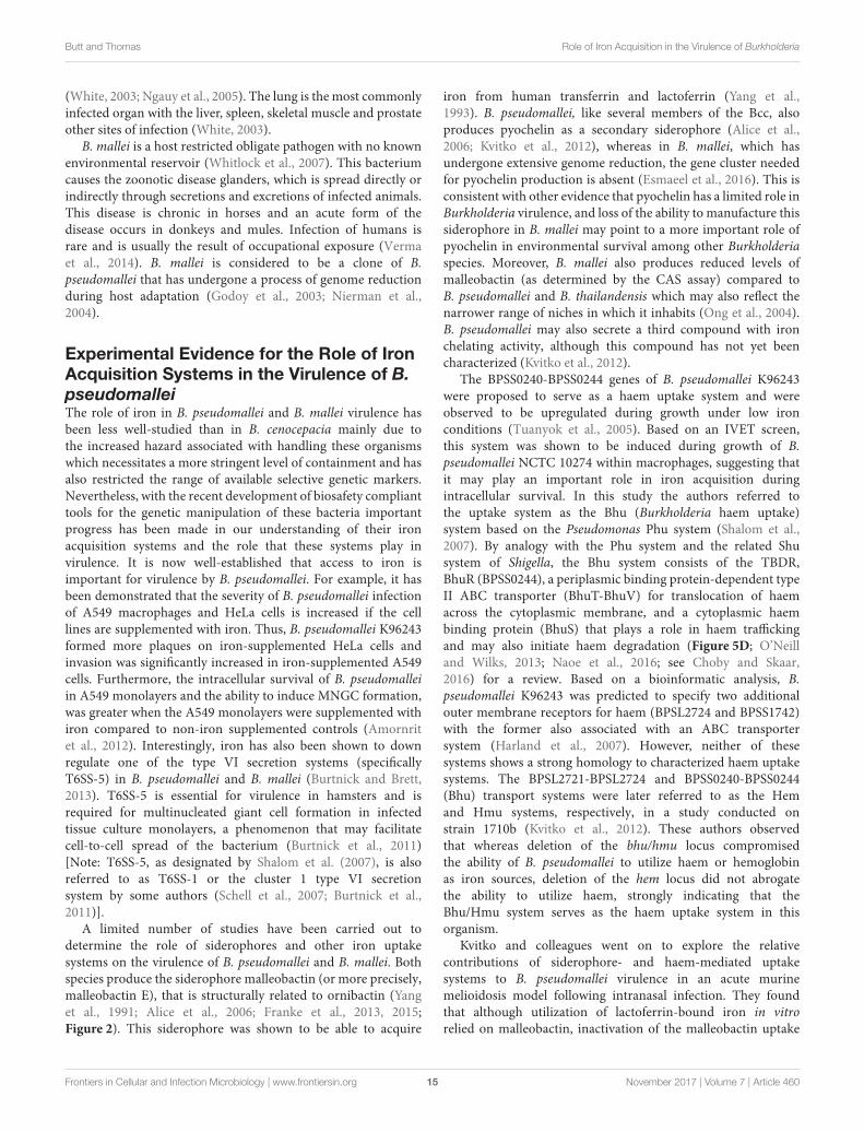
Butt and Thomas Role of Iron Acquisition in the Virulence of Burkholderia
(White, 2003; Ngauy et al., 2005). The lung is the most commonlyinfected organ with the liver, spleen, skeletal muscle and prostateother sites of infection (White, 2003).
B. mallei is a host restricted obligate pathogen with no knownenvironmental reservoir (Whitlock et al., 2007). This bacteriumcauses the zoonotic disease glanders, which is spread directly orindirectly through secretions and excretions of infected animals.This disease is chronic in horses and an acute form of thedisease occurs in donkeys and mules. Infection of humans israre and is usually the result of occupational exposure (Vermaet al., 2014). B. mallei is considered to be a clone of B.pseudomallei that has undergone a process of genome reductionduring host adaptation (Godoy et al., 2003; Nierman et al.,2004).
Experimental Evidence for the Role of IronAcquisition Systems in the Virulence of B.pseudomalleiThe role of iron in B. pseudomallei and B. mallei virulence hasbeen less well-studied than in B. cenocepacia mainly due tothe increased hazard associated with handling these organismswhich necessitates a more stringent level of containment and hasalso restricted the range of available selective genetic markers.Nevertheless, with the recent development of biosafety complianttools for the genetic manipulation of these bacteria importantprogress has been made in our understanding of their ironacquisition systems and the role that these systems play invirulence. It is now well-established that access to iron isimportant for virulence by B. pseudomallei. For example, it hasbeen demonstrated that the severity of B. pseudomallei infectionof A549 macrophages and HeLa cells is increased if the celllines are supplemented with iron. Thus, B. pseudomallei K96243formed more plaques on iron-supplemented HeLa cells andinvasion was significantly increased in iron-supplemented A549cells. Furthermore, the intracellular survival of B. pseudomalleiin A549 monolayers and the ability to induce MNGC formation,was greater when the A549 monolayers were supplemented withiron compared to non-iron supplemented controls (Amornritet al., 2012). Interestingly, iron has also been shown to downregulate one of the type VI secretion systems (specificallyT6SS-5) in B. pseudomallei and B. mallei (Burtnick and Brett,2013). T6SS-5 is essential for virulence in hamsters and isrequired for multinucleated giant cell formation in infectedtissue culture monolayers, a phenomenon that may facilitatecell-to-cell spread of the bacterium (Burtnick et al., 2011)[Note: T6SS-5, as designated by Shalom et al. (2007), is alsoreferred to as T6SS-1 or the cluster 1 type VI secretionsystem by some authors (Schell et al., 2007; Burtnick et al.,2011)].
A limited number of studies have been carried out todetermine the role of siderophores and other iron uptakesystems on the virulence of B. pseudomallei and B. mallei. Bothspecies produce the siderophore malleobactin (or more precisely,malleobactin E), that is structurally related to ornibactin (Yanget al., 1991; Alice et al., 2006; Franke et al., 2013, 2015;Figure 2). This siderophore was shown to be able to acquire
iron from human transferrin and lactoferrin (Yang et al.,1993). B. pseudomallei, like several members of the Bcc, alsoproduces pyochelin as a secondary siderophore (Alice et al.,2006; Kvitko et al., 2012), whereas in B. mallei, which hasundergone extensive genome reduction, the gene cluster neededfor pyochelin production is absent (Esmaeel et al., 2016). This isconsistent with other evidence that pyochelin has a limited role inBurkholderia virulence, and loss of the ability to manufacture thissiderophore in B. mallei may point to a more important role ofpyochelin in environmental survival among other Burkholderiaspecies. Moreover, B. mallei also produces reduced levels ofmalleobactin (as determined by the CAS assay) compared toB. pseudomallei and B. thailandensis which may also reflect thenarrower range of niches in which it inhabits (Ong et al., 2004).B. pseudomallei may also secrete a third compound with ironchelating activity, although this compound has not yet beencharacterized (Kvitko et al., 2012).
The BPSS0240-BPSS0244 genes of B. pseudomallei K96243were proposed to serve as a haem uptake system and wereobserved to be upregulated during growth under low ironconditions (Tuanyok et al., 2005). Based on an IVET screen,this system was shown to be induced during growth of B.pseudomallei NCTC 10274 within macrophages, suggesting thatit may play an important role in iron acquisition duringintracellular survival. In this study the authors referred tothe uptake system as the Bhu (Burkholderia haem uptake)system based on the Pseudomonas Phu system (Shalom et al.,2007). By analogy with the Phu system and the related Shusystem of Shigella, the Bhu system consists of the TBDR,BhuR (BPSS0244), a periplasmic binding protein-dependent typeII ABC transporter (BhuT-BhuV) for translocation of haemacross the cytoplasmic membrane, and a cytoplasmic haembinding protein (BhuS) that plays a role in haem traffickingand may also initiate haem degradation (Figure 5D; O’Neilland Wilks, 2013; Naoe et al., 2016; see Choby and Skaar,2016) for a review. Based on a bioinformatic analysis, B.pseudomallei K96243 was predicted to specify two additionalouter membrane receptors for haem (BPSL2724 and BPSS1742)with the former also associated with an ABC transportersystem (Harland et al., 2007). However, neither of thesesystems shows a strong homology to characterized haem uptakesystems. The BPSL2721-BPSL2724 and BPSS0240-BPSS0244(Bhu) transport systems were later referred to as the Hemand Hmu systems, respectively, in a study conducted onstrain 1710b (Kvitko et al., 2012). These authors observedthat whereas deletion of the bhu/hmu locus compromisedthe ability of B. pseudomallei to utilize haem or hemoglobinas iron sources, deletion of the hem locus did not abrogatethe ability to utilize haem, strongly indicating that theBhu/Hmu system serves as the haem uptake system in thisorganism.
Kvitko and colleagues went on to explore the relativecontributions of siderophore- and haem-mediated uptakesystems to B. pseudomallei virulence in an acute murinemelioidosis model following intranasal infection. They foundthat although utilization of lactoferrin-bound iron in vitrorelied on malleobactin, inactivation of the malleobactin uptake
Frontiers in Cellular and Infection Microbiology | www.frontiersin.org 15 November 2017 | Volume 7 | Article 460

Butt and Thomas Role of Iron Acquisition in the Virulence of Burkholderia
system in strain 1710b did not cause attenuation in the murinemelioidosis model (Kvitko et al., 2012). Interestingly, a strainthat was defective for malleobactin, pyochelin, and haem uptakewas also fully virulent in the melioidosis model, although thetitres of bacteria recovered from some organs was significantlylower. As this mutant could still grow with ferritin as aniron source, it was suggested that another iron uptake systemis present that could compensate for the loss of the otheruptake pathways. There are at least two possibilities that couldaccount for this. First, the CAS agar assay indicated thatB. pseudomallei mutants lacking the ability to biosynthesisemalleobactin and pyochelin specify an additional secreted iron-chelating compound of unknown identity (Kvitko et al., 2012).Secondly, B. pseudomallei encodes the FtrABCD system whichmay play a role in iron acquisition during infection (Mathewet al., 2014). Perhaps surprisingly, although B. pseudomallei708a (a strain containing a >130 kb genome deletion thatremoves the malleobactin synthesis genes) was virulent inthe mouse model used by Kvitko and colleagues, it wasattenuated in G. mellonella (Wand et al., 2011; Kvitko et al.,2012).
The outer membrane receptors required for uptake of ferric-siderophore complexes and haem require the action of thecytoplasmic membrane-anchored TonB-ExbB-ExbD complex toenergize transport of these iron sources. A B. mallei tonBmutant, which is unable to internalize ferric-malleobactin orhaem, was shown to be completely attenuated in mice at 105
CFU, in comparison to an LD50 of 7.4 x 10⁴ CFU for thewild type, and it also exhibited lower titres in target organs(lungs and spleen). Supplementation of the medium with ferrousiron, which is assimilated by a TonB-independent mechanism,partially restored virulence and led to higher titres of tonBmutantbacteria in the spleen, indicating that the inability to acquire ironis the main reason for the loss of virulence in the mutant (Mottet al., 2015). However, as TonB systems in other species havebeen shown to be involved in transport of other large molecules,including some enzyme cofactors (thiamine, cobalamin) andsaccharides (sialic acid, sucrose, maltodextrins) the possibilityexists that there are other transport requirements served by the B.mallei TonB system that must be met for full virulence (Schaueret al., 2008; Roy et al., 2010).
Another study in which potential novel therapeutics werescreened for their ability to counteract B. pseudomallei killingof C. elegans suggested an important role for iron acquisitionin virulence. Exposure of B. pseudomallei to the plant alkaloidcurcumin (diferuloylmethane), prior to their administration toC. elegans, significantly enhanced the survival of the worms(Eng and Nathan, 2015). Curcumin is structurally related tobis-catecholates, which include bacterial siderophores such asazotochelin, cepaciachelin, and serratiochelin, and is itself aknown iron chelator that may serve to cause a decrease iniron availability (Jiao et al., 2006). Accordingly, microarray datashowed that B. pseudomallei treated with curcumin upregulatedgenes for iron transport, including those for malleobactin,pyochelin and haem uptake, as well as genes encoding theTonB system. Moreover, genes encoding the biosynthesis ofmalleobactin and pyochelin were upregulated and increased
secretion of siderophores by treated B. pseudomallei wasobserved. The authors suggest that the anti-infective effectsobserved by curcumin are due to the bacteria diverting theirmetabolism away from virulence and toward iron uptake toensure growth and maintenance. These observations providesuggestive evidence for the importance of iron acquisition forvirulence in this organism.
BURKHOLDERIA GLADIOLI
B. gladioli was originally identified as a pathogen of Iridaceae,specifically irises and gladioli, and then later was recognized asthe cause of infections in certain groups of immunocompromisedpatients, particularly among those with CF and CGD (Boyantonet al., 2005; Kennedy et al., 2007). The iron acquisition systemsof this organism have yet to be determined. However, thegenome sequence offers a few clues. We note that it does notcontain orthologs of the genes for the biosynthesis of ornibactin,malleobactin, pyochelin, or cepaciachelin (Table 1). However, itdoes contain orthologs of the bhuRSTUV genes. Unusually, thesegenes are organized into two separate operons that are present onchromosome 1 (bhuRST) and chromosome 2 (bhuUV) (resultsnot shown), but nevertheless suggest that B. gladioli may be ableto utilize haem as an iron source during infection of human hosts.The B. gladioli genome also encodes a FtrABCD system. It is notknown whether one or both of these systems is important forestablishing an infection in humans.
CLOSING REMARKS
To conclude, we are still far from ascertaining the relativeimportance of the different iron acquisition mechanismsavailable to the Burkholderia that are brought to bear duringinfection of a human host. A number of studies have been carriedout with B. cenocepacia to address this question which haveprovided what might at first appear to be conflicting results.However, as themodel systems are so varied, and the iron contentand/or availability in these systems is also markedly different,we believe that most of these apparent contradictory results canbe rationalized. Nonetheless, in terms of whether high affinityiron acquisition systems are essential for successful colonizationof the CF lung and which ones are deployed, further studiesare required. Due to the difficulty of working safely with B.pseudomallei and B. mallei fewer studies have been conducted,but it would appear that neither the known siderophores ofB. pseudomallei nor its haem uptake system are important forsystemic melioidosis. There is great scope for further work intothe role of iron acquisition in the virulence of this importantgroup of bacteria.
AUTHOR CONTRIBUTIONS
AB and MT contributed equally to writing the manuscript. MTprepared the figures, table and Supplementary Material.
Frontiers in Cellular and Infection Microbiology | www.frontiersin.org 16 November 2017 | Volume 7 | Article 460

Butt and Thomas Role of Iron Acquisition in the Virulence of Burkholderia
ACKNOWLEDGMENTS
We would like to thank the BBSRC who are supporting thecurrent work of AB and MT on the regulation of iron acquisitionin Burkholderia (research grant BB/M003531/1).
SUPPLEMENTARY MATERIAL
The Supplementary Material for this article can be foundonline at: https://www.frontiersin.org/articles/10.3389/fcimb.2017.00460/full#supplementary-material
REFERENCES
Adler, C., Corbalan, N. S., Seyedsayamdost, M. R., Pomares, M. F., De Cristobal,
R. E., Clardy, J., et al. (2012). Catecholate siderophores protect bacteria from
pyochelin toxicity. PLoS ONE 7:e46754. doi: 10.1371/journal.pone.0046754
Agnoli, K., Lowe, C. A., Farmer, K. L., Husnain, S. I., and Thomas, M. S.
(2006). The ornibactin biosynthesis and transport genes of Burkholderia
cenocepacia are regulated by an extracytoplasmic function sigma factor
which is a part of the Fur regulon. J. Bacteriol. 188, 3631–3644.
doi: 10.1128/JB.188.10.3631-3644.2006
Alice, A. F., Lopez, C. S., Lowe, C. A., Ledesma, M. A., and Crosa, J.
H. (2006). Genetic and transcriptional analysis of the siderophore
malleobactin biosynthesis and transport genes in the human pathogen
Burkholderia pseudomallei K96243. J. Bacteriol. 188, 1551–1566.
doi: 10.1128/JB.188.4.1551-1566.2006
Amornrit,W.M. V., andWangteeraprasert, T., and Korbsrisate, S. (2012). Elevated
intracellular levels of iron in host cells promotes Burkholderia pseudomallei
infection. Asian Biomed. 6, 465–471. doi: 10.5372/1905-7415.0603.078
Andersen, S. B., Marvig, R. L., Molin, S., Johansen, H. K., and Griffin, A. S. (2015).
Long-term social dynamics drive loss of function in pathogenic bacteria. Proc.
Natl. Acad. Sci. U.S.A. 112, 10756–10761. doi: 10.1073/pnas.1508324112
Andrews, S. C., Robinson, A. K., and Rodriguez-Quinones, F. (2003).
Bacterial iron homeostasis. FEMS Microbiol. Rev. 27, 215–237.
doi: 10.1016/S0168-6445(03)00055-X
Asghar, A. H., Shastri, S., Dave, E., Wowk, I., Agnoli, K., Cook, A. M.,
et al. (2011). The pobA gene of Burkholderia cenocepacia encodes a
group I Sfp-type phosphopantetheinyltransferase required for biosynthesis
of the siderophores ornibactin and pyochelin. Microbiology 157, 349–361.
doi: 10.1099/mic.0.045559-0
Balla, J., Jacob, H. S., Balla, G., Nath, K., Eaton, J. W., and Vercellotti, G.
M. (1993). Endothelial-cell heme uptake from heme proteins: induction of
sensitization and desensitization to oxidant damage. Proc. Natl. Acad. Sci.
U.S.A. 90, 9285–9289. doi: 10.1073/pnas.90.20.9285
Baltz, R. H. (2011). Function of MbtH homologs in nonribosomal peptide
biosynthesis and applications in secondary metabolite discovery. J. Ind.
Microbiol. Biotechnol. 38, 1747–1760. doi: 10.1007/s10295-011-1022-8
Banin, E., Vasil, M. L., and Greenberg, E. P. (2005). Iron and Pseudomonas
aeruginosa biofilm formation. Proc. Natl. Acad. Sci. U.S.A. 102, 11076–11081.
doi: 10.1073/pnas.0504266102
Barelmann, I., Meyer, J. M., Taraz, K., and Budzikiewicz, H. (1996). Cepaciachelin,
a new catecholate siderophore from Burholderia (Pseudomonas) cepacia. Z.
Nat. 51, 627–630.
Barth, A. L., and Pitt, T. L. (1996). The high amino-acid content of sputum
from cystic fibrosis patients promotes growth of auxotrophic Pseudomonas
aeruginosa. J. Med. Microbiol. 45, 110–119. doi: 10.1099/00222615-45-2-110
Baysse, C., De Vos, D., Naudet, Y., Vandermonde, A., Ochsner, U., Meyer,
J. M., et al. (2000). Vanadium interferes with siderophore-mediated iron
uptake in Pseudomonas aeruginosa. Microbiology 146 (Pt 10), 2425–2434.
doi: 10.1099/00221287-146-10-2425
Berlutti, F., Morea, C., Battistoni, A., Sarli, S., Cipriani, P., Superti, F., et al. (2005).
Iron availability influences aggregation, biofilm, adhesion and invasion of
Pseudomonas aeruginosa and Burkholderia cenocepacia. Int. J. Immunopathol.
Pharmacol. 18, 661–670. doi: 10.1177/039463200501800407
Beukes, C. W., Palmer, M., Manyaka, P., Chan, W. Y., Avontuur, J. R., Van Zyl,
E., et al. (2017). Genome data provides high support for generic boundaries
in Burkholderia sensu lato. Front Microbiol 8:1154. doi: 10.3389/fmicb.2017.
01154
Boucher, R. C. (2007). Airway surface dehydration in cystic
fibrosis: pathogenesis and therapy. Annu. Rev. Med. 58, 157–170.
doi: 10.1146/annurev.med.58.071905.105316
Boyanton, B. L. Jr., Noroski, L. M., Reddy, H., Dishop, M. K., Hicks, M. J.,
Versalovic, J., et al. (2005). Burkholderia gladioli osteomyelitis in association
with chronic granulomatous disease: case report and review. Pediatr. Infect. Dis.
J. 24, 837–839. doi: 10.1097/01.inf.0000177285.44374.dc
Braud, A., Hoegy, F., Jezequel, K., Lebeau, T., and Schalk, I. J. (2009).
New insights into the metal specificity of the Pseudomonas aeruginosa
pyoverdine-iron uptake pathway. Environ. Microbiol. 11, 1079–1091.
doi: 10.1111/j.1462-2920.2008.01838.x
Brickman, T. J., and Armstrong, S. K. (2012). Iron and pH-responsive FtrABCD
ferrous iron utilization system of Bordetella species. Mol. Microbiol. 86,
580–593. doi: 10.1111/mmi.12003
Brickman, T. J., and McIntosh, M. A. (1992). Overexpression and purification
of ferric enterobactin esterase from Escherichia coli. Demonstration of
enzymatic hydrolysis of enterobactin and its iron complex. J. Biol. Chem. 267,
12350–12355.
Britigan, B. E., Rasmussen, G. T., and Cox, C. D. (1997). Augmentation of oxidant
injury to human pulmonary epithelial cells by the Pseudomonas aeruginosa
siderophore pyochelin. Infect. Immun. 65, 1071–1076.
Britigan, B. E., Serody, J. S., and Cohen, M. S. (1994). The role of lactoferrin
as an anti-inflammatory molecule. Adv. Exp. Med. Biol. 357, 143–156.
doi: 10.1007/978-1-4615-2548-6_14
Burtnick, M. N., and Brett, P. J. (2013). Burkholderia mallei and
Burkholderia pseudomallei cluster 1 type VI secretion system gene
expression is negatively regulated by iron and zinc. PLoS ONE 8:e76767.
doi: 10.1371/journal.pone.0076767
Burtnick, M. N., Brett, P. J., Harding, S. V., Ngugi, S. A., Ribot, W. J., Chantratita,
N., et al. (2011). The cluster 1 type VI secretion system is a major virulence
determinant in Burkholderia pseudomallei. Infect. Immun. 79, 1512–1525.
doi: 10.1128/IAI.01218-10
Cao, J., Woodhall, M. R., Alvarez, J., Cartron, M. L., and Andrews, S. C. (2007).
EfeUOB (YcdNOB) is a tripartite, acid-induced and CpxAR-regulated, low-pH
Fe2+ transporter that is cryptic in Escherichia coli K-12 but functional in E. coli
O157:H7.Mol. Microbiol. 65, 857–875. doi: 10.1111/j.1365-2958.2007.05977.x
Caza, M., and Kronstad, J. W. (2013). Shared and distinct mechanisms of iron
acquisition by bacterial and fungal pathogens of humans. Front. Cell Infect.
Microbiol. 3:80. doi: 10.3389/fcimb.2013.00080
Celia, H., Noinaj, N., Zakharov, S. D., Bordignon, E., Botos, I., Santamaria, M.,
et al. (2016). Structural insight into the role of the Ton complex in energy
transduction. Nature 538, 60–65. doi: 10.1038/nature19757
Chipperfield, J. R., and Ratledge, C. (2000). Salicylic acid is not a
bacterial siderophore: a theoretical study. Biometals 13, 165–168.
doi: 10.1023/A:1009227206890
Choby, J. E., and Skaar, E. P. (2016). Heme synthesis and acquisition in bacterial
pathogens. J. Mol. Biol. 428, 3408–3428. doi: 10.1016/j.jmb.2016.03.018
Chu, B. C., Garcia-Herrero, A., Johanson, T. H., Krewulak, K. D., Lau, C.
K., Peacock, R. S., et al. (2010). Siderophore uptake in bacteria and the
battle for iron with the host; a bird’s eye view. Biometals 23, 601–611.
doi: 10.1007/s10534-010-9361-x
Coffman, T. J., Cox, C. D., Edeker, B. L., and Britigan, B. E. (1990). Possible role
of bacterial siderophores in inflammation. Iron bound to the Pseudomonas
siderophore pyochelin can function as a hydroxyl radical catalyst. J. Clin. Invest.
86, 1030–1037. doi: 10.1172/JCI114805
Cohen, T. S., and Prince, A. (2012). Cystic fibrosis: a mucosal immunodeficiency
syndrome. Nat. Med. 18, 509–519. doi: 10.1038/nm.2715
Frontiers in Cellular and Infection Microbiology | www.frontiersin.org 17 November 2017 | Volume 7 | Article 460

Butt and Thomas Role of Iron Acquisition in the Virulence of Burkholderia
Cornelis, P., and Matthijs, S. (2002). Diversity of siderophore-mediated iron
uptake systems in fluorescent pseudomonads: not only pyoverdines. Environ.
Microbiol. 4, 787–798. doi: 10.1046/j.1462-2920.2002.00369.x
Cornelis, P., Wei, Q., Andrews, S. C., and Vinckx, T. (2011). Iron homeostasis and
management of oxidative stress response in bacteria. Metallomics 3, 540–549.
doi: 10.1039/c1mt00022e
Cosgrove, S., Chotirmall, S. H., Greene, C. M., and McElvaney, N. G.
(2011). Pulmonary proteases in the cystic fibrosis lung induce interleukin 8
expression from bronchial epithelial cells via a heme/meprin/epidermal growth
factor receptor/Toll-like receptor pathway. J. Biol. Chem. 286, 7692–7704.
doi: 10.1074/jbc.M110.183863
Courtney, J. M., Dunbar, K. E., McDowell, A., Moore, J. E., Warke, T. J., Stevenson,
M., et al. (2004). Clinical outcome of Burkholderia cepacia complex infection in
cystic fibrosis adults. J. Cyst. Fibros. 3, 93–98. doi: 10.1016/j.jcf.2004.01.005
Cox, C. D., and Graham, R. (1979). Isolation of an iron-binding compound from
Pseudomonas aeruginosa. J. Bacteriol. 137, 357–364.
Cuiv, P. O., Clarke, P., Lynch, D., and O’connell, M. (2004). Identification of rhtX
and fptX, novel genes encoding proteins that show homology and function in
the utilization of the siderophores rhizobactin 1021 by Sinorhizobium meliloti
and pyochelin by Pseudomonas aeruginosa, respectively. J. Bacteriol. 186,
2996–3005. doi: 10.1128/JB.186.10.2996-3005.2004
Cunrath, O., Gasser, V., Hoegy, F., Reimmann, C., Guillon, L., and Schalk,
I. J. (2015). A cell biological view of the siderophore pyochelin iron
uptake pathway in Pseudomonas aeruginosa. Environ. Microbiol. 17, 171–185.
doi: 10.1111/1462-2920.12544
Cuppels, D., Stipanovic, R., Stoessl, A., and Stothers, J. (1987). The constitution
and properties of a pyochelin–zinc complex. Can. J. Chem. 65, 2126–2130.
doi: 10.1139/v87-354
Darling, P., Chan, M., Cox, A. D., and Sokol, P. A. (1998). Siderophore production
by cystic fibrosis isolates of Burkholderia cepacia. Infect. Immun. 66, 874–877.
Davies, J. C., Alton, E. W. F. W., and Bush, A. (2007). Cystic fibrosis. BMJ 335,
1255–1259. doi: 10.1136/bmj.39391.713229.AD
De Vos, D., De Chial, M., Cochez, C., Jansen, S., Tummler, B., Meyer, J. M., et al.
(2001). Study of pyoverdine type and production by Pseudomonas aeruginosa
isolated from cystic fibrosis patients: prevalence of type II pyoverdine isolates
and accumulation of pyoverdine-negative mutations. Arch. Microbiol. 175,
384–388. doi: 10.1007/s002030100278
Deng, P., Wang, X., Baird, S. M., Showmaker, K. C., Smith, L., Peterson, D. G.,
et al. (2016). Comparative genome-wide analysis reveals that Burkholderia
contaminansMS14 possesses multiple antimicrobial biosynthesis genes but not
major genetic loci required for pathogenesis. Microbiologyopen 5, 353–369.
doi: 10.1002/mbo3.333
Depoorter, E., Bull, M. J., Peeters, C., Coenye, T., Vandamme, P., and
Mahenthiralingam, E. (2016). Burkholderia: an update on taxonomy and
biotechnological potential as antibiotic producers. Appl. Microbiol. Biotechnol.
100, 5215–5229. doi: 10.1007/s00253-016-7520-x
Dobritsa, A. P., and Samadpour, M. (2016). Transfer of eleven species of
the genus Burkholderia to the genus Paraburkholderia and proposal of
Caballeronia gen. nov. to accommodate twelve species of the genera
Burkholderia and Paraburkholderia. Int. J. Syst. Evol. Microbiol. 66, 2836–2846.
doi: 10.1099/ijsem.0.001065
Doring, G., and Gulbins, E. (2009). Cystic fibrosis and innate immunity: how
chloride channel mutations provoke lung disease. Cell Microbiol. 11, 208–216.
doi: 10.1111/j.1462-5822.2008.01271.x
Doring, G., Parameswaran, I. G., and Murphy, T. F. (2011). Differential adaptation
of microbial pathogens to airways of patients with cystic fibrosis and
chronic obstructive pulmonary disease. FEMS Microbiol. Rev. 35, 124–146.
doi: 10.1111/j.1574-6976.2010.00237.x
Drevinek, P., and Mahenthiralingam, E. (2010). Burkholderia cenocepacia in cystic
fibrosis: epidemiology and molecular mechanisms of virulence. Clin. Microbiol.
Infect. 16, 821–830. doi: 10.1111/j.1469-0691.2010.03237.x
Drevinek, P., Holden, M. T., Ge, Z., Jones, A. M., Ketchell, I., Gill, R. T.,
et al. (2008). Gene expression changes linked to antimicrobial resistance,
oxidative stress, iron depletion and retained motility are observed when
Burkholderia cenocepacia grows in cystic fibrosis sputum. BMC Infect. Dis.
8:121. doi: 10.1186/1471-2334-8-121
Elborn, J. S. (2016). Cystic fibrosis. Lancet 388, 2519–2531.
doi: 10.1016/S0140-6736(16)00576-6
Elhassanny, A. E., Anderson, E. S., Menscher, E. A., and Roop, R. M. II. (2013).
The ferrous iron transporter FtrABCD is required for the virulence of Brucella
abortus 2308 in mice.Mol. Microbiol. 88, 1070–1082. doi: 10.1111/mmi.12242
Eng, S. A., and Nathan, S. (2015). Curcumin rescues Caenorhabditis elegans
from a Burkholderia pseudomallei infection. Front. Microbiol. 6:290.
doi: 10.3389/fmicb.2015.00290
Esmaeel, Q., Pupin, M., Kieu, N. P., Chataigné, G., Béchet, M., Deravel, J., et al.
(2016). Burkholderia genome mining for nonribosomal peptide synthetases
reveals a great potential for novel siderophores and lipopeptides synthesis.
Microbiologyopen 5, 512–526. doi: 10.1002/mbo3.347
Franke, J., Ishida, K., and Hertweck, C. (2015). Plasticity of the malleobactin
pathway and its impact on siderophore action in human pathogenic bacteria.
Chemistry 21, 8010–8014. doi: 10.1002/chem.201500757
Franke, J., Ishida, K., Ishida-Ito, M., and Hertweck, C. (2013). Nitro versus
hydroxamate in siderophores of pathogenic bacteria: effect of missing
hydroxylamine protection in malleobactin biosynthesis. Angew. Chem. Int. Ed.
Engl. 52, 8271–8275. doi: 10.1002/anie.201303196
Fung, C., Naughton, S., Turnbull, L., Tingpej, P., Rose, B., Arthur, J., et al. (2010).
Gene expression of Pseudomonas aeruginosa in a mucin-containing synthetic
growth medium mimicking cystic fibrosis lung sputum. J. Med. Microbiol. 59,
1089–1100. doi: 10.1099/jmm.0.019984-0
Godoy, D., Randle, G., Simpson, A. J., Aanensen, D. M., Pitt, T. L., Kinoshita,
R., et al. (2003). Multilocus sequence typing and evolutionary relationships
among the causative agents of melioidosis and glanders, Burkholderia
pseudomallei and Burkholderia mallei. J. Clin. Microbiol. 41, 2068–2079.
doi: 10.1128/JCM.41.5.2068-2079.2003
Hannauer, M., Barda, Y., Mislin, G. L., Shanzer, A., and Schalk, I. J.
(2010). The ferrichrome uptake pathway in Pseudomonas aeruginosa
involves an iron release mechanism with acylation of the siderophore
and recycling of the modified desferrichrome. J. Bacteriol. 192, 1212–1220.
doi: 10.1128/JB.01539-09
Hare, N. J., Solis, N., Harmer, C.,Marzook, N. B., Rose, B., Harbour, C., et al. (2012).
Proteomic profiling of Pseudomonas aeruginosa AES-1R, PAO1 and PA14
reveals potential virulence determinants associated with a transmissible cystic
fibrosis-associated strain. BMCMicrobiol. 12:16. doi: 10.1186/1471-2180-12-16
Harland, D. N., Dassa, E., Titball, R. W., Brown, K. A., and Atkins, H. S. (2007).
ATP-binding cassette systems in Burkholderia pseudomallei and Burkholderia
mallei. BMC Genomics 8:83. doi: 10.1186/1471-2164-8-83
Harrison, F. (2007). Microbial ecology of the cystic fibrosis lung.Microbiology 153,
917–923. doi: 10.1099/mic.0.2006/004077-0
Holden, M. T., Seth-Smith, H. M., Crossman, L. C., Sebaihia, M., Bentley, S. D.,
Cerdeno-Tarraga, A. M., et al. (2009). The genome of Burkholderia cenocepacia
J2315, an epidemic pathogen of cystic fibrosis patients. J. Bacteriol. 191,
261–277. doi: 10.1128/JB.01230-08
Hunt, T. A., Kooi, C., Sokol, P. A., and Valvano, M. A. (2004). Identification of
Burkholderia cenocepacia genes required for bacterial survival in vivo. Infect.
Immun. 72, 4010–4022. doi: 10.1128/IAI.72.7.4010-4022.2004
Hunter, R. C., Asfour, F., Dingemans, J., Osuna, B. L., Samad, T., Malfroot, A., et al.
(2013). Ferrous iron is a significant component of bioavailable iron in cystic
fibrosis airways.Mbio 4:e00557–13. doi: 10.1128/mBio.00557-13
Inahashi, Y., Zhou, S., Bibb, M. J., Song, L., Al-Bassam, M. M., Bibb, M. J., et al.
(2017). Watasemycin biosynthesis in Streptomyces venezuelae: thiazoline C-
methylation by a type B radical-SAM methylase homologue. Chem. Sci. 8,
2823–2831. doi: 10.1039/C6SC03533G
Iobbi-Nivol, C., and Leimkuhler, S. (2013). Molybdenum enzymes, their
maturation andmolybdenum cofactor biosynthesis in Escherichia coli. Biochim.
Biophys. Acta 1827, 1086–1101. doi: 10.1016/j.bbabio.2012.11.007
Isles, A., Maclusky, I., Corey, M., Gold, R., Prober, C., Fleming, P., et al. (1984).
Pseudomonas cepacia infection in cystic fibrosis: an emerging problem. J.
Pediatr. 104, 206–210. doi: 10.1016/S0022-3476(84)80993-2
Jiao, Y., Wilkinson, J. T., Christine Pietsch, E., Buss, J. L., Wang, W., Planalp, R.,
et al. (2006). Iron chelation in the biological activity of curcumin. Free Radic.
Biol. Med. 40, 1152–1160. doi: 10.1016/j.freeradbiomed.2005.11.003
Jones, A. M., Dodd, M. E., Govan, J. R., Barcus, V., Doherty, C. J., Morris, J., et al.
(2004). Burkholderia cenocepacia and Burkholderia multivorans: influence on
survival in cystic fibrosis. Thorax 59, 948–951. doi: 10.1136/thx.2003.017210
Jung, W. H., Sham, A., White, R., and Kronstad, J. W. (2006). Iron
regulation of the major virulence factors in the AIDS-associated pathogen
Frontiers in Cellular and Infection Microbiology | www.frontiersin.org 18 November 2017 | Volume 7 | Article 460

Butt and Thomas Role of Iron Acquisition in the Virulence of Burkholderia
Cryptococcus neoformans. PLoS Biol. 4:e410. doi: 10.1371/journal.pbio.
0040410
Kennedy, M. P., Coakley, R. D., Donaldson, S. H., Aris, R. M., Hohneker, K.,
Wedd, J. P., et al. (2007). Burkholderia gladioli: five year experience in a
cystic fibrosis and lung transplantation center. J. Cyst. Fibros. 6, 267–273.
doi: 10.1016/j.jcf.2006.10.007
Kolpen, M., Hansen, C. R., Bjarnsholt, T., Moser, C., Christensen, L. D., Van
Gennip, M., et al. (2010). Polymorphonuclear leucocytes consume oxygen in
sputum from chronic Pseudomonas aeruginosa pneumonia in cystic fibrosis.
Thorax 65, 57–62. doi: 10.1136/thx.2009.114512
Konings, A. F., Martin, L. W., Sharples, K. J., Roddam, L. F., Latham, R., Reid, D.
W., et al. (2013). Pseudomonas aeruginosa uses multiple pathways to acquire
iron during chronic infection in cystic fibrosis lungs. Infect. Immun. 81,
2697–2704. doi: 10.1128/IAI.00418-13
Krewulak, K. D., andVogel, H. J. (2008). Structural biology of bacterial iron uptake.
Biochim. Biophys. Acta 1778, 1781–1804. doi: 10.1016/j.bbamem.2007.07.026
Kvitko, B. H., Goodyear, A., Propst, K. L., Dow, S. W., and Schweizer, H. P.
(2012). Burkholderia pseudomallei known siderophores and hemin uptake
are dispensable for lethal murine melioidosis. PLoS Negl. Trop. Dis. 6:e1715.
doi: 10.1371/journal.pntd.0001715
Lamothe, J., and Valvano, M. A. (2008). Burkholderia cenocepacia-induced delay
of acidification and phagolysosomal fusion in cystic fibrosis transmembrane
conductance regulator (CFTR)-defective macrophages. Microbiology 154,
3825–3834. doi: 10.1099/mic.0.2008/023200-0
Lau, C. K., Krewulak, K. D., and Vogel, H. J. (2016). Bacterial ferrous
iron transport: the Feo system. FEMS Microbiol. Rev. 40, 273–298.
doi: 10.1093/femsre/fuv049
Lipuma, J. J. (2005). Update on the Burkholderia cepacia complex. Curr. Opin.
Pulm. Med. 11, 528–533. doi: 10.1097/01.mcp.0000181475.85187.ed
Loutet, S. A., and Valvano, M. A. (2010). A decade of Burkholderia
cenocepacia virulence determinant research. Infect. Immun. 78, 4088–4100.
doi: 10.1128/IAI.00212-10
Loveridge, E. J., Jones, C., Bull, M. J., Moody, S. C., Kahl, M. W., Khan, Z., et al.
(2017). Reclassification of the specialized metabolite producer Pseudomonas
mesoacidophilaATCC 31433 as a member of the Burkholderia cepacia complex.
J. Bacteriol. 199:e00125-17. doi: 10.1128/JB.00125-17
Madeira, A., Dos Santos, S. C., Santos, P. M., Coutinho, C. P., Tyrrell, J., McClean,
S., et al. (2013). Proteomic profiling of Burkholderia cenocepacia clonal isolates
with different virulence potential retrieved from a cystic fibrosis patient during
chronic lung infection. PLoS ONE 8:e83065. doi: 10.1371/journal.pone.0083065
Mahenthiralingam, E., Vandamme, P., Campbell, M. E., Henry, D. A., Gravelle,
A. M., Wong, L. T., et al. (2001). Infection with Burkholderia cepacia complex
genomovars in patients with cystic fibrosis: virulent transmissible strains of
genomovar III can replace Burkholderia multivorans. Clin. Infect. Dis. 33,
1469–1475. doi: 10.1086/322684
Martin, L. W., Reid, D. W., Sharples, K. J., and Lamont, I. L. (2011). Pseudomonas
siderophores in the sputum of patients with cystic fibrosis. Biometals 24,
1059–1067. doi: 10.1007/s10534-011-9464-z
Marvig, R. L., Damkiaer, S., Khademi, S. M. H., Markussen, T. M., Molin, S., and
Jelsbak, L. (2014). Within-host evolution of Pseudomonas aeruginosa reveals
adaptation toward iron acquisition from hemoglobin. Mbio 5:e00966-14.
doi: 10.1128/mBio.00966-14
Mathew, A., Eberl, L., and Carlier, A. L. (2014). A novel siderophore-independent
strategy of iron uptake in the genus Burkholderia.Mol. Microbiol. 91, 805–820.
doi: 10.1111/mmi.12499
Matsui, H., Grubb, B. R., Tarran, R., Randell, S. H., Gatzy, J. T., Davis, C. W.,
et al. (1998). Evidence for periciliary liquid layer depletion, not abnormal ion
composition, in the pathogenesis of cystic fibrosis airways disease. Cell 95,
1005–1015. doi: 10.1016/S0092-8674(00)81724-9
Matsui, H., Verghese, M. W., Kesimer, M., Schwab, U. E., Randell, S. H., Sheehan,
J. K., et al. (2005). Reduced three-dimensional motility in dehydrated airway
mucus prevents neutrophil capture and killing bacteria on airway epithelial
surfaces. J. Immunol. 175, 1090–1099. doi: 10.4049/jimmunol.175.2.1090
Mesureur, J., Feliciano, J. R., Wagner, N., Gomes, M. C., Zhang, L., Blanco-
Gonzalez, M., et al. (2017). Macrophages, but not neutrophils, are critical
for proliferation of Burkholderia cenocepacia and ensuing host-damaging
inflammation. PLoS Pathog. 13:e1006437. doi: 10.1371/journal.ppat.1006437
Meyer, J. M., and Abdallah, M. A. (1978). The fluorescent pigment of
pseudomonas fluorescens: biosynthesis, purification and physicochemical
properties.Microbiology 107, 319–328. doi: 10.1099/00221287-107-2-319
Meyer, J. M., Hohnadel, D., and Halle, F. (1989). Cepabactin from Pseudomonas
cepacia, a new type of siderophore. J. Gen. Microbiol. 135, 1479–1487.
doi: 10.1099/00221287-135-6-1479
Meyer, J.-M., Van Van, T., Stintzi, A., Berge, O., and Winkelmann, G. (1995).
Ornibactin production and transport properties in strains of Burkholderia
vietnamiensis and Burkholderia cepacia (formerly Pseudomonas cepacia).
Biometals 8, 309–317. doi: 10.1007/BF00141604
Mira, N. P., Madeira, A., Moreira, A. S., Coutinho, C. P., and Sa-Correia, I. (2011).
Genomic expression analysis reveals strategies of Burkholderia cenocepacia to
adapt to cystic fibrosis patients’ airways and antimicrobial therapy. PLoS ONE
6:e28831. doi: 10.1371/journal.pone.0028831
Mott, T. M., Vijayakumar, S., Sbrana, E., Endsley, J. J., and Torres, A. G. (2015).
Characterization of the Burkholderia mallei tonB mutant and its potential as
a backbone strain for vaccine development. PLoS Negl. Trop. Dis. 9:e0003863.
doi: 10.1371/journal.pntd.0003863
Nairz, M., Schroll, A., Sonnweber, T., and Weiss, G. (2010). The struggle for
iron - a metal at the host-pathogen interface. Cell Microbiol. 12, 1691–1702.
doi: 10.1111/j.1462-5822.2010.01529.x
Naoe, Y., Nakamura, N., Doi, A., Sawabe, M., Nakamura, H., Shiro, Y., et al. (2016).
Crystal structure of bacterial haem importer complex in the inward-facing
conformation. Nat. Commun. 7:13411. doi: 10.1038/ncomms13411
Neilands, J. B. (1995). Siderophores: structure and function of microbial
iron transport compounds. J. Biol. Chem. 270, 26723–26726.
doi: 10.1074/jbc.270.45.26723
Ngauy, V., Lemeshev, Y., Sadkowski, L., and Crawford, G. (2005).
Cutaneous melioidosis in a man who was taken as a prisoner of war
by the Japanese during World War II. J. Clin. Microbiol. 43, 970–972.
doi: 10.1128/JCM.43.2.970-972.2005
Nguyen, A. T., O’Neill, M. J., Watts, A. M., Robson, C. L., Lamont, I. L., Wilks,
A., et al. (2014). Adaptation of iron homeostasis pathways by a Pseudomonas
aeruginosa pyoverdine mutant in the cystic fibrosis lung. J. Bacteriol. 196,
2265–2276. doi: 10.1128/JB.01491-14
Nierman, W. C., Deshazer, D., Kim, H. S., Tettelin, H., Nelson, K. E., Feldblyum,
T., et al. (2004). Structural flexibility in the Burkholderia mallei genome. Proc.
Natl. Acad. Sci. U.S.A. 101, 14246–14251. doi: 10.1073/pnas.0403306101
Noinaj, N., Guillier, M., Barnard, T. J., and Buchanan, S. K. (2010). TonB-
dependent transporters: regulation, structure, and function. Annu. Rev.
Microbiol. 64, 43–60. doi: 10.1146/annurev.micro.112408.134247
O’Grady, E. P., and Sokol, P. A. (2011). Burkholderia cenocepacia differential
gene expression during host-pathogen interactions and adaptation to the host
environment. Front. Cell. Infect. Microbiol. 1:15. doi: 10.3389/fcimb.2011.00015
O’Neill, M. J., and Wilks, A. (2013). The P. aeruginosa heme binding protein PhuS
is a heme oxygenase titratable regulator of heme uptake. ACS Chem. Biol. 8,
1794–1802. doi: 10.1021/cb400165b
Ong, C., Ooi, C. H., Wang, D., Chong, H., Ng, K. C., Rodrigues, F., et al. (2004).
Patterns of large-scale genomic variation in virulent and avirulent Burkholderia
species. Genome Res. 14, 2295–2307. doi: 10.1101/gr.1608904
Ong, K. S., Aw, Y. K., Lee, L. H., Yule, C. M., Cheow, Y. L., and Lee, S. M.
(2016). Burkholderia paludis sp. nov., an antibiotic-siderophore producing
novel Burkholderia cepacia complex species, isolated from Malaysian tropical
peat swamp soil. Front. Microbiol. 7:2046. doi: 10.3389/fmicb.2016.02046
Oren, A., and Garrity, A. G. (2015). Proposal to modify rule 27 of the international
code of nomenclature of prokaryotes. Int. J. Syst. Evol. Microbiol. 65:2342.
doi: 10.1099/ijs.0.000288
Palmer, K. L., Aye, L. M., and Whiteley, M. (2007). Nutritional cues control
Pseudomonas aeruginosa multicellular behavior in cystic fibrosis sputum. J.
Bacteriol. 189, 8079–8087. doi: 10.1128/JB.01138-07
Palmer, K. L., Mashburn, L. M., Singh, P. K., and Whiteley, M. (2005).
Cystic fibrosis sputum supports growth and cues key aspects of
Pseudomonas aeruginosa physiology. J. Bacteriol. 187, 5267–5277.
doi: 10.1128/JB.187.15.5267-5277.2005
Parke, J. L., and Gurian-Sherman, D. (2001). Diversity of the Burkholderia cepacia
complex and implications for risk assessment of biological control strains.
Annu. Rev. Phytopathol. 39, 225–258. doi: 10.1146/annurev.phyto.39.1.225
Frontiers in Cellular and Infection Microbiology | www.frontiersin.org 19 November 2017 | Volume 7 | Article 460

Butt and Thomas Role of Iron Acquisition in the Virulence of Burkholderia
Parrow, N. L., Fleming, R. E., and Minnick, M. F. (2013). Sequestration
and scavenging of iron in infection. Infect. Immun. 81, 3503–3514.
doi: 10.1128/IAI.00602-13
Peeters, E., Sass, A., Mahenthiralingam, E., Nelis, H., and Coenye, T. (2010).
Transcriptional response of Burkholderia cenocepacia J2315 sessile cells to
treatments with high doses of hydrogen peroxide and sodium hypochlorite.
BMC Genomics 11:90. doi: 10.1186/1471-2164-11-90
Perez, A., Issler, A. C., Cotton, C. U., Kelley, T. J., Verkman, A. S.,
and Davis, P. B. (2007). CFTR inhibition mimics the cystic fibrosis
inflammatory profile. Am. J. Physiol. Lung Cell Mol. Physiol. 292, L383–L395.
doi: 10.1152/ajplung.00403.2005
Perumal Samy, R., Stiles, B. G., Sethi, G., and Lim, L. H. K. (2017). Melioidosis:
clinical impact and public health threat in the tropics. PLoS Negl. Trop. Dis.
11:e0004738. doi: 10.1371/journal.pntd.0004738
Pessi, G., Braunwalder, R., Grunau, A., Omasits, U., Ahrens, C. H., and Eberl, L.
(2013). Response of Burkholderia cenocepacia H111 to micro-oxia. PLoS ONE
8:e72939. doi: 10.1371/journal.pone.0072939
Ratledge, C., and Dover, L. G. (2000). Iron metabolism in pathogenic bacteria.
Annu. Rev. Microbiol. 54, 881–941. doi: 10.1146/annurev.micro.54.1.881
Reid, D. W., Anderson, G. J., and Lamont, I. L. (2009). Role of lung iron in
determining the bacterial and host struggle in cystic fibrosis. Am. J. Physiol.
Lung Cell. Mol. Physiol. 297, L795–L802. doi: 10.1152/ajplung.00132.2009
Reid, D. W., Carroll, V., O’may, C., Champion, A., and Kirov, S. M.
(2007). Increased airway iron as a potential factor in the persistence of
Pseudomonas aeruginosa infection in cystic fibrosis. Eur. Respir. J. 30, 286–292.
doi: 10.1183/09031936.00154006
Reid, D. W., Lam, Q. T., Schneider, H., and Walters, E. H. (2004). Airway iron
and iron-regulatory cytokines in cystic fibrosis. Eur. Respir. J. 24, 286–291.
doi: 10.1183/09031936.04.00104803
Reid, D. W., Withers, N. J., Francis, L., Wilson, J. W., and Kotsimbos, T.
C. (2002). Iron deficiency in cystic fibrosis: relationship to lung disease
severity and chronic Pseudomonas aeruginosa infection. Chest 121, 48–54.
doi: 10.1378/chest.121.1.48
Reik, R., Spilker, T., and Lipuma, J. J. (2005). Distribution of Burkholderia
cepacia complex species among isolates recovered from persons
with or without cystic fibrosis. J. Clin. Microbiol. 43, 2926–2928.
doi: 10.1128/JCM.43.6.2926-2928.2005
Rhodes, K. A., and Schweizer, H. P. (2016). Antibiotic resistance in Burkholderia
species. Drug Resist. Update 28, 82–90. doi: 10.1016/j.drup.2016.07.003
Rose, H., Baldwin, A., Dowson, C. G., and Mahenthiralingam, E. (2009). Biocide
susceptibility of the Burkholderia cepacia complex. J. Antimicrob. Chemother.
63, 502–510. doi: 10.1093/jac/dkn540
Rottig, M., Medema, M. H., Blin, K., Weber, T., Rausch, C., and Kohlbacher, O.
(2011). NRPSpredictor2–a web server for predicting NRPS adenylation domain
specificity. Nucleic Acids Res. 39, W362–W367. doi: 10.1093/nar/gkr323
Roy, S., Douglas, C. W., and Stafford, G. P. (2010). A novel sialic acid utilization
and uptake system in the periodontal pathogenTannerella forsythia. J. Bacteriol.
192, 2285–2293. doi: 10.1128/JB.00079-10
Runyen-Janecky, L. J. (2013). Role and regulation of heme iron acquisition
in gram-negative pathogens. Front. Cell. Infect. Microbiol. 3:55.
doi: 10.3389/fcimb.2013.00055
Sanchez, M., Sabio, L., Galvez, N., Capdevila, M., and Dominguez-Vera, J.
M. (2017). Iron chemistry at the service of life. IUBMB Life 69, 382–388.
doi: 10.1002/iub.1602
Sawana, A., Adeolu, M., and Gupta, R. S. (2014). Molecular signatures and
phylogenomic analysis of the genus Burkholderia: proposal for division of this
genus into the emended genus Burkholderia containing pathogenic organisms
and a new genus Paraburkholderia gen. nov. harboring environmental species.
Front. Genet. 5:429. doi: 10.3389/fgene.2014.00429
Schauer, K., Rodionov, D. A., and De Reuse, H. (2008). New substrates for TonB-
dependent transport: do we only see the “tip of the iceberg”? Trends Biochem.
Sci. 33, 330–338. doi: 10.1016/j.tibs.2008.04.012
Schell, M. A., Ulrich, R. L., Ribot,W. J., Brueggemann, E. E., Hines, H. B., Chen, D.,
et al. (2007). Type VI secretion is amajor virulence determinant in Burkholderia
mallei. Mol. Microbiol. 64, 1466–1485. doi: 10.1111/j.1365-2958.2007.
05734.x
Shalom, G., Shaw, J. G., and Thomas, M. S. (2007). In vivo expression technology
identifies a type VI secretion system locus in Burkholderia pseudomallei that
is induced upon invasion of macrophages. Microbiology 153, 2689–2699.
doi: 10.1099/mic.0.2007/006585-0
Singh, P. K., Parsek, M. R., Greenberg, E. P., and Welsh, M. J. (2002). A
component of innate immunity prevents bacterial biofilm development.Nature
417, 552–555. doi: 10.1038/417552a
Skaar, E. P. (2010). The battle for iron between bacterial pathogens and their
vertebrate hosts. PLoS Pathog. 6:e1000949. doi: 10.1371/journal.ppat.1000949
Smith, E. E., Buckley, D. G., Wu, Z., Saenphimmachak, C., Hoffman, L. R.,
D’argenio, D. A., et al. (2006). Genetic adaptation by Pseudomonas aeruginosa
to the airways of cystic fibrosis patients. Proc. Natl. Acad. Sci. U.S.A. 103,
8487–8492. doi: 10.1073/pnas.0602138103
Smith, M. D., Wuthiekanun, V., Walsh, A. L., andWhite, N. J. (1995). Quantitative
recovery of Burkholderia pseudomallei from soil in Thailand. Trans. R. Soc.
Trop. Med. Hyg. 89, 488–490. doi: 10.1016/0035-9203(95)90078-0
Sokol, P. A. (1986). Production and utilization of pyochelin by clinical isolates of
Pseudomonas cepacia. J. Clin. Microbiol. 23, 560–562.
Sokol, P. A., and Woods, D. E. (1988). Effect of pyochelin on
Pseudomonas cepacia respiratory infections. Microb. Pathog. 5, 197–205.
doi: 10.1016/0882-4010(88)90022-8
Sokol, P. A., Darling, P., Woods, D. E., Mahenthiralingam, E., and Kooi, C.
(1999). Role of ornibactin biosynthesis in the virulence of Burkholderia cepacia:
characterization of pvdA, the gene encoding L-ornithine N(5)-oxygenase.
Infect. Immun. 67, 4443–4455.
Son, M. S., Matthews, W. J. Jr., Kang, Y., Nguyen, D. T., and Hoang, T. T.
(2007). In vivo evidence of Pseudomonas aeruginosa nutrient acquisition
and pathogenesis in the lungs of cystic fibrosis patients. Infect. Immun. 75,
5313–5324. doi: 10.1128/IAI.01807-06
Song, E., Jaishankar, G. B., Saleh, H., Jithpratuck, W., Sahni, R., and
Krishnaswamy, G. (2011). Chronic granulomatous disease: a review of
the infectious and inflammatory complications. Clin. Mol. Allergy 9:10.
doi: 10.1186/1476-7961-9-10
Song, Y., Salinas, D., Nielson, D. W., and Verkman, A. S. (2006). Hyperacidity of
secreted fluid from submucosal glands in early cystic fibrosis. Am. J. Physiol.
Cell Physiol. 290, C741–C749. doi: 10.1152/ajpcell.00379.2005
Sousa, S. A., Ramos, C. G., and Leitao, J. H. (2011). Burkholderia cepacia complex:
emerging multihost pathogens equipped with a wide range of virulence factors
and determinants. Int. J. Microbiol. 2011:607575. doi: 10.1155/2011/607575
Stephan, H., Freund, S., Beck, W., Jung, G., Meyer, J. M., and Winkelmann,
G. (1993). Ornibactins–a new family of siderophores from Pseudomonas.
Biometals 6, 93–100. doi: 10.1007/BF00140109
Stites, S. W., Walters, B., O’brien-Ladner, A. R., Bailey, K., and Wesselius, L. J.
(1998). Increased iron and ferritin content of sputum from patients with cystic
fibrosis or chronic bronchitis. Chest 114, 814–819. doi: 10.1378/chest.114.3.814
Sun, G. X., Zhou, W. Q., and Zhong, J. J. (2006). Organotin decomposition
by pyochelin, secreted by Pseudomonas aeruginosa even in an iron-
sufficient environment. Appl. Environ. Microbiol. 72, 6411–6413.
doi: 10.1128/AEM.00957-06
Tang, A. C., Turvey, S. E., Alves, M. P., Regamey, N., Tummler, B., and Hartl, D.
(2014). Current concepts: host-pathogen interactions in cystic fibrosis airways
disease. Eur. Respir. Rev. 23, 320–332. doi: 10.1183/09059180.00006113
Thomas, M. S. (2007). Iron acquisition mechanisms of the Burkholderia cepacia
complex. Biometals 20, 431–452. doi: 10.1007/s10534-006-9065-4
Thomas, S. R., Ray, A., Hodson, M. E., and Pitt, T. L. (2000). Increased
sputum amino acid concentrations and auxotrophy of Pseudomonas
aeruginosa in severe cystic fibrosis lung disease. Thorax 55, 795–797.
doi: 10.1136/thorax.55.9.795
Tuanyok, A., Kim, H. S., Nierman, W. C., Yu, Y., Dunbar, J., Moore, R.
A., et al. (2005). Genome-wide expression analysis of iron regulation in
Burkholderia pseudomallei and Burkholderia mallei using DNA microarrays.
FEMS Microbiol. Lett. 252, 327–335. doi: 10.1016/j.femsle.2005.09.043
Tunney, M. M., Field, T. R., Moriarty, T. F., Patrick, S., Doering, G., Muhlebach,
M. S., et al. (2008). Detection of anaerobic bacteria in high numbers in sputum
from patients with cystic fibrosis. Am. J. Respir. Crit. Care Med. 177, 995–1001.
doi: 10.1164/rccm.200708-1151OC
Tyrrell, J., Whelan, N., Wright, C., Sa-Correia, I., McClean, S., Thomas,
M., et al. (2015). Investigation of the multifaceted iron acquisition
strategies of Burkholderia cenocepacia. Biometals 28, 367–380.
doi: 10.1007/s10534-015-9840-1
Frontiers in Cellular and Infection Microbiology | www.frontiersin.org 20 November 2017 | Volume 7 | Article 460

Butt and Thomas Role of Iron Acquisition in the Virulence of Burkholderia
Uehlinger, S., Schwager, S., Bernier, S. P., Riedel, K., Nguyen, D. T., Sokol, P.
A., et al. (2009). Identification of specific and universal virulence factors
in Burkholderia cenocepacia strains by using multiple infection hosts. Infect.
Immun. 77, 4102–4110. doi: 10.1128/IAI.00398-09
Valvano, M. A. (2015). Intracellular survival of Burkholderia cepacia complex in
phagocytic cells. Can. J. Microbiol. 61, 607–615. doi: 10.1139/cjm-2015-0316
Vargas-Straube, M. J., Camara, B., Tello, M., Montero-Silva, F., Cardenas, F., and
Seeger, M. (2016). Genetic and functional analysis of the biosynthesis of a non-
ribosomal peptide siderophore in Burkholderia xenovorans LB400. PLoS ONE
11:e0151273. doi: 10.1371/journal.pone.0151273
Vergunst, A. C., Meijer, A. H., Renshaw, S. A., and O’callaghan, D. (2010).
Burkholderia cenocepacia creates an intramacrophage replication niche in
zebrafish embryos, followed by bacterial dissemination and establishment of
systemic infection. Infect. Immun. 78, 1495–1508. doi: 10.1128/IAI.00743-09
Verma, A. K., Saminathan, M., Neha, Tiwari, R., Dhama, K., and Vir Singh, S.
(2014). Glanders-A re-emerging zoonotic disease: a review. J. Biol. Sci. 14,
38–51. doi: 10.3923/jbs.2014.38.51
Visca, P., Ciervo, A., Sanfilippo, V., and Orsi, N. (1993). Iron-regulated
salicylate synthesis by Pseudomonas spp. J. Gen. Microbiol. 139, 1995–2001.
doi: 10.1099/00221287-139-9-1995
Visca, P., Colotti, G., Serino, L., Verzili, D., Orsi, N., and Chiancone, E. (1992).
Metal regulation of siderophore synthesis in Pseudomonas aeruginosa and
functional effects of siderophore-metal complexes.Appl. Environ.Microbiol. 58,
2886–2893.
Visser, M. B., Majumdar, S., Hani, E., and Sokol, P. A. (2004). Importance
of the ornibactin and pyochelin siderophore transport systems in
Burkholderia cenocepacia lung infections. Infect. Immun. 72, 2850–2857.
doi: 10.1128/IAI.72.5.2850-2857.2004
Wand, M. E., Muller, C. M., Titball, R. W., and Michell, S. L. (2011). Macrophage
and Galleria mellonella infection models reflect the virulence of naturally
occurring isolates of B. pseudomallei, B. thailandensis and B. oklahomensis.
BMC Microbiol. 11:11. doi: 10.1186/1471-2180-11-11
Wang, J., Lory, S., Ramphal, R., and Jin, S. (1996). Isolation and characterization
of Pseudomonas aeruginosa genes inducible by respiratory mucus
derived from cystic fibrosis patients. Mol. Microbiol. 22, 1005–1012.
doi: 10.1046/j.1365-2958.1996.01533.x
Whitby, P. W., Vanwagoner, T. M., Springer, J. M., Morton, D. J., Seale, T. W., and
Stull, T. L. (2006). Burkholderia cenocepacia utilizes ferritin as an iron source. J.
Med. Microbiol. 55, 661–668. doi: 10.1099/jmm.0.46199-0
White, N. J. (2003). Melioidosis. Lancet 361, 1715–1722.
doi: 10.1016/S0140-6736(03)13374-0
Whitlock, G. C., Estes, D. M., and Torres, A. G. (2007). Glanders: off to
the races with Burkholderia mallei. FEMS Microbiol. Lett. 277, 115–122.
doi: 10.1111/j.1574-6968.2007.00949.x
Wiersinga, W. J., Currie, B. J., and Peacock, S. J. (2012). Melioidosis. N. Engl. J.
Med. 367, 1035–1044. doi: 10.1056/NEJMra1204699
Wiersinga,W. J., VanDer Poll, T.,White, N. J., Day, N. P., and Peacock, S. J. (2006).
Melioidosis: insights into the pathogenicity of Burkholderia pseudomallei. Nat.
Rev. Microbiol. 4, 272–282. doi: 10.1038/nrmicro1385
Wolpert, M., Gust, B., Kammerer, B., and Heide, L. (2007). Effects of deletions
of mbtH-like genes on clorobiocin biosynthesis in Streptomyces coelicolor.
Microbiology 153, 1413–1423. doi: 10.1099/mic.0.2006/002998-0
Worlitzsch, D., Tarran, R., Ulrich, M., Schwab, U., Cekici, A., Meyer, K. C.,
et al. (2002). Effects of reduced mucus oxygen concentration in airway
Pseudomonas infections of cystic fibrosis patients. J. Clin. Invest. 109, 317–325.
doi: 10.1172/JCI0213870
Yabuuchi, E., Kosako, Y., Oyaizu, H., Yano, I., Hotta, H., Hashimoto, Y.,
et al. (1992). Proposal of Burkholderia gen. nov. and transfer of seven
species of the genus Pseudomonas homology group II to the new genus,
with the type species Burkholderia cepacia (Palleroni and Holmes 1981)
comb. nov.Microbiol. Immunol. 36, 1251–1275. doi: 10.1111/j.1348-0421.1992.
tb02129.x
Yang, H. M., Chaowagul, W., and Sokol, P. A. (1991). Siderophore production by
Pseudomonas pseudomallei. Infect. Immun. 59, 776–780.
Yang, H., Kooi, C. D., and Sokol, P. A. (1993). Ability of Pseudomonas pseudomallei
malleobactin to acquire transferrin-bound, lactoferrin-bound, and cell-derived
iron. Infect. Immun. 61, 656–662.
Yoder-Himes, D. R., Konstantinidis, K. T., and Tiedje, J. M. (2010).
Identification of potential therapeutic targets for Burkholderia cenocepacia by
comparative transcriptomics. PLoS ONE 5:e8724. doi: 10.1371/journal.pone.
0008724
Yoon, S. S., Coakley, R., Lau, G. W., Lymar, S. V., Gaston, B., Karabulut, A. C.,
et al. (2006). Anaerobic killing of mucoid Pseudomonas aeruginosa by acidified
nitrite derivatives under cystic fibrosis airway conditions. J. Clin. Invest. 116,
436–446. doi: 10.1172/JCI24684
Youard, Z. A., Wenner, N., and Reimmann, C. (2011). Iron acquisition
with the natural siderophore enantiomers pyochelin and enantio-pyochelin
in Pseudomonas species. Biometals 24, 513–522. doi: 10.1007/s10534-010-
9399-9
Yuhara, S., Komatsu, H., Goto, H., Ohtsubo, Y., Nagata, Y., and Tsuda, M.
(2008). Pleiotropic roles of iron-responsive transcriptional regulator
Fur in Burkholderia multivorans. Microbiology 154, 1763–1774.
doi: 10.1099/mic.0.2007/015537-0
Zeng, X., Mo, Y., Xu, F., and Lin, J. (2013). Identification and characterization
of a periplasmic trilactone esterase, Cee, revealed unique features of ferric
enterobactin acquisition in Campylobacter. Mol. Microbiol. 87, 594–608.
doi: 10.1111/mmi.12118
Zlosnik, J. E., Zhou, G., Brant, R., Henry, D. A., Hird, T. J., Mahenthiralingam,
E., et al. (2015). Burkholderia species infections in patients with
cystic fibrosis in British Columbia, Canada. 30 years’ experience.
Ann. Am. Thorac. Soc. 12, 70–78. doi: 10.1513/AnnalsATS.201408-
395OC
Conflict of Interest Statement: The authors declare that the research was
conducted in the absence of any commercial or financial relationships that could
be construed as a potential conflict of interest.
Copyright © 2017 Butt and Thomas. This is an open-access article distributed
under the terms of the Creative Commons Attribution License (CC BY). The
use, distribution or reproduction in other forums is permitted, provided the
original author(s) or licensor are credited and that the original publication in
this journal is cited, in accordance with accepted academic practice. No use,
distribution or reproduction is permitted which does not comply with these
terms.
Frontiers in Cellular and Infection Microbiology | www.frontiersin.org 21 November 2017 | Volume 7 | Article 460