irene say, rachana tyagi, smit shah abstract · irene say, rachana tyagi, smit shah abstract...
TRANSCRIPT
CASE SERIES PEER REVIEWED | OPEN ACCESS
www.edoriumjournals.com
International Journal of Case Reports and Images (IJCRI)International Journal of Case Reports and Images (IJCRI) is an international, peer reviewed, monthly, open access, online journal, publishing high-quality, articles in all areas of basic medical sciences and clinical specialties.
Aim of IJCRI is to encourage the publication of new information by providing a platform for reporting of unique, unusual and rare cases which enhance understanding of disease process, its diagnosis, management and clinico-pathologic correlations.
IJCRI publishes Review Articles, Case Series, Case Reports, Case in Images, Clinical Images and Letters to Editor.
Website: www.ijcasereportsandimages.com
Novel adaptation of the AxiEM electromagnetic neuronavigation system for intraoperative tracking of
neuroendoscope during intraventricular surgery
Irene Say, Rachana Tyagi, Smit Shah
ABSTRACT
Introduction: Endoscopic third ventriculostomy is a minimally invasive neurosurgical procedure which is the most commonly used to treat hydrocephalus via creating an opening in the floor of the third ventricle which allows excess cerebrospinal fluid to flow into surrounding basal cisterns by bypassing obstructions. Use of electromagnetic (AxiEM) neuronavigation to assess precise anatomical landmarks intraoperatively is gaining more importance to achieve accurate results. Endoscopic third ventriculostomy and neuroendoscopic intraventricular surgery overcome the persistent risk of infection and hardware failure associated with ventriculoperitoneal shunting for the treatment of hydrocephalus. However, the surgical technique is associated with endoscopic third ventriculostomy (ETV) risks neurovascular catastrophe. Case Series: The aim of this case series is to assess the safety and effectiveness in surgical outcomes of adding neuronavigation tracking to endoscopic visualization for intraventricular surgery. A retrospective chart review (case series) of adult and pediatric patients treated with neuronavigation-guided endoscopic third ventriculostomy (ETV) or intraventricular cyst fenestration for radiographically confirmed and clinically significant congenital or acquired hydrocephalus in university hospital setting between 2012–2014; n = 21 patients was performed. Herein, we present our surgical outcomes and complications with an average follow-up of 20 months. Conclusion: Intraoperative neuronavigation provides a safe corridor for neuroendoscopy and avoids the complications of skull fixation in both adult and pediatric patients. Adding image guidance to neuroendoscopy increases safety margins for targeting accuracy, especially for patients with challenging anatomic landmarks.
(This page in not part of the published article.)
International Journal of Case Reports and Images, Vol. 9 No. 1, January 2018. ISSN: 0976-3198
Int J Case Rep Images 2018;9(1):9–15. www.ijcasereportsandimages.com
Say et al. 9
CASE SERIES PEER REVIEWED | OPEN ACCESS
Novel adaptation of the AxiEM electromagnetic neuronavigation system for intraoperative tracking of
neuroendoscope during intraventricular surgery
Irene Say, Rachana Tyagi, Smit Shah
ABSTRACT
Introduction: Endoscopic third ventriculostomy is a minimally invasive neurosurgical procedure which is the most commonly used to treat hydrocephalus via creating an opening in the floor of the third ventricle which allows excess cerebrospinal fluid to flow into surrounding basal cisterns by bypassing obstructions. Use of electromagnetic (AxiEM) neuronavigation to assess precise anatomical landmarks intraoperatively is gaining more importance to achieve accurate results. Endoscopic third ventriculostomy and neuroendoscopic intraventricular surgery overcome the persistent risk of infection and hardware failure associated with ventriculoperitoneal shunting for the treatment of hydrocephalus. However, the surgical technique is associated with endoscopic third ventriculostomy (ETV) risks neurovascular catastrophe. Case Series: The aim of this case series is to assess the safety and effectiveness in surgical outcomes of adding neuronavigation tracking to endoscopic visualization for intraventricular surgery. A retrospective chart review (case series) of adult and pediatric patients treated with neuronavigation-guided endoscopic third
Irene Say1, Rachana Tyagi2, Smit Shah2
Affiliations: 1Rutgers, New Jersey Medical School, Depart-ment of Neurosurgery, Newark, New Jersey, USA; 2Rutgers Robert Wood Johnson University Hospital, Department of Neurosurgery, New Brunswick, New Jersey, USA.Corresponding Author: Smit Shah, 112 Montgomery Street, Apt 3G, City: Highland Park, New Jersey, USA - 08904; Email: [email protected]
Received: 14 October 2017Accepted: 01 November 2017Published: 01 January 2018
ventriculostomy (ETV) or intraventricular cyst fenestration for radiographically confirmed and clinically significant congenital or acquired hydrocephalus in university hospital setting between 2012–2014; n = 21 patients was performed. Herein, we present our surgical outcomes and complications with an average follow-up of 20 months. Conclusion: Intraoperative neuronavigation provides a safe corridor for neuroendoscopy and avoids the complications of skull fixation in both adult and pediatric patients. Adding image guidance to neuroendoscopy increases safety margins for targeting accuracy, especially for patients with challenging anatomic landmarks.
Keywords: Electromagnetic, Endoscopic, Neu-ronavigation, Shunt, Ventriculoperitoneal, Ven-triculostomy
How to cite this article
Say I, Tyagi R, Shah S. Novel adaptation of the AxiEM electromagnetic neuronavigation system for intraoperative tracking of neuroendoscope during intraventricular surgery. Int J Case Rep Images 2018;9(1):9–15.
Article ID: Z01201801CS10094IS
*********
doi: 10.5348/ijcri-201801-CS-10094
INTRODUCTION
Patients with hydrocephalus traditionally require surgical implantation of a ventricular shunt system for cerebrospinal fluid diversion and are at risk for early
International Journal of Case Reports and Images, Vol. 9 No. 1, January 2018. ISSN: 0976-3198
Int J Case Rep Images 2018;9(1):9–15. www.ijcasereportsandimages.com
Say et al. 10
and late complications of shunt failure and infection. Hospital charges in the US related to these shunts exceed $2 billion per year and patients may suffer neurologic deficit, while losing quality of life and productivity. Ventriculoperitoneal shunts have the highest complication rate of any procedure in neurosurgery, and complications can have devastating outcomes including damage to fornices, hypothalamus, subdural hygromas and cranial nerve palsies [1]. Shannon et al. also showed patients had higher out of pocket expenditure for ventriculoperitoneal shunt failure which increases the financial burden on patients and their family [2, 3].
Endoscopic third ventriculostomy (ETV) has emerged as a promising answer to the problem of hydrocephalus, whereby an alternative pathway of cerebrospinal fluid (CSF) flow is created through a fenestration in the floor of third ventricle [4–6]. However, success with ETV and the neuroendoscope demands high technical skill given the acute, yet rare risks of catastrophic injury, including basilar artery injury, memory loss, and endocrine dysfunction. Advances in preoperative imaging have allowed for better patient selection and technical advances in neuroendoscopy have improved visualization of critical anatomy [2, 7–10]. However, there remains a higher need to improve patient outcomes of ETV by ensuring better accuracy, thus further reducing the risk of catastrophic neurovascular injury and providing patients with independence from ventricular shunt systems.
CASE SERIES
We performed a retrospective chart review of adult and pediatric patients treated with neuronavigation-guided endoscopic third ventriculostomy (ETV) or intraventricular cyst fenestration for radiographically confirmed, clinically significant congenital or acquired hydrocephalus between 2012–2014; n = 21 patients. All patients underwent successful completion of this procedure without clinically significant acute or long-term complications. IRB (Internal Review Board) approval was acquired and patient information was stored and retrieved from a password protected database.
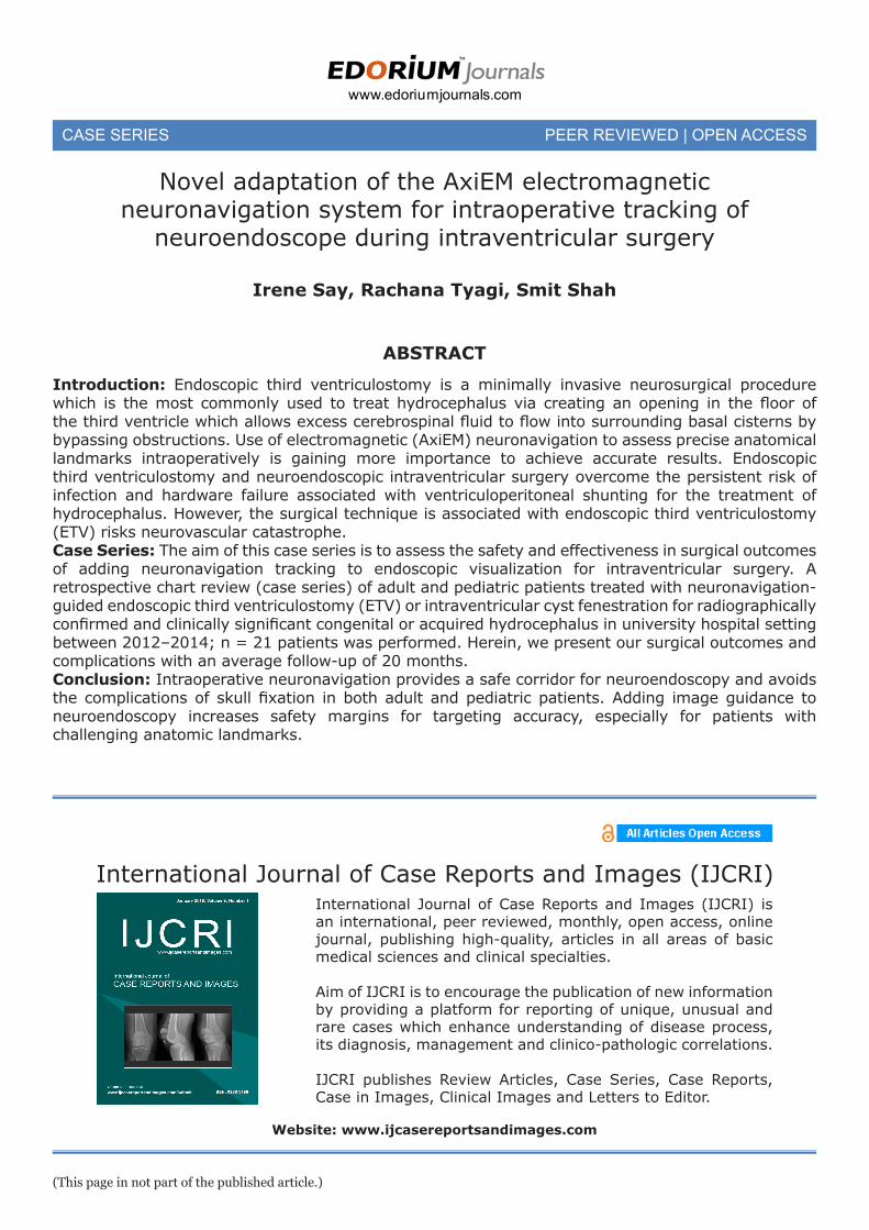
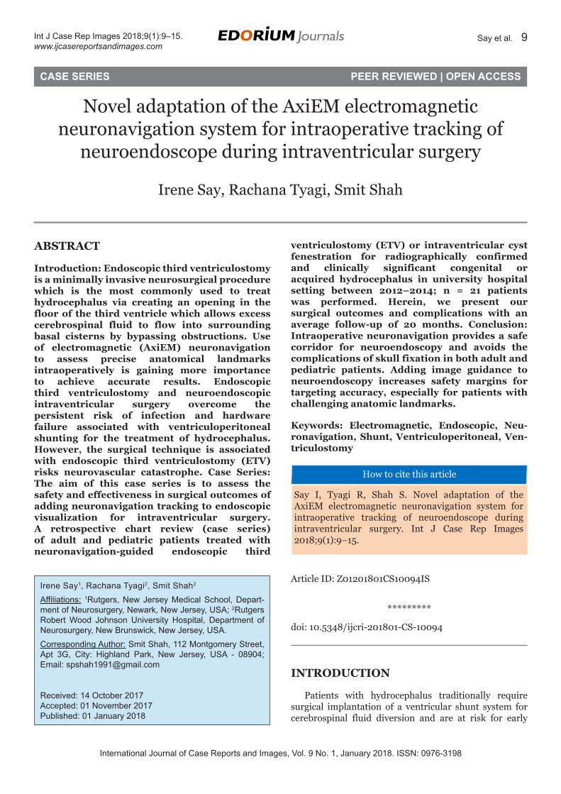
We followed the standard procedure of registration of the Medtronic AxiEM electromagnetic tracking system to the preoperative MRI Stealth with contrast during which head was positioned in a gel donut without pin fixation under general anesthesia. The entry site was pre-planned to allow the best approach through skull, parenchyma and lateral ventricle through the foramen of Monroe to the floor of third ventricle avoiding injury to cortical vessels and the fornix in addition to targeting area anterior to the basilar artery without damaging it. A double-screen set-up allowed simultaneous endoscopy and neuronavigation, irrigation with lactated Ringer’s, and balloon catheter set-up (Figure 1 and Figure 2). We cannulated the ventricle using live navigation with the AxiEM stylet in a 14F peel-away sheath (model # PLVW-
Figure 1: (A) Stylet in sheath and (B) Stylet with peel away trochar not in endoscope.
Figure 2: Double screen simultaneous neuroendoscopy and neuronavigation for continuous real time orientation. Tip of the endoscope is within the ventricle, oriented along the preplanned trajectory with a clear view of the planned fenestration site.
16.0-38) using the Trajectory 1, Trajectory 2 and guidance views to maintain perfect alignment with the pre-planned trajectory. The projection is set to 10 mm as the tip of the stylet sits 1 cm proximal to the tip of the trocar. The endoscope is also 1 cm longer than the stylet and no further adjustment of the projection is required when navigating the endoscope. Once the ventricle is entered, the sheath is advanced to the tip of the trocar which is then removed. Cerebrospinal fluid return confirms appropriate placement.
The stylet is then placed in the working port of endoscope for simultaneous real-time multi-planar neuronavigation confirmation of the endoscope position relative to the pre-planned trajectory and visualized anatomic landmarks are obtained as well.
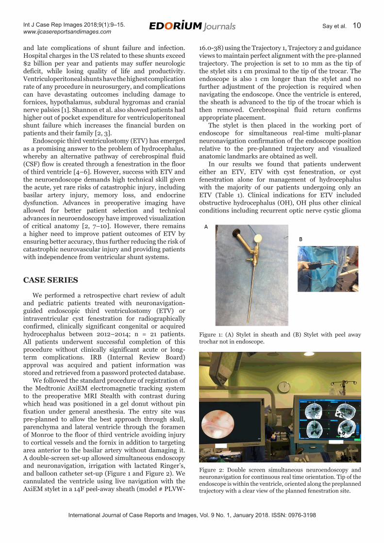
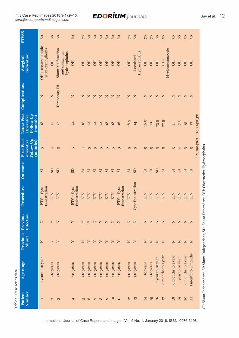
In our results we found that patients underwent either an ETV, ETV with cyst fenestration, or cyst fenestration alone for management of hydrocephalus with the majority of our patients undergoing only an ETV (Table 1). Clinical indications for ETV included obstructive hydrocephalus (OH), OH plus other clinical conditions including recurrent optic nerve cystic glioma
International Journal of Case Reports and Images, Vol. 9 No. 1, January 2018. ISSN: 0976-3198
Int J Case Rep Images 2018;9(1):9–15. www.ijcasereportsandimages.com
Say et al. 11
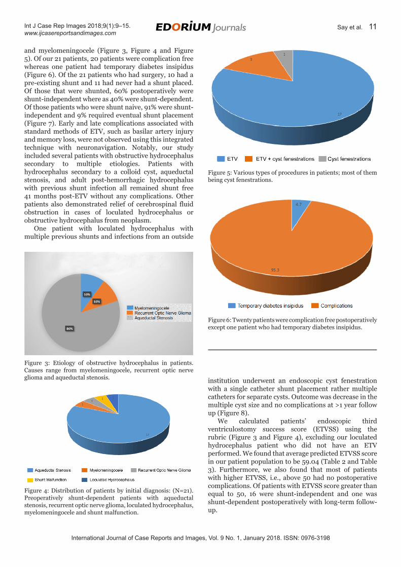
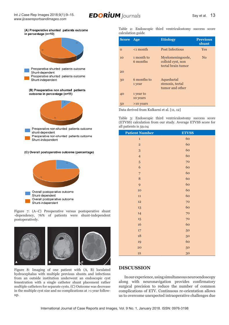
and myelomeningocele (Figure 3, Figure 4 and Figure 5). Of our 21 patients, 20 patients were complication free whereas one patient had temporary diabetes insipidus (Figure 6). Of the 21 patients who had surgery, 10 had a pre-existing shunt and 11 had never had a shunt placed. Of those that were shunted, 60% postoperatively were shunt-independent where as 40% were shunt-dependent. Of those patients who were shunt naive, 91% were shunt-independent and 9% required eventual shunt placement (Figure 7). Early and late complications associated with standard methods of ETV, such as basilar artery injury and memory loss, were not observed using this integrated technique with neuronavigation. Notably, our study included several patients with obstructive hydrocephalus secondary to multiple etiologies. Patients with hydrocephalus secondary to a colloid cyst, aqueductal stenosis, and adult post-hemorrhagic hydrocephalus with previous shunt infection all remained shunt free 41 months post-ETV without any complications. Other patients also demonstrated relief of cerebrospinal fluid obstruction in cases of loculated hydrocephalus or obstructive hydrocephalus from neoplasm.
One patient with loculated hydrocephalus with multiple previous shunts and infections from an outside
Figure 5: Various types of procedures in patients; most of them being cyst fenestrations.
Figure 6: Twenty patients were complication free postoperatively except one patient who had temporary diabetes insipidus.
Figure 3: Etiology of obstructive hydrocephalus in patients. Causes range from myelomeningocele, recurrent optic nerve glioma and aqueductal stenosis.
Figure 4: Distribution of patients by initial diagnosis: (N=21). Preoperatively shunt-dependent patients with aqueductal stenosis, recurrent optic nerve glioma, loculated hydrocephalus, myelomeningocele and shunt malfunction.
institution underwent an endoscopic cyst fenestration with a single catheter shunt placement rather multiple catheters for separate cysts. Outcome was decrease in the multiple cyst size and no complications at >1 year follow up (Figure 8).
We calculated patients’ endoscopic third ventriculostomy success score (ETVSS) using the rubric (Figure 3 and Figure 4), excluding our loculated hydrocephalus patient who did not have an ETV performed. We found that average predicted ETVSS score in our patient population to be 59.04 (Table 2 and Table 3). Furthermore, we also found that most of patients with higher ETVSS, i.e., above 50 had no postoperative complications. Of patients with ETVSS score greater than equal to 50, 16 were shunt-independent and one was shunt-dependent postoperatively with long-term follow-up.
International Journal of Case Reports and Images, Vol. 9 No. 1, January 2018. ISSN: 0976-3198
Int J Case Rep Images 2018;9(1):9–15. www.ijcasereportsandimages.com
Say et al. 12Ta
ble
1: C
ase
seri
es d
ata
Pat
ien
t N
um
ber
Age
ran
geP
revi
ous
Sh
un
tP
revi
ous
Infe
ctio
nP
roce
du
reO
utc
ome
Fir
st P
ost
oper
ativ
e F
ollo
w U
p
(mon
ths)
Lat
est
Pos
t O
per
ativ
e F
ollo
w U
p
(mon
ths)
Com
pli
cati
ons
Su
rgic
al
Ind
icat
ion
ET
VS
S
11
year
to 1
0 ye
arN
NE
TV +
Cys
t Fe
nest
rati
onSI
518
NO
H +
rec
urre
nt o
ptic
ne
rve
cyst
ic g
liom
a60
2>
10 y
ears
YY
ETV
SD4
24N
OH
60
3>
10 y
ears
YN
ETV
SD5
24Te
mpo
rary
DI
Shun
t Mal
func
tion
an
d co
ngen
ital
hy
droc
epha
lus
60
4>
10 y
ears
YY
ETV
+ C
yst
Fene
stra
tion
SD5
24N
OH
60
5>
10 y
ears
NY
ETV
SI4
18N
OH
70
6>
10 y
ears
YY
ETV
SI5
24N
OH
60
7>
10 y
ears
YN
E
TVSI
524
NO
H60
8>
10 y
ears
YY
ETV
SI5
24N
OH
60
9>
10 y
ears
YY
ETV
SI5
18N
OH
60
10>
10 y
ears
YN
ETV
SI5
18N
OH
60
11>
10 y
ears
YN
ETV
+ C
yst
Fene
stra
tion
SI3
16N
OH
60
12>
10 y
ears
NN
ETV
SI5
18.5
NO
H70
13>
10 y
ears
YN
Cys
t Fen
estr
atio
nSD
514
NLo
cula
ted
Hyd
roce
phal
us60
14>
10 y
ears
NN
ETV
SI5
19.5
NO
H70
15>
10 y
ears
NN
ETV
SI5
21N
OH
70
161
year
to 1
0 ye
arN
NE
TVSD
523
.5N
OH
60
176
mon
ths
to 1
yea
rN
NE
TVSI
522
.5N
OH
+
Mye
lom
enin
goce
le50
186
mon
ths
to 1
yea
rN
NE
TVSI
524
NO
H50
191
year
to 1
0 ye
arN
NE
TVSI
417
.5N
OH
60
206
mon
ths
to 1
yea
rN
NE
TVSI
515
NO
H50
211
mon
th to
6 m
onth
sN
NE
TVSI
517
NO
H30
4.76
1904
762
20.2
1428
571
SI: S
hunt
Ind
epen
dent
, SI:
Shu
nt I
ndep
ende
nt, S
D: S
hunt
Dep
ende
nt, O
H: O
bstr
ucti
ve H
ydro
ceph
alus
International Journal of Case Reports and Images, Vol. 9 No. 1, January 2018. ISSN: 0976-3198
Int J Case Rep Images 2018;9(1):9–15. www.ijcasereportsandimages.com
Say et al. 13
Figure 7: (A–C) Preoperative versus postoperative shunt -dependency, 76% of patients were shunt-independent postoperatively.
Figure 8: Imaging of one patient with (A, B) loculated hydrocephalus with multiple previous shunts and infections from an outside institution underwent an endoscopic cyst fenestration with a single catheter shunt placement rather multiple catheters for separate cysts. (C) Outcome was decrease in the multiple cyst size and no complications at >1 year follow-up.
Table 2: Endoscopic third ventriculostomy success score calculation guide
Score Age Etiology Previous shunt
0 <1 month Post Infectious Yes
10 1 month to 6 months
Myelomeningocele, colloid cyst, non tectal brain tumor
No
20
30 6 months to 1 year
Aqueductal stenosis, tectal tumor and other
40 1 year to 10 years
50 >10 years
Data derived from Kulkarni et al. [11, 12]
Table 3: Endoscopic third ventriculostomy success score (ETVSS) calculation from our study. Average ETVSS score for all patients is 59.04
Patient Number ETVSS
1 60
2 60
3 60
4 60
5 70
6 60
7 60
8 60
9 60
10 60
11 60
12 70
13 60
14 70
15 70
16 60
17 50
18 50
19 60
20 50
21 30
DISCUSSION
In our experience, using simultaneous neuroendoscopy along with neuronavigation provides confirmatory surgical precision to reduce the number of common complications of ETV. Continuous re-orientation allows us to overcome unexpected intraoperative challenges due
International Journal of Case Reports and Images, Vol. 9 No. 1, January 2018. ISSN: 0976-3198
Int J Case Rep Images 2018;9(1):9–15. www.ijcasereportsandimages.com
Say et al. 14
to abnormal anatomy throughout the procedure. Most importantly, our technique has provided durable shunt-independence, thus improving our patients’ quality of life and reducing the socioeconomic burden of shunt hardware.
Of note, at long-term follow-up of average 41 months, there was no observed incidence of basilar artery injury, infection, CSF leak, or forniceal injury. Failure to identify the location of the basilar artery with the endoscope can increase the risk of vascular injury, particularly with abnormal ventricular anatomy. Excessive manipulation and traction of the parenchyma while exploring the ventricles can lead to CSF leak, or forniceal injury [11–13]. The added neuronavigation provides the ability to confirm accurate localization even without normal anatomic landmarks to reduce the intraoperative risks.
Published success rates for ETV [14, 15], as measured by shunt-independence are about 73% as compared to our rate which is 76% during first postoperative average follow-up of 4.76 months. To further validate this, we will have to perform more ETV cases and also follow up patients over a longer time period to gauge our improvement. As compared to on our calculation of predicted ETVSS of 59.04%, our actual postoperative ETV success rate was 81% (17/21). However, we would still like to optimize and validate our success score by performing more cases and getting more surgical experience in patients with different variety of indication of obstructive hydrocephalus.
This surgical technique can also be used in a larger patient population including those with myelomeningocele [5]. Our future studies will evaluate the long-term cost-effectiveness of this quality improvement technique by examining the length of stay, length of surgery, and rate of re-hospitalization.
CONCLUSION
From the perspective of surgical quality improvement, adapting frameless electromagnetic neuronavigation provides real-time, multi-planar orientation during neuroendoscopic intraventricular surgery and reduces the risk of injury to critical brain structures such as the fornix and intracerebral vessels, which are typically vulnerable during standard endoscopic third ventriculostomy techniques. Intraoperative navigation provides a safe corridor for neuroendoscopy and avoids many complications of skull fixation in both adult and pediatric patients. Adding image guidance to neuroendoscopy increases safety margins for targeting accuracy, especially for patients with challenging anatomic landmarks. This available, safe addition to the recent advances in endoscopic neurosurgery will augment our access to critical regions of the brain for tumor resection and treatment of hydrocephalus, while reducing the risk of known common complications such as basilar artery rupture, cerebrospinal fluid leak and infections.
REFERENCES
1. http://www.hydroassoc.org/complications-of-etv-and-etvcpc/
2. Schroeder HW, Wagner W, Tschiltschke W, Gaab MR. Frameless neuronavigation in intracranial endoscopic neurosurgery. J Neurosurg 2001 Jan;94(1):72–9.
3. Shannon CN, Simon TD, Reed GT, et al. The economic impact of ventriculoperitoneal shunt failure. J Neurosurg Pediatr 2011 Dec;8(6):593–9.
4. Guzman R, Pendharkar AV, Zerah M, Sainte-Rose C. Use of the NeuroBalloon catheter for endoscopic third ventriculostomy. J Neurosurg Pediatr 2013 Mar;11(3):302–6.
5. Hayhurst C, Byrne P, Eldridge PR, Mallucci CL. Application of electromagnetic technology to neuronavigation: A revolution in image-guided neurosurgery. J Neurosurg 2009 Dec;111(6):1179–84.
6. Oertel JM, Vulcu S, Schroeder HW, Konerding MA, Wagner W, Gaab MR. Endoscopic transventricular third ventriculostomy through the lamina terminalis. J Neurosurg 2010 Dec;113(6):1261–9.
7. Longatti P, Fiorindi A, Feletti A, Baratto V. Endoscopic opening of the foramen of magendie using transaqueductal navigation for membrane obstruction of the fourth ventricle outlets: Technical note. J Neurosurg 2006 Dec;105(6):924–7.
8. Tao C. Complications of endoscopic third ventriculostomy. [Available at: http://w w w .hydroassoc .org/w p -conte nt/up loads/kbase/Complicat ions-of-Endoscopic-Third-Ventriculostomy.pdf]
9. Rohde V, Behm T, Ludwig H, Wachter D. The role of neuronavigation in intracranial endoscopic procedures. Neurosurg Rev 2012 Jul;35(3):351–8.
10. Vogel TW, Bahuleyan B, Robinson S, Cohen AR. The role of endoscopic third ventriculostomy in the treatment of hydrocephalus. J Neurosurg Pediatr 2013 Jul;12(1):54–61.
11. Kulkarni AV, Drake JM, Mallucci CL, Sgouros S, Roth J, Constantini S. Endoscopic third ventriculostomy in the treatment of childhood hydrocephalus. J Pediatr 2009 Aug;155(2):254–9.e1.
12. Kulkarni AV, Riva-Cambrin J, Browd SR. Use of the ETV success score to explain the variation in reported endoscopic third ventriculostomy success rates among published case series of childhood hydrocephalus. J Neurosurg Pediatr 2011 Feb;7(2):143–6.
13. Wang Y, Gao J, Zhang D, Ding Q. Complications of endoscopic third ventriculostomy for hydrocephalus. [Article in Chinese]. Zhonghua Yi Xue Za Zhi 2015 May 5;95(17):1338–40.
14. Grand W, Leonardo J, Chamczuk AJ, Korus AJ. Endoscopic third ventriculostomy in 250 adults with hydrocephalus: Patient selection, outcomes, and complications. Neurosurgery 2016 Jan;78(1):109–19.
15. Beuriat PA, Szathmari A, Grassiot B, Plaisant F, Rousselle C, Mottolese C. Role of endoscopic third ventriculostomy in the management of myelomeningocele-related hydrocephalus: A retrospective study in a single french institution. World Neurosurg 2016 Mar;87:484–93.
International Journal of Case Reports and Images, Vol. 9 No. 1, January 2018. ISSN: 0976-3198
Int J Case Rep Images 2018;9(1):9–15. www.ijcasereportsandimages.com
Say et al. 15
SUGGESTED READING
• https://academic.oup.com/neurosurgery/article/78/1/124/2453606/Commentary-Endoscopic-Third-Ventriculostomy-in-250
• Sindou M. Practical Handbook of Neurosurgery: From Leading Neurosurgeons. Volume, 1. New York: Springer Wien; 2009. p. 38.
*********
Author ContributionsIrene Say – Substantial contributions to conception and design, Acquisition of data, Analysis and interpretation of data, Drafting the article, Revising it critically for important intellectual content, Final approval of the version to be publishedRachana Tyagi – Substantial contributions to conception and design, Acquisition of data, Analysis and interpretation of data, Drafting the article, Revising it critically for important intellectual content, Final approval of the version to be published
Smit Shah – Substantial contributions to conception and design, Acquisition of data, Analysis and interpretation of data, Drafting the article, Revising it critically for important intellectual content, Final approval of the version to be published
Guarantor of SubmissionThe corresponding author is the guarantor of submission.
Source of SupportNone
Conflict of InterestAuthors declare no conflict of interest.
Copyright© 2018 Irene Say et al. This article is distributed under the terms of Creative Commons Attribution License which permits unrestricted use, distribution and reproduction in any medium provided the original author(s) and original publisher are properly credited. Please see the copyright policy on the journal website for more information.
Access full text article onother devices
Access PDF of article onother devices
EDORIUM JOURNALS OPEN ACCESS
Edorium Journals: On Web
About Edorium JournalsEdorium Journals is a publisher of international, high-quality, open access, scholarly journals covering subjects in basic sciences and clinical specialties and subspecialties.
Edorium Journals www.edoriumjournals.com
Edorium Journals et al.
Edorium Journals: An introduction
Why should you publish with Edorium Journals?In less than 10 words: “We give you what no one does”.
Vision of being the bestWe have the vision of making our journals the best and the most authoritative journals in their respective special-ties. We are working towards this goal every day.
Exceptional servicesWe care for you, your work and your time. Our efficient, personalized and courteous services are a testimony to this.
Editorial reviewAll manuscripts submitted to Edorium Journals undergo pre-processing review followed by multiple rounds of stringent editorial reviews.
Peer reviewAll manuscripts submitted to Edorium Journals undergo anonymous, double-blind, external peer review.
Early view versionEarly View version of your manuscript will be published in the journal within 72 hours of final acceptance.
Manuscript statusFrom submission to publication of your article you will get regular updates about status of your manuscripts.
Our Commitment
Favored author programOne email is all it takes to become our favored author. You will not only get 15% off on all manuscript but also get information and insights about scholarly publishing.
Institutional membership programJoin our Institutional Memberships program and help scholars from your institute make their research acces-sible to all and save thousands of dollars in publication fees.
Our presenceWe have high quality, attractive and easy to read publica-tion format. Our websites are very user friendly and en-able you to use the services easily with no hassle.
Something more...We request you to have a look at our website to know more about us and our services. Please visit: www.edoriumjournals.com
We welcome you to interact with us, share with us, join us and of course publish with us.
Browse Journals
CONNECT WITH US
Invitation for article submissionWe sincerely invite you to submit your valuable research for publication to Edorium Journals.
Six weeksWe give you our commitment that you will get first deci-sion on your manuscript within six weeks (42 days) of submission. If we fail to honor this commitment by even one day, we will give you a 75% Discount Voucher for your next manuscript.
Four weeksWe give you our commitment that after we receive your page proofs, your manuscript will be published in the journal within 14 days (2 weeks). If we fail to honor this commitment by even one day, we will give you a 75% Discount Voucher for your next manuscript.
This page is not a part of the published article. This page is an introduction to Edorium Journals.