involvement of the visual pathway is not a risk factor of visual · pdf fileinvolvement of the...
TRANSCRIPT
RESEARCH Open Access
Involvement of the visual pathway is not arisk factor of visual field deficits in patientswith occipital arteriovenous malformations:an fMRI studyXianzeng Tong1,2,3,4, Jun Wu1,2,3,4, Fuxin Lin1,2,3,4, Yong Cao1,2,3,4, Yuanli Zhao1,2,3,4, Zhen Jin5 and Shuo Wang1,2,3,4*
Abstract
Background: Occipital arteriovenous malformations (AVMs) are still one of neurosurgery’s most intriguing andchallenging pathologies. In this study, we reviewed our series of patients with occipital AVMs admitted in BeijingTiantan Hospital from June 2013 through January 2015 and attempted to evaluate the risk factors of visual fielddeficits (VFDs) in these patients at presentation.
Methods: Forty-two consecutive patients with occipital AVMs were included in our study. Patient parameters(age, sex, and history of hemorrhage) and AVM characteristics (size, side, venous drainage, Spetzler-Martingrade, and diffuseness) were collected. VFDs were quantified using an Octopus perimetry. Conventional MRI,blood oxygen level dependent fMRI (BOLD-fMRI) of the visual cortex, and diffusion tensor imaging (DTI) ofthe optic radiation were performed. The least distances from the AVM to the optic radiation (AVM-OR) andfrom the AVM to the visual cortex (AVM-VC) were measured. Univariate analyses were used to correlate initialVFDs with patient parameters, AVM characteristics, AVM-OR, and AVM-VC distances.
Results: VFDs were identified in 14 patients, among which 12 patients presented with a history of hemorrhage and2 patients presented with nonhemorrhagic chronic headache. VFDs were more common (P = 0.000003) in patientswith ruptured AVMs. VFD frequency was not associated with patient age, sex, and AVM characteristics (size, side,venous drainage, S-M grade, and diffuseness). Unlike other lesions involving the optic radiation and visual cortex, thefrequency of VFDs in occipital AVMs did not correlate with the AVM-OR and AVM-VC distances (P = 0.640 and 0.638,respectively).
Conclusions: A history of hemorrhage is an independent risk factor of VFDs in occipital AVMs. Most unrupturedoccipital AVMs may present with chronic headache and seizures other than VFDs. The distances from the AVMs to theoptic radiation and the visual cortex are not associated with preexisting VFDs. Our results prompt us to probe into theplasticity of the visual pathway in patients with this congenital vascular anomaly.
Keywords: Arteriovenous malformations (AVMs), Visual field deficits (VFDs), fMRI, DTI
* Correspondence: [email protected] of Neurosurgery, Beijing Tiantan Hospital, Capital MedicalUniversity, Beijing 100050, People’s Republic of China2China National Clinical Research Center for Neurological Diseases, Beijing,People’s Republic of ChinaFull list of author information is available at the end of the article
© 2015 Tong et al. Open Access This article is distributed under the terms of the Creative Commons Attribution 4.0International License (http://creativecommons.org/licenses/by/4.0/), which permits unrestricted use, distribution, andreproduction in any medium, provided you give appropriate credit to the original author(s) and the source, provide a link tothe Creative Commons license, and indicate if changes were made. The Creative Commons Public Domain Dedication waiver(http://creativecommons.org/publicdomain/zero/1.0/) applies to the data made available in this article, unless otherwise stated.
Tong et al. Chinese Neurosurgical Journal (2015) 1:10 DOI 10.1186/s41016-015-0010-7
BackgroundOptic radiation is a group of axons passing from the lateralgeniculate nucleus (LGN) anteriorly over the roof of thelateral ventricle and then turning backwards to the visualcortex. Patients with occipital arteriovenous malforma-tions (AVMs) involving the optic radiation and visual cor-tex tend to present with visual field disturbances andmigraine-like headaches [4, 15, 25]. The risk factors of pre-existing visual field deficits (VFDs) of occipital AVMs havebeen discussed in previous studies [2, 5, 16, 22, 23]. A his-tory of hemorrhage may be an independent risk factor ofVFDs at presentation [2, 16]. Medial occipital AVMs maybe associated with more preexisting VFDs [5]. VFDs mightoccur as a result of occipital lobe seizures [2, 16, 22]. Forunruptured occipital AVMs, the etiology of preexistingVFDs may be related to ischemia and/or vascular steal inparenchyma adjacent to the arteriovenous malformation(AVM) [22, 23]. VFDs may also occur as a result of masseffect [2]. Surgical management of an occipital AVM mayalso cause new or worse VFDs.Diffusion tensor imaging (DTI) tractography is cur-
rently the only noninvasive technique that enables invivo visualization of the course and characterization ofwhite matter fiber tracts. With DTI, study has demon-strated that the least distance from the lesion to theoptic radiation was associated with preexisting VFDs[24], but their series predominantly included gliomas in-volving the optic radiation. Distinct from brain tumorssuch as gliomas and other congenital vascular lesionssuch as vein of Galen malformations and dural AVMs,brain AVMs are described as a congenitally abnormalcomplex of afferent arteries communicating with drain-ing veins [11]. Risk factors of preexisting VFDs in occipi-tal AVMs may differ from those caused by tumors andother pathologies. Till now, the literature provides fewsystematic accounts of the relationship between preexist-ing VFDs and the distance from the AVMs to the opticradiation or visual cortex. Is it possible that the preexist-ing VFD is associated with the distance from the AVMsto the optic radiation or visual cortex? What are the riskfactors of preexisting VFDs in patients with occipitalAVMs? In this study, we will attempt to answer thesetwo questions and discuss our preliminary results.
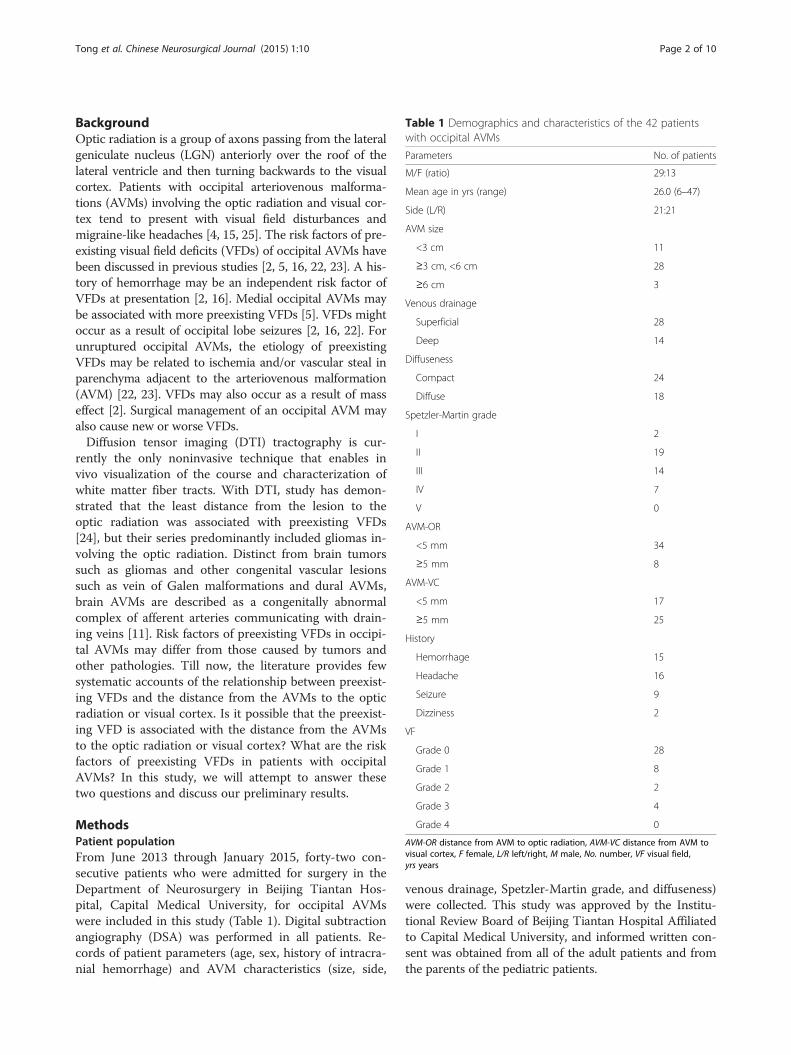
MethodsPatient populationFrom June 2013 through January 2015, forty-two con-secutive patients who were admitted for surgery in theDepartment of Neurosurgery in Beijing Tiantan Hos-pital, Capital Medical University, for occipital AVMswere included in this study (Table 1). Digital subtractionangiography (DSA) was performed in all patients. Re-cords of patient parameters (age, sex, history of intracra-nial hemorrhage) and AVM characteristics (size, side,
venous drainage, Spetzler-Martin grade, and diffuseness)were collected. This study was approved by the Institu-tional Review Board of Beijing Tiantan Hospital Affiliatedto Capital Medical University, and informed written con-sent was obtained from all of the adult patients and fromthe parents of the pediatric patients.
Table 1 Demographics and characteristics of the 42 patientswith occipital AVMs
Parameters No. of patients
M/F (ratio) 29:13
Mean age in yrs (range) 26.0 (6–47)
Side (L/R) 21:21
AVM size
<3 cm 11
≥3 cm, <6 cm 28
≥6 cm 3
Venous drainage
Superficial 28
Deep 14
Diffuseness
Compact 24
Diffuse 18
Spetzler-Martin grade
I 2
II 19
III 14
IV 7
V 0
AVM-OR
<5 mm 34
≥5 mm 8
AVM-VC
<5 mm 17
≥5 mm 25
History
Hemorrhage 15
Headache 16
Seizure 9
Dizziness 2
VF
Grade 0 28
Grade 1 8
Grade 2 2
Grade 3 4
Grade 4 0
AVM-OR distance from AVM to optic radiation, AVM-VC distance from AVM tovisual cortex, F female, L/R left/right, M male, No. number, VF visual field,yrs years
Tong et al. Chinese Neurosurgical Journal (2015) 1:10 Page 2 of 10

Imaging acquisitionA 3 T Siemens Tim Trio magnetic resonance imaging(MRI) scanner with a 12-channel head coil was used forscanning protocol. T1-weighted structural imaging, bloodoxygen level dependent functional magnetic resonanceimaging (BOLD-fMRI), time of flight magnetic resonanceangiography (TOF-MRA), and DTI were collected foreach patient. All imaging and image processing were per-formed by two technical assistants at the Medical ImagingCenter, the 306th Hospital of PLA. All data analyzing pro-cedures were done by two physicians at our institution.T1-weighted three-dimensional magnetization-prepared
rapid acquisition gradient echo sequence was measuredwith a echo time (TE) of 2.98 ms, a repetition time (TR)of 2300 ms, a matrix size of 256 × 256, a field of view of250 × 250 mm, a slice thickness of 1 mm, and a slab sizeof 17.6 cm.For BOLD-fMRI, maps of neural activity within the vis-
ual cortex were generated in each patient. Visual taskgiven was “black white checkerboard”. A “block” designwas used in which there were 80 dynamics and every 10dynamics of activity was alternated with 10 dynamics ofrest, beginning with rest. The total time of the task was240 s. The fMRI sequence was a fast-field echo single-shotecho planar imaging technique with an echo planar im-aging factor of 79, a TR of 3000 ms, a TE of 30 ms, and aflip angle of 90. Contiguous slices, 6 mm thick, weretaken, and 30 slices were obtained. The field of view was210 × 210 mm, with a matrix size of 64 mm, resulting inan in-plane resolution of 3.3 mm. Voxel size was 3.3 ×3.3 × 4.0 mm. For each slice, 80 dynamic time points werecollected, providing a total imaging time of 4 min and 8 s,30.1 s for each run.Time of flight MRA (TOF-MRA) was obtained by the
following parameters: TR, 22 ms; TE, 3.86 ms; matrix size,384 × 256; field of view, 220 × 180 mm; bandwidth,178 Hz per pixel; voxel size, 0.8 × 0.6 × 1.0 mm; flip angle;18. 30 slices were obtained, and the total imaging timewas 3 min and 29 s.For DTI, we applied a single-shot spin-echo diffusion-
weighted echo planar imaging sequence (TE, 93 ms; TR,6100 ms; matrix size, 128 × 128; field of view, 230 ×230 mm; bandwidth, 1396 Hz per pixel; voxel size, 1.8 ×1.8 × 3 mm and diffusion-encoding gradients in 12 direc-tions using b values of 0 and 1000 s/mm2). We used 45slices with no intersection gap. The slice thickness was3 mm, and each slice was angulated by 40° anteriorly tocover the optic chiasm, the occipital lobe, the corpus cal-losum, and the upper two thirds of the temporal lobe. Thetime needed for image acquisition was 6 min and 38 s.
Data analysisThe MRI data were saved as DICOM format. The datawere transferred to an off-line iPlan 3.0 workstation
(Brainlab, Feldkirchen, Germany) for analysis. Motioncorrection was done in raw BOLD-fMRI. The raw DTIimages were corrected for eddy currents. The TOF-MRA, BOLD-fMRI, and DTI images then automaticallyintegrated with T1 structural images. Using the generallinear model (GLM), the BOLD data were analyzed ac-cording to the stimuli parameters of visual task. The 3Dmodel of the visual area was reconstructed according tothe activated volumes that were considered to consist ofP = 0.001 activated areas. For fiber tracking of the opticradiation, the ipsilateral lateral geniculate was selected asthe first seed volume. All the fibers that cross this vol-ume of interest (VOI) were tracked. The second seedvolume was the activated visual cortex. Fiber trackingwas initiated with fractional anisotropy (FA) of 0.2. Atlast, the 3D anatomic structure of the AVMs was recon-structed by threshold adjustment of the TOF-MRA data.
Acquisition and labeling of important parametersThrough the iPlan 3.0 software, the maximum diametersof the AVMs were measured from the structural MRI onthe axial, coronary, and sagittal directions. The distancesof AVM from the optic radiation (AVM-OR) and visualcortex (AVM-VC) were measured respectively on theaxial, coronary, and sagittal directions. The least distancewas what this study required.
Visual field assessmentVisual fields testing of the central 30° were performedwith an Octopus field analyzer (Haag Streit International,Koeniz, Switzerland) by an experienced ophthalmologistwho was blinded to the results of the neuroimaging find-ings. As to Kupersmith et al. [16], VFDs were graded asnormal VFs (grade 0), incomplete quadrantanopsia (grade1), complete quadrantanopsia (grade 2), incomplete hemi-anopsia (grade 3), and complete hemianopsia (grade 4).Mild defects were defined as grade 1 and grade 2, whilegrade 3 and grade 4 were referred to as severe defects.
Statistical analysisStatistical analyses were performed with the StatisticalPackage for the Social Sciences software (version 20.0;SPSS, Inc, Chicago, IL). Univariate analyses were used toassess the risk factors of preexisting VFDs. Statistical sig-nificance was established at a value of P < 0.05.
ResultsDemographic data and clinical presentationsThe baseline characteristics of the patients and AVMs arelisted in Table 1. Besides VFDs, none of our patients pre-sented with more than one symptom. Fifteen (36 %) pa-tients presented with a history of hemorrhage, 9 (21 %)with seizures, 16 (38 %) with nonhemorrhagic chronicheadaches, and 2 (5 %) with intermittent dizziness. In ten
Tong et al. Chinese Neurosurgical Journal (2015) 1:10 Page 3 of 10
patients with nonhemorrhagic headache, there was an ab-solute preference for the side ipsilateral to the AVM. Theheadache was described as throbbing in seven cases andassociated with visual phenomena (blurred vision in hemi-field or flickering) in six cases.The AVMs ranged in diameter from 13.5 to
70.0 mm (mean 39.3 mm). The size was <3 cm in 11cases and ≥3 cm in 31 (including ≥6 cm in 3, Table 1).The primary feeding arteries to the AVM werethrough the cortical branches of the posterior cerebralartery, but all AVMs ≥3 cm in diameter had multiplefeeding arteries involving the anterior circulation. Ofthe 42 cases in this study, 14 had deep venous drain-age or both deep and superficial venous drainagewhile the other 28 had only superficial venous drain-age. AVM configuration (compact vs. diffuse) was de-fined according to previous study [17].
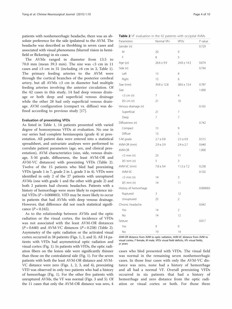
Evaluation of preexisting VFDsAs listed in Table 1, 14 patients presented with varieddegree of homonymous VFDs at evaluation. No one inour series had complete hemianopsia (grade 4) at pres-entation. All patient data were entered into a statisticalspreadsheet, and univariate analyses were performed tocorrelate patient parameters (age, sex, and clinical pres-entation), AVM characteristics (size, side, venous drain-age, S-M grade, diffuseness, the least AVM-OR andAVM-VC distances) with preexisting VFDs (Table 2).Twelve of the 15 patients who bled had preexistingVFDs (grade 1 in 7, grade 2 in 1, grade 3 in 4). VFDs wereidentified in only 2 of the 27 patients with unrupturedAVMs (one with grade 1 and the other with grade 2) andboth 2 patients had chronic headaches. Patients with ahistory of hemorrhage were more likely to experience ini-tial VFDs (P = 0.000003). VFD may be more likely to occurin patients that had AVMs with deep venous drainage.However, that difference did not reach statistical signifi-cance (P = 0.165).As to the relationship between AVMs and the optic
radiation or the visual cortex, the incidence of VFDswas not associated with the least AVM-OR distances(P = 0.640) and AVM-VC distances (P = 0.238) (Table 2).Asymmetry of the optic radiation or the activated visualcortex occurred in 38 patients (Figs. 1, 2, and 3). All 14 pa-tients with VFDs had asymmetrical optic radiation andvisual cortex (Fig. 1). In patients with VFDs, the optic radi-ation fibers on the lesion side were significantly thinnerthan those on the contralateral side (Fig. 1). For the sevenpatients with both the least AVM-OR distance and AVM-VC distance were zero (Figs. 1, 2, 3, and 4), preexistingVFD was observed in only two patients who had a historyof hemorrhage (Fig. 1). For the other five patients withunruptured AVMs, the VF was normal (Figs. 3 and 5). Ofthe 11 cases that only the AVM-OR distance was zero, 4
cases who bled presented with VFDs. The visual fieldwas normal in the remaining seven nonhemorrhagiccases. In those four cases with only the AVM-VC dis-tance was zero, none had a history of hemorrhageand all had a normal VF. Overall preexisting VFDsoccurred in six patients that had a history ofhemorrhage and zero distance from the optic radi-ation or visual cortex or both. For those three
Table 2 VF evaluation in the 42 patients with occipital AVMs
Parameters Normal VFs VFDs P value
Gender (n) 0.729
M 20 9
F 8 5
Age (ys) 26.6 ± 9.9 24.8 ± 14.3 0.674
Side (n) 0.744
Left 13 8
Right 15 6
Size (mm) 39.8 ± 12.8 38.6 ± 13.4 0.787
Size 1.000
<3 cm (n) 7 4
≥3 cm (n) 21 10
Venous drainage (n) 0.165
Superficial 21 7
Deep 7 7
Diffuseness (n) 0.742
Compact 15 9
Diffuse 13 5
S-M grade 2.7 ± 0.8 2.5 ± 0.9 0.515
AVM-OR (mm) 2.9 ± 3.9 2.4 ± 2.7 0.640
AVM-OR 1.000
<5 mm (n) 23 11
≥5 mm (n) 5 3
AVM-VC (mm) 7.8 ± 9.4 11.3 ± 7.2 0.238
AVM-VC 0.102
<5 mm (n) 14 3
≥5 mm (n) 14 11
History of hemorrhage 0.000003
Ruptured 3 12
Unruptured 25 2
Chronic headache 0.042
Yes 14 2
No 14 12
Seizure 0.017
Yes 9 0
No 19 14
AVM-OR distance from AVM to optic radiation, AVM-VC distance from AVM tovisual cortex, F female, M male, VFDs visual field deficits, VFs visual fields,ys years
Tong et al. Chinese Neurosurgical Journal (2015) 1:10 Page 4 of 10
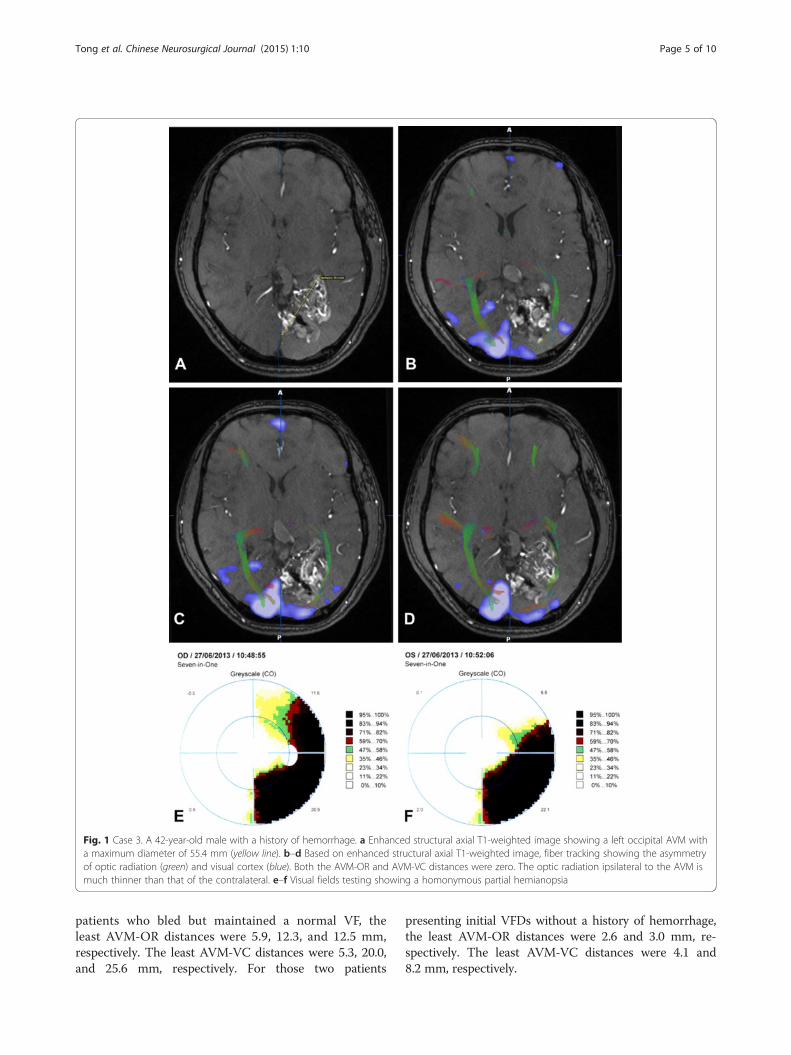
patients who bled but maintained a normal VF, theleast AVM-OR distances were 5.9, 12.3, and 12.5 mm,respectively. The least AVM-VC distances were 5.3, 20.0,and 25.6 mm, respectively. For those two patients
presenting initial VFDs without a history of hemorrhage,the least AVM-OR distances were 2.6 and 3.0 mm, re-spectively. The least AVM-VC distances were 4.1 and8.2 mm, respectively.
Fig. 1 Case 3. A 42-year-old male with a history of hemorrhage. a Enhanced structural axial T1-weighted image showing a left occipital AVM witha maximum diameter of 55.4 mm (yellow line). b–d Based on enhanced structural axial T1-weighted image, fiber tracking showing the asymmetryof optic radiation (green) and visual cortex (blue). Both the AVM-OR and AVM-VC distances were zero. The optic radiation ipsilateral to the AVM ismuch thinner than that of the contralateral. e–f Visual fields testing showing a homonymous partial hemianopsia
Tong et al. Chinese Neurosurgical Journal (2015) 1:10 Page 5 of 10
DiscussionOccipital AVMs are still one of neurosurgery’s intriguingand challenging pathologies because of their proximityto the visual cortex and optic radiations. The prevalenceof VFDs at presentation has been reported by severalstudies. Pollock et al. reported 7 (21 %) of 34 patientswith postgeniculate visual pathway AVMs had VFDs be-fore stereotactic radiotherapy [19]. Anderson et al. [1]reported that 13 (50 %) of 26 patients with occipitalAVMs presented with VFDs. Martin and Wilson [18] re-ported that 9 (56 %) of 16 patients had preexisting VFDsbefore surgical excision. Bartolomei et al. [2] reported afrequency of preexisting VFDs in 10 (43 %) of 23 pa-tients. Similar prevalence occurred in our series and 14(33 %) of the 42 patients presented with VFDs.
Risk factors of preexisting VFDsStudies reported that contralateral homonymous VFDsoccurred in 67–90 % of patients with ruptured occipitalAVMs [2, 16, 18, 25]. But in a series of 42 patients who
received curative treatment of occipital AVMs, 22 (52.4 %)of the patients presented with VFDs and only 11 (50.0 %)of these patients had a history of hemorrhage [23]. In ourseries of 42 patients, 15 (35.7 %) had a history ofhemorrhage and 12 (80 %) patients had preexisting VFDs.Our results showed that a history of hemorrhage was anindependent risk factor of preexisting VFDs in occipitalAVMs. Three patients with a history of hemorrhage didnot present with VFDs at evaluation. One reason might bethat the AVM-OR or the AVM-VC distance was far awayenough to avoid VFDs after hemorrhage. The other reasonmight be that VFDs secondary to hemorrhage had spon-taneously recovered to normal VFs over time [16]. For thetwo patients who presented with VFDs but had nonhe-morrhagic chronic headache, the etiology may be associ-ated with ischemia or vascular steal in parenchymaadjacent to the AVM [23].Studies have shown that the frequency of VFDs varied
with the locations of the AVMs. Dehdashti and colleaguesfound that patients with medial occipital AVMs were more
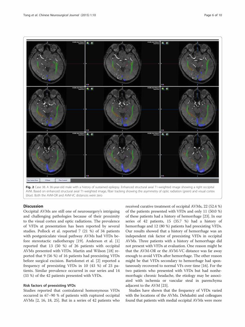
Fig. 2 Case 38. A 36-year-old male with a history of sustained epilepsy. Enhanced structural axial T1-weighted image showing a right occipitalAVM. Based on enhanced structural axial T1-weighted image, fiber tracking showing the asymmetry of optic radiation (green) and visual cortex(blue). Both the AVM-OR and AVM-VC distances were zero
Tong et al. Chinese Neurosurgical Journal (2015) 1:10 Page 6 of 10
likely to experience a preexisting VFD [5]. Kupersmithet al. [16] reported that VFD occurred in 75 % of thepatients with medial occipital AVMs, 50 % of those withlateral and 43.5 % of those with anterior occipital AVMs.But no location was associated with a significantly higher
frequency of visual defects. In our study, we did not classifythe location of AVMs as medial, lateral, anterior, or thewhole occipital lobe. Instead, we used the DTI tractogra-phy and BOLD-MRI to evaluate the relationship be-tween the AVMs and the optic radiation or the visual
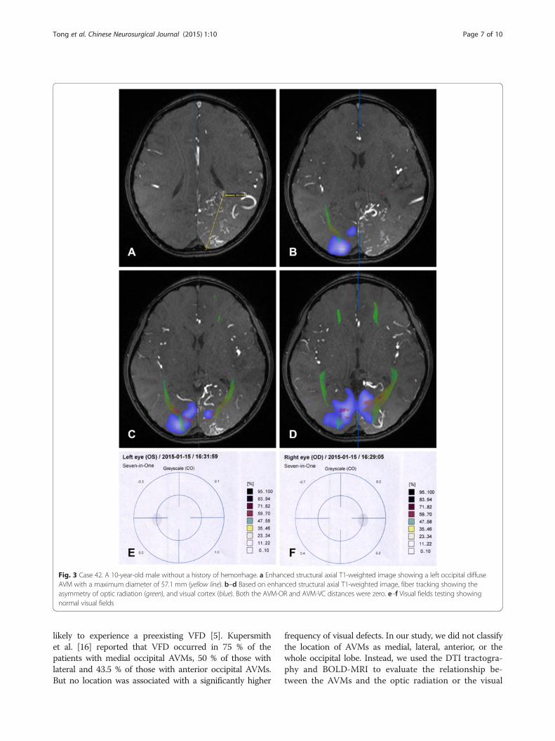
Fig. 3 Case 42. A 10-year-old male without a history of hemorrhage. a Enhanced structural axial T1-weighted image showing a left occipital diffuseAVM with a maximum diameter of 57.1 mm (yellow line). b–d Based on enhanced structural axial T1-weighted image, fiber tracking showing theasymmetry of optic radiation (green), and visual cortex (blue). Both the AVM-OR and AVM-VC distances were zero. e–f Visual fields testing showingnormal visual fields
Tong et al. Chinese Neurosurgical Journal (2015) 1:10 Page 7 of 10
cortex [24]. Our study was the first to use DTI andBOLD-MRI to evaluate the relationship between preexist-ing VFDs and occipital AVM locations. With optic fibertracking, we found that the VFD frequency was not associ-ated with the distance from the AVM to the optic radi-ation or the visual cortex. Eleven (32.4 %) of the 34patients with AVM-OR distance <5 mm and 3 (37.5 %) ofthe 8 patients with that distance ≥5 mm experiencedpreexisting VFDs; VFDs were identified in 17.6 % (3/17) ofthe patients with AVM-VC distance <5 mm and 44 % (11/25) of patients with that distance ≥5 mm. The differencesdid not reach statistical significance. This result is not
consistent with the previous studies that predominantlydealt with gliomas involving the optic radiation. Previousstudy has found that 55.6 % (15/27) of the patients inwhom the distance from the lesion to the optic radiationwas <5 mm had various degrees of preoperative VFDs,and all nine patients in whom the distance from the lesionto the optic radiation was >5 mm had preoperative normalVFs [24]. In our study with AVMs, the fiber tract adjacentto the lesion can often be displaced. While in the studypredominately dealing with gliomas, the gliomas tendedto infiltrate or destruct the white matter diffusely. The dif-ferent results from brain tumors and AVMs are probably
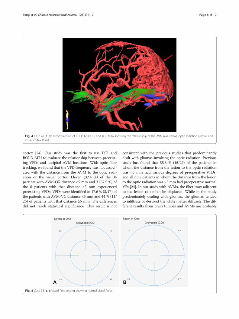
Fig. 4 Case 42. A 3D reconstruction of BOLD-MRI, DTI, and TOF-MRA showing the relationship of the AVM (red arrow), optic radiation (green), andvisual cortex (blue)
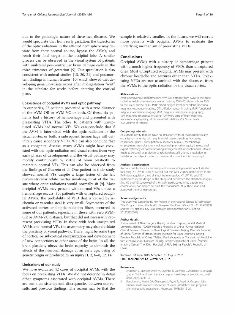
Fig. 5 Case 38. a, b Visual field testing showing normal visual fields
Tong et al. Chinese Neurosurgical Journal (2015) 1:10 Page 8 of 10
due to the pathologic nature of these two diseases. Wewould speculate that from early gestation, the trajectoriesof the optic radiations in the affected hemisphere may de-viate from their normal course, bypass the AVMs, andreach their final target in the occipital lobe. A similarprocess can be observed in the visual system of patientswith unilateral peri-ventricular brain damage early in thethird trimester of gestation [9]. Our speculation is alsoconsistent with animal studies [13, 20, 21] and postmor-tem findings in human fetuses [10] which showed that de-veloping geniculo-striate axons after mid-gestation “wait”in the subplate for weeks before entering the corticalplate.
Coexistence of occipital AVMs and optic pathwayIn our series, 22 patients presented with a zero distanceof the AVM-OR or AVM-VC or both. Of these, six pa-tients had a history of hemorrhage and presented withpreexisting VFDs. The other 16 patients with unrup-tured AVMs had normal VFs. We can conclude that ifthe AVM is intermixed with the optic radiation or thevisual cortex or both, a subsequent hemorrhage will def-initely cause secondary VFDs. We can also conclude thatas a congenital disease, many AVMs might have coex-isted with the optic radiation and visual cortex from veryearly phases of development and the visual pathway maymodify continuously by virtue of brain plasticity tomaintain normal VFs. This can also be observed fromthe findings of Guzzeta et al. One patient in their studyshowed normal VFs despite a large lesion of the leftperi-ventricular white matter involving most of the tis-sue where optic radiations would normally sit [9]. Mostoccipital AVMs may present with normal VFs unless ahemorrhage occurs. For patients with unruptured occipi-tal AVMs, the probability of VFD that is caused by is-chemia or vascular steal is very small. Asymmetry of theactivated cortex and optic radiation fibers occurred insome of our patients, especially in those with zero AVM-OR or AVM-VC distance, but that did not necessarily rep-resent preexisting VFDs. In those with both unrupturedAVMs and normal VFs, the asymmetry may also elucidatethe plasticity of visual pathway. There might be some typeof cortical or subcortical reorganization and developmentof new connections to other areas of the brain. In all, thebrain plasticity obeys the brain capacity to diminish theeffects of the neuronal damage at an early age, being ofgenetic origin or produced by an injury [1, 3, 6–8, 12, 14].
Limitations of our studyWe have evaluated 42 cases of occipital AVMs with thefocus on preexisting VFDs. We did not describe in detailother symptoms associated with occipital AVMs. Thereare some consistency and discrepancies between our re-sults and previous findings. The reason may be that the
sample is relatively smaller. In the future, we will recruitmore patients with occipital AVMs to evaluate theunderlying mechanism of preexisting VFDs.
ConclusionsOccipital AVMs with a history of hemorrhage presentwith a much higher frequency of VFDs than unrupturedones. Most unruptured occipital AVMs may present withchronic headache and seizures other than VFDs. Preex-isting VFDs are not associated with the distances fromthe AVMs to the optic radiation or the visual cortex.
AbbreviationsAVM: arteriovenous malformation; AVM-OR: distance from AVM to the opticradiation; AVMs: arteriovenous malformations; AVM-VC: distance from AVMto the visual cortex; BOLD-fMRI: blood oxygen level dependent functionalmagnetic resonance imaging; DTI: diffusion tensor imaging; fMRI: functionalmagnetic resonance imaging; MRA: magnetic resonance angiography;MRI: magnetic resonance imaging; TOF-MRA: time of flight magneticresonance angiography; VFDs: visual field deficits; VFs: Visual fields;VOI: volume of interest.
Competing interestsAll authors certify that we have no affiliations with or involvement in anyorganization or entity with any financial interest (such as honoraria;educational grants; participation in speakers’ bureaus; membership,employment, consultancies, stock ownership, or other equity interest; andexpert testimony or patent-licensing arrangements), or nonfinancial interest(such as personal or professional relationships, affiliations, knowledge, orbeliefs) in the subject matter or materials discussed in this manuscript.
Authors’ contributionsAuthor contributions to the study and manuscript preparation include thefollowing. XT, JW, FL, and ZJ carried out the fMRI studies, participated in thefMRI data acquisition, and drafted the manuscript. XT, JW, FL, and YCparticipated in the design of the study and performed the statistical analysis.SW, YC, and YZ conceived of the study, participated in its design andcoordination, and helped to draft the manuscript. All authors read andapproved the final manuscript.
AcknowledgementsThis study was supported by Key Projects in the National Science & TechnologyPillar Program during the Twelfth Five-year Plan Period (Grant No. 2011BAI08B08)and the 973 National Key Basic Research Development Plan (Grant No.2012CB720704).
Author details1Department of Neurosurgery, Beijing Tiantan Hospital, Capital MedicalUniversity, Beijing 100050, People’s Republic of China. 2China NationalClinical Research Center for Neurological Diseases, Beijing, People’s Republicof China. 3Center of Stroke, Beijing Institute for Brain Disorders, Beijing,People’s Republic of China. 4Beijing Key Laboratory of Translational Medicinefor Cerebrovascular Diseases, Beijing, People’s Republic of China. 5MedicalImaging Center, The 306th Hospital of PLA, Beijing, People’s Republic ofChina.
Received: 30 June 2015 Accepted: 31 August 2015
References1. Anderson V, Spencer-Smith M, Leventer R, Coleman L, Anderson P, Williams
J, et al. Childhood brain insult: can age at insult help us predict outcome?Brain. 2009;132:45–56.
2. Bartolomei J, Wecht DA, Chaloupka J, Fayad P, Awad IA. Occipital lobevascular malformations: prevalence of visual field deficits and prognosisafter therapeutic intervention. Neurosurg. 1998;43:415–21.
Tong et al. Chinese Neurosurgical Journal (2015) 1:10 Page 9 of 10
3. Berker EA, Berker AH, Smith A. Translation of Broca’s 1865 report.Localization of speech in the third left frontal convolution. Arch Neurol.1986;43:1065–72.
4. Bruyn GW. Intracranial arteriovenous malformation and migraine.Cephalalgia. 1984;4:191–207.
5. Dehdashti AR, Thines L, Willinsky RA, terBrugge KG, Schwartz ML, TymianskiM, et al. Multidisciplinary care of occipital arteriovenous malformations:effect on nonhemorrhagic headache, vision, and outcome in a series of 135patients. J Neurosurg. 2010;113:742–8.
6. Gall C, Prilloff S, Sabel BA. Recovery of function and plasticity after lesions ofthe central visual pathway. J Neurol Neurosur Ps. 2010;11:18–30.
7. Gallegos-Duarte M, Moguel-Ancheita S, Mendiola-Santibañez JD, Morales-Tlalpan V, Saldaña C. Plasticity of the visual pathway and neuroimaging.In: Erondu OF, editor. Medical Imaging in Clinical Practice. Rijeka,Croatia - EUROPEAN UNION: InTech; 2013. p 309–326.
8. Guzzetta A. Plasticity of the visual system after congenital brain damage: a fewweeks can matter. Dev Med Child Neurol. 2010;52:699.
9. Guzzetta A, D’Acunto G, Rose S, Tinelli F, Boyd R, Cioni G. Plasticity of thevisual system after early brain damage. Dev Med Child Neurol.2010;52(14):891–900.
10. Hevner RF. Development of connections in the human visual system duringfetal mid-gestation: a DiI-tracing study. J Neuropathol Exp Neurol.2000;59:385–92.
11. Joint Writing Group of the Technology Assessment Committee AmericanSociety of Interventional and Therapeutic Neuroradiology, Joint Section onCerebrovascular Neurosurgery a Section of the American Association ofNeurological Surgeons and Congress of Neurological Surgeons, Section ofStroke and the Section of Interventional Neurology of the American Academyof Neurology, Atkinson RP, Awad IA, Batjer HH, Dowd CF, Furlan A, et al.Reporting terminology for brain arteriovenous malformation clinical andradiographic features for use in clinical trials. Stroke. 2001;32:1430–42.
12. Kennard M, Fulton JF. Age and reorganization of central nervous system.Mt Sinai J Med. 1942;9:594–606.
13. Kostovic I, Rakic P. Developmental history of the transient subplate zone inthe visual and somatosensory cortex of the macaque monkey and humanbrain. J Comp Neurol. 1990;297:441–70.
14. Krageloh-Mann I, Horber V. The role of magnetic resonance imaging inelucidating the pathogenesis of cerebral palsy: a systematic review. DevMed Child Neurol. 2007;49:144–51.
15. Kupersmith MJ. Neurovascular neuro-ophthalmology. Heidelberg: Springer;1993. p. 307–24. pp 443–444.
16. Kupersmith MJ, Vargas ME, Yashar A, Madrid M, Nelson K, Seton A, et al.Occipital arteriovenous malformations: visual disturbances and presentation.Neurology. 1996;46:953–7.
17. Lawton MT, Kim H, McCulloch CE, Mikhak B, Young WL. A supplementarygrading scale for selecting patients with brain arteriovenous malformationsfor surgery. Neurosurg. 2010;66:702–13.
18. Martin NA, Wilson CB. Medial occipital arteriovenous malformations: surgicaltreatment. J Neurosurg. 1982;56:798–802.
19. Pollock BE, Lunsford LD, Kondziolka D, Bissonette DJ, Flickinger JC.Stereotactic radiosurgery for postgeniculate visual pathway arteriovenousmalformation. J Neurosurg. 1996;84:437–41.
20. Rakic P. Prenatal genesis of connections subserving ocular dominance inthe rhesus monkey. Nature. 1976;261:467–71.
21. Rakic P. Prenatal development of the visual system in rhesus monkey. PhilosTrans R Soc Lond B Biol Sci. 1977;278:245–60.
22. Schlosser MJ, McCarthy G, Fulbright RK, Gore JC, Awad IA. Cerebral vascularmalformations adjacent to sensorimotor and visual cortex: functionalmagnetic resonance imaging studies before and after therapeuticintervention. Stroke. 1997;28:1130–7.
23. Sinclair J, Marks MP, Levy RP, Adler JR, Chang SD, Lopez JR, et al. Visual fieldpreservation after curative multi-modality treatment of occipital lobearteriovenous malformations. Neurosurg. 2005;57:655–67.
24. Sun GC, Chen XL, Zhao Y, Wang F, Hou BK, Wang YB, et al. Intraoperativehigh-field magnetic resonance imaging combined with fiber tractneuronavigation-guided resection of cerebral lesions involving opticradiation. Neurosurg. 2011;69:1070–84.
25. Troost BT, Newton TH. Occipital lobe arteriovenous malformations: clinicaland radiologic features in 26 cases with comments on differentiation frommigraine. Arch Ophthalmol. 1975;93:250–6.
Submit your next manuscript to BioMed Centraland take full advantage of:
• Convenient online submission
• Thorough peer review
• No space constraints or color figure charges
• Immediate publication on acceptance
• Inclusion in PubMed, CAS, Scopus and Google Scholar
• Research which is freely available for redistribution
Submit your manuscript at www.biomedcentral.com/submit
Tong et al. Chinese Neurosurgical Journal (2015) 1:10 Page 10 of 10