involvement of prohibitin upregulation in abrin-triggered apoptosis
TRANSCRIPT
Hindawi Publishing CorporationEvidence-Based Complementary and Alternative MedicineVolume 2012, Article ID 605154, 11 pagesdoi:10.1155/2012/605154
Research Article
Involvement of Prohibitin Upregulation inAbrin-Triggered Apoptosis
Yu-Huei Liu,1, 2, 3 Konan Peck,4 and Jung-Yaw Lin1
1 Institute of Biochemistry and Molecular Biology, College of Medicine, National Taiwan University, Taipei 100, Taiwan2 Graduate Institute of Integrated Medicine of Chinese Medicine, China Medical University, Taichung 404, Taiwan3 Department of Medical Genetics and Medical Research, China Medical University Hospital, Taichung 404, Taiwan4 Institute of Biomedical Sciences, Academia Sinica, Taipei 115, Taiwan
Correspondence should be addressed to Jung-Yaw Lin, [email protected]
Received 20 May 2011; Accepted 18 July 2011
Academic Editor: Senthamil R. Selvan
Copyright © 2012 Yu-Huei Liu et al. This is an open access article distributed under the Creative Commons Attribution License,which permits unrestricted use, distribution, and reproduction in any medium, provided the original work is properly cited.
Abrin (ABR), a protein purified from the seeds of Abrus precatorius, induces apoptosis in various types of cancer cells. However,the detailed mechanism remains largely uncharacterized. By using a cDNA microarray platform, we determined that prohibitin(PHB), a tumor suppressor protein, is significantly upregulated in ABR-triggered apoptosis. ABR-induced upregulation of PHBis mediated by the stress-activated protein kinase/c-Jun NH2-terminal kinase (SAPK/JNK) pathway, as demonstrated by chemicalinhibitors. In addition, ABR significantly induced the expression of Bax as well as the activation of caspase-3 and poly(ADP-ribose)polymerase (PARP) in Jurkat T cells, whereas the reduction of PHB by specific RNA interference delayed ABR-triggered apoptosisthrough the proapoptotic genes examined. Moreover, our results also indicated that nuclear translocation of the PHB-p53 complexmay play a role in the transcription of Bax. Collectively, our data show that PHB plays a role in ABR-induced apoptosis, which maybe helpful for the development of diagnostic or therapeutic agents.
1. Introduction
Abrin (ABR), purified from the seeds of Abrus precatorius,belongs to the family of type II ribosome-inactivatingproteins (RIPs) that contain 2 subunits. These include a toxicA chain with RNA N-glycosidase activity and a galactose-binding B chain with lectin activity [1]. Like ricin fromRicinus communis, the A chain of ABR functions via theinhibition of protein biosynthesis through depurinationof a single adenine residue (A4324) of the 28S ribosomalRNA [2, 3]. In contrast, the B chain of ABR functions byinteracting with the galactose moiety of glycoproteins orglycolipids on the cell membrane and is internalized intocells through receptor-mediated endocytosis. Several reportshave documented that ABR is mitogenic [4], antifertility[5], antitumoral [6, 7], and immunopotentiating [8–12]agent. In addition to its ability to inhibit protein synthesis,ABR is believed to adopt alternative mechanisms to triggercell apoptosis [13]; despite this, it is relatively less toxic tonormal cells than to cancer cells [10, 14]. Our previous
studies have implied that apoptosis induced by ABR couldbe partially independent of its RNA N-glycosidase activityand instead be mediated by its binding and the decrease ofantioxidant protein-1 (AOP-1), increase of reactive oxygenspecies production, and release of cytochrome c into thecytosol [15].
Prohibitin (PHB) is localized on the cell membrane,mitochondria, and nucleus; this localization may play apivotal role in its regulation of cell-cycle progression by theinhibition of DNA replication in multiple cell types [16].The protumorigenic versus antitumorigenic role of PHB incancer cells remains controversial. An oncogenic role hasbeen identified for PHB in different kinds of cancer cells,including those of the breast [17], bladder [18], gastric[19], ovary [20], and prostate [21], whereas PHB’s role asa tumor suppressor has been demonstrated in esophagealsquamous cell carcinoma [22–26]. These opposing effectsof PHB in cancer may be due to 2 possible mechanisms.One is a polymorphism in PHB [27]. The other involves its

2 Evidence-Based Complementary and Alternative Medicine
subcellular localization; increased levels of PHB on the cellmembrane facilitates tumorigenesis through its interactionwith c-Raf induced by the Ras oncogene [28], whereasincreased levels of PHB in the nucleus induces apoptosisby increasing the transcriptional activity of p53 and itstranslocation to the cytoplasm [29].
In order to understand the genetic basis of the apoptoticsignaling exerted by ABR, a microarray platform was usedto investigate the expression profiles of genes in Jurkat Tcells after ABR exposure. Among the genes identified, PHBwas significantly upregulated; however, it has yet to be deter-mined whether PHB plays a role in ABR-triggered apoptosis.Here, we report that overexpression of PHB is involved inABR-triggered Jurkat T cell apoptosis. Upregulation of PHBthrough the JNK/SAPK pathway activated the pro-apoptoticgene Bax via the accumulation and translocation of thePHB-p53 complex to the cytoplasm. The elucidation of thechanges in gene expression and the cellular mechanismsof the response to ABR exposure may be helpful for thedevelopment of diagnostic or therapeutic agents.
2. Materials and Methods
2.1. Isolation of ABR. ABR was isolated from seeds of thered variety of A. precatorius using Sepharose 6B affinitycolumn chromatography and purified as described in aprevious study [30]. The purity and molecular weight of ABRprotein were confirmed by Coomassie blue staining (datanot shown). The stock protein solution was diluted withphosphate-buffered saline (PBS, pH 7.2) to a concentrationof 100 μM.
2.2. Cells and Culture Conditions. The human Jurkat Tleukemia cancer cell line was obtained from American TypeCulture Collection and maintained in RPMI-1640 mediumsupplemented with 10% heat-inactivated fetal bovine serum,100 U/mL penicillin, and 100 μg/mL streptomycin. Cellswere grown in suspension in a 5% CO2 humidified atmo-sphere at 37◦C.
2.3. Cell Proliferation Assay. Cells were seeded in 96-wellplates at a density of 5 × 10³ cells per well and were treatedwith (various doses) or without ABR. At the indicated times,viable cells were analyzed by measuring the conversion of thetetrazolium salt 4-[3-(4-iodophenyl)-2-(4-nitrophenyl)-2H-5-tetrazolio]-1,3-benzene disulfonate (WST-1) to formazan.The formazan dye produced by metabolically active cellswas measured with a scanning multiwell spectrophotometerafter 4 h of incubation, according to the manufacturer’sinstructions.
2.4. The cDNA Microarray System. The gene expressionpatterns regulated by ABR were analyzed with a previouslyestablished cDNA microarray platform (9600 probes) [31].The mRNA from cells with or without ABR treatmentwas extracted using an Oligotex-dT column (Qiagen). A2 mg quantity of each mRNA sample was labeled with
biotin or digoxigenin for membrane hybridization, dual-color detection, and image analysis as described previously[31].
2.5. Antibodies and Chemical Inhibitors. An antibody specificto PHB was purchased from Lab Vision Corporation.An antibody specific to p53 was obtained from SantaCruz Biotechnology. Antibodies specific to cleaved-caspase-3, and cleaved-poly(ADP-ribose) polymerase (PARP) werepurchased from Cell Signaling. Antibodies against Bax andactin were obtained from Chemicon. The chemical inhibitorsPD 98059, SB 203580, and SP600125 were purchased fromSigma.
2.6. Immunoblotting. Total cell lysates were collected in lysisbuffer (50 mM HEPES-KOH, pH 7.5, 1% Triton X-100,150 mM NaCl, and protease inhibitor cocktail (Roche)).The extracts were centrifuged at 14000 rpm for 20 min, andthen the clear supernatant was separated by using 10%SDS-PAGE. After transferred, the separated proteins to apolyvinyldene fluoride (PVDF) membrane (Immunobilon-P, 0.45 mm; Millipore, Billerica, Mass, USA) by using theNA-1512 semi-dry transfer apparatus (NIHON EIDO), themembranes were blocked with 5% skim milk in trisbufferedsaline containing 1% Tween 20 (TBST, pH 7.4) at roomtemperature for 30 min and then incubated overnight at4◦C with primary antibodies. The membranes were washed4 times with TBST for 10 min each at room temperatureand incubated with HRP-conjugated secondary antibodiesfor 1 h at room temperature. The membranes were thenwashed 4 times with TBST. The proteins were visualizedusing the SuperSignal West Femto Chemiluminescent Kit(Thermo Scientific) and exposed to an X-ray film (Kodak).The Image J program (http://rsb.info.nih.gov/) was used forquantization the expression fold. For western blot analysis,the fold increase of the indicated proteins was determinedby normalizing to corresponding actin expression. For IP-western analysis, the densitometry readings of the bands werenormalized to control.
2.7. Enzyme-Linked Immunosorbent Assay (ELISA) for Phos-phor MAPK Detection. Cells were treated with or withoutABR after pretreating with the indicated inhibitors or vehicle(DMSO) alone. After the indicated period of time, cells wereharvested and lysed as above. The PathScan MAP KinaseMulti-Target Sandwich ELISA kit was used to determinephosphor ERK, -p38, and-JNK/SAPK levels according tomanufacturer’s instruction (Cell Signaling).
2.8. Short Interfering RNA . Short interfering RNAs (siRNAs)against PHB (sc-37629) and the negative control siRNA(sc-37007) were obtained from Santa Cruz Biotechnology.A total of 2 × 10⁵ cells were plated in a 6-well platefor 24 h, and siRNA transfection was carried out usingthe Lipofectamine 2000 kit according to the manufacturer’sinstructions (Invitrogen).

Evidence-Based Complementary and Alternative Medicine 3
2.9. Terminal Deoxynucleotidyl Transferase-Catalyzed Deox-yuridine Triphosphate (dUTP)-Nick End Labeling (TUNEL)Method [15]. Apoptotic cell death was examined by TUNELmethod as manufactory’s suggestion (Roche Molecular Bio-chemicals). Each sample with 1 × 10⁴ events was analyzedwith a Becton-Dickinson FACSCalibur, and the distributionof cells was determined.
2.10. Chromatin Immunoprecipitation (ChIP) Assays. Cellstreated with or without ABR for the indicated time periodswere examined. After fixing the protein-DNA complex usingformaldehyde (1% final concentration) at room temperaturefor 10 min, the reaction was stopped with glycine. Afterwashed and lysed the cells, the lysates were sonicated andcentrifuged, and the supernatants were used for immuno-precipitation of Bax with PHB antibody or control IgG.Antibody-bound protein/DNA complexes were precipitatedand eluted in 300 μL of elution buffer (1% SDS, 50 mMNaHCO3). Cross-linking was reversed by heating at 65◦Cfor 4 h. The DNA was resuspended in 200 μL of distilledwater and treated with 30 μg of proteinase K at 37◦C for1 h, followed by phenol/chloroform extraction and ethanolprecipitation. PCR was conducted using 100 ng of DNA asthe template. The following PCR primers were used forthe Bax promoter: forward primer 5′-CCGGGAATTCCA-GACTGCA-3′ and reverse primer 5′-AGCTCTCCCCAG-CGCAGAA-3′. Each band was quantitatively determinedusing the Image J program (http://rsb.info.nih.gov/). Thedensitometry readings of the bands were normalized to theinput.
2.11. Statistical Analysis. SPSS 12.0 for Windows (SPSSInc.) was used to analyze the data. A two-tailed paired-samples Student’s t-test was used for statistical analysis of thecomparative data from the two groups. P-value <0.05 valueswere considered statistically significant.
3. Results
3.1. ABR Induces Upregulation of PHB in Human Jurkat TCells. To evaluate the effect of ABR on leukemia cells invitro, Jurkat T leukemia cells were exposed to 0.01–100 nMof ABR for 24 h. The cell viability was then determined byWST-1 assay. As shown in Figure 1(a), the growth of JurkatT cells was reduced by ABR in a dose-dependent manner. Thevalue of the 50% cytotoxic concentration (CC50) for the 24 htreatment was determined for the water fraction to be 0.32± 0.06 nM. The data are represented as mean ± SD from 3independent experiments.
Microarray analysis was used to identify novel candidatesthat are differentially expressed after 1 nM ABR treatmentfor 3 h. A total of 128 genes, out of the 9600 probes tested,were significantly altered by >1.5-fold in response to ABR(P < 0.05). The top 10 significant up-/down-regulated genesare listed in Table 1, sorted by fold increase or decrease.PHB, a significantly upregulated gene with diverse cellularfunctions, was selected for further investigation. To explorethe potential role of PHB in ABR-treated apoptosis, Jurkat
T cells were first treated with ABR (0.1–10 nM). As shownin Figure 1(b), ABR significantly increased the expressionof PHB protein after treatment for 9 h. Cells were furthertreated with 1 nM ABR for different time periods. An initialincrease of PHB protein was observed at the 3 h time pointand was sustained for up to 18 h after ABR treatment(Figure 1(c)). To determine whether PHB upregulation dueto ABR is because of increased transcription or increasedRNA stability, the RNA synthesis inhibitor actinomycinD or the protein synthesis inhibitor cyclohexamide waspreincubated with cells for 1 h before ABR was added.The results show that not only actinomycin D but alsocyclohexamide significantly diminished ABR-induced PHBupregulation (Figure 1(d)). This finding suggests that ABR-induced PHB upregulation requires de novo RNA synthesis.
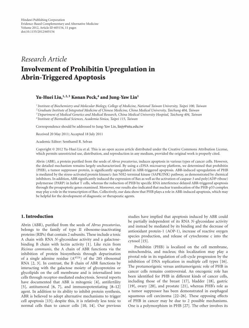
3.2. ABR Upregulates PHB Expression through the SAPK/JNKPathway. To explore which of the signaling pathways arerequired for ABR-induced upregulation of the PHB gene,several specific chemical inhibitors were used. The effectof these inhibitors was examined using an ELISA-baseddetection system (Figure 2(a)). Jurkat T cells were pretreatedwith PD98059 (PD, MEK inhibitor; 20 μM), SB203580 (SB,p38 MAPK inhibitor; 20 μM), or SP600125 (SP, JNK/SAPKinhibitor; 30 μM) for 1 h, followed by treatment with ABR forthe time indicated; total protein was used to determine PHBexpression. SP significantly reduced the ABR-induced PHBexpression, whereas the 2 other kinase inhibitors PD andSB rarely affected on the upregulation of PHB (Figure 2(b)).These results suggest the possible involvement of JNK/SAPK,but not of ERK1/2 or p38 MAPK, in the regulation of PHBexpression in Jurkat T cells treated with ABR.
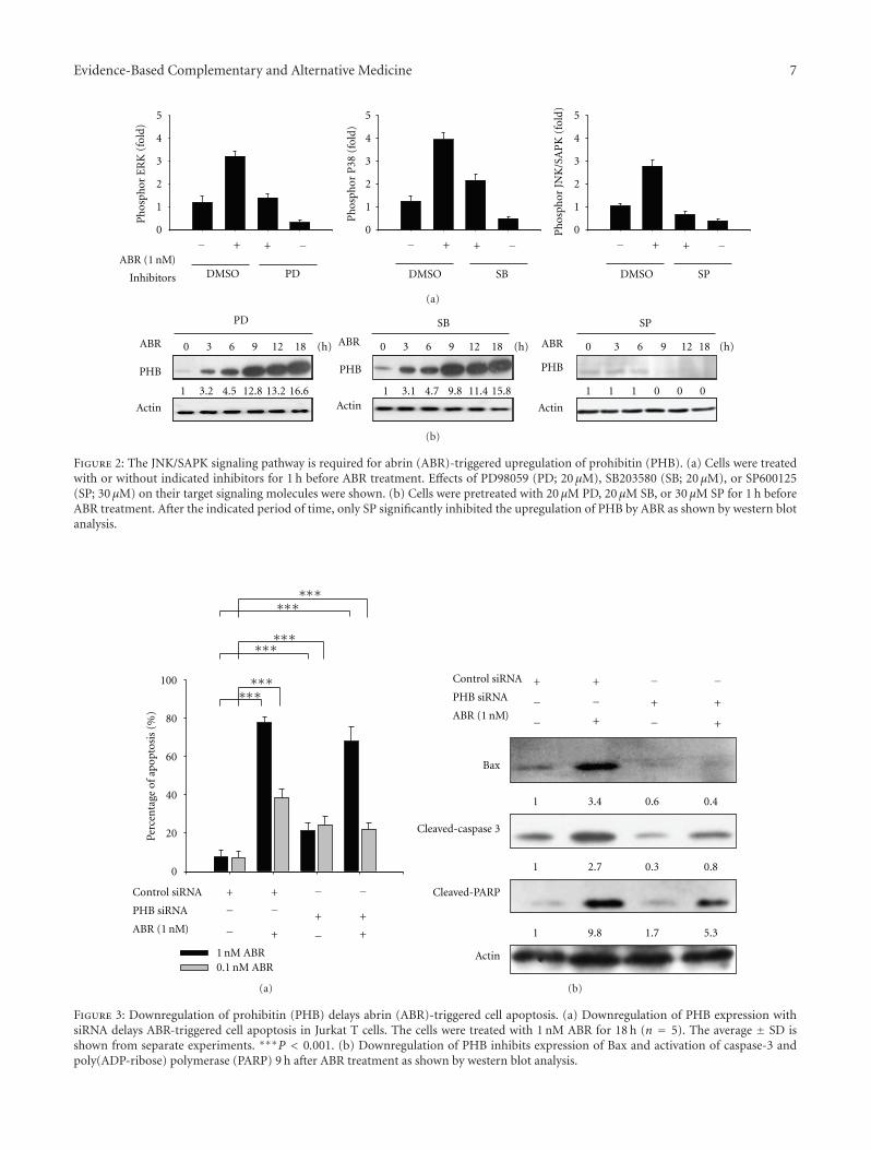
3.3. PHB Is Involved in ABR-Induced Cell Apoptosis. SinceABR induced the expression of PHB, a tumor suppressorgene that may induce cell apoptosis by arresting the cellcycle at the G1/S phase, we focused on determining whetherPHB participates in the apoptotic signaling triggered by ABR.Apoptosis induced by ABR (0.1 and 1 nM) was investigatedusing TUNEL method. As shown in Figure 3(a), group 1and 2, the maximal apoptotic response was achieved 18 hafter treating cells with ABR (77.7% apoptosis after 1 nMABR treatment versus 38.5% apoptosis after 0.1 nM ABRtreatment; P < 0.001). Although the specific PHB RNA inter-ference (PHB siRNA) did significantly enhance cell apoptosis(PHB siRNA induces 22.8% apoptosis versus control siRNAinduces 7.4% apoptosis, P < 0.001; Figure 3(a) groups 1and 3), knockdown PHB reduced ABR-induced apoptosis(PHB siRNA reduced 1 nM ABR-induced apoptosis by 9.7%,P < 0.001, and PHB siRNA reduced 0.1 nM ABR-inducedapoptosis by 16.6%, P < 0.001; Figure 3(a) groups 2 and4). Apoptosis-related genes including Bax, caspase-3, andPARP were also examined. ABR significantly induced theexpression of Bax (6.3-fold; P < 0.001) as well as theactivation of caspase-3 and PARP in the Jurkat T cells(Figure 3(b), lanes 1 and 2). However, the activation wasreduced when the PHB expression reduced (Figure 3(b),

4 Evidence-Based Complementary and Alternative Medicine
0 0.01 0.1 1 10 100
Concentration (nM)
0
20
40
60
80
100
120
Cel
lsu
rviv
al(%
ofco
ntr
ol)
(a)
PHB
Dose 00 .1 1 10 (nM)
1 10.5 12.7 8
Actin
Western blot
ABR
(b)
Western blot
PHB
Actin
Time0 3 6 9 12 18 (h)
1 5.8 7.9 12.4 13.6 16.8
ABR ( 1 )nM
(c)
1 4.9 0.4 0.7 0.3 1.2 Western blot
CHX
− −−−
− − −− −+
+ − + − +
+
+ +
ActD
ABR ( 1 )
PHB
Actin
nM
(d)
Figure 1: Abrin (ABR) upregulates prohibitin (PHB) expression through transcriptional regulation in Jurkat T cells. (a) ABR-inducedcytotoxic activity in a dose-dependent manner in Jurkat T cells after 24 h treatment. The data are represented as mean ± SD from 3independent experiments. (b) ABR (0.1–10 nM) significantly increased the expression of PHB after treatment for 9 h. (c) ABR (1 nM)-induced upregulation of PHB in a time-dependent manner. (d) ABR-induced PHB upregulation requires de novo RNA synthesis.
lanes 3 and 4). The data showed that PHB is involved in ABR-triggered apoptosis.
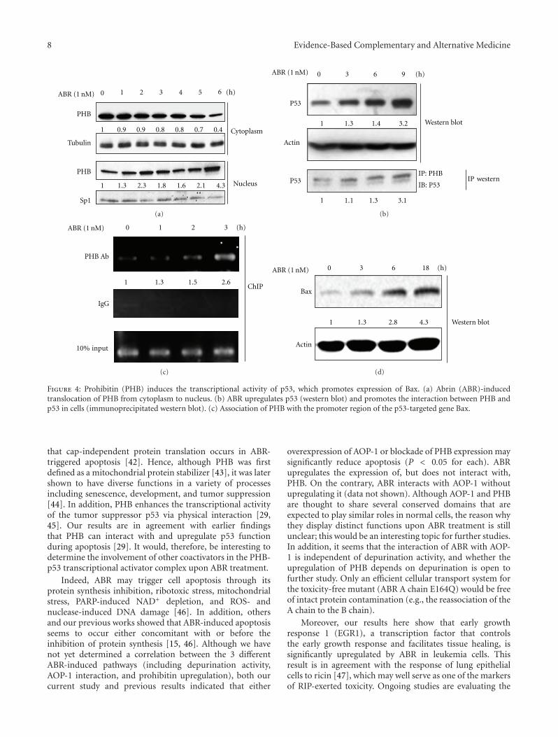
3.4. Upregulation of Human Bax Expression through Translo-cation of the PHB-p53 Complex from the Cytoplasm tothe Nucleus. The previously described results raised thepossibility that PHB may be involved in the apoptoticprocesses triggered by ABR. On the other hand, increasedlevels of PHB in the nucleus may interact with the tumorsuppressor protein p53 by which it exerts its apoptotic effect.Therefore, we attempted to determine whether there was aninteraction between PHB and p53. As shown in Figure 4(a),PHB was translocated from the cytoplasm to the nucleusafter ABR treatment for 6 h. Furthermore, a 1.3-, 1.4-, and3.2-fold increase in p53 and a 1.1-, 1.3-, and 3.1-fold increasein the interaction with PHB were observed when cells weretreated with ABR for 3, 6, and 9 h, respectively (Figure 4(b)).These results indicated that ABR may induce a physicalinteraction between PHB and p53 in the early stage ofABR-induced cell apoptosis. Since Bax is known to be oneof the transcriptional regulation targets for PHB and p53,a ChIP assay was performed by using specific primers toamplify a potential p53-binding region in Bax. As shown inFigure 4(c), p53 was recruited to the promoter regions ofBax in a time-dependent manner. These results suggest that
ABR induces the formation of the PHB-p53 complex in thenucleus, which enhances the transcriptional activity of p53on Bax following apoptosis.
4. Discussion
Studies have shown that some proteins, including ABR, ricin,modeccin, diphtheria toxin, shiga toxin, and pseudomonastoxin, are apoptosis inducers [32–34]. Although ABR hasbeen clearly identified as an inducer of apoptotic cell death byactivating caspase-3 in several kinds of cancer cells [15, 35–38], the mechanisms of its involvement in cell apoptosisremain to be investigated. In this study, PHB is shown tobe upregulated in a dose-dependent manner during ABRtreatment and might play a potent role in ABR-triggeredapoptosis by enhancing the activity and expression of p53.To the best of our knowledge, this is the first study to explainand demonstrate the role of PHB in ABR-induced apoptosisin human leukemia cells. The potential clinical applicationsof ABR may involve the enhancement of drug targeting aswell as a decrease in side effects on noncancerous cells.
ABR is a RIP, which induces a shutdown of proteinsynthesis in target cells [33, 39]. However, previous reportsalso showed that the apoptosis-related protein Bax can beupregulated by ABR [40, 41]. Our results also indicatedan overexpression of PHB and p53. One explanation is

Evidence-Based Complementary and Alternative Medicine 5
Ta
ble
1:To
p10
up-
/dow
nre
gula
ted
gen
esch
angi
ng
inre
spon
seto
abri
nex
posu
rear
ran
ged
byfo
ldch
ange
.G
ene
com
mon
nam
e,de
scri
ptio
n,
and
gen
eon
tolo
gycl
assi
fica
tion
(wh
ere
know
n)
are
liste
d.
Un
igen
en
um
ber
Com
mon
nam
eD
escr
ipti
onFo
ldch
ange
GO
biol
ogic
alpr
oces
sG
Om
olec
ula
rfu
nct
ion
GO
cellu
lar
proc
ess
Upr
egu
lati
onH
s.32
6035
EG
R1
Ear
lygr
owth
resp
onse
112
.9Tr
ansc
ript
ion
,DN
Ade
pen
den
tTr
ansc
ript
ion
acti
vato
rac
tivi
tyN
ucl
eus
Hs.
5027
69SL
C3A
2So
lute
carr
ier
fam
ily3
(act
ivat
ors
ofdi
basi
can
dn
eutr
alam
ino
acid
tran
spor
t),m
embe
r2
3.0
Tran
smem
bran
etr
ansp
ort
Cat
alyt
icac
tivi
tyP
lasm
am
embr
ane
Hs.
2178
HIS
T2H
2BE
His
ton
ecl
ust
er2,
H2b
e2.
5N
ucl
eoso
me
asse
mbl
yB
indi
ng
toD
NA
and
prot
ein
Nu
cleu
s
Hs.
8296
3G
NR
H1
Gon
adot
ropi
n-r
elea
sin
gh
orm
one
1(l
ute
iniz
ing-
rele
asin
gh
orm
one)
2.4
Mu
ltic
ellu
lar
orga
nis
mal
deve
lopm
ent
Hor
mon
eac
tivi
tyE
xtra
cellu
lar
Hs.
4674
08T
RIM
28Tr
ipar
tite
mot
if-c
onta
inin
g28
2.3
Tran
scri
ptio
n,D
NA
depe
nde
nt
Tran
scri
ptio
nco
acti
vato
r/co
repr
esso
rac
tivi
tyN
ucl
eus
Hs.
5143
03P
HB
Pro
hib
itin
2.3
Neg
ativ
ere
gula
tion
ofce
llpr
olif
erat
ion
,gen
e-sp
ecifi
ctr
ansc
rip
tion
from
RN
Apo
lym
eras
eII
prom
oter
byco
mpe
titi
vepr
omot
erbi
ndi
ng;
regu
lati
onof
apop
tosi
s;si
gnal
tran
sdu
ctio
n
Tran
scri
ptio
nac
tiva
tor/
repr
esso
rac
tivi
tyC
ytop
lasm
a,pl
asm
am
embr
ane,
mit
och
ondr
ia,n
ucl
eus
Hs.
5344
04R
PL1
0R
ibos
omal
prot
ein
L10
2.2
Tran
slat
ion
Stru
ctu
ralc
onst
itu
ent
ofri
boso
me
Cyt
osol
Hs.
5120
DY
NL
L1
Dyn
ein
,lig
ht
chai
n,L
C8-
typ
e1
2.2
Indu
ctio
nof
apop
tosi
sM
otor
acti
vity
Cyt
osol
Hs.
2263
90R
RM
2R
ibon
ucl
eoti
dere
duct
ase
M2
2.1
DN
Are
plic
atio
nO
xido
redu
ctas
eac
tivi
tyC
ytos
olH
s.20
2207
OSC
P1
Org
anic
solu
teca
rrie
rpa
rtn
er1
2.1
Tran
spor
tP
lasm
am
embr
ane
Hs.
2552
4P
TP
N23
Pro
tein
tyro
sin
eph
osph
atas
e,n
on-
rece
ptor
typ
e23
2.1
Cel
lpro
ject
ion
orga
niz
atio
nH
ydro
lase
acti
vity
Cyt
opla
sma

6 Evidence-Based Complementary and Alternative Medicine
Ta
ble
1:C
onti
nu
ed.
Un
igen
en
um
ber
Com
mon
nam
eD
escr
ipti
onFo
ldch
ange
GO
biol
ogic
alpr
oces
sG
Om
olec
ula
rfu
nct
ion
GO
cellu
lar
proc
ess
Dow
nre
gula
tion
Hs.
7259
87T
UB
A1C
Tubu
lin,a
lph
a1c
−1.8
Cel
lula
rpr
otei
nm
etab
olic
proc
ess
Stru
ctu
ralm
olec
ule
acti
vity
Cyt
osol
Hs.
5351
92E
EF1
A1
Eu
kary
otic
tran
slat
ion
elon
gati
onfa
ctor
1al
pha
1−1
.6Tr
ansl
atio
nTr
ansl
atio
nel
onga
tion
fact
orac
tivi
tyC
ytos
ol
Hs.
5145
81A
CT
G1
Act
in,g
amm
a1
−1.6
Cel
lula
rco
mpo
nen
tm
ovem
ent
Pro
tein
bin
din
gC
ytos
ol
Hs.
5662
GN
B2L
1G
uan
ine
nu
cleo
tide
-bin
din
gpr
o-te
in(G
prot
ein
),be
tap
olyp
epti
de2-
like
1−1
.5Po
siti
vere
gula
tion
ofap
opto
sis
Pro
tein
bin
din
gC
ytos
ol
Hs.
5343
46R
PS7
Rib
osom
alpr
otei
nS7
−1.5
Tran
slat
ion
Stru
ctu
ralc
onst
itu
ent
ofri
boso
me
Cyt
osol
Hs.
4334
27R
PS1
7R
ibos
omal
prot
ein
S17
−1.5
Tran
slat
ion
Stru
ctu
ralc
onst
itu
ent
ofri
boso
me
Cyt
osol
Hs.
5662
GN
B2L
1G
uan
ine
nu
cleo
tide
-bin
din
gpr
o-te
in(G
prot
ein
),be
tap
olyp
epti
de2-
like
1−1
.5N
egat
ive
regu
lati
onof
cell
grow
thP
rote
inbi
ndi
ng
Nu
cleu
s,cy
toso
l
Hs.
4444
67E
EF1
GE
uka
ryot
ictr
ansl
atio
nel
onga
tion
fact
or1
gam
ma
−1.5
Tran
slat
ion
alel
onga
tion
Tran
slat
ion
elon
gati
onfa
ctor
acti
vity
Cyt
osol
Hs.
5145
81A
CT
G1
Act
in,g
amm
a1
−1.5
Cel
lula
rco
mpo
nen
tm
ovem
ent
Stru
ctu
ralc
onst
itu
ento
fcyt
oske
leto
nC
ytos
ol
Hs.
5097
36H
SP90
AB
1H
eat
shoc
kpr
otei
n90
kDa
alph
a(c
ytos
olic
),cl
ass
Bm
embe
r1
−1.5
Reg
ula
tion
ofty
pe
Iin
terf
eron
-med
iate
dsi
gnal
ing
path
way
Un
fold
edpr
otei
nbi
ndi
ng
Cyt
osol
Evidence-Based Complementary and Alternative Medicine 7
Ph
osph
orP
38
Ph
osph
orJN
K/S
AP
K
Ph
osph
orE
RK
DMSO DMSO DMSOPD SB SP
0
1
2
3
4
5
0
1
2
3
4
5
0
1
2
3
4
5
− + −+ − + −+ − + −+
ABR (1 )nM
Inhibitors
(fol
d)
(fol
d) (fol
d)
(a)
PHB
Actin
ABR
PHB
Actin
ABR
PHB
Actin
ABR0 3 6 9 12 18 (h) 0 3 6 9 12 18 (h) 0
0 0 0
3 6 9 12 18 (h)
PD SB SP
1 1 1 1 13.2 4.5 12.8 13.2 16.6 3.1 4.7 9.8 11.4 15.8
(b)
Figure 2: The JNK/SAPK signaling pathway is required for abrin (ABR)-triggered upregulation of prohibitin (PHB). (a) Cells were treatedwith or without indicated inhibitors for 1 h before ABR treatment. Effects of PD98059 (PD; 20 μM), SB203580 (SB; 20 μM), or SP600125(SP; 30 μM) on their target signaling molecules were shown. (b) Cells were pretreated with 20 μM PD, 20 μM SB, or 30 μM SP for 1 h beforeABR treatment. After the indicated period of time, only SP significantly inhibited the upregulation of PHB by ABR as shown by western blotanalysis.
1 nM ABR0.1 nM ABR
Control siRNA
PHB siRNA
− −−−
−+ +
+
+ − +
+
∗∗∗∗∗∗
∗∗∗∗∗∗
∗∗∗∗∗∗
ABR (1 )nM
Perc
enta
geof
apop
tosi
s(%
)
0
20
40
60
80
100
(a)
Control siRNA
PHB siRNA
− −−−
−+ +
+
+ − +
+ABR (1 )nM
Bax
Cleaved-caspase 3
Cleaved-PARP
Actin
1
1
1
3.4 0.6 0.4
2.7 0.3 0.8
9.8 1.7 5.3
(b)
Figure 3: Downregulation of prohibitin (PHB) delays abrin (ABR)-triggered cell apoptosis. (a) Downregulation of PHB expression withsiRNA delays ABR-triggered cell apoptosis in Jurkat T cells. The cells were treated with 1 nM ABR for 18 h (n = 5). The average ± SD isshown from separate experiments. ∗∗∗P < 0.001. (b) Downregulation of PHB inhibits expression of Bax and activation of caspase-3 andpoly(ADP-ribose) polymerase (PARP) 9 h after ABR treatment as shown by western blot analysis.
8 Evidence-Based Complementary and Alternative Medicine
0 1 2 3 4 5 6 (h)ABR (1 nM)
PHB
Tubulin
PHB
Sp1
1
1
0.9 0.9 0.8 0.8 0.7 0.4
1.3 2.3 1.8 1.6 2.1 4.3
Cytoplasm
Nucleus
(a)
ABR (1 nM) 0 3 6 9 (h)
Western blot
IP: PHB
IB: P53
11
1
.3
1 1. 1.3 1.3
1.4 3.2
P53
P53
Actin
IP esternw
(b)
ABR (1 nM) 0 1 2 3
1 1.3 1.5 2.6
IgG
ChIP
PHB Ab
10% nputi
(h)
(c)
ABR (1 nM) 0 3 6 18 (h)
Bax
Actin
2.81 1.3 4.3 Western blot
(d)
Figure 4: Prohibitin (PHB) induces the transcriptional activity of p53, which promotes expression of Bax. (a) Abrin (ABR)-inducedtranslocation of PHB from cytoplasm to nucleus. (b) ABR upregulates p53 (western blot) and promotes the interaction between PHB andp53 in cells (immunoprecipitated western blot). (c) Association of PHB with the promoter region of the p53-targeted gene Bax.
that cap-independent protein translation occurs in ABR-triggered apoptosis [42]. Hence, although PHB was firstdefined as a mitochondrial protein stabilizer [43], it was latershown to have diverse functions in a variety of processesincluding senescence, development, and tumor suppression[44]. In addition, PHB enhances the transcriptional activityof the tumor suppressor p53 via physical interaction [29,45]. Our results are in agreement with earlier findingsthat PHB can interact with and upregulate p53 functionduring apoptosis [29]. It would, therefore, be interesting todetermine the involvement of other coactivators in the PHB-p53 transcriptional activator complex upon ABR treatment.
Indeed, ABR may trigger cell apoptosis through itsprotein synthesis inhibition, ribotoxic stress, mitochondrialstress, PARP-induced NAD+ depletion, and ROS- andnuclease-induced DNA damage [46]. In addition, othersand our previous works showed that ABR-induced apoptosisseems to occur either concomitant with or before theinhibition of protein synthesis [15, 46]. Although we havenot yet determined a correlation between the 3 differentABR-induced pathways (including depurination activity,AOP-1 interaction, and prohibitin upregulation), both ourcurrent study and previous results indicated that either
overexpression of AOP-1 or blockade of PHB expression maysignificantly reduce apoptosis (P < 0.05 for each). ABRupregulates the expression of, but does not interact with,PHB. On the contrary, ABR interacts with AOP-1 withoutupregulating it (data not shown). Although AOP-1 and PHBare thought to share several conserved domains that areexpected to play similar roles in normal cells, the reason whythey display distinct functions upon ABR treatment is stillunclear; this would be an interesting topic for further studies.In addition, it seems that the interaction of ABR with AOP-1 is independent of depurination activity, and whether theupregulation of PHB depends on depurination is open tofurther study. Only an efficient cellular transport system forthe toxicity-free mutant (ABR A chain E164Q) would be freeof intact protein contamination (e.g., the reassociation of theA chain to the B chain).
Moreover, our results here show that early growthresponse 1 (EGR1), a transcription factor that controlsthe early growth response and facilitates tissue healing, issignificantly upregulated by ABR in leukemia cells. Thisresult is in agreement with the response of lung epithelialcells to ricin [47], which may well serve as one of the markersof RIP-exerted toxicity. Ongoing studies are evaluating the

Evidence-Based Complementary and Alternative Medicine 9
Mitochondria
ABRA
ABRA
Cytochrome C
release
Caspase 9
Caspase 3
AOP-1
PARP
ΔΨm ↓ROS ↑
Lipid
peroxidation
Translation inhibitionCellular factors?
ABRB
Endosome
Golgi
Endoplasmic
JNK/SAPKactivation
DNA fragmentation
PHB
PHBPHB gene exprression ↑
P53
Bax gene expression ↑
Other factors?
Nucleus
Extracellular
Cytoplasm
eticulumr
Apoptosis
Figure 5: A model of abrin (ABR)-triggered apoptosis. ABR-induced apoptosis may occur through at least 3 pathways: first, inhibition ofprotein synthesis by its N-glycosidase activity; second, modulation of the function of mitochondria by specific interaction with antioxidantprotein-1 (AOP-1); and third, interference with the transcription regulated by prohibitin (PHB). Repression of prohibitin attenuates ABR-triggered apoptosis via preventing the expression of BAX, cleaved-caspase 3, and cleaved-poly(ADP-ribose) polymerase (PARP). Once PHBis upregulated by ABR through the JNK/SAPK signaling pathway, the expression of proapoptotic gene Bax is turned on through the nucleartranslocation and p53 interaction of PHB, by which activates the caspase cascade, and finally, apoptosis occurs.
potential of these ABR-related genes for clinical intervention.Nevertheless, as Abrus precatorius is labeled as a biologicalweapon which may be fatal if eaten, development of a passivevaccine or an antidote for ABR is necessary but underinvestigation [48, 49]. More understanding of the molecularmechanisms exerted by RIP family proteins may acceleratetheir clinical applications.
In conclusion, we propose the model shown in Figure 5.ABR exhibits biological functions involving at least 3 path-ways: translational inhibition, mitochondrial dysfunction,and transcriptional interfere through the upregulation ofPHB. Since the downregulation of PHB significantly delaysapoptosis induced by ABR, PHB could be employed inreducing the toxicity of immunotoxins and, hence, improvethe efficiency of cancer chemotherapy.
Acknowledgments
This study was supported by Grants from the Committeeon Chinese Medicine and Pharmacy (CCMP-96-RD-203-1)and the Department of Health (DOH97-TD-I-111-TM018,DOH98-TD-I-111-TM020), executive Yuan, Taiwan.
References
[1] J. Y. Lin, K. Y. Tserng, C. C. Chen, L. T. Lin, and T. C. Tung,“Abrin and ricin: new anti-tumour substances,” Nature, vol.227, no. 5255, pp. 292–293, 1970.
[2] J. U. Baenziger and D. Fiete, “Structural determinantsof Ricinus communis agglutinin and toxin specificity foroligosaccharides,” Journal of Biological Chemistry, vol. 254, no.19, pp. 9795–9799, 1979.
[3] F. Stirpe, L. Barbieri, M. G. Battelli, M. Soria, and D. A. Lappi,“Ribosome-inactivating proteins from plants: present statusand future prospects,” Nature Biotechnology, vol. 10, no. 4, pp.405–412, 1992.
[4] S. J. Kaufman and A. McPherson, “Abrin and hurin: two newlymphocyte mitogens,” Cell, vol. 4, no. 3, pp. 263–268, 1975.
[5] V. P. Kamboj and B. N. Dhawan, “Research on plants forfertility regulation in India,” Journal of Ethnopharmacology,vol. 6, no. 2, pp. 191–226, 1982.
[6] J. Y. Lin, T. C. Lee, and T. C. Tung, “Inhibitory effects of fourisoabrins on the growth of sarcoma 180 cells,” Cancer Research,vol. 42, no. 1, pp. 276–279, 1982.
[7] R. Hegde, T. K. Maiti, and S. K. Podder, “Purification andcharacterization of three toxins and two agglutinins fromAbrus precatorius seed by using lactamyl-sepharose affinitychromatography,” Analytical Biochemistry, vol. 194, no. 1, pp.101–109, 1991.

10 Evidence-Based Complementary and Alternative Medicine
[8] S. Tripathi and T. K. Maiti, “Stimulation of murinemacrophages by native and heat-denatured lectin from Abrusprecatorius,” International Immunopharmacology, vol. 3, no. 3,pp. 375–381, 2003.
[9] V. Ramnath, G. Kuttan, and R. Kuttan, “Immunopotentiatingactivity of abrin, a lectin from Abrus precatorius Linn,” IndianJournal of Experimental Biology, vol. 40, no. 8, pp. 910–913,2002.
[10] V. Ramnath, G. Kuttan, and R. Kuttan, “Antitumour effectof abrin on transplanted tumours in mice,” Indian Journal ofPhysiology and Pharmacology, vol. 46, no. 1, pp. 69–77, 2002.
[11] S. Tripathi and T. K. Maiti, “Immunomodulatory role ofnative and heat denatured agglutinin from Abrus precatorius,”International Journal of Biochemistry and Cell Biology, vol. 37,no. 2, pp. 451–462, 2005.
[12] A. Tilwari, N. P. Shukla, and U. D. Pathirissery, “Immunomod-ulatory activity of the aqueous extract of seeds of Abrusprecatorius Linn (Jequirity) in mice,” Iranian Journal ofImmunology, vol. 8, no. 2, pp. 96–103, 2011.
[13] K. J. Dickers, S. M. Bradberry, P. Rice, G. D. Griffiths, and J.A. Vale, “Abrin poisoning,” Toxicological Reviews, vol. 22, no.3, pp. 137–142, 2003.
[14] L. N. L. Chan, J. S. Li, and S. Y. Liu, “Differential effectsof abrin on normal and tumor cells,” Journal of CellularPhysiology, vol. 123, no. 1, pp. 132–138, 1985.
[15] S. F. Shih, Y. H. Wu, C. H. Hung, H. Y. Yang, and J. Y.Lin, “Abrin triggers cell death by inactivating a thiol-specificantioxidant protein,” Journal of Biological Chemistry, vol. 276,no. 24, pp. 21870–21877, 2001.
[16] W. Rizwani, M. Alexandrow, and S. Chellappan, “Prohibitinphysically interacts with MCM proteins and inhibits mam-malian DNA replication,” Cell Cycle, vol. 8, no. 10, pp. 1621–1629, 2009.
[17] S. Manjeshwar, D. E. Branam, M. R. Lerner, D. J. Brackett,and E. R. Jupe, “Tumor suppression by the prohibitin gene3’untranslated region RNA in human breast cancer,” CancerResearch, vol. 63, no. 17, pp. 5251–5256, 2003.
[18] T. F. Wu, H. Wu, Y. W. Wang et al., “Prohibitin in thepathogenesis of transitional cell bladder cancer,” AnticancerResearch, vol. 27, no. 2, pp. 895–900, 2007.
[19] X. Kang, L. Zhang, J. Sun et al., “Prohibitin: a potentialbiomarker for tissue-based detection of gastric cancer,” Journalof Gastroenterology, vol. 43, no. 8, pp. 618–625, 2008.
[20] R. C. Gregory-Bass, M. Olatinwo, W. Xu et al., “Prohibitinsilencing reverses stabilization of mitochondrial integrity andchemoresistance in ovarian cancer cells by increasing theirsensitivity to apoptosis,” International Journal of Cancer, vol.122, no. 9, pp. 1923–1930, 2008.
[21] R. Ummanni, H. Junker, U. Zimmermann et al., “Prohibitinidentified by proteomic analysis of prostate biopsies distin-guishes hyperplasia and cancer,” Cancer Letters, vol. 266, no.2, pp. 171–185, 2008.
[22] H. Z. Ren, J. S. Wang, G. Q. Pan et al., “Comparativeproteomic analysis of β-catenin-mediated malignant progres-sion of esophageal squamous cell carcinoma,” Diseases of theEsophagus, vol. 23, no. 2, pp. 175–184, 2010.
[23] M. J. Nuell, D. A. Stewart, L. Walker et al., “Prohibitin, an evo-lutionarily conserved intracellular protein that blocks DNAsynthesis in normal fibroblasts and HeLa cells,” Molecular andCellular Biology, vol. 11, no. 3, pp. 1372–1381, 1991.
[24] T. Sato, H. Saito, J. Swensen et al., “The human prohibitin genelocated on chromosome 17q21 is mutated in sporadic breastcancer,” Cancer Research, vol. 52, no. 6, pp. 1643–1646, 1992.
[25] R. T. Dell’Orco, J. K. McClung, E. R. Jupe, and X. T.Liu, “Prohibitin and the senescent phenotype,” ExperimentalGerontology, vol. 31, no. 1-2, pp. 245–252, 1996.
[26] E. Ikonen, K. Fiedler, R. G. Parton, and K. Simons, “Prohibitin,an antiproliferative protein, is localized to mitochondria,”FEBS Letters, vol. 358, no. 3, pp. 273–277, 1995.
[27] E. R. Jupe, X. T. Liu, J. L. Kiehlbauch, J. K. McClung, andR. T. Dell’Orco, “Prohibitin in breast cancer cell lines: lossof antiproliferative activity is linked to 3’ untranslated regionmutations,” Cell Growth and Differentiation, vol. 7, no. 7, pp.871–878, 1996.
[28] K. Rajalingam and T. Rudel, “Ras-Raf signaling needs pro-hibitin,” Cell Cycle, vol. 4, no. 11, pp. 1503–1505, 2005.
[29] G. Fusaro, P. Dasgupta, S. Rastogi, B. Joshi, and S. Chellappan,“Prohibitin induces the transcriptional activity of p53 and isexported from the nucleus upon apoptotic signaling,” Journalof Biological Chemistry, vol. 278, no. 48, pp. 47853–47861,2003.
[30] P. P. Knowles and P. E. Thorpe, “Purification of immunotoxinscontaining ricin A-chain and abrin A-chain using bluesepharose CL-6B,” Analytical Biochemistry, vol. 160, no. 2, pp.440–443, 1987.
[31] J. J. W. Chen, R. Wu, P. C. Yang et al., “Profiling expressionpatterns and isolating differentially expressed genes by cDNAmicroarray system with colorimetry detection,” Genomics, vol.51, no. 3, pp. 313–324, 1998.
[32] N. Komatsu, T. Oda, and T. Muramatsu, “Involvement ofboth caspase-like proteases and serine proteases in apoptoticcell death induced by ricin, modeccin, diphtheria toxin, andpseudomonas toxin,” Journal of Biochemistry, vol. 124, no. 5,pp. 1038–1044, 1998.
[33] K. Sandvig and B. van Deurs, “Toxin-induced cell lysis: pro-tection by 3-methyladenine and cycloheximide,” ExperimentalCell Research, vol. 200, no. 2, pp. 253–262, 1992.
[34] S. K. Kochi and R. J. Collier, “DNA fragmentation andcytolysis in U937 cells treated with diphtheria toxin or otherinhibitors of protein synthesis,” Experimental Cell Research,vol. 208, no. 1, pp. 296–302, 1993.
[35] S. Narayanan, A. Surolia, and A. A. Karande, “Ribosome-inactivating protein and apoptosis: abrin causes cell death viamitochondrial pathway in Jurkat cells,” Biochemical Journal,vol. 377, no. 1, pp. 233–240, 2004.
[36] H. Ohba, S. Moriwaki, R. Bakalova, S. Yasuda, and N.Yamasaki, “Plant-derived abrin-a induces apoptosis in cul-tured leukemic cell lines by different mechanisms,” Toxicologyand Applied Pharmacology, vol. 195, no. 2, pp. 182–193, 2004.
[37] X. Qu and L. Qing, “Abrin induces HeLa cell apoptosisby cytochrome c release and caspase activation,” Journal ofBiochemistry and Molecular Biology, vol. 37, no. 4, pp. 445–453, 2004.
[38] V. Ramnath, P. S. Rekha, G. Kuttan, and R. Kuttan, “Regulationof Caspase-3 and Bcl-2 expression in Dalton’s lymphomaascites cells by abrin,” Evidence-Based Complementary andAlternative Medicine, vol. 6, no. 2, pp. 233–238, 2009.
[39] G. D. Griffiths, M. D. Leek, and D. J. Gee, “The toxicplant proteins ricin and abrin induce apoptotic changesin mammalian lymphoid tissues and intestine,” Journal ofPathology, vol. 151, no. 3, pp. 221–229, 1987.
[40] S. K. Bhutia, S. K. Mallick, S. Maiti, D. Mishra, and T. K. Maiti,“Abrus abrin derived peptides induce apoptosis by targetingmitochondria in HeLa cells,” Cell Biology International, vol. 33,no. 7, pp. 720–727, 2009.

Evidence-Based Complementary and Alternative Medicine 11
[41] S. K. Bhutia, S. K. Mallick, S. Maiti, and T. K. Maiti,“Inhibitory effect of Abrus abrin-derived peptide fractionagainst Dalton’s lymphoma ascites model,” Phytomedicine, vol.16, no. 4, pp. 377–385, 2009.
[42] T. E. Graber and M. Holcik, “Cap-independent regulation ofgene expression in apoptosis,” Molecular Biosystems, vol. 3, no.12, pp. 825–834, 2007.
[43] X. Wang, X. Zuo, B. Kucejova, and X. J. Chen, “Reducedcytosolic protein synthesis suppresses mitochondrial degener-ation,” Nature Cell Biology, vol. 10, no. 9, pp. 1090–1097, 2008.
[44] J. K. McClung, E. R. Jupe, X. T. Liu, and R. T. Dell’Orco,“Prohibitin: potential role in senescence, development, andtumor suppression,” Experimental Gerontology, vol. 30, no. 2,pp. 99–124, 1995.
[45] S. Rastogi, B. Joshi, P. Dasgupta, M. Morris, K. Wright, andS. Chellappan, “Prohibitin facilitates cellular senescence byrecruiting specific corepressors to inhibit E2F target genes,”Molecular and Cellular Biology, vol. 26, no. 11, pp. 4161–4171,2006.
[46] S. Narayanan, K. Surendranath, N. Bora, A. Surolia, and A.A. Karande, “Ribosome inactivating proteins and apoptosis,”FEBS Letters, vol. 579, no. 6, pp. 1324–1331, 2005.
[47] L. DaSilva, D. Cote, C. Roy et al., “Pulmonary gene expressionprofiling of inhaled ricin,” Toxicon, vol. 41, no. 7, pp. 813–822,2003.
[48] K. Surendranath and A. A. Karande, “A neutralizing antibodyto the A chain of abrin inhibits abrin toxicity both in vitro andin vivo,” Clinical and Vaccine Immunology, vol. 15, no. 5, pp.737–743, 2008.
[49] J. Tang, T. Yu, L. Guo, J. Xie, N. Shao, and Z. He, “In vitroselection of DNA aptamer against abrin toxin and aptamer-based abrin direct detection,” Biosensors and Bioelectronics,vol. 22, no. 11, pp. 2456–2463, 2007.

Submit your manuscripts athttp://www.hindawi.com
Stem CellsInternational
Hindawi Publishing Corporationhttp://www.hindawi.com Volume 2014
Hindawi Publishing Corporationhttp://www.hindawi.com Volume 2014
MEDIATORSINFLAMMATION
of
Hindawi Publishing Corporationhttp://www.hindawi.com Volume 2014
Behavioural Neurology
EndocrinologyInternational Journal of
Hindawi Publishing Corporationhttp://www.hindawi.com Volume 2014
Hindawi Publishing Corporationhttp://www.hindawi.com Volume 2014
Disease Markers
Hindawi Publishing Corporationhttp://www.hindawi.com Volume 2014
BioMed Research International
OncologyJournal of
Hindawi Publishing Corporationhttp://www.hindawi.com Volume 2014
Hindawi Publishing Corporationhttp://www.hindawi.com Volume 2014
Oxidative Medicine and Cellular Longevity
Hindawi Publishing Corporationhttp://www.hindawi.com Volume 2014
PPAR Research
The Scientific World JournalHindawi Publishing Corporation http://www.hindawi.com Volume 2014
Immunology ResearchHindawi Publishing Corporationhttp://www.hindawi.com Volume 2014
Journal of
ObesityJournal of
Hindawi Publishing Corporationhttp://www.hindawi.com Volume 2014
Hindawi Publishing Corporationhttp://www.hindawi.com Volume 2014
Computational and Mathematical Methods in Medicine
OphthalmologyJournal of
Hindawi Publishing Corporationhttp://www.hindawi.com Volume 2014
Diabetes ResearchJournal of
Hindawi Publishing Corporationhttp://www.hindawi.com Volume 2014
Hindawi Publishing Corporationhttp://www.hindawi.com Volume 2014
Research and TreatmentAIDS
Hindawi Publishing Corporationhttp://www.hindawi.com Volume 2014
Gastroenterology Research and Practice
Hindawi Publishing Corporationhttp://www.hindawi.com Volume 2014
Parkinson’s Disease
Evidence-Based Complementary and Alternative Medicine
Volume 2014Hindawi Publishing Corporationhttp://www.hindawi.com