involvement of nitric oxide in regulation of the medullary respiratory
TRANSCRIPT
Involvement of nitric oxide in regulation of the medullary respiratory rhythm in neonatal rats
Denis Volgin, Alla Volgina and Mykhailo Seredenko
Department of Hypoxic States, 0.O.Bogomolets Institute of Physiology, Ukrainian National Academy of Sciences, 4 Bogomolets St., 01024 Kiev, Ukraine, Email: mms @ serv.biph.kiev.ua
Abstract. The NADPH-diaphorase (as a neuronal NO-synthase) reactivity in the medullary structures of the respiratory rhythm (RR) generator and the role of NO in the regulation of respiratory activity in the phrenic nerve of artificially superfused semi-isolated medulla-spinal cord preparations were investigated in newborn rats. NADPH-diaphorase positive neurons were found in all nuclei of both dorsal and ventral respiratory groups of neurons. The maximal density of stained cells was present within the rostral part of the ventrolateral medulla (VLM), in the region of the lateral paragigantocellular reticular nucleus. It was found that endogenous NO mediates the mechanism of tonic inhibitory control of the RR frequency located in the rostral VLM under normal and hypoxic conditions, and appears to be involved in generation of the basic RR by the more caudal structures of VLM. It was shown that NO biosynthesis mediates the effect of NMDA receptors activation on the RR.
Key words: NO, NO-synthase, NMDA, NADPH-diaphorase, respiratory rhythm, semi-isolated medulla-spinal cord preparation, ventrolateral medulla, hypoxia

176 D. Volgin et al.
INTRODUCTION
Nitric oxide (NO) is a free radical gas molecule in- volved as a chemical messenger in different physiologi- cal systems (Dawson and Snyder 1994). In the nervous system NO appears to modulate neurotransmission, ac- ting in an intercellular or "retrograde" manner to increase or depress the presynaptic transmitter release or modify the activity of glutamate receptors (Garthwaite 199 1 , Dawson and Snyder 1994, Garthwaite and Boulton 1995). NO is thought to be released from neurons con- taining NO synthase (NOS) isozyme I or neuronal NOS. Such neurons were found in different regions of mam- malian brain including pons and medulla (Dun et al. 1994). Reduced nicotinamide adenine dinucleotide phosphate (NADPH)-diaphorase was shown to be a mar- ker enzyme for NOS-containing neurons (Hope et al. 1991, Vincent and Kimura 1992). NOS-immunoreactiv- ity and NADPH-diaphorase reactivity were localized in the structures of the respiratory central pattern generator in adult animals (Vincent and Kimura 1992, Dun et al. 1994), yet not in neonate. NO formation in the brain is stimulated by N-methyl-D-aspartate (NMDA) receptor activation (Garthwaite et al. 1989). In adult cats NO was shown to be involved in the pontine inspiratory off- -switch mechanism mediated by NMDA receptor acti- vation (Ling et al. 1992). Similarly, Funk et al. (1993) have demonstrated that NMDA receptors were indirect- ly involved in regulation of the respiratory rhythm (RR) generated by medullary slices from newborn rats. The existing results of experimental studies on adult animals provide insight into NO as a central excitatory mess- enger involved in the augmentation of ventilation during hypoxia (Ogawa et al. 1995, Prabhakar et al. 1995). On the other hand, the role of NO in the regulation of RR in newborn animals remains obscure. The aim of the pres- ent study was: (1) to examine the distribution of NADPH reactivity in the medullary structures involved in respir- atory rhythmogenesis in newborn rats, and (2) to examine the effect of NO-related drugs on RR generated by superfused medullo-spinal preparations from new- born rats under normal and hypoxic conditions.
METHODS
Objects
One hundred and thirty 3- to 4-day-old Wistar rats from the breeding colony of the Bogomolets Institute of
Physiology, Kiev, were used in the experiments. All sur- gical manipulations were performed on animals under deep anesthesia by inhalation of ether. For preparations from the rat pups in situ the method of Marchenko et al. (1 995) was used, which may avoid some defects of well- -known methods of preparation of completely isolated medullo-spinal preparation (IMSP) in vitro by Suzue (1984) and Onimary et al. (1987). The last methods are connected with the traumatic manipulations by isolation and transportation of the preparation into the experimen- tal chamber, and semi-isolated medullo-spinal prepara- tions (SIMSP) practically have no distinctions from IMSP on superfusion quality (Marchenko et al. 1995).
Histochemistry
A modified NADPH-diaphorase histochemistry method (Vincent and Kimura 1992, Petrovicky and Nemcova 1995) was used to localize the distribution of NOS-containing neurons in the medullary nuclei in- volved in respiratory rhythmogenesis. In anesthetized animals the posterior surface of the skull was opened and the cerebellum was aspirated under sustained superfu- sion by cool (lO°C) Krebs solution. Then the medulla was superfused by freshly prepared 4% paraformalde- hyde in 0.1 M phosphate buffer, pH 7.3. After 10-min- -long superfusion the medulla was removed, postfixed at 4-5' C for 2 h and cryoprotected with 15% sucrose for 24 h. Cryostat sections 50 ym thick were cut and col- lected in 0.1 M phosphate buffer, pH 7.3. To demonstrate the NADPH-diaphorase reaction, the free-floating sec- tions were incubated in 0.1 phosphate buffer, pH 7.3, containing 0.3% Triton X-100, 0.5 mglml nitroblue te- trazolium, 1.0 mglml beta-NADPH and 1.2 mglml so- dium malate (reagents from Sigma, USA) at 37' C for 60 min. Then the sections were rinsed in phosphate buffer, pH 7.3, and mounted onto gelatin-coated slides. After placing the coverslips, the sections were examined under a light microscope.
Electrophysiology
Semi-isolated medullo-spinal preparations were made and left in situ (Fig. 1). Such modification of the preparation allowed us to reduce the trauma of excising a totally isolated preparation and its replacement into a chamber.
In anesthetized animals the posterior surface of the skull was opened and the cerebellum was removed. Then

Nitric oxide in respiratory rhythm 177
a dorsal laminectomy down to the lumbar level was per- formed, the dura rnater was opened, and the dorsal spinal roots and VI-XI cranial nerves were bilaterally tran- sected. After the wound had been sutured, the animal was turned, ventral surface up. The brain basal surface was opened, and the brainstem was transected at the level of the VI cranial nerve roots (Fig. 1). Above the level of transection tissue was aspirated. Then all body parts below the diaphragm were removed, the right phrenic nerve was prepared, and all thoracic organs were also removed (Marchenko et al. 1995). After ventral laminec- tomy and opening of the dura rnater, the spinal cord was transected at the C6-C7 level. All ventral spinal roots (ex- cluding the C3-Cs roots forming the right phrenic nerve) and the XI1 cranial nerves were also transected.
In one series of the experiments the VLM was tran- sected at the anterior border of a bundle of the roots of the IX-X cranial nerves, i.e., between the chemosensitive M and S zones (Figs. 1 and 2). In newborn rats transec- tioning at this level results in an increase of the frequency of RR in the phrenic nerve. This fact demonstrates the mechanism of tonic inhibitory control of RR located in the rostra1 VLM (Marchenko et al. 1995). Transection- ing was performed with a blade fixed in a micromanipu- lator. To avoid artifacts resulting from mechanical damage, sectioning was made at superfusion of the prep-
arations with the solution cooled to 10' C. Recording of inspiratory activity was begun 10 min after recovery of working solution temperature.
Electrical activity in the phrenic nerve (inspiratory discharges, ID) was recorded with bipolar Ag - AgCl - electrodes (spacing of 5.0 mm) assembled in aplexiglass box. The nerve was covered with paraffin oil to prevent its drying up. Electrical signals recorded from the phrenic nerve were amplified with an AC amplifier (a gain of 10"10~ within the bandpass from 5.0 Hz to lo4 Hz). Then the signals were discriminated, integrated with an amplitude-frequency integrator (a time constant of 30.0 ms), and through a 12-category AD-convertor addressed to the port of a PC. The software used allowed us to select and statistically process the experimental data and to present the results in a graphical form; it was developed in our laboratory.
Solutions
Preparations were superfused with a modified Krebs solution containing (mM): NaCl, 124.0; KCl, 5.0; KH2P04, 1.2; CaC12, 2.4; MgS04, 1.3; NaHC03, 26.0; glucose, 30.0 (pH 7.4). The solution was saturated with carbogene (95% 0 2 and 5% CO2) for 10 min and 1 literlmin rate. Hypoxia was modelled by 3-min-long epi-
Fig. I . Scheme of a superfused semi- -isolated medullo-spinal preparation of 3- to 4-day-old rats. A and B, dorsal and ventral surfaces, respectively. V-XI1 are the roots of respective cranial nerv- es; CI-Cg are the cervical spinal roots. 1 , the level of transection of the brain- stem; 2, that of the ventrolateral medul- la; 3, the obex; 4, the phrenic nerve.

*OO man 1- I
Fig. 2. NADPH-diaphorase-positive neurons within the Cday-old rat's medulla (n = 8). 7, n.facialis; Amb, nucl. amiguus; CVL, nucl. caudoventrolateralis reticularis; Gi, nucl. gigantocellularis reticularis; LPGi, nucl. paragigantocellularis reticularis; MdV, nucl. medullaris reticularis ventralis; Ramb, nucl. retroambigualis; RVL,nucl. rostroventrolateralis reticularis; Sol , nucl. tractus solitarii.

Nitric oxide in respiratory rhythm 179
sodes of superfusion of preparations with the solution saturated under the same conditions with a gas mixture containing 95% N? and 5% CO?. The oxygen pressure (PO') in the solution on the medullary surface was pola- rographically controlled during the experiment and maintained within 43 1 k 34 mm Hg in control and 36 + 9 rnm Hg in hypoxia. The working temperature of the sol- utions was controlled with a semi-conductive thermo- transducer and maintained at a 26.S°C level. The rate of superfusion was 4.0 mllmin.
Drugs
G ( 1 ) N -nitro-L-arginine methyl ether. a concurrent
NO-synthase inhibitor (L-NAME, 10.0 mcM) (Roche et al. 1996, Prickaert et al. 1998, Kalish et al. 1999); (2) hemoglobine. a NO scavenger (Hb. 0.30 mcM): (3) so- dium nitroprusside. an exogenous donor (SNP. 10.0 mcM): (4) L-arginine. a substrate of NO biosynthesis (L-Arg, 10.0 mcM); and (5) NMDA (5.0 mcM) were used in the experiments. All drugs were dissolved in the superfusing solution and applied or, the medullary sur- face of SIMSP for 3 min. Applications of the NMDA. hypoxic and acidotic solutions followed 10-min-long superfusions with the solution containing a used drug. In addition, we reversed the preparations by subsequent superfusion with L-arginine solution.
Data analysis
The distribution of NADPH-diaphorase neurons was characterized by their average density in the medullary nuclei (number of cells per 0.1 mm'). Integrated inspir- atory activity was recorded within 9-min-long intervals (3 ~ n i n of control. 3 rnin of superfusion with the testing solution. and 3 ~ n i n of washing out). It was characterized by its frequency (Fi. min-') and amplitude of the ID (ar- bitrary units. a. u.). One arbitrary unit corresponds to one point of the PC display (frequency of 5.0 Hz and ampli- tude of 5.0 mcV of the integrated electrical signal during one AD-convertor cycle). All results were compared using Student's t-test (P<0.05).
the rostra1 level the most pronounced density of stained multipolar, medium-sized (15-25 mcm) cells was found within the ventral reticular nuclei. i.e.. n. paragigantocel- lularis lateralis (27.9 k 2 .6 cells per 0.1 mm'). n. ro- stroventrolateralis reticularis ( 14.2 k 2.1 cells per 0.1 mm') and n. gigantocellularis (12.7 k 1.2 cells per 0.1 mm') (Figs . 2A. B and 3). This group of cells was dis- played down to the caudal border of the IV ventricle with the maximal density of stained cells at the level of the caudal part of n. facialis. At the more caudal levels stained neurons were present within the ventral parts of paramedian reticular nucleus ( 10.1 k 1.3 cells per 0.1 mm') and ventral medullary reticular nucleus (4.3 + 0.7 cells per 0.1 mm') (Figs. 2C and 3). Single. srnall-to-me- dium sized. stained cells were scattered at all rostrocau- dal levels of n. alnbiguus (2.9 + 0.3 cells per 0.1 mm') (Figs. 2D. E and 3). Groups of multipolar. medium- sized neurons were observed within the ventrolateral parts of n. tractus solitarii (6.2 k 0.4 cells per 0.1 mm') (Figs. 2F and 3).
S 01 ,bnb LP Gi RESULTS
Histochemistry Fig. 3. Veraged density of the NADPH-diaphorase-positive neurons (cell per 0.1 mm2) within the main nuclei involved in
A number of NADPH-diaphorase-positive neurons respiratory rhythmogenesis. Designations are the same as in were present at all rostrocaudal le\.els of the medulla. At Fig. 2 ( H = 8).

180 D. Volgin et al.
L-NAME 13 pM
5 0 a u I
L-NAME 10 ILM 50 a .u . I
1 min I
Fig. 4. Effects of L-NAME (11 = 18). hemoglobine ( n = 20). L-arginine ( n = 14) and sodium nitroprusside (11 = 15) on in- tegrated respiratory activity in the n.phrenicus of medullo-spi- nal preparations. A, C. E, G. recordings from preparations with intact ventrolateral medulla: B. D. F. H. from those with a transection through level 2 (see Fig. 1).
LArg 10 pM 50 a.u. I 1 min I
LArg 10 pM
G
SNP 10 pM I 1 min I
H I
SNP 10 pM 50 a.u. I I min I

Electrophysiology
THE EFFECT OF NO-RELATED DRUGS ON SIMSP'S RESPIRATORY RHYTHM IN CONTROL
Nitric oxide in respiratory rhythm 181
A
In initial SIMSP configuration, superfusion of the preparations with L-NAME significantly increased the ID frequency (by 34.6%); simultaneously their ampli- tude dropped by 14.0% (Figs. 4 A and 5). After the ros- tral VLM (zone M) had been separated, application of L-NAME began to evoke opposite changes in the ID fre- quency: it dropped by 41.7% (Figs. 4B and 5). The same changes were evoked after Hb application (Figs. 4C, D and 5). Three-min-long application of L-Arg on intact SIMSP's resulted in a trend toward the decrease in the frequency (by 6.2%) and a significant increase in the am- plitude of ID by 18.1 % (Figs. 4E and 5). After separation of the rostral VLM the L-Arg application resultzd in a significant increase in ID frequency (Figs. 4F and 5). At SNP applications the respiratory pattern generated by SIMSP significantly changed in the same fashion (the ID frequency dropped by 2 1.1 % and the rise of ID ampli- tude by 14.9%). After rostral VLM separation the same SNP application significantly increased the ID fre- quency (Figs. 4G, H and 5).
THE EFFECT OF NO ON REALIZATION OF NMDAS INFLUENCE ON SIMSP'S RESPIRATORY RHYTHM
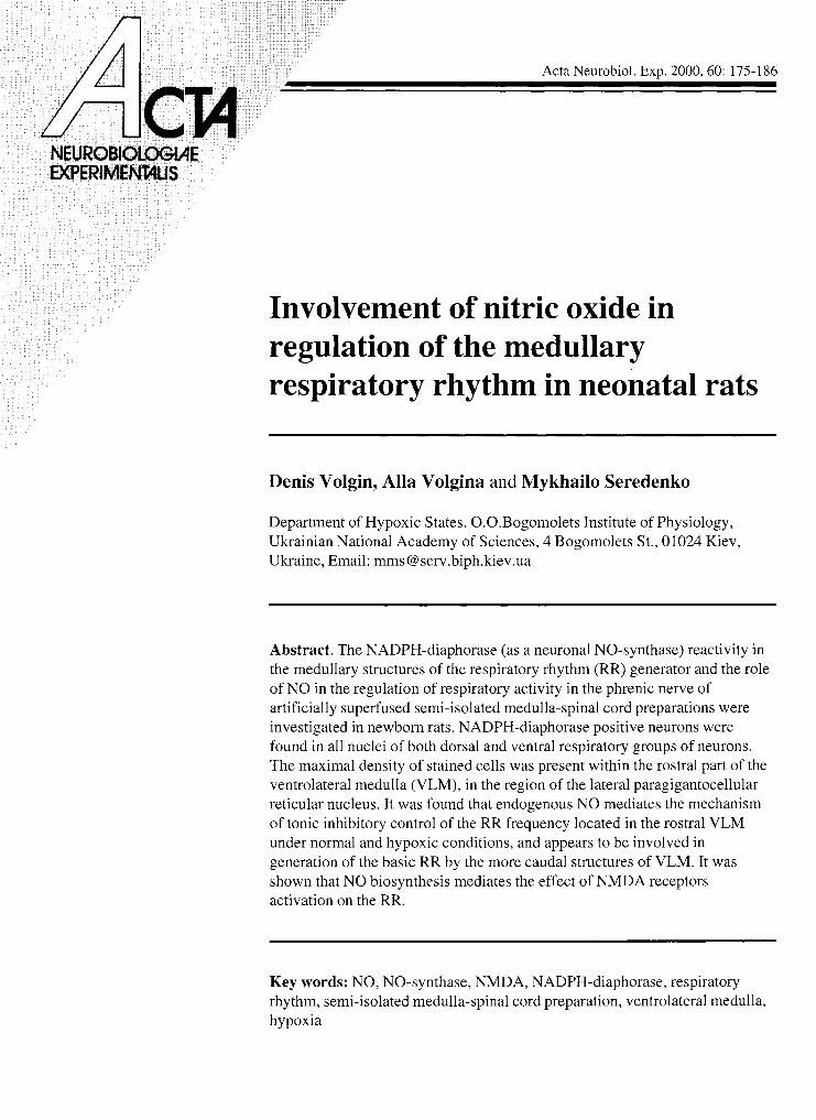
Three-min-long application of NMDA resulted in a significant (by 49.1%) rise in the ID frequency: a trend toward an increase in the ID amplitude was also noticed (Figs. 6A and 7). Preliminary 10-min-long superfusion with a L-NAME-containing solution resulted in an ab- sence of significant changes in the ID parameters when NMDA was applied (Figs. 6E and 7). The same results were obtained when Hb was earlier administered (Figs. 6 C and 7). NMDA applied against the background of 10- -min-long L-Arg application evoked a more prominent increase in the ID frequency (2.25 times). as compared with the control reaction (Figs. 6D and 7).
Fig. 5 . Effects of L-NAME (n = 18), hemoglobine (11 = 20) , L-arginine (11 = 14) and sodium nitroprusside (n = 15) on fre- quency (A ) and amplitude ( B ) of integrated inspiratory dis- charges in the n. phrenicus of medullo-spinal preparations. Data averaged from intact preparations (open bars) and from those with a transection through level 2, Fig. 1 (filled bars). Significant differences (P<0.05) from the control values are shown by asterisks.
Control L-NAME Hb L-Arg SNP
Control L-NAME Hb L-Arg SNP
182 D. Volgin et al.
NMDA 5 pM l
A F, min -'
1 min 1
Control PIMDA L N A h l E Hh a NMDA MMDA NMDA
L-NAME 10 pM NMDA 5 pM 5 0 a u I
1 min 1
I C
Hb 0,3 pM NMDA 5 pM 50 a.u.
I min I
L-Arg 10 pM NMDA 5 pM Control NMDA L N A h l E -
NMDA H b
NMDA NMDA
Fig. 7. Effects of L-NAAME ( 1 1 = 18). hemoglobine ( n = 20) and L-arginine ( 1 1 = 14) on frequency (A) and amplitude (B) of in-
Fig. 6. Effects of NMDA on integrated respiratory activity in tegrated inspilatory discharges in the n. phrenicus of medul- the n. phrenicus of medullo-spinal preparations. In B-D. L- lo-spinal preparations. measured under NMDA application. -NAME. hemoglobine andL-arginine. respectively. werepre- Significant differences (P<().()s) from the control ~ ' a lues are liminarily applied. shown by asterisks.
Nitric oxide it1 respiratory rhy thm 183
A F, min -1
- -
L-NAME 10 pM 95% N2 + 5% C02 50 a.u. I
I mln I Control Hypoxia L.NAME Hb L-Arg hypoxla typoxla hypoxla
I min 1
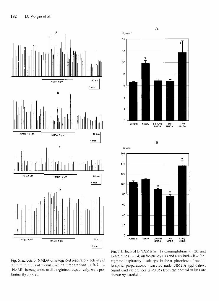
Fig. 8. Effects of hypoxia on integrated respiratory activity in the phrenic of medullo-spinal preparations ( 1 1 = 27) . In B-D. L-NAME. hemoglobine and L-arginine. respecti\,ely. were preliminarily applied.
Control Hypoxia L-NAME Hb L.Arg hypoxia l~ypoxia lrypoxia
Fig. 9. Effects of L-NAME. hemoglobine and L-arginine on frequency (A) and alnplitude (B) of integrated inspiratory dis- charges in the n. phrenicus of medullo-spinal preparations. measured under hypoxia (n = 27). Significant differences (P<0.05) from the control values are shown by asterisks. and those between the "normal" and "treated" preparations under hypoxia - crosses.

184 D. Volgin et al.
THE EFFECT OF NO ON SIMSP'S RESPIRATORY RHYTHM UNDER HYPOXIA
In intact preparations three-min-long superfusion by the anoxic isocapnic solution evoked a significant drop of averaged ID frequency (by 23.2%) (Figs. 8A and 9). After 10-min-long L-NAME application the ID fre- quency was significantly increased (by 66.8%) and the amplitude was significantly dropped (by 10.4%) during the subsequent hypoxic test (Figs. SB and 9). The pre- liminary Hb application resulted in a similar effect under hypoxia (Figs. 8C and 9). Indices of ID under hypoxic condition after preliminary 10-min-long L-Arg applica- tion were changed in an opposite fashion: a significant drop in the frequency (by 31.4%) and rise of amplitude (by 7.0%) were observed (Figs. 8D and 9).
DISCUSSION
In our experirnents the maximal density of NADPH- -diaphorase positive neurons were observed in the re- ticular formation within the lateral paragigantocellular nucleus (LPGi). I t is shown that in adult rats a group of stained cells is present within the same region (Vincent and Kimura 1992). Von Euler ( 1986) suggested that the LPGi may be involved in RR generation. The proposal was based on neuroanatomic evidence (retrograde trans- port of horseradish peroxidase from the n. ambiguus). and the reversible apnea induced by focal cooling of the LPGi (Andrezik et al. 1981, Budzinska et al. 1985). It cannot be ruled out that the LPGi region is the most es- sential source of endogenous NO modulating respiratory rhythmogenesis in newborn rats. Our data is compatible with the results obtained by Dun et al. (1994). who dem- onstrated the presence of NOS-positive cells within the LPGi in adult rats.
We have found NADPH-diaphorase-containing neu- rons within the main parts of ventral (n. arnbiguus) and dorsal (n. tractus solitarii) respiratory groups. These re- sults demonstrate that in the newborn rats neuronal sub- strates related to respiratory rhythrnogenesis are capable of generating NO.
Results of our electrophysiological experiments allow us to conclude that in newborn rats endogenous NO mediates the influence of NMDA receptor activation on medullary RR. It was shown that the levels of NMDA re- ceptor mRNA in NOS-containing neurons were higher than in non-NOS units (Price et al. 1993). In our previous experiments NMDA receptor blockade evoked signifi-
cant changes in KR generated by SIMSP's with intact rnedulla from newborn rats (Volgin et al. 1998). How- ever. NMDA receptors are not essential in generation of respiratory activity in the 400-mcm-thick medullary slices at the level of the pre-Botzinger complex (Funk et al. 1993). In this respect the LPGi as a more rostral me- dullary structure does not appear to be involved in the respiratory rhythmogenesis. NO is shown to increase glutamate release within the n. tractus solitarii in adult rats (Ogawa et al. 1995). It seems possible that the glu- tarnatergic neurotransrnission and NO formation, related to NMDA receptor activation, within the intermediate and caudal VLM excite respiratory neurons responsible for generation of RR. It should be taken into account that NMDA receptors are not directly involved in the func- tioning of the rhythm-generation network (Funk et al. 1993). but they are probably responsible for trans- mission of tonically active inputs to this network from the reticular formation neurons. It also cannot be ruled out that NO produced with NMDA receptor activation is able to amplify such activating signals due to intensifi- cation of the glutamate release and activation of non- -NMDA receptors directly involved in the generation of RR (Funk et al. 1993).
In early postnatal rats the most rostral VLM portions. which projectionally correspond to the M chemosensitive zone. are known to tonically inhibit the RR-generating network (Marchenko et al. 1995), yet neurotransmitter mechanisms of this phenomenon remain obscure. In this respect we should mention that glutamatergic trans- mission is involved in the tonic inhibitory mechanism of the rostral VLM which supresses the respiratory hypoxic reflex (Dillon et al. 1991). Our data on the influence of the NO synthase blocking effect on RR generated by SIMSP before and after separation of the rostral VLM suggest the involvement of NO in this tonic inhibitory mechanism in newborn rats.
Perinatal hypoxia evokes a cascade of modifications in the brain tissue, which includes increased release of glutamate, activation of the corresponding receptors and an elevation in the intracellular ca2+. These changes re- sult in the development of hypoxic-ischemic encephalo- pathy manifested by cramps. hypotonia and depression of respiration (Johnston 1997). We believe that, in addi- tion to other mechanisms. the inhibitory NO-mediated mechanism of the rostral VLM is involved in hypoxic depression of respiration in neonates. It is known that NO. which is synthetized in carotid chemoreceptors, in- hibits this activity in norrnoxia and hypoxia (Prabhakar

Nitric oxide in respiratory rhythm 185
et al. 1995, Trzebski et al. 1995). It should be added that NO plays a considerable role of inhibitory neuromodu- lation in the rostral VLM regions involved in cardiovas- cular control (Moibenko et al. 1997). Recent data suggest that the hypoxic respiratory responses were augmented in mutant mice deficient in the neuronal NOS (Kline et al. 1998).
These results and our findings allow us to conclude that NO is an important central neuromodulator of the respiratory rhythm in newborn rats.
ACKNOWLEDGEMENT
This research was partly supported by grant PSU074134 of International Soros Support Education Program (ISSEP).
REFERENCES
Andrezik J.A.. Chan-Palay V . . Palay S.L. (198 1 ) The nucleus paragigantocellularis lateralis in the rat. Demonstration af- ferent~ by the retrograde transport o f horseradish peroxi- dase. Anat. Embryol. 16 1 : 373-390.
Budzinska K.. von Euler C.. Kao F .F . . Pantaleo I.. Yamamoto Y . ( 1985) Effects o f graded focal cold block in rostral areas o f the medulla. Acta Physiol. Scand. 124: 329-340.
Dawson T.M. , Snyder S.H. (1994) Gases as biological mess- engers: nitric oxide and carbon monoxide in the brain. J. Neurosci. 14: 5 147-5 159.
Dillon G.H.. Welsh D.E., Waldrop T.G. (1991 )Modulation o f respiratory reflexes by an excitatory amino acid mech- anism in the ventsolateral medulla. Respir. Physiol. 85: 55-72.
Dun N.J.. Dun S.L.. Forsterman U . (1994) Nitric oxide syn- thare immunoreactivity in rat pontine medullary neurones. Neuroscience 59: 429-445.
Euler C. , von (1986) Brain stem mechanisms for generation and control o f breathing pattern. In: Handbook o f physio- logy. The respiratory system. Control o f breathing (Eds. H.Rahn and W.O.Fenn) . Vol . 2. Am. Physiol. Soc.. Wash- ington. D.C. p. 1-67.
Funk G.D.. Smith J.C.. Feldman J.L. (1993) Generation and transmission o f respiratory oscillations in medullary slices: role o f excitatory amino acids. J. Neurophysiol. 70: 1497- 1515.
Garthwaite J. (199 1 ) Glutamate, nitric oxide and cell-cell sig- nalling in the nervous system. Trends Neurosci. 14: 60-67.
Garthwaite J..Boulton C.L. (1995) Nitric oxide signalling in the central nervous system. Annu. Rev. Physiol. 57: 683- 706.
Garthwaite J.. Garthuaite G.. Palmer R.M.J.. Moncada S. (1989) NMDA receptor activation induced nitric oxide
synthesis from arginine in the rat brain slices. Eur. J . Phar- macol. 172: 4 13-4 16.
Hope B.T., Michael G.J.. Knigge K.M.. Vincent S.R. ( 1 99 1 ) Neuronal NADPH-diaphorase is a nitric oxide synthase. Proc. Natl. Acad. Sci. U S A . 88: 281 1-2814.
Johnston M.V. (1997) Hypoxic and ischemic disorders o f in- fants and children. Brain Dev. 19: 235-239.
Kalish B.E.. Jhaniandas K.. Beninger R.J., Boegman R.J. ( 1 999) Modulation o f quinolitic acid-induced depletion o f striatal NADPH diaphorase and enkephalinergic neurons by inhibition o f nitric oxide synthase. Brain Res. 8 17: 15 1 - 162.
Kline D.D.. Yang T.. Huang P.L.. Prabhakar N.R. (1998) Al- tered respiratory responses to hypoxia in mutant mice defi- cient in neuronal nitric oxide syn:hase. J . Physiol. 5 1 1 : 273-288.
Ling L. , Karius D.R., Fiseus R.R.. Speck D.F. ( 1992) En- dogenous nitric oxide required for an integrative respir- atory function in the cat brain. J. Neurophysiol. 68: 1910-1912.
Marchenko V.A.. Fenik V.B., Preobrazhenski N.N., Seredenko M.M. (1995) Respiratory activity generated by semi-iso- lated medullo-spinal preparation and recorded from the phrenic nerve o f newborn rats. Neurophysiology 27: 385- 395.
Moibe!lko A.A. , Sagach V.F.. Shapoval L.N. et al. (1997) Role o f endothelium and endothelium-produced biologi- cally active substances in regulation o f circulation and car- diac function (in Ukrainian). Fiziol. Zh. 43: 3-18.
Ogaua H . . Mizusax&a A,. Kikychi Y . et al . ( 1 995) Nitric oxide as a retrograde messenger in the nucleus tri~ctus solitarii o f rats during hypoxia. J . Physiol. 486: 495-504.
Onimary H.. Arata A,. Homma J. (1987) Localization o f res- piratory rhythm-generating neurones in the medulla o f brainstem-spinal cord preparations from newborn rats. Neurosci. Lett. 78: 152- 155.
Petrovicky P.. Nemcova V . (1995) Topographical organiza- tion o f NADPH-diaphorase positive neurons and fibres in dorsal ponto-mesencephalic teglnentum in the rat brain. J . Brain Res. 36: 539-545.
Prabhakar N.R.. Cherniack N.S.. Haxhiu M.A. (1995) Inhibi- tory and excitatory effects o f nitric oxide on respiratory re- sponses to hypoxia. In: Ventral brainstem mechanisms and control o f respiration and blood pressure (Eds. C.O.Trouth, R.M.Millis, H.F.Kiwull-Schone and M.E.Schlafke). Mar- cel - Dekker. Inc., New York. p. 393-404.
Price R.H.. Mayer B., Beitz A.J. (1993) Nitric oxide synthase neurons in rat brain express more NMDA receptor mRNA than non-NOS neurons. NeuroReport. 4: 807-8 10.
Prickaert J.. De Vente J.. Markerink-van Ittersum M.. Stein- busch H.W. (1998) Behavioural. neurochemical and neuroanatomical effects o f chronic postnatal N-nitro-L-ar- ginine methyl ether treatment in neonatal and adult rats. Neuroscience 87: 18 1 - 195.

186 D. Volgin et al.
Roche A.K.. Cook M.. Wilcox G.L., Kajander K.C. ( 1 996) A nitric oxide synthesis inhibitor (L-NAME) reduces licking behavior and Fos-labeling in the spinal cord of rats during formalin-induced inflammation. Pain 66: 33 1-341.
Suzue T. (1984) Respiratory rhythm generation in the in vitro brain stem-spinal cord preparation of the neonatal rat. J. Physiol. 354: 173-1 84.
Trzebski A,. Sato Y.. Suzuki A,, Sato A. (1995) Inhibition of nitric oxide synthesis potentiates the responsiveness of ca- rotid chemoreceptors to systemic hypoxia in the rat. Neu- rosci. Lett. 190: 29-32.
Vincent S.R.. Kimura H. (1992) Histochemical mapping of ni- tric oxide synthase in the rat brain. Neuroscience 46: 755- 784.
Volgin D.V., Marchenko V.A., Seredenko M.M.. Vasilenko D.A. ( 1998) Involvement of NMDA receptors in the con- trol of respiratory rhythm generated by medullo-spinal preparations of early postnatal rats. Neurophysiology 30: 101-106.
Receii~ed 2 Febnran 1999, accepted I Mcrr-ch 2000