invited review the microanatomy of calcium stores in human
TRANSCRIPT

Hislol Hislopalhol (1997) 12: 479-490
001: 10.14670/HH-12.479
http://www.hh.um.es
Histology and Histopathology
From Cell Biology to Tissue Engineering
Invited Review
The microanatomy of calcium stores in human neutrophils: Relationship of structure to function E.J. Pettit, E.V. Davies and M.B. Hallett Molecular Signalling Group, Department of Surgery, University of Wales College of Medicine, Cardiff, U.K
Summary. As changes in cytosolic free Ca2+ play key roles in coupling responses in neutrophils, it is important to locate and identify Ca2+ storage sites within these cells. Here, recent data is presented which highlights the functional link between microanatomical structure and cell signalling function. Fluorescent optical probes for cytosolic free Ca2+ have been used, together with organelle specific markers. We present evidence from conventional fluorescence microscopy, together with ratiometric and confocal laser scanning fluorescence microscopy, which pin-points two cellular locations for Ca2+ within the neutrophil; one within the nuclear lobes, and the other towards the cell periphery. Knowledge of these two locations provides a clear insight into how signalling in this cell type is regulated and provides a framework for explaining how specific stimuli act to produce specific responses.
Key words: Neutrophils, Calcium stores, Confocal microscopy, Microanatomy, Ca2+ signalling
Introduction
Neutrophils are essential white blood cells of the immune system, involved in the inflammatory response occurring during any breach in body defences, such as bacterial attack. In addition to this life-saving function, neutrophils are also the mediators of damaging inflammatory diseases, such as rheumatoid arthritis. A review by Leff and Repine (1993) listed seventeen serious diseases in which neutrophil malfunction plays the major role. Understanding the mechanisms underlying neutrophil activity is thus an important goal for medical research.
Neutrophil extravasation and migration through tissues is the essential first step in any inflammatory response (Marsh et aI., 1967). This process is dependent upon neutrophil recognition of adhesion molecules, such
Offprint requests to: Dr. Elizabeth J . Pettit, Molecular Signalling Group,
Department of Surgery, University of Wales College of Medicine, Heath
Park, Cardiff CF4 4XN, UK
as the selectins, and ICAM-I (Kishimoto and Rothlein, 1994). Neutrophils then respond to a range of chemotactic factors, such as the bacterial peptide, f-metleu-phe, complement component C5a, and leukotriene B4, which direct their migration towards the site of damage or attack. Here, neutrophils engulf and destroy any pathogens present, using mechanisms which include recognition of opsonised bacteria by specialised receptors in the plasma membrane, such as the Fc and complement receptors. Recognition results in phagocytosis (Brown, 1994), and bacterial killing by release of toxic components from neutrophil granules into the phagosome (Zeya and Spitznagel , 1972) . When inflammation has subsided, or a critical number of bacteria have been ingested, the neutrophil begins the apoptotic process, leading to the safe disposal of the spent cell and contents (Haslett, 1992).
Fundamental to each one of these functional processes are changes in neutrophil cytosolic free Ca2+ concentration, which are important signals in the mediation between ligand recognition and end response (Pozzan et aI, 1983). Thus , the study of molecular processes occurring during neutrophil activity, and especially Ca2+ signalling, are central to an understanding of both normal and disease states of inflammation, and to the design of novel therapeutic regimes during inflammation.
Neutrophil microanatomy
The neutrophil is a unique cell, specifically designed for an individual role in the body. It is terminally differentiated , and undertakes a minimal amount of novel protein synthesis during the typical 36-hour life span. The anatomy of this small cell perfectly reflects neutrophil function.
Human neutrophils are 10-15 11m in diameter (Wintrobe, 1981). 20% of the cell volume consists of multi-lobed nucleus, and another 20%, of the dense granules necessary for bacterial killing (Fig. 1). Other typical mammalian cell constituents, such as mitochondria and ER, make up less than 1 % of the volume of an average neutrophil (Hallett, 1989). The only

Calcium stores in human neutrophils
prorninent membranous stmcture in the neutrophil is a region of vestigial, Golgi-like ER, situated towards the centre of the cell, between the lobes of the nucleus (Fig. 1).
Neutrophil Caz+ signalling
Like al1 mamrnalian cells, neutrophils contain a high concentration of calcium (ImM), but the majority is stored in a bound form (Petroski et al., 1963). In the resting neutrophil, the free ca2+ concentration is
approximately 100nM, but stimulation can result in the concentration rising to 1 pm (Hallett, 1989). There are two mechanisms for increasing cytosolic free ca2+ in the neutrophil, namely calcium store release, and ca2+ influx, via channels in the plasma membrane (Demareux et al., 1994). The spatial and temporal relationship between these two types of activity, and especially the location and functional roles of calcium store release will be dealt with in greater detail later, but first it is necessary to have a rudimentary understanding of signalling processes in neutrophils.
F., Fig. l. Elecm" h micmgraph of a mature ,. neutrophil, showing the ,'&: nuclear lobes (N),
w *M.* granules (g), and Golgi- I like near-nuclear calcium , store ( X ) . x 20,000

Calcium stores in human neutrophils
(i) Receptor-cytosol signalling
Occupation of neutrophil receptors with their ligands (fMLP, C5a, CDllblCD18, PAF, 11-8, etc) results in signal transduction across the membrane. In the case of the seven membrane-spanning region chemoattractant receptors, this occurs via heterotrimeric G-proteins coupled to the receptor (Bommakanti et al., 1995). The chemoattractants produce different cellular responses, which depend on their concentration. For example, fMLP at pM-nM concentrations causes priming of the respiratory burst without any detectable change in cytosolic free ca2+, whilst pm concentrations directly activate the NADPH oxidase, and cause both calcium releases from stores, and influx (Al-Mohanna and Hallett, 1990; Thelen et al., 1993). The mechanisms for other ligands are less well understood, and may, or may not, involve heterotrimenc- or monomeric G-proteins.
The initial interaction between the ligand and receptor results in the activation of one of the range of different types of heterotrimeric G-proteins situated on the inner surface of the membrane - Gi2, in the case of fMLP. The G-protein with GDP bound to it is inactive, and conversion to the active form involves dissociation of the By subunits from the a subunit, which contains the nucleotide binding site; GDP is released, and GTP binds to the empty site. The protein is then in the active state, until the GTP is hydrolysed to form GDP again (Fig. 2). Enzymes in the cytosol control the rate of the association-dissociation reaction. Activated G-proteins couple to intracellular effector molecules, and thus cause a cascade of intracellular events, mediated by particular effector molecules coupled to that G-protein.
(ii) Intracellular signalling
One of the enzymes activated by the active form of the heterotrimeric G-proteins is phospholipase C (B
form, PLC; Cockroft and Gomperts, 1985). The function of PLCB is to hydrolyse the plasma membrane lipid, phosphatidylinositol-4,5-bisphosphate. Inositol 1,4,5 trisphosphate (IP3), is then free to diffuse into the cytoplasm, leaving the lipid diacylglycerol (DAG) in the membrane (Berridge and Irvine, 1989).
The response to fMLP causes IP3 diffusion to a calcium store by this pathway, leading to occupation of IP3 receptors in the store membrane. The IP3 receptor is a calcium channel consisting of four subunits, and IP3 binding causes channel opening, releasing free calcium ions into the cytosol (Berridge and Irvine, 1984). Phosphorylation of IP3 generates IP4, and although its actions are not well established, it has been suggested that it may open ca2+ channels on the plasma membrane (Meldolesi and Pozzan, 1987). The inositol phosphate signalling pathway was virtually unknown twenty years ago (Michell, 1995). The generation of an increase in cytosolic free calcium concentration is summarised in Figure 3.
Calcium is cleared from the cytosol by the ca2+- ATPase pumps, both in the store membranes, and the plasma membrane (Lagast et al., 1984). Rapid clearing of the calcium ions allows the neutrophil to prepare for a second wave of calcium activity, which occurs in some situations such as integrin mediated signals (Pettit and Hallett, 1996a,b), and during chernotaxis. There is littie evidence to suggest that calcium-induced calcium release (CICR) pathways operate in neutrophils, m r do neutrophils posses caffeine, or ryanodine-sensitive receptors, typical of other mammalian cells such as adrenal medullary cells (Cheek et al., 1990).
The effects of an increase in cytosolic free calcium on the neutrophil are very varied. Calcium ions activate protein kinase C (PKCy), via its translocation to the plasma membrane, and association with DAG (Dang et al., 1995). Protein kinase C is responsible for the phosphorylation of proteins important in neutrophil
Rg. 2 The ¡-un between hgae-ric O- pmwm and m OhernaPaeELc
witwlluihv stgnai. Tke MLP-type W r (M trafmwnbrane domahs), ooupled to the G-protein (a&. causes ihe exchenge of gJmIdhie d- (m.d g lmtd ' i
(@TP) to w a me (3- ~IVteharrPlaause
EFFECTOR

Calcium stores in human neutrophils
activities, such as NADPH oxidase components (respiratory burst), and vimentin (cytoskeleton). Calcium is involved in regulating the assembly of F-actin, and pseudopod formation, which occurs during chemotaxis (Fernandez-Segura et al., 1995), phagocytosis, and adhesion (Westlin e2 al., 1992). An increase in cytosolic free atlcium is associated with phagocytic events, but not necasarily essential for them to occur (Theler et al., 1995). Release of calcium ions during phagocytosis is mediated by the occupation of the Fc y receptors. Phagocytic events may also result in the movement of calcium stores to the periphagosornal region of the cell (Sawyer et al., 1985; Stendahl et al., 1994; Theler et al., 1995). Cytosolic free caicium increases during non- specific adherence (Jaconi et al., 1988), and can be correlated to neutrophil spreading (Kruskal et al., 1986; Hendry and Maxfield, 1993; Theler et al., 1995). The effwt of cytosolic free calcium concentration on actin filament assembly and disassembly are mediated by gelsolin (Howard et al., 1990), and profilin (Goldschmidt-Clermont, 1990). The range of actions of calcium and IP3 me reviewed by Albritton et al. (1992).
(iii) Other signaíling mechanisms.
The active form of the heterotrimeric G-proteins can also be coupled to other effector molecules, and may cause the fonnation of a number of other inkrcellular, or possibly intraceiiular messengers, in addition to calcium. Phospholipase A2 hydrolyses phosphatidylcholine and
phosphatidylethanolarnine to produce arachidonic acid. This, with DAG and calcium ions, activates protein kinase C, and is a precursor for the formation of leukotriene B4 (LTB4), secreted as a chemoattractant to recruit more neutrophils (Ford-Hutchinson et al., 1980). Phospholipase D hydrolyses phosphatidylcholine, to produce DAG and choline. DAG can be enzymatically converted to phosphatidic acid, which may be a second messenger for oxidase activation (Agwu et al., 1991; Dana et al., 1994).
Another activity of G-proteins in neutrophils, which remains unclear, is the modulation of the cytosolic cyclic-AMP concentration (Tsu et al., 1995). In other cells responding to hormonal stimulation, different receptbrs coupled to G-proteins can either activate, or inhibit the adenylate cyclase enzyme, which in its active form, causes an increase in cAMP levels. The cAMP then either activates, or inhibits, protein kinases, causing different intracellular effects. One of the functions of calcium-calrnodulin is to activak the phosphodiesterase enzyme, which decreases the c M P concentration. The presence of cAMP has been demonstrated in neutrophils, but little is known about its functions.
The importante of the Caz+ store in neutrophils
The use of increasingly effective fluorescent rnicroscopic techniques, and the wide variety of calcium indicators, buffers and chelators, has resulted in exciting new developments in calcium signalling research in
INTRACELLULAR EFFECTS
Flg. 3. Intrmllular calcium signalling. Generation of inositol trisphospate (IP3) by activated phospholipase CY (PKC) causes calcium. ion release (Caz+) from intracellular stores (St). PKC is activated by the active form of the G-pmtein, generated by the receptor interaction (R). 1 : phosphatidyl- inositol-4,s- bisphosphate; 2: diacyl- glycerol; CIF: calcium influx factor; a: activated G- protein.
Calcium stores in human neutrophils
hurnan neutrophils. Recent research (Parekh et al., 1993; Randriamampita and Tsein, 1993; Davies and Hallett, 1995a) has suggested that "calcium influx factor" (CIF) may be liberated from stores at the sarne time as calcium itself. CIF is thought to diffuse to the plasma membrane, and cause opening of calcium channels there, allowing extracellular calcium ions to flood the cytosol, and replenish the emptied stores. There has been lively debate concerning the truth, and relevance, of these observations (Bemdge, 1996; Hallett et al., 1996).
Closely tied to the CIF debate, has been the observation by severa1 workers of discrete locations within the neutrophil from which, calcium is released after cell stimulation by a variety of pathways (Burgoyne et al., 1989; Stendahl et al., 1994). The two stimulation pathways that have formed the basis of these observations are mediated by the two main receptor types on the neutrophil surface. The chemoattractant receptors have been the classical model in neutrophil signalling. Almost al1 of our present knowledge of neutrophil signalling processes is derived from the study of this type of receptor, and its agonists. Recently, however, a second type of receptor has been more widely studied. This class of receptor includes the functionally important Fc receptors, and the 4 (CR3, Macl) integrin family of adhesion/complement receptors. The fundamental difference between these types of receptor are that a one-to-one occupation of the second type does not result in signal transduction. Ligand recognition must be followed by some form of physical connection - "clustering" - before signal transduction is initiated. Experimentally, this has usually been achieved by crosslinking, using a secondary antibody.
Recently, crosslinking of the FcyRII (CD32) on the surface of neutrophils, by immune complexes or by secondary antibody has been reported to increase cytosolic free ca2+ (Davies et al., 1994; Davies and Hallett, 1995b). Similarly, the B2 integrin family were originally thought to be adhesion receptors, with no role in signal transduction or neutrophil activation. However, severa1 workers have reported that crosslinking the integrin causes cytosolic free calcium concentration to increase (Ng-Sikorski et al., 1991; Petersen et al., 1993). Recently, novel techniques have pinpointed the intraceílular origin of calcium store release mediated by the engagement, or engagement and clustering, of the types of receptor discussed above (Pettit and Hallett, 1996a,b). Here, we present recent findings concerning the location, and functional roles, of different calcium stores in human neutrophils.
Calcium store location
Functionally, calcium stores can be defined as membrane bound organelles containing calcium at a higher concentration than the cytosol. This is achieved by both an inwardly-facing ~ a ~ + - ~ ~ ~ a s e , which "pumps" calcium into the store, and also a calcium
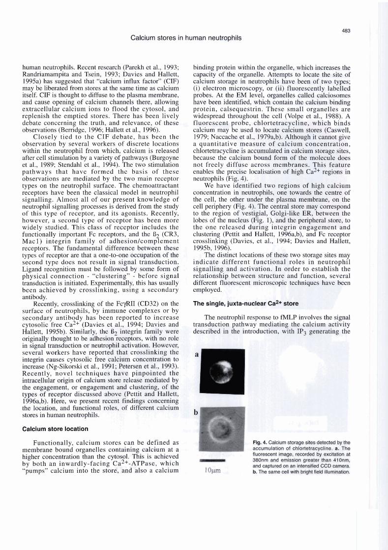
binding protein within the organelle, which increases the capacity of the organelle. Attempts to locate the site of calcium storage in neutrophils have been of two types; (i) electron microscopy, or (ii) fluorescently labelled probes. At the EM level, organelles called calciosomes have been identified, which contain the calcium binding protein, calsequestrin. These small organelles are widespread throughout the cell (Volpe et al., 1988). A fluorescent probe, chlortetracycline, which binds calcium may be used to locate calcium stores (Caswell, 1979; Naccache et al., 1979a,b). Although it cannot give a quantitative measure of calcium concentration, chlortetracycline is accumulated in calcium storage sites, because the calcium bound form of the molecule does not freely diffuse across membranes. This feature enables the precise localisation of high ca2+ regions in neutrophils (Fig. 4).
We have identified two regions of high calcium concentration in neutrophils, one towards the centre of the cell, the other under the plasma membrane, on the cell periphery (Fig. 4). The central store rnay correspond to the region of vestigial, Golgi-like ER, between the lobes of the nucleus (Fig. l), and the peripheral store, to the one released during integrin engagement and clustering (Pettit and Hallett, 1996a,b), and Fc receptor crosslinking (Davies, et al., 1994; Davies and Hallett, 1995b, 1996).
The distinct locations of these two storage sites may indicate different functional roles in neutrophil signalling and activation. In order to establish the relationship between structure and function, severa1 different fluorescent microscopic techniques have been employed.
The single, juxta-nuclear Ca2+ store
The neutrophil response to fMLP involves the signal transduction pathway mediating the calcium activity described in the introduction, with IP3 generating the
- 380nm and eGssion greater- than 41 ünm, and captured on an intensified CCD camera.
1 o~lm b. The same cell with bright field illumination.

Calcium stores in human neutrophils
store release signal. This release is followed almost visualisation of store release by preventing the calcium instantaneously by calcium influx, so that the two influx (Davies, et al., 1991, 1992; Pettit and Hallett, activities cannot normally be resolved. However, the 1995). Under these conditions, ratiometric imaging of removal of calcium ions from the extracellular medium neutrophils loaded with Fura-2 has shown that cytosolic by a chelator, such as EGTA, Qr prevention of calcium free ca2+ rises initially from a single focus within the influx by the channel blocker, SKF96365, allows the neutrophil (Fig. 5).
acriaine orange AV - u i u ~ 6 \ 3 )
Fig. 5. The juxta-nuclear Ca2+ release site in neutrophil. The figure shows the localised release of Ca2+ frorn the store induced by 1-rnet-leu-phe (1pM) in the absence of extracellular Caz+ (a) as a ratiornetric irnage of a fura2 loaded neutrophil, where blue and green represents resting and high cytosolic free Caz+ respectively, and (b) as a pseudo-3D rnap where the xy plane are the dirnensions of the cell and the z axis is proporlional to the cytosolic free Can+ concentration. In the lower panel, the sarne cell is stained with (i) acridine orange, as a rnarker for the position of the nucleus, (ii) DiOC6(3) as a rnarker for rnernbranous organelles, and (iii) by subtraction of irnage (i) frorn irnage (ii) the non-nuclear rnernbranous structures. In the final irnage (iii), a "hot spot" of mernbrane staining (') is obsewed at a position which corresponds to the position of elevated Caz+ (') in the Ca2+ irnage (a).
Calcium stores in human neutrophils
Recently, rapid time scan confocal microscopy of neutrophils loaded with the calcium indicator, Fluo3 (Merritt et al, 1991; Pettit and Hallett, 1996b) has prodded further details of the location of the release site,
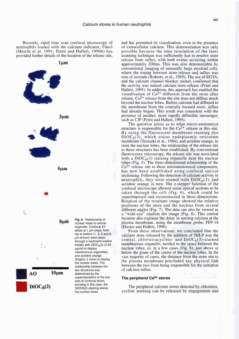
Fig. 6. Relationship ol nuclear lobes to central organelle. Confocal XY slices at 1 pm steps, from top to bottorn (1,3,6 and 8 pm shown) were taken through a neutrophil loaded initially with DiOC6(3) (0.25 pglrnl) lo display rnembranous organelles, and acridine orange (5ng/rnl, 5 rnins) to display the nuclear lobes. The relationship between the two structures was
1 A 0 [email protected] deterrnined by the 1 - - superirnpositkn of the two sets of wnfocal slices, showing in lhis case, the DiOB(3) staining above the nuclear lobes.
and has permitted its visuaiisation, even in the presence of extracellular calcium. This demonstration was only possible because the time resolution of the laser scanning technique was sufficiently fast to resolve store release from influx, with both events occurring within approximately 200ms. This was also demonstrable by conventional imaging of unusually large myeloid cells, where the timing between store release and influx was tens of seconds (Roberts, et al., 1995). The use of EGTA, and the calcium channel blocker, nickel, confmed that the activity was indeed caicium store release (Pettit and Hallett, 1995). In addition, this approach has enabled the visualisation of ca2+ diffusion from the store after release. ca2+ release from the site does not diffuse much beyond the nuclear lobes. Before calcium had diffused to the membrane from the centrally located store, influx had already begun. This result was consistent with the presence of another, more rapidly diffusible messenger, such as CIF (Pettit and Hallett, 1995).
The question arises as to what micro-anatornical structure is responsible for the ca2+ release at this site. By using the fluorescent membrane-staining dye DiOC6(3), which stains endoplasmic reticulum membrane (Terasaki et al., 1984), and acridine orange, to stain the nuclear lobes, the relationship of the release site to these structures has been established. By conventionai fluorescence microscopy, the release site was associated with a DiOC6(3) staining organeiie near the nuclear lobes (Fig. 5). The three-dimensional relationship of the ca2+ release site to these microanatomical components, has now been established using confocal optical sectioning. Following the detection of calcium activity in neutrophils, they were stained with DiOC6(3), and acridine orange in turn. The z-stepper function of the confocai microscope allowed serial optical sections to be taken through the cell (Fig. 6), which could be superimposed and reconstructed in three dimensions. Rotation of the resultant image showed the relative positions of the store and the nucleus from severa1 different angles (Fig. 7). The data can also be viewed as a "wide-eye" random dot image (Fig. 8). This central location also explains the delay in sensing caicium at the plasma membrane, using the membrane probe, FFP-18 (Davies and Hallett, 1996).
From these observations, we concluded that the calcium store released by the addition of fMLP was the central, chlortetracycline- and DiOC6(3)-stained membranous organelle, nestled in the space between the nuclear lobes, or, in a few cases (Fig. 6), just above or below the plane of the centre of the nuclear lobes. In the vast majority of cases, the distance from the store site to the plasma membrane precluded any physical link between the two from being responsible for the initiation of calcium influx.
* - ~4.' .e\ 1,; - 8 - r r The peripheral Caz+ &ores " h Q' 0 ' W'&44 *Si W I L W
The peripheral calcium stores detected by chlortetr cycline staining can be released by engagement an t


. . , , , - ,. . , : L Calcium stores in human neutrophils ta<<;;i.lci t ; ;.>.<;,; '+ , .: $k t 2 w ; . * . r . . - l , - - . ' 8 n:.:.'i:. c ‘ ;.-;" - - L * L. , .
. S . - . +.. , .', .,, g ; ,. ' .
that there are different pathways for store release, and different relationships between store release and infiux in each case. One difference between the two signal transduction mechanisms is that massive tyrosine phosphorylation activity occurs in response to integrin (Burridge, et d., 1992; Fuortes et al., 1994), or FcylUI clustering, but not in response to fA4LP. Localised phosphorylation events could sensitise peripheral store IP3 receptors, which in the absence of phosphorylation remain insensitive, leaving the central store to respond in this case (Jayraman, 19%). The site of the central store, between the nuclear lobes, is aiso the location of other interesting organelles in the neutrophil, such as the Golgi-like region, and the rnicrotubule organising centre (MTOC). Although most workers quote the Golgi region as the calcium store, the MTOC has also been identified as playing a role in calcium release in other cells (Brundage et al., 1993). It will be possible to resolve which of these two structures is responsible for the calcium release, by using electron microscopic immuno- localisation of molecules such as IP3 receptor and ca2+ ATPase. This will provide the link between the structure, and its function in calcium signalling. a; JJ -
.< . - - = > í C~nclusion y,$., ; a 3 , ; r- .- J , y . . 'r, ,--. . . 1 ' L '
The evidence for a "two-stores, two-pathways" signalling arrangement in human neutrophils is graduaily accumulating. There is release of caicium from a single, centraily located srore upon fMLP addition, and recent work by several groups has documented the presence of
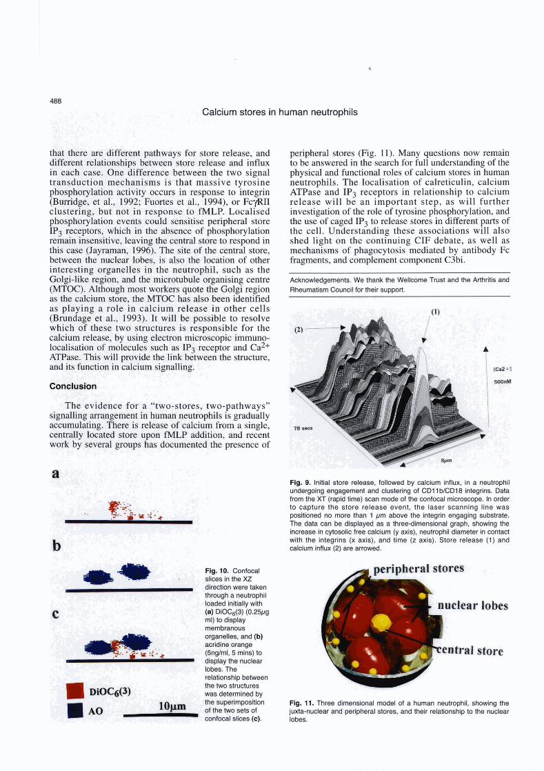
Fig. 10. Confocal slices in the XZ direction were taken through a neutrophil loaded initially with
C (a) DiOC6(3) (0.25~9 ml) to display membranous organelles, and (b) acridine orange (5ng/ml, 5 mins) to display the nuclear lobes. The relationshi~ between
peripheral stores (Fig. 11). Many questions now remain to be answered in the search for full understanding of the physical and functionai roles of caicium stores in human neutrophils. The localisation of calreticulin, calcium ATPase and IP3 receptors in relationship to calcium release will be an important step, as will further investigation of the role of tyrosine phosphorylation, and the use of caged IP3 to release stores in different parts of the cell. Understanding these associations will also shed light on the continuing CIF debate, as well as mechanisms of phagocytosis mediated by antibody Fc fragments, and complement component C3bi.
Acknowiedgements. We thank the Welicome Trust and the Arthritis and Rheumatism Council for their support.
Flg. 9. lnitial store release, followed by calcium influx, in a neutrophil undergoing engagement and clustering of CDl 1 biCD18 integrins. Data from lhe XT (rapid time) scan mode of the confocal microsoope. In order lo capture the store release event, the laser scanning line was positioned no more than 1 pm above the integrin engaging substrate. The data can be displayed as a three-dimensional graph, showing the increase in cytosolic free calcium (y axis), neutrophil diameter in contad with the integrins (x axis), and time (z axis). Store release (1) and calcium influx (2) are amwed.
entra1 store F the iwo st~udures was determined by the superimposition Flg. 11. Three dimensional model of a human neutrophil, showing the "prn of the two sets of juxta-nuclear and penpheral stores, and their relationship to the nuclear confocal slices (e). lobes.
Calcium stores in human neutrophils
Agwu D.E., McPhail L.C., Sozzani S., Bass D.A. and McCall C.E. (1991). Phosphatidic acid as a second messenger in human poly- morphonuclear leukocytes: effects on activation of the NADPH oxidase. J. Clin. Invest. 88, 531-539.
Albritton N.L., Meyer T. and Stryer L. (1992). Range of messenger action of calcium ion and inositol 1,4,btrisphosphate. Science 258, 1812-1814.
Al-Mohanna F.A. and Hallett M.B. (1990). Actin polymerisation in neutrophils is triggered without a requirement for a rise in cytosolic free calcium. Biochem. J. 266, 669-674.
Berridge M.J., (1996). Capacitative calcium entry - sifting through the evidence for CIF. Biochem. J. 314, 1055.
Berridge M.J. and Irvine R.F. (1984). lnositol 1,4,5-trisphosphate, a novel second messenger in cellular signal transduction. Nature 312, 315-321
Bemdge M.J. and Irvine R.F. (1989). lnositol 1,4,5-trisphosphates and cell signalling. Nature 341, 197-205.
Burridge K., Turner C.E. and Romer L.H. (1992). Tyrosine phosphorylation of paxillin and pp125fak accompanies adherente to extracellular matrix: a role for cytoskeletal assembly? J. Cell Biol. 1 19,893-903.
Bommakanti R.K., Dratz E.A., Siemsen D.W. and Jesaitis A.J. (1995). Extensive contact between Gi2 and N-formyl peptide receptor of human neutrophils: mapping of binding siteg using receptor-mimetic peptides. Biochemistry 34, 6720-6728.
Brown E.J. (1 994). Phagocytosis. BioEssays 17, 109-1 17. Brundage R.A., Fogarty K.E., Tuft R.A. and Fay F.S. (1993).
Chemotaxis of newt eosinophils: calcium regulation of chemotactic response. Am. J. Physlol. 265, C1527-C1543.
Burgoyne R.D., Cheek T.R., Morgan A., O'Sullivan A.J., Moreton R.B., Mata A.M., Colyer J., Lee A.G. and East J.M. (1989). Distribution of two distinct calcium-ATPase like proteins and their relationship to the agonist sensitive calcium store in adrenal chromaffin cells. Nature 342,72-74.
Caswell A.H. (1979). Methods of measuring intracellular calcium. Int. Rev. Cytol. 56, 145-181.
Cheek T.R., O'Sullivan A.J., Moreton R.B., Berridge M.J. and Burgoyne R.D. (1990). The caffeine sensitive calcium &ore in bovine adrenal chromaffin cells: an examination of its role in triggering secretion and calcium homeostasis. FEBS Lett. 266, 91-95.
Cockroft S. and Gomperts B.D. (1985). Role of guanidine nucleotide binding proteinin the activation of polyphosphoinositide phospho- diesterase. Nature 314, 534-536.
Dana R., Malech H.L. and Levy R. (1994). The requirement for phospholipase A2 for activation of the assembled NADPH oxidase in human neutrophils. Biochem. J. 297,217-223.
Dang P.M-C., Rais S., Hakim J. and Perianin A. (1995). Redistribution of protein kinase C isoforms in human neutrophils stimulated by formyl peptides and phorbol myristate acetate. Biochem. Biophys. Res. Commun. 212,644-872.
Davies E.V. and Hallen M.B. (1995a). A soluble factor directly stimulates Ca2+ entry in neutrophils. Biochem. Biophys. Res. Commun. 206, 348-354.
Davies E.V. and Hallett M.B. (1995b). A novel pathway for calcium signalling in neutrophils by immune complexes. lmmunology 85, 538-543.
Davies E.V. and Hallett M.B. (1996). Near-membrane calcium changes
in response to store release measured using FFP-18. Cell Calcium (in press).
Davles E.V., Hallett M.B. and Campbell A.K. (1991). Localised superoxide reiease by neutrophils can be provoked by a eytosolic oalcium 'cloud'. lmmunology 73,228-234.
Davies E.V., Campbell A.K. and Hallett M.B. (1992). Dissociatlon of store release from transmembrane influx of calcium in human neutrophils. FEBS Lett. 31 3, 121 -1 25.
Davies E.V., Campbell A.K. and Hallett M.B. (1894). Calcium oscillations in neutrophils triggered by immune complexes result from calcium influx. lmmunology 82,57432.
Demareux N., Monod A., Lew D.P. and Krause K.H. (1994). Characterisation of receptor-rnedlated and store regulated calcium influx in human neutrophils. Biochem. J. 297,595-601.
Femandez-Segura E., Garcia J.M., Santos J.L. ami Campos A. (1995). Shape, f-actin and surface morphology chmges during chemotactic peptide-induced polarlty in human neutrophils. Anat. Rec. 241, 519- 528.
Ford-Hutchinson A.W., Bray M.A., Doig M.V., Sh'pley M.E. and Smith M.J.H. (1980). Leukotriene 64, a potent chemoklnetic and agreggating substance released from polymorphonuclear leukocytes. Nature 286,264-266.
Fuortes M., Jin W-W. and Nathan C. (1994). 82 Integrin-dependent tyrosine phosphorylation of paxillin in human neutrophils treated with TNF. J. Csll Biol. 127, 1477-1 483.
Goldschmidt-Ciermont P.J., Machesky L.M., Baldassare J.J. and Pollard T.D. (1990). The actin-binding protein profilin binds to PIP2 and inhibii its hydrolysis by phospholipaae C. Science 247,1575-1578.
Hallen M.B. (1989). The significance of stimulus-response coupling in the neutrophil for physiology and patholagy. In: The neutrophil: cellular biochemistry and physiology. Halietl M.B. (ed). CR Press. pp 1-22.
Halleti M.B., Pettit E.J. and Davies E.V. (1996). Capac~ive calcium influx and a diffusible influx factor. Biaehgm. J. 314, 1054-1055.
Hasleti C. (1992). Resolution of acute inflammation and the role of apoptosis in the tissue fate of granuiocytes. Clin. Sci. 83, 639-848.
Hendry W. and Mañffeld F.R. (1993). Regulation of neutrophil matility and adhesion by intracellular calcium transients. Blood Cells 19, 143-1 64.
Howard T.. Chaponnier C., Yin H. and Stossel T. (1990). Gelsolin-acün interaction and actin polymerisation in human neutrophils. J. Cell Biol. 11 0, 1983-1 991.
Jaconi M.E., Rivest R.W., Schlegel W., Woflheim C.B., PHtet D. and Lew P.D. (1988). Spontaneous and chemoattractant-induced oscillations of cytosolic free calcium in single adheremt human neutrophils. J. Biol. Chem. 263, 10557-10560.
Jayaraman T., Ondrias K., Ondriasova E. and Marks A.R. (1996). Regulation of the inositol 1,4,5-trisphosphate receptor by tyrosine phosphorylation. Science 272, 1492-1494.
Kishlmoto T.K. and Rothlein R. (1994). Integrins, ICAMs and selectins: role and regulation of adhesion molecules in neutrophil recrultment to inflammatory sites. Adv. Phamacol. 25,117-169.
Kruskal B.A., Shak S. and Maxfield F.R. (1986). Spreading of neutrophils is immediately preceded by a large increase in cytosolic free calcium. Proc. Natl. Acad. Sci. USA 83,2919-2923.
Lagast H., Lew D.P. and Waldvogel F.A. (1984). ATP-dependent calcium pump in the plasma membrana d guinea pig and human neutrophils. J. Clin. Invest. 73, 107-115.
Leff J.A. and Repine J.E. (1993). Neutrophil-mediated tissue injury. In:

Calcium stores in human neutrophils
The neutrophil. Abramson J.G. and Wheeler J.S. (eds). Oxford University Press. pp 229-262.
Marsh J.C., Boggs D.R., Camright G.E. and Wintrobe M.M. (1967) Neutrophil kinetics in acute infection. J. Clin. Invest. 46, 1943-1953.
Meldolesi J. and Pozzan T. (1987). Pathways of calcium influx at the plasma membrane - Voltage-, receptor- and second messenger- operated channels. Exp. Cell Res. 171,271 -283.
Merritt J.E., McCarthy S.A., Davies M.P.A. and Moores K.E. (1 991). Use of fluo-3 to measure cytosolic Cae+ in platelets and neutrophils. Biochem. J. 269,513-519.
Michell B. (1995). Early steps along the road to inositol-lipid-based signalling. TlBS 20,326-329.
Naccache P.H., Volpi M., Showell H.J., Becker E.L. and Sha'afi R.I. (1979a). Chemotactic factor-induced release of membrane calcium in rabbit neutrophils. Science 203, 461-453.
Naccache P.H., Showell H.J., Becker E.L. and Sha'afi R.I. (1979b). lnvolvement of membrane calcium in the response of rabbit neutrophils to chemotactlc factors as evidenced by the fluorescente of chlortetracycline. J. Cell Biol. 83, 179-190.
Ng-Sikorski J., Andersson R., Patanoyo M. and Andersson T. (1991). Calcium signalling capacity of the CDllWCD18 integrin on human neutrophils. Exp. Cell Res. 195, 504-508.
Parekh A.B., Terlau H. and Stuhmer W. (1993). Depletion of InsP3 stores activates a Caz+ and K+ current by means of a phosphatase and a diffusible messenger. Nature 364,814-818.
Petersen M., Williams J.D. and Hallett M.B. (1993). Cross-linking of CD l l b or CD18 signals release of localised calcium from intra- oellular stores in neutrophils. lmmunology 80, 157-159.
Pettit E.J. and Hallett M.B. (1995). Early calcium signalling events in neutrophils detected by rapid confocal laser scanning. Biochem. J. 31 O, 445-448.
Pettit E.J. and Hallett M.B. (1996a). Localised and global cytosolic calcium changes in neutrophils dunng engagement of CDl 1 bICD18 integrin visualked using confocal laser scgnning reconstruction. J. Cell Sci. 109,1689-1 694.
Pettit E.J. and Hallett M.B. (1996b). Dynamic imaging of cytosolic free calcium in human neutrophils using confocal laser scanning microscopy. In: Ion measurements in cells: microscopic measurement and biological activities. Morgan A.J., Sigee D., Sumner A.T. and Warley A. (eds). Portland Press. (in press).
Petroski R.J., Naccache P.H., Becker E.L. and Sha'afi R.I. (1963). Effect of the chemotactic peptkie and cytochalasin B on the cellular level of calcium in rabbit neutrophils. FEBS Lett. 100, 161 -1 64.
Pozzan T., Lew D.P., Wollheim, C.B. and Tsien R.Y. (1983). Is cytosolic ionised calcium regulating neutrophil activation? Science 221, 1413-
1415. Randriamampita C. and Tsein R.Y. (1993). Emptying of intracellular
Caz+ stores releases a novel small messenger that stimulates Caz+ influx. Nature 364,809-814.
Roberts G.M., Davies E.V. and Hallett M.B. (1995). Slow calcium waves in large myeloid cells as result of a diffusible cytosolic factor. J. Leuk. Biol. 57,837-841.
Sawyer D.W., Sullivan J.A. and Mandell G.L. (1985). lntracelllular free calcium localisation in neutrophils during phagocytosis. Science 230, 663-666.
Stendahl O., Krause K-H., Krischer J., Jerstrom P., Theler J-M., Clark R.A., Carpentier J-L. and Lew D.P. (1994). Redistribution of intra- cellular Ca2+ stores during phagocytosis in human neutrophils. Scienca 265, 1439-1441.
Terasaki M., Song J., Wong J.R., Weiss M.J. and Chen L.B. (1984). Localization of endoplasmic reticulum in living and glutaraldehyde- fixed cells with fluoresecent dyes. Cell38, 101-1 10.
Thelen M., Dewald B. and Baggiolini M. (1993). Neutrophil signal trans- duction and activation of the respiratory burst. Physiol. Rev. 73, 797- 822.
Theler J-M., Lew D.P., Jaconi M.E., Krause K-H., Wollheim C.B. and Schlegel W. (1995). lntracellular pattern of cytosolic calcium changes during adhesion and multiple phagocytosis in human neutrophils. Dynamics of intracellular calcium stores. Blood 85, 21 94-2201.
Tsu R.C., Lai H.W.L., Allen R.A. and Wong Y.H. (1995). Differential coupling of the formyl peptide receptor to adenylate cyclase and phospholipase C by the pertussis-toxin insensitive Gz protein. Biochem. J. 309,331 -339.
Volpe P., Krause K.H., Hashimoto K.S., Zorzato F., Pozzan T., Meldolesi J. and Lew D.P. (1988). Calciosome, a cytoplasmic organelle: the inositol 1,4,5 trisphosphate-sensitive calcium store of non-rnuscle cells? Proc. Natl. Acad. Sci. USA 85, 1091- 1095.
Westlin W.F., Keily J.M. and Gimbrone M.A. (1992). Interleukin-8 induces changes in neutrophil actin conformation and distribution: relationship to inhibition of adhesion to cytokine-activated endothelium. J. Leuk. Biol. 52, 43-51.
Wintrobe M.M., Lee G.R., Boggs D.R., Bithell T.C., Foerster J., Athens J.W. and Lukens J.N. (1981). Granulocytes: Neutrophils, eosinophils and basophils. In: Clinical haematology. Lea and Febiger. pp 192- 238.
Zeya H.I. and Spitznagel J.K. (1972). Cationic protein-bearing granules of polymorphonuclear leukocytes: separation from enzyme-rich granules. Science 163, 1069-1 071.