invited review huanglongbing: a · pdf filepling diseases denoted “greening “ in...
TRANSCRIPT
SUMMARY
A detailed account is given of the history, aetiology,biology, epidemiology, detection, geographical distribu-tion, and control of huanglongbing (HLB), a destructivedisease of citrus that represents a major threat to theworld citrus industry, and is slowly invading new citrus-growing areas. HLB, whose name in Chinese means“yellow dragon disease”, was first reported from south-ern China in 1919 and is now known to occur in next to40 different Asian, African, Oceanian, South and NorthAmerican countries. The agent is a phloem-restricted,non cultured, Gram-negative bacterium causing crip-pling diseases denoted “greening “ in South Africa,“mottle leaf” in the Philippines, “dieback” in India,“vein phloem degeneration” in Indonesia. The HLBbacterium belongs to the genus Candidatus Liberibac-ter, three species of which are currently known, Candi-datus Liberibacter asiaticus, occurring in Asian coun-tries and, to a lesser extent, in Brazil and the USA(Florida), Candidatus Liberibacter africanus with itssubspecies “capensis”, recorded from African countries,and Candidatus Liberibacter americanus present inBrazil. The suggestion is that each liberibacter specieshas evolved in the continent after which it is named.HLB symptoms are virtually the same wherever the dis-ease occurs. Infected trees show a blotchy mottle condi-tion of the leaves that results in the development of yel-low shoots, the early and very characteristic symptom ofthe disease. Trees are stunted, declining and bear a few,small-sized, and deformed (lop-sided) fruits, that arepoorly coloured (greening) and with coloration startingat the peduncular end (colour inversion). HLB can betransmitted by grafting from citrus to citrus and by dod-der to periwinkle. The psyllids Trioza erytreae and Di-aphorina citri are natural vectors. Two different types of
Corresponding author: J.M. BovéFax: +33.557.122369E-mail: [email protected]
1 This review is dedicated to the memory of Professor S.P. Capoor(1912-1993), one of the most eminent phytopathologists of India, whocarried out most of his work on virus and virus-like diseases of citrus andother crops at the Virus Research Center in Poona, Maharashtra, India.
HLB are known: the heat-sensitive African form trans-mitted by T. erytreae, which develops at temperatures of22-25°C, and the heat-tolerant Asian form, transmittedby D. citri, which stands temperatures well above 30°C.Although the HLB pathogen can be identified by elec-tron microscopy, other laboratory methods are used forroutine detection. ELISA with monoclonal antibodies isnot recommended. Better systems are dot blot hy-bridization with a DNA probe, and various PCR for-mats (one-step, nested, multiplex) using species-specificprimers based on 16S rRNA or rplKAJL-rpoBC operonsequences. Because no curative methods of HLB areavailable, control is preventive and largely based on in-oculum elimination by removal of infected trees andchemical treatments against vectors. Strict quarantinemeasures must be implemented to impair further inter-national spread of HLB agents and their vectors.
Key words: Citrus, huanglongbing, liberibacter, psyl-lid vectors, diagnosis, control.
INTRODUCTION
Huanglongbing (HLB), a destructive disease of cit-rus, is caused by endogenous, sieve tube-restricted bac-teria, named liberibacters, which are transmitted fromtree to tree by citrus psyllid insect vectors: Diaphorinacitri in Asia and America, and Trioza erytreae in Africa.Practically all commercial citrus species and cultivarsare sensitive, regardless of rootstocks. Another destruc-tive affection of citrus is tristeza disease, due to Citrustristeza virus (CTV), which induces quick decline anddeath of citrus trees grafted on sour orange (Citrus au-rantium L.) rootstock. Control of tristeza disease isachieved by replacing sour orange by rootstocks givingtolerant combinations with the scions. Many importantcitrus regions, where CTV is endemic, have learned tolive with it. For HLB however, no control is known, ex-cept preventing the trees from becoming infected.Therefore, HLB is probably the most serious disease ofcitrus, much more serious than tristeza, and it repre-sents a dangerous threat for regions still free of the dis-ease, such as the Mediterranean basin, Western Asia,
Journal of Plant Pathology (2006), 88 (1), 7-37 Edizioni ETS Pisa, 2006 7
INVITED REVIEW
HUANGLONGBING: A DESTRUCTIVE, NEWLY-EMERGING,CENTURY-OLD DISEASE OF CITRUS1
J.M. Bové
Laboratoire de Biologie Cellulaire et Moléculaire, Institut de Biologie Végétale Moléculaire (IBVM), Unité Mixtede Recherche en Génomique, Développement et Pouvoir Pathogène, Centre de Recherche INRA de Bordeaux, 71, Ave.Edouard Bourlaux, 33883 Villenave d’Ornon Cedex, France, and Fundecitrus, 201, Av. Dr. Adhemar Pereira de Barros,
Araraquara S.P., Brazil
Australia and Pacific Ocean islands. Until recently, America was also free of HLB, but in
March 2004 and August 2005, symptoms of the diseasewere recognized, respectively in the State of São Paulo,Brasil, and in Florida, USA, two of the largest citrusgrowing regions in the world. This discovery has given arenewed interest in HLB, as witnessed by the organisa-tion of an international workshop on HLB in November2005 in Florida.
Even though HLB is newly emerging in America, it isprobably one of the oldest known diseases of citrus. Re-views on HLB in Asia and Africa have appeared (Gar-nier and Bové, 1993; da Graça, 1991; da Graça and Ko-rsten, 2004; Halbert and Manjunath, 2004). In this re-view, America will be covered as well. Until 1995, thedisease was generally known under the South Africanname “greening”. Today, the official designation is“huanglongbing”, the most appropriate Chinese name(see below). Here, the abbreviation “HLB” will be usedthroughout as the generic name of the disease, regard-less of the country where it occurs.
The mycoplasmal agent of citrus stubborn disease(Igwegbe and Calavan, 1970; Laflèche and Bové,1970b) and the bacterial agent of HLB (Laflèche andBové, 1970a) were discovered by electron microscopy(EM) in the same year, 1970. The stubborn agent wasavailable in culture by 1971, characterized as Spiroplas-ma citri by 1973 (Saglio et al., 1973), and its genome se-quence completed by 2005. Even today, the HLB bac-terium has not yet been obtained in culture. This ex-plains why characterisation of the HLB agent has notprogressed as fast as that of S. citri. Molecular tech-niques had to become available to finally characterisethe organism at the phylogenetic and taxonomic level,and this review will focus in particular on the nature ofthe HLB bacteria and their detection for HLB identifi-cation and confirmation. As the phytoplasmas and theHLB liberibacters have not yet been cultured, Koch’spostulates could not be fulfilled. However, from over-whelming but indirect evidence, it is assumed that theyare the causal agents of the diseases with which they areassociated. This assumption will be followed here.
HISTORICAL BACKGROUND
Huanglongbing in China. Reinking (1919), evaluat-ing diseases of economic plants in southern China, usedEnglish to report, in 1919, on “yellow shoot” of citrus, adisease which he thought to be of little importance inthose days. However, later surveys showed that by 1936the disease had spread to become a serious problem.The most extensive work on HLB in southern Chinawas to be conducted from 1941 to 1955 by Lin KungHsiang (Fig. 1A). Born in 1910 in Cunxia, within theFujian citrus belt, he obtained his Ph.D. at Cornell Uni-
versity, USA, in the late 1930s (see Lin Kung Shun* etal., 1996). On his return to China, he joined the Christ-ian University of Lingnan in Guangzhou, later to be-come the South China Agricultural University, and de-voted most of his time to the study of HLB. Between1941 and 1955, he carried out several surveys and fieldvisits (Fig. 2). For instance, in 1943 he visited Taiwan,which he found affected by HLB, or rather “likubin”,the name for HLB in the island. From the discussionshe had with the farmers of the Chaozhou county inGuangdong province (see Fig. 2), he learned that HLBhad been there since the 1870s, and on the basis of vari-ous observations he estimated that HLB in South Chinaoriginated from that area.
In the Chaozhou district, the name given by thefarmers to the disease was “huang long bing”, “bing”standing for disease, “huang” meaning yellow, and“long”, dragon, hence: yellow dragon disease. However,in other districts, different names were used. Lin con-sidered that “huang long” was the most appropriatename, because “huang long”, i. e. yellow dragon or yel-low shoot, is the name given by the farmers to the newflush of growth on infected trees, and represents a char-acteristic, early symptom of the disease. Fig. 6 showssuch yellow shoots or dragons. The most significant re-sult obtained by Lin was the demonstration, by preciseexperimental work, that HLB is a graft-transmissible,infectious disease, and should neither be attributed tophysiological disorders such as mineral deficiencies orwater logging, nor to soil-borne diseases such as nema-tode infestation or Fusarium infection (Lin, 1956). InLin’s experiments, transmission of the HLB pathogenwas rightly obtained by graft-inoculation. Previously,Chen Chi-bao (1943) had also obtained “transmission”,but only by graft- propagation of HLB-affected shoots.As pointed out by Lin (1956), Chen failed to provideevidence for the systemic, infectious nature of HLB.
Lin’s work was carried out during difficult times inChina. The results were published in 1956 (Lin, 1956). Itis often said that Lin’s results remained practically un-known to the scientific community out of China. This isprobably so, but there was at least one “Western” phy-topathologist, Prof. Antonio Ciccarone, who not onlyknew the work of Lin, for having visited the Guangzhouarea in 1956, but who also tried to make it known bypublishing a note on “yellow shoot” in the Italian journalRivista di Agrumicoltura 2: 45-50, 1957, in which he gavethe reference to Lin’s 1956 publication. In his note, An-tonio Ciccarone pointed out rightly that in Lin’s trans-mission experiments, which were all done in the open,some of the uninoculated control plants showed HLBsymptoms. Lin was well aware of this situation, and thisprompted him to undertake, early 1951, a second trans-
8 Huanglongbing of Citrus Journal of Plant Pathology (2006), 88 (1), 7-37
* Lin Kung Shun is the brother of Lin Kung Hsiang.

Journal of Plant Pathology (2006), 88 (1), 7-37 Bové 9
Fig. 1. Pioneering studies on HLB were conducted by: (A) Lin Kung Hsiang (1910-1986) in China (Ph.D. from Cornell Universi-ty, USA), (B) A.P.D. McClean (1900-1995) in South Africa (Ph.D. from Natal University, South Africa), (C) S.P. Capoor (1912-1993) in India (Ph.D. from London University, UK), and (D) Monique Garnier (1949-2003) in France (Ph.D. from Bordeaux 2University, France).
Fig. 2. Places visited by Lin Kung Hsiang between 1941 and 1955 in southern China and Taiwan. Found affected by HLB: (*),with no clear evidence for HLB: (o).
mission experiment involving a Tankan mandarin blockand a Ponkan mandarin block. At the end of 1954, inthe Ponkan block for instance, none of the 112 uninocu-lated control trees and none of the 50 uninoculatedguard row trees were symptomatic, while as many as 83of 94 inoculated trees showed HLB symptoms, and only4 of 22 uninoculated guard row trees were symptomatic.These data clearly demonstrated transmission of theHLB pathogen by graft inoculation. In addition, in orderto explain the fact that some uninoculated trees becameaffected, Lin assumed vector transmission to occur.
It has become evident that Lin was the first todemonstrate, by graft inoculation, the infectious natureof the disease, for which he appropriately used thename “huanglongbing”. For these reasons, the Interna-tional Organization of Citrus Virologists (IOCV) pro-posed in 1995 at the 13th conference of the Organiza-tion in Fuzhou (Fujian, China), that the official name ofthe disease be huanglongbing (HLB), and this proposalwas accepted. Today, HLB is widely used for theAfrican, American, and Asian forms of the disease.
Greening in South Africa. A disease similar to HLB,was observed in 1928 under the name “yellow shoot” inthe western Transvaal, while the name “greening” pre-vailed in the eastern Transvaal.(Oberholzer et al., 1965).However, the true nature of the disease was not immedi-ately recognized, and, in the first description of “green-ing” in 1937, the problem was still assumed to be miner-al toxicity (van der Merwe and Andersen, 1937). It wasthe collaboration between a phytopathologist, A.P.D.McClean (Fig. 1B), and a horticulturist, P.C.J. Oberholz-er, that resulted in 1965 in the demonstration that green-ing was transmissible by graft inoculation (McClean andOberholzer, 1965a) as well as by the African citrus psyl-la, T. erytreae (McClean and Oberholzer, 1965b). The in-fectious nature of “greening” was thus established, how-ever ten years after that of “huanglongbing”.
Mottle leaf in the Philippines. The disease was de-scribed in 1921, and thought to be related to zinc defi-ciency (Lee, 1921). It became a serious problem in thelate 1950s. In 1966, Salibe and Cortez (1968) and Mar-tinez and Wallace (1968) stressed the similarities be-tween the symptoms of “mottle leaf” and those of HLBin China and Taiwan, and “greening” in South Africa.The major contribution of the Philippines to HLB wasthe demonstration, in 1967, that “mottle leaf” could betransmitted by the Asian citrus psylla, D. citri (Martinezand Wallace, 1967). In the same year, it was also report-ed in India that HLB could be transmitted by D. citrifrom trees affected by “citrus dieback”.
Dieback and HLB in India. India is apparently anoth-er country where HLB seems to have a long history. Asindicated by Capoor (1963), citrus in India has been
known to suffer seriously from certain disorders resultingin low production, twig dieback, slow death and evensudden wilting. These symptoms had been attributed to“dieback”, a disease that was first observed by RoghojiBhonsale (cited by Capoor, 1963) in the 18th Century inthe Central Provinces, soon after the introduction of cit-rus in India. It was also observed by Bonavia (1888) inAssam. However, the problem with Indian “dieback”comes from the fact that, as pointed out by Asana (1958),“dieback in citrus is not a specific disease”. Similarly, forCapoor (1963), dieback was merely a symptom picture,and many factors, including soil disorders, nutritional de-ficiencies, twig fungi, and viruses, were evoked to ac-count for it. In some cases, the major cause was CTV. In-deed, in the State of Bombay, it was clearly proven by theVirus Research Center at Poona that some type ofdieback was due to CTV (Vasudeva and Capoor, 1958;Capoor, 1963). In other cases, HLB might have been in-volved. This was most certainly the case in the Coorg re-gion, north of Mysore. As indicated by Asana (1958), inthat area “the most pronounced symptom of the diseaseis the characteristic mottling of leaves”. This was a verypertinent observation, as mottling is indeed, as knowntoday, the most characteristic symptom of HLB. Severalyears later, positive dot blot hybridisation confirmedHLB in mottled Coorg mandarin leaves (Varma et al.,1993), and members of the 12th conference of the IOCVin India in 1992 were able to see very severe HLB on the“Coorg” mandarin trees and other citrus cultivars in Go-nicoppal and Chitalli.
In 1966, further support to the HLB hypothesis camefrom the work of Fraser and co-workers (Fraser et al.,1966; Fraser and Singh, 1968). Questioning tristeza as thegeneral cause of dieback, they indicated that CTV wasnot invariably present in dieback-affected trees. Many ofthe citrus species affected in India are tolerant to tristezain other countries. Moreover, deficiencies of zinc andother minor elements were often implicated in diebackon a symptom basis, but applications of these elementsfailed to cure the dieback condition, although sometimesslight and temporary improvements were obtained.
These observations suggested that HLB might havebeen involved in dieback, and a survey for “dieback” inall major citrus areas of India was made. A striking fea-ture of dieback was observed during the survey, namelythe fact that, in early stages of the disease, leaf symp-toms were often restricted to one or a few limbs. Thissymptom had already been described for “greening” inSouth Africa by McClean and Oberholzer (1965), andfor “huanglongbing” by Lin (1956) in China, but Lin’swork had remained unknown, as mentioned above. Itwas thus concluded that dieback was “caused by thevirus responsible for greening disease of citrus in SouthAfrica”. However, this conclusion was based only onsymptom observations, and was not confirmed by ex-perimental work.
10 Huanglongbing of Citrus Journal of Plant Pathology (2006), 88 (1), 7-37

Unquestionable proof for the presence of HLB in In-dia was eventually obtained at the virus Research Centerat Poona by Capoor (Fig. 1 C) and co-workers, whenthey succeeded in transmitting the HLB pathogen bythe Asian psylla, D. citri (Capoor et al., 1967). With thistechnique at hand, they were able to show that treeswith dieback symptoms invariably proved positive forHLB. Among the trees positive for HLB, some carriedCTV others did not. Finally, in 1971, Bové and co-workers were able to detect the HLB bacterium in aMusambi sweet orange seedling, which had been exper-imentally infected by D. citri nymphs with the “Poona”strain of HLB (and was thus free of CTV), and hadbeen kindly provided by Prof. Capoor in 1969 (Laflècheand Bové, 1970b; Bové and Saglio, 1974). Many resultson HLB in the Bordeaux laboratory have been obtainedwith the Poona strain.
Phloem necrosis and Vein phloem degeneration inIndonesia. In Indonesia, HLB is a major problem (Au-bert et al., 1985; Bové et al., 2000b). Interestingly, thedisease is called “Vein phloem degeneration” (Tirtawid-jaja et al., 1965).
In an anatomical study of greening-affected sweet or-ange shoots from South Africa, Schneider (1968) foundlocalized pockets of necrotic phloem scattered through-out the vascular system of mature leaves, that blockedthe translocation stream. Other anatomical aberrationswere observed and believed to be reactions to theblockage, i.e. massive accumulation of starch in theplastids, disordered cambial activity with excessive for-mation of phloem, soon to become necrotic. Leaf mottlesymptoms associated with HLB are very probably theconsequence of these alterations.
Destruction of citrus by HLB in Thailand. HLB firstappeared in Thailand in the 1960s and was so severethat the length of time between the onset of the diseaseand debilitation of the entire tree was about two years(Schwarz et al., 1973). Roistacher (1996) has stressedthe seriousness of the disease and its destruction of cit-rus in Thailand. HLB destroys 10 to 15% of tangerinetrees each year and, in the northern regions, many citrusareas have gone out of production. In addition to beingspread by the Asian psyllid vector, D. citri, the disease isalso propagated by infected nursery productions. In-deed, Thailand was known as one of the rare countriesin the world to use marcotting (or air layering) on alarge scale to produce nursery trees.
ELECTRON MICROSCOPY AND THE BACTERIALNATURE OF THE HLB AGENT
The HLB pathogen is a Gram negative bacterium.By 1967, at a time when the name “HLB” was not yet
used, it became established that “greening” was trans-missible by graft inoculation as well as by the two citruspsyllids. These results suggested that the pathogen caus-ing greening was a virus, the only plant agent known atthat time to be transmitted in these ways, and it becamefashionable to speak of the “greening virus”. For thesame reasons, the expression “stubborn virus” enteredthe literature (see for instance Fraser and Singh, 1968;Salibe and Cortez, 1968). It was even thought thatgreening and stubborn, which had some common symp-toms, were caused by different strains of the same virus.
In 1967, mycoplasma like organisms (MLOs) werefound to be associated with plant diseases, the aetiolo-gies of which were previously thought to be of virus na-ture (Doi et al., 1967). Mycoplasmas are special bacte-ria, which lack a cell wall and are surrounded only by asingle cytoplasmic membrane. Most of the “MLO-dis-eases” were of the “yellows” type, and their symptomsresembled those of greening and/or stubborn. For thesereasons, search for such MLOs in greening-, and stub-born-affected sweet orange leaves was initiated by elec-tron microscopy (EM), first in Versailles, and later inBordeaux, France.
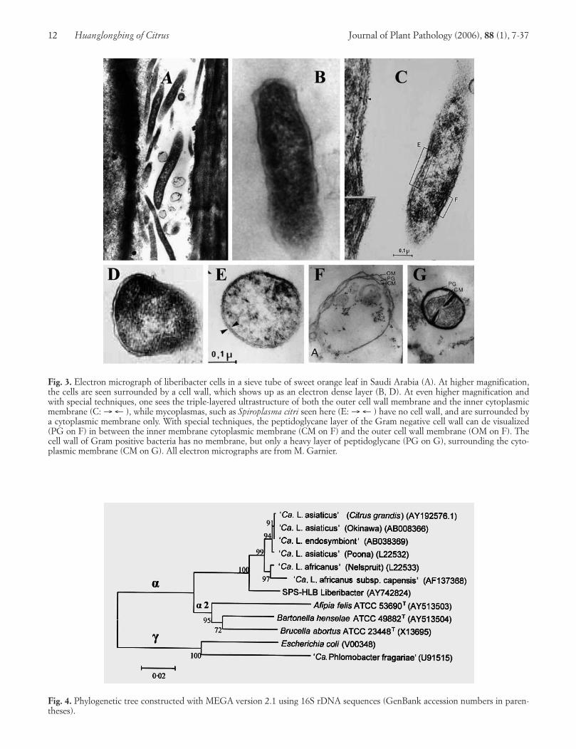
In both cases, micro-organisms were detected in thephloem sieve tubes of symptomatic leaves, but not inthose of healthy leaves. In the case of stubborn, the mi-cro-organism was soon obtained in culture, and turnedout to be a new type of mycoplasma, one with helicalmorphology and motility, and named Spiroplasma citri(Saglio et al., 1973). In the case of the greening organ-ism, which could not be obtained in culture, it was ini-tially thought that it was also a mycoplasma (Laflècheand Bové, 1970a, b), but the agent was soon found to beenclosed by a 25-nm-thick envelope, which was muchthicker than the 7 to 10-nm-thick cytoplasmic mem-brane envelope, characteristic of mycoplasmas, includ-ing S. citri (Fig. 3 E) (Saglio et al., 1971; Garnier et al.,1976). These properties suggested that the HLB organ-ism possessed, in addition to its cytoplasmic membrane,a bacterial cell wall. This was indeed found to be thecase, and it could be shown that the cell wall was of theGram negative type, composed of a membrane and apeptidoglycane layer (Garnier and Bové, 1977; Garnieret al., 1984a). Thus, the HLB agent was a Gram nega-tive bacterium (Garnier et al., 1984b). As shown byGarnier and Bové (1977), in classic specimen fixationprocedures for EM, the cell wall of the HLB bacteriumappears as a characteristic, electron-dense layer sur-rounding the bacterial cell (Fig. 3 B, D), and this suf-fices for HLB identification. With special fixation meth-ods, cell membranes reveal their triple layered ultra-structure, and the HLB bacterium is seen as surroundedby two such triple layered membranes (Fig. 3 C), i.e. theinner, cytoplasmic membrane, and the outer membrane,as part of the Gram negative cell wall (Garnier andBové, 1977; see also Moll and Martin, 1974). Under
Journal of Plant Pathology (2006), 88 (1), 7-37 Bové 11
12 Huanglongbing of Citrus Journal of Plant Pathology (2006), 88 (1), 7-37
Fig. 3. Electron micrograph of liberibacter cells in a sieve tube of sweet orange leaf in Saudi Arabia (A). At higher magnification,the cells are seen surrounded by a cell wall, which shows up as an electron dense layer (B, D). At even higher magnification andwith special techniques, one sees the triple-layered ultrastructure of both the outer cell wall membrane and the inner cytoplasmicmembrane (C: → ← ), while mycoplasmas, such as Spiroplasma citri seen here (E: → ← ) have no cell wall, and are surrounded bya cytoplasmic membrane only. With special techniques, the peptidoglycane layer of the Gram negative cell wall can de visualized(PG on F) in between the inner membrane cytoplasmic membrane (CM on F) and the outer cell wall membrane (OM on F). Thecell wall of Gram positive bacteria has no membrane, but only a heavy layer of peptidoglycane (PG on G), surrounding the cyto-plasmic membrane (CM on G). All electron micrographs are from M. Garnier.
Fig. 4. Phylogenetic tree constructed with MEGA version 2.1 using 16S rDNA sequences (GenBank accession numbers in paren-theses).
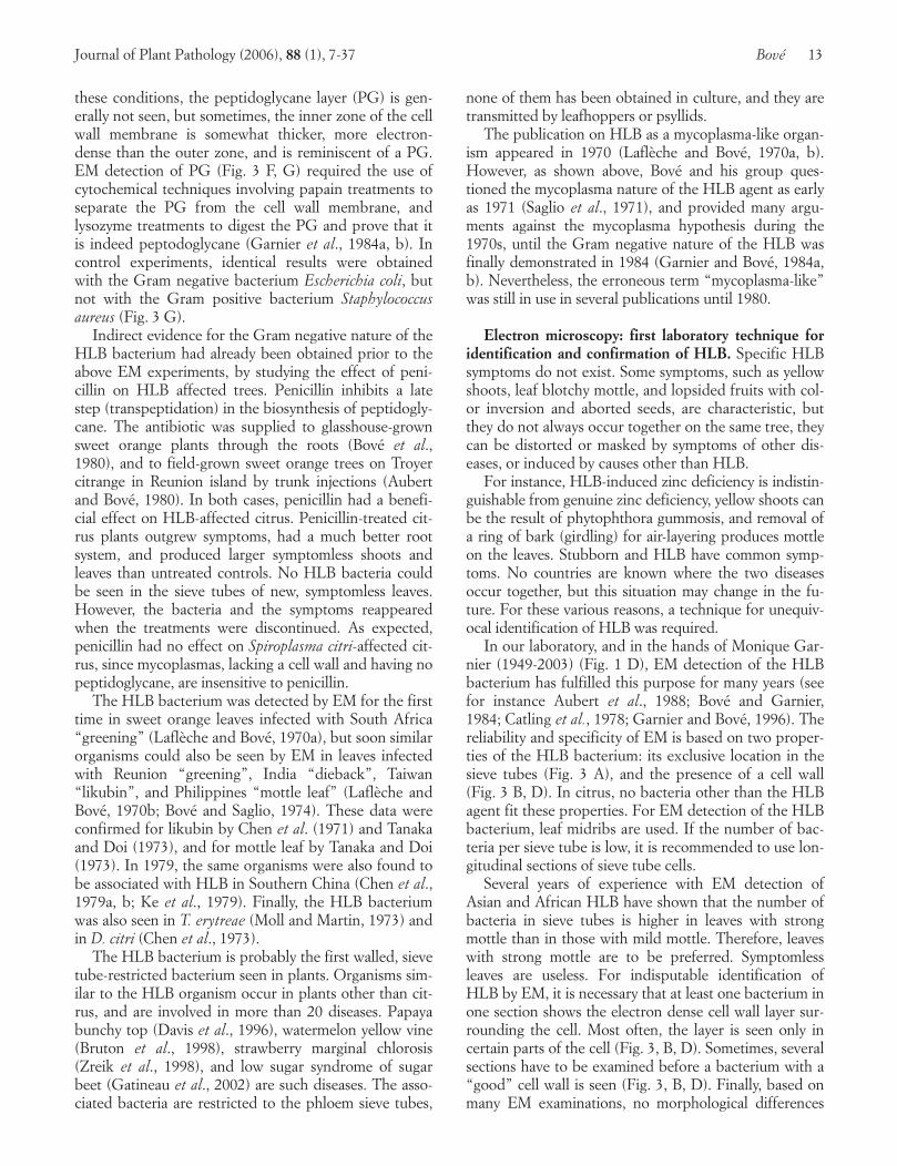
these conditions, the peptidoglycane layer (PG) is gen-erally not seen, but sometimes, the inner zone of the cellwall membrane is somewhat thicker, more electron-dense than the outer zone, and is reminiscent of a PG.EM detection of PG (Fig. 3 F, G) required the use ofcytochemical techniques involving papain treatments toseparate the PG from the cell wall membrane, andlysozyme treatments to digest the PG and prove that itis indeed peptodoglycane (Garnier et al., 1984a, b). Incontrol experiments, identical results were obtainedwith the Gram negative bacterium Escherichia coli, butnot with the Gram positive bacterium Staphylococcusaureus (Fig. 3 G).
Indirect evidence for the Gram negative nature of theHLB bacterium had already been obtained prior to theabove EM experiments, by studying the effect of peni-cillin on HLB affected trees. Penicillin inhibits a latestep (transpeptidation) in the biosynthesis of peptidogly-cane. The antibiotic was supplied to glasshouse-grownsweet orange plants through the roots (Bové et al.,1980), and to field-grown sweet orange trees on Troyercitrange in Reunion island by trunk injections (Aubertand Bové, 1980). In both cases, penicillin had a benefi-cial effect on HLB-affected citrus. Penicillin-treated cit-rus plants outgrew symptoms, had a much better rootsystem, and produced larger symptomless shoots andleaves than untreated controls. No HLB bacteria couldbe seen in the sieve tubes of new, symptomless leaves.However, the bacteria and the symptoms reappearedwhen the treatments were discontinued. As expected,penicillin had no effect on Spiroplasma citri-affected cit-rus, since mycoplasmas, lacking a cell wall and having nopeptidoglycane, are insensitive to penicillin.
The HLB bacterium was detected by EM for the firsttime in sweet orange leaves infected with South Africa“greening” (Laflèche and Bové, 1970a), but soon similarorganisms could also be seen by EM in leaves infectedwith Reunion “greening”, India “dieback”, Taiwan“likubin”, and Philippines “mottle leaf” (Laflèche andBové, 1970b; Bové and Saglio, 1974). These data wereconfirmed for likubin by Chen et al. (1971) and Tanakaand Doi (1973), and for mottle leaf by Tanaka and Doi(1973). In 1979, the same organisms were also found tobe associated with HLB in Southern China (Chen et al.,1979a, b; Ke et al., 1979). Finally, the HLB bacteriumwas also seen in T. erytreae (Moll and Martin, 1973) andin D. citri (Chen et al., 1973).
The HLB bacterium is probably the first walled, sievetube-restricted bacterium seen in plants. Organisms sim-ilar to the HLB organism occur in plants other than cit-rus, and are involved in more than 20 diseases. Papayabunchy top (Davis et al., 1996), watermelon yellow vine(Bruton et al., 1998), strawberry marginal chlorosis(Zreik et al., 1998), and low sugar syndrome of sugarbeet (Gatineau et al., 2002) are such diseases. The asso-ciated bacteria are restricted to the phloem sieve tubes,
none of them has been obtained in culture, and they aretransmitted by leafhoppers or psyllids.
The publication on HLB as a mycoplasma-like organ-ism appeared in 1970 (Laflèche and Bové, 1970a, b).However, as shown above, Bové and his group ques-tioned the mycoplasma nature of the HLB agent as earlyas 1971 (Saglio et al., 1971), and provided many argu-ments against the mycoplasma hypothesis during the1970s, until the Gram negative nature of the HLB wasfinally demonstrated in 1984 (Garnier and Bové, 1984a,b). Nevertheless, the erroneous term “mycoplasma-like”was still in use in several publications until 1980.
Electron microscopy: first laboratory technique foridentification and confirmation of HLB. Specific HLBsymptoms do not exist. Some symptoms, such as yellowshoots, leaf blotchy mottle, and lopsided fruits with col-or inversion and aborted seeds, are characteristic, butthey do not always occur together on the same tree, theycan be distorted or masked by symptoms of other dis-eases, or induced by causes other than HLB.
For instance, HLB-induced zinc deficiency is indistin-guishable from genuine zinc deficiency, yellow shoots canbe the result of phytophthora gummosis, and removal ofa ring of bark (girdling) for air-layering produces mottleon the leaves. Stubborn and HLB have common symp-toms. No countries are known where the two diseasesoccur together, but this situation may change in the fu-ture. For these various reasons, a technique for unequiv-ocal identification of HLB was required.
In our laboratory, and in the hands of Monique Gar-nier (1949-2003) (Fig. 1 D), EM detection of the HLBbacterium has fulfilled this purpose for many years (seefor instance Aubert et al., 1988; Bové and Garnier,1984; Catling et al., 1978; Garnier and Bové, 1996). Thereliability and specificity of EM is based on two proper-ties of the HLB bacterium: its exclusive location in thesieve tubes (Fig. 3 A), and the presence of a cell wall(Fig. 3 B, D). In citrus, no bacteria other than the HLBagent fit these properties. For EM detection of the HLBbacterium, leaf midribs are used. If the number of bac-teria per sieve tube is low, it is recommended to use lon-gitudinal sections of sieve tube cells.
Several years of experience with EM detection ofAsian and African HLB have shown that the number ofbacteria in sieve tubes is higher in leaves with strongmottle than in those with mild mottle. Therefore, leaveswith strong mottle are to be preferred. Symptomlessleaves are useless. For indisputable identification ofHLB by EM, it is necessary that at least one bacterium inone section shows the electron dense cell wall layer sur-rounding the cell. Most often, the layer is seen only incertain parts of the cell (Fig. 3, B, D). Sometimes, severalsections have to be examined before a bacterium with a“good” cell wall is seen (Fig. 3, B, D). Finally, based onmany EM examinations, no morphological differences
Journal of Plant Pathology (2006), 88 (1), 7-37 Bové 13
could be found to distinguish the Asian HLB bacteriumfrom his African counterpart.
VARIOUS FORMS OF HLB: ASIAN, AFRICAN, ANDAMERICAN
Symptoms of citrus stubborn disease and HLB areinfluenced by temperatures at which affected treesgrow. Symptoms of stubborn are severe in hot, dry areasof California, Arizona, Morocco, Iran, and Iraq. InSouth Africa, HLB and the African psyllid vector, T.erytreae, occur in the cool, “highveld” areas of Transvaaland Swaziland, but not in the hotter,” lowveld” areas ofSwaziland. However, HLB and T. erytreae are present inthe Cape Town region (Garnier et al., 2000a), where thesouthern latitude compensates for the low altitude, andis responsible for a temperate climate.
In Madagascar, HLB and T. erytreae only occur onthe central, high plateau (1200 to 1500 m a.s.l.), but noton the low lying coastal areas. In Kenya, HLB and T.erytreae are not seen below an altitude of 600 to 700m.On the contrary, in Asia, HLB and the Asian psyllidvector, D. citri, are found in hot low altitude areas. Forinstance, in Indonesia, in 1984, HLB and D. citri werepresent in the whole of Java coastal lowlands, but manytrees in orchards located between 800 and 1200 m a.s.l.were still healthy (Aubert et al., 1985). In North Bali,HLB was commonly present below an altitude of 650m, but was rarely seen above 1000 m, and its distribu-tion reflected that of D. citri (Bové et al., 2000b). Simi-larly, in the Ningnan county of Sichuan (South China),100% of trees were affected by HLB in orchards locat-ed at altitudes of 1090 to 1200 m, where D citri wasabundant, while between 1385 and 1620 m, the per-centage went down to 3% and no psyllids could befound (Zhao, 1981).
In the frame of an international cooperation experi-ment started in 1969, the influence of temperature onHLB symptoms was also studied under phytotron condi-tions in two temperature controlled chambers: a “cool”chamber (24°C with a 16-hr light period and 22°C with a8-hr dark period) and a “warm” chamber (32°C with a16-hr light period and 27°C with a 8-hr dark period)(Bové et al., 1974). Sweet orange plants graft-inoculatedwith the following HLB sources were used: South Africa(Nelspruit) “greening”, India (Poona) “dieback”, andPhilippines (Lipa City) “mottle leaf”. The presence ofHLB bacteria in inoculated plants was established by EM.
With South African HLB, severe symptoms were ob-tained after 30 weeks in the cool chamber, and the plantsaveraged 35 cm in height; no symptoms developed in thewarm chamber, and the plants reached 190 cm. When,after 40 weeks in the cool chamber, severely affectedplants were transferred to the warm conditions, theyquickly produced new, vigorous growth, recovered, and
remained symptomless during the remaining 10 monthsof the experiment. With both Indian HLB and Philip-pine HLB, symptoms were as pronounced at 22-24°C asat 27-32°C. After 30 weeks in the warm chamber, symp-tomatic seedlings infected with Indian HLB and Philip-pine HLB measured 25 and 40 cm, respectively, whilehealthy controls had grown to 180 and 140 cm.
This experiment as well as the field observations showthat HLB in Africa is heat-sensitive, and occurs only incool areas, with temperatures remaining below 30-32°C.Similarly, the African psyllid vector, T. erytreae, thrivesonly in cool environments, and is also sensitive to hightemperature combined with low relative humidity(Catling, 1969c). On the contrary, HLB in Asia is heat-tolerant, and symptoms occur even when temperaturesare well above 30°C. The Asian psyllid vector, D. citri,has similar properties, and is also heat-tolerant.
The South African Nelspruit strain, and the IndianPoona strain of the HLB bacterium could be transmittedto periwinkle (Catharanthus roseus) plants (see below).Temperature experiments conducted with infected peri-winkle plants gave the same results than those obtainedabove with HLB-affected citrus seedlings: with the Indi-an HLB bacterium, symptom expression occurred atboth 25 and 32°C, but only at 25°C with the SouthAfrican strain (Garnier and Bové, 1983). The effect oftemperature on symptom expression is thus the same inperiwinkle and citrus. Therefore, the temperature effectis due to the HLB bacterium and not to the plant, theAfrican HLB bacterium being heat sensitive and theAsian HLB bacterium, heat tolerant. This biological dif-ference indicates that the African and Asian HLB bacte-ria are not identical. Indeed, as shown below, they repre-sent different bacterial species: Candidatus Liberibacterafricanus and Candidatus Liberibacter asiaticus .
The fact that the African HLB bacterium and T. ery-treae are both heat-sensitive, and the Asian HLB bac-terium and D. citri, both heat-tolerant, seems to be agood example of adaptation between vector andpathogen. It has to be remembered however, that, ex-perimentally at least, each one of the two vectors cantransmit each one of the two HLB bacterial species(Massonié et al., 1976; Lallemand et al., 1986).
In the State of São Paulo, Brazil, Ca L. asiaticus hasbeen found in less than 10% of the HLB-affected trees.Indeed, a third bacterial species has been discovered:Candidatus Liberibacter americanus (see below), whichaffects more than 90% of the trees. In São Paulo State,the Asian psyllid vector, D. citri, was reported as early as1942, and transmits not only Ca L. asiaticus, but probablyalso Ca L. americanus. Indeed, the new liberibacter couldbe detected by PCR within D. citri psyllids collected onCa L. americanus-infected trees. Furthermore, in theglasshouse, sweet orange seedlings, graft-inoculated withCa L. americanus, showed severe HLB leaf mottle at bothcool (22-24°C) and warm (27-32°C) conditions (Teixeira
14 Huanglongbing of Citrus Journal of Plant Pathology (2006), 88 (1), 7-37

et al., 2005c). These observations suggest that the SouthAmerican (Brazil) HLB is of the heat-tolerant form.
In Florida, the presence of D. citri was reported in1998 (Halbert, 1998), HLB has been observed in theSouthern part of the State in August 2005, and only CaL. asiaticus has been detected in HLB-affected trees.Therefore, the North American (USA) HLB is probablyalso of the heat-tolerant form.
TRANSMISSION OF THE HLB BACTERIUM FROMCITRUS TO PERIWINKLE BY DODDER
In citrus, the HLB bacterium is present only in smallnumbers, but in dodder (Cuscuta campestris) the organ-ism reaches high titers (Ghosh et al.,1977). Periwinkle(Catharanthus roseus) is a host plant in which also manymycoplasma-like organisms multiply actively in sievetubes. These observations suggested to attempt transmis-sion of the HLB bacterium from citrus to periwinklethrough dodder. Positive transmissions were indeed ob-tained (Garnier and Bové, 1983). In the case of IndianHLB, one of four periwinkle plants connected via dod-der to a HLB-affected sweet orange seedling developedpeculiar yellowing symptoms, and contained HLB-likebacteria in sieve tubes, some of which were particularlyrich in bacteria. Shoots from the symptomatic periwinklewere top-grafted onto healthy periwinkle plants, whichdeveloped the characteristic symptoms after 3 months at25°C. In this way, large numbers of symptomatic, HLB-infected periwinkle plants were produced. Similar re-sults were obtained with South African, Chinese, andPhilippine strains of the HLB organism. It must bestressed that periwinkle plants naturally infected withthe HLB agent have never been observed. Citrus psyllidsdislike periwinkle, are unable to transmit the HLB agentto this plant, and die when forced to stay on it.
Ultrathin sections from HLB-infected periwinkleplants were used in EM to demonstrate the Gram nega-tive nature of the bacterium (Garnier et al., 1984a, b)(see above). As the HLB bacterium is not available inculture, great numbers of infected periwinkles have beenused as convenient sources for the phylogenetic and tax-onomic characterization of the HLB bacterium, the pro-duction of monoclonal antibodies, and the developmentof molecular detection techniques (see below).
Transmission of the HLB bacterium to tobacco(Nicotiana tabacum Xanthi) by dodder has also been ob-tained (Garnier and Bové, 1993).
PHYLOGENETIC AND TAXONOMIC CHARACTERIZA-TION OF THE AFRICAN AND ASIAN HLB BACTERIA
16S rDNA. To determine the phylogenetic positionof the HLB bacterium, the 16S ribosomal DNAs (16S
rDNA) of the South African Nelspruit strain and theAsian Poona strain were obtained from total DNA ofHLB-infected periwinkle plants, by polymerase chainreaction (PCR)-amplification, using the universal PCR-primers f-D1/r-P1 for amplification of prokaryotic 16SrDNA (Weisburg et al., 1991). In these experiments,care must be taken to prevent interference by periwin-kle mitochondrial and chloroplast 16S rDNAs. Thiscan be achieved by cutting mitochondrial 16S rDNAwith endonuclease Bcl1 before PCR amplification. Theamplified DNA, about 1500 base pairs (bp) in size,contains bacterial as well as chloroplast 16S rDNA.The presence of bacterial DNA can be ascertained byEcoRI, which cuts it into two fragments (~650bp and~850bp), while chloroplast DNA is untouched. The16S rDNAs were cloned from the ~1500bp amplifiedproducts, and sequenced. Hybridization and PCR ex-periments performed with oligonucleotides specific forthe amplified sequences revealed that the DNAs ob-tained were indeed the 16S rDNAs of the HLB bacte-ria, and not the DNA of a contaminating organism(Jagoueix et al., 1994).
Comparisons with 16S rDNA sequences obtainedfrom the GeneBank database showed that the Africanand Asian HLB bacteria belonged to the α subdivisionof the class Proteobacteria. Even though their closest rel-atives were members of the α-2 subgroup, the HLBbacteria were distinct from this subgroup, as the level of16S rDNA sequence identity was only 87.5%. There-fore, the two HLB bacteria represented a new lineage inthe α subdivision of the class Proteobacteria. As themembers of this class are Gram negative bacteria, theseresults confirmed the Gram negative nature of the HLBbacterium, a property which had already been deducedfrom earlier EM studies by Garnier et al. (1984a, b).
The α subdivision of Proteobacteria is a diversegroup of microbes that includes both plant pathogensor symbionts with some distinctive properties (Agrobac-terium tumefaciens, Bradyrhizobium spp.) and humanpathogens (Rochalimea spp., Bartonella baciliformis,Brucella abortus, Afipia spp., etc). Organisms in thisgroup live in intimate association with eukariotic cellsand, in many cases, have acquired the ability to surviveand grow within an arthropod vector. The HLB organ-ism fits this description remarkably well. Indeed, itgrows in a specialized niche in its eukariotic plant host,the phloem sieve tubes, and it is transmitted by twoarthropod vectors, the psyllids T. erytreae and D. citri, inwhich it multiplies in the hemolymph and within thesalivary glands.
RplKAJL-rpoBC operon and DNA probes. Using to-tal DNA from periwinkle plants infected with the AsianPoona (India) strain of the HLB bacterium, severalDNA fragments of the bacterial genome could be ob-tained by random cloning (Villechanoux et al., 1992).
Journal of Plant Pathology (2006), 88 (1), 7-37 Bové 15
Fragment In-2.6 (2.6 kbp), when used as a probe inSouthern- or dot-hybridisations, hybridised at highstringency with all tested strains of the Asian HLB bac-terium, but not with the African strain. At low or inter-mediate stringency, some hybridisation was also seenwith the African strain. The Asian strains that gave posi-tive hybridisation signals with probe Poona In-2.6 werethose from Poona (India) (homologous strain), NakhomPathom (Thailand), Lipa city (Philippines), Fujian (Chi-na), Taiwan, and Bali (Indonesia).
By sequencing, In-2.6 was found to be part of therplKAJL-rpoBC gene cluster, the well-known, bacterialβ-operon, which codes for ribosomal proteins K, A, J,and L, and RNA polymerase subunits β and β’ (Vil-lechanoux et al., 1993). From the sequence of In-2.6,two PCR primers were designed, f-1898 and r-1897, andused to amplify part of the rplKAJL-rpoBC operon ofthe African Nelspruit HLB bacterium (Planet et al.,1995). A clear DNA band of about 1700 bp was ob-tained. Upon cloning and sequencing, the DNA fromthe African strain (1676 bp) was indeed found to corre-spond to part of the expected β-operon. It was calledAS-1.7. The AS-1.7 DNA hybridised at high stringencywith DNA from periwinkle or citrus plants infectedwith the African Nelspruit strain, but no hybridisationwas observed in the case of the Asian strains tested(Planet et al., 1995). As indicated above, opposite re-sults were obtained when In-2.6 was used as the probe:no hybridisation with the African strain, but strong hy-bridisation with all Asian strains tested. The overall nu-cleotide identity between In-2.6 and AS-1.7 was 74.2%.This relatively low homology for similar organisms ex-plains why no hybridisation was observed between In-2.6 and DNA from periwinkle plants infected with theAfrican Nelspruit HLB bacterium, and vice versa. Thislow homology also suggested that the African strains ofthe HLB bacterium and the Asian strains were membersof two different species of the same genus (Jagoueix etal., 1994; Planet et al., 1995).
Monoclonal Antibodies. Since 1987, only thirteendifferent monoclonal antibodies (MA) specific for theHLB bacteria have been produced (Garnier et al., 1991;Gao et al., 1993). This low number is explained by thefact that the HLB bacteria are not available in culture.The first ten MAs were raised using as immunogen ho-mogenates of phloem tissue from HLB-affected peri-winkle plants. Of these MAs, two (including MA 10A6)were against the Indian Poona strain, five against astrain from China (Fujian), and three against the SouthAfrican Nelspruit strain. The use of these MAs for thedetection of HLB-bacteria has shown that each MA isvery specific for the strain used for immunisation and,therefore, they cannot be used for generalized diagnosisof HLB (Garnier et al., 1991).
In attempting to produce antibodies recognizing most
or all strains of the HLB bacterium, an antigenic proteinof the Indian Poona strain was purified by immunoaffin-ity-chromatography using MA 10A6 directed against thisprotein, and used for in vitro immunisation of spleencells (Gao et al., 1993). Three MAs were obtained, oneof which (1A5) recognized all Asian strains tested exceptthe Chinese one, whereas the other two recognized mostof the Asian, but not the Chinese strain. None of thethree MAs reacted with the South African strain. Theseresults agreed with those obtained with DNA probes,confirming that Asian and African HLB bacteria aremembers of two different bacterial species.
MA 10A6, coupled to CNBr-activated sepharose 4B,has also been successfully used to purify the PoonaBLO by immunoaffinity (Villechanoux et al., 1990). Inthis way, purified cells of the HLB bacterium could beobserved for the first time in the electron microscope
Candidatus genera and species: Candidatus Liberi-bacter africanus and Candidatus Liberibacter asiaticus.Bacteriologists have had a conservative attitude when itcame to give Latin binomial names to non-cultured or-ganisms. However, with the development of DNA am-plification by PCR and DNA sequencing, it became pos-sible to characterise such organisms at the molecular andphylogenetic level. On the basis of such considerations,Murray and Schleifer (1994) proposed the “Candidatus”designation as an interim taxonomic status, to provide aproper allocation of sequence-based potential new taxaat the genus and species level. One of the first non-cul-tured bacteria to benefit from the Candidatus proposalwas the HLB bacterium, shown by 16S rDNA sequencecomparisons to be the first member of a new subgroupin the α subdivision of the Proteobacteria (see above).The trivial name liberobacter (Jagoueix et al., 1994), lat-er replaced by liberibacter (Garnier et al., 2000b) (fromthe Latin liber [bark} and bacter [bacterium]), was givento organisms in this new subgroup.
As indicated above, the HLB liberibacter strainsfrom Africa can be distinguished from those in Asia onthe basis of temperature sensitivity (Bové et al., 1974),DNA hybridizations and genomic properties (Vil-lechanoux et al., 1992, 1993), and serology (Garnier etal., 1991; Gao et al., 1993). For these reasons, they rep-resent two different species. Therefore, following theCandidatus proposal of Murray and Schleifer, the HLBliberibacters from Asia should be denoted CandidatusLiberibacter asiaticus, and the HLB liberibacters fromAfrica Candidatus Liberibacter africanus (Jagoueix etal., 1994; Garnier et al., 2000b).
The sequence variability of the region between the 16SrRNA gene and the 23S rRNA gene in the ribosomaloperons is useful for differentiating species within gen-era. This 16S/23S intergenic region was obtained for twoAsian strains of the HLB liberibacter, the Indian Poonastrain and a strain from Fuzhou, Sichuan (China), as well
16 Huanglongbing of Citrus Journal of Plant Pathology (2006), 88 (1), 7-37

as for the African Nelspruit liberibacter (Jagoueix et al.,1997). The intergenic regions of the two Asian liberibac-ters had 100% sequence identity, even though they be-long to different serotypes and were isolated from geo-graphically distant areas. However, the sequence identitybetween an Asian liberibacter strain and the Africanliberibacter was only 79.46%, confirming the fact thatthe African liberibacter and the Asian liberibacter be-long to two different species. Similar results have alsobeen obtained by Subandiyah et al. (2000).
Candidatus Liberibacter africanus subsp. capensis.A third liberibacter was detected by PCR in an orna-mental rutaceous tree, Cape chestnut (Calodendrumcapense), in the Cape region of South Africa. Leaves ofthe affected tree showed characteristic mottling. Thenew liberibacter was characterized by serology and thesequences of its 16S rDNA, the intergenic 16S/23S rDNA region, and ribosomal protein genes of the βoperon. Phylogenetic analysis showed the new liberib-acter to be more closely related to Ca. L. africanus thanto Ca. L. asiaticus, and a subspecies status was assignedto it: Candidatus Liberibacter africanus subsp. capensis(Garnier et al., 2000b).
16S rDNA-based phylogeny tree. Fig. 4 shows the16S rDNA-based phylogeny tree of liberibacters. Thetree was constructed from 16S rDNA sequences ob-tained from GenBank. It can be seen that all four Ca. L.asiaticus strains cluster together, and the “asiaticus”cluster is close to the “africanus” cluster. The “asiati-cus” cluster and the “africanus” cluster form the “asiati-cus”/“africanus” liberibacter group. The new liberibac-ter from São Paulo State, Ca. L americanus (see below),is not part of this group, but forms a separate branch,indicating that it is a species different from Ca. L. asiati-cus and Ca. L. africanus. All liberibacters are membersof the α subdivision of the class Proteobacteria, andtheir closest relatives are members of the α2 subgroup.The tree also shows two members of the γ subdivision.
Recently, a phylogenetic tree was constructed fromthe omp gene sequences of the African and Asianliberibacters, and confirmed the phylogeny based on16S rDNA sequences (Bastianel et al., 2005). The ompgene sequences were also used to study the diversity ofseveral strains of Ca. L. asiaticus. Each strain could becharacterised by a specific PCR-RFLP profile.
IDENTIFICATION OF HLB BY DETECTION OF THEAFRICAN AND ASIAN LIBERIBACTERS
Until 1992, electron microscopy visualization of thewalled HLB organisms in the sieve tubes of citrus leavesshowing blotchy mottle was the only reliable method ofdetection, and was widely used (Garnier and Bové,
1996). However, the technique was heavy, and unable todistinguish between Asian and African liberibacters.This differentiation has now become possible with thedevelopment of molecular techniques such as DNA hy-bridization and PCR. As mentioned, monoclonal anti-bodies are too specific to be used for diagnosis.
DNA Hybridisation. In dot hybridisation, probe In-2.6 (see above) gives positive hybridisation signals withDNA isolated from citrus leaves infected with Asianliberibacter strains, while probe AS 1.7 reacts positivelywith African liberibacter strains (Villechanoux et al.,1992, 1993; Planet et al., 1995). These probes can alsobe used very efficiently to detect liberibacters in psyllidinsect vectors. Individual insects are crushed onto a ny-lon membrane and the membrane or “crush-blot” issubmitted to hybridisation with one or the other probe(Bové et al., 1993) Non-radioactive probes have beendeveloped (Hocquellet et al., 1997).
PCR. Two PCR systems have been used. The first isbased on the amplification of a 1160 bp fragment ofliberibacter 16S rDNA (Jagoueix et al., 1996). Theprimer pair OI1/OI2c is able to amplify the rDNA ofboth liberibacter species, while the pair OA1/OI2c am-plifies preferentially the African liberibacter rDNA. Incountries were the two liberibacter species are knownor suspected to be present, it is advisable to use the twoforward primers, OI1 + OA1, and the common reverseOI2c primer in the same PCR mixture. Sequence analy-sis shows that the rDNA amplified from the Asianliberibacter has one Xba1 restriction site, and yields, up-on Xba1 treatment, two fragments 520 bp and 640 bp.in size, respectively. The rDNA amplified from theAfrican liberibacter has an additional site, and yieldsthree fragments with a size of 520 bp, 506 bp, and 130bp. Hence, by Xba1 treatment of the amplified DNA, itis easy to identify the liberibacter species present in agiven sample (Jagoueix et al., 1996).
The second PCR system is based on the sequence ofthe rplKAJL-rpoBC operon, which is slightly differentfrom one liberibacter species to the other. In particular,the intergenic region between genes rplA and rplJ is 34bp larger in the Asian than in the African liberibacter.With forward primer f-rplA2, selected in the rplA gene,and reverse primer r-rplJ5 from the rplJ gene, a 703 bpDNA is amplified from the Asian liberibacter, while a669 bp DNA is obtained with the African liberibacter.When both liberibacter species are present in the samesample, amplification of the two DNAs is obtained, andupon agarose gel electrophoresis, two DNA bands areseen, the upper (703 bp) corresponding to the Asianliberibacter, and the lower (669 bp), to the Africanliberibacter (Hocquellet et al., 1999). Finally, a PCR as-say was developed, based on the β operon sequence andspecific for the detection of Candidatus Liberibacter
Journal of Plant Pathology (2006), 88 (1), 7-37 Bové 17

africanus subsp. capensis (Garnier et al., 2000b).By the use of these molecular techniques, the pres-
ence of HLB has been clearly established in severalAfrican and Asian countries (Bové et al., 1993; Bové etal., 1996; Bové et al., 2000b; Doe Doe et al., 2003; Gar-nier and Bové, 1996; Garnier and Bové, 2000; Garnieret al., 2000a; Korsten et al., 1996; Regmi et al., 1996;Varma et al., 1993). The presence of both liberibacterspecies, sometimes in the same trees, was confirmed inReunion and Mauritius islands (Garnier et al., 1996).
HLB IN SÃO PAULO STATE, BRAZIL: OLD AND NEWLIBERIBACTERS
Even though HLB was reported in São Paulo State(SPS) only in 2004, the Asian psyllid vector of HLB, Dcitri, has been present in Brazil at least since 1942, whenit was reported for the first time (Lima, 1942). Speci-mens of D. citri from Brazil were present at the BritishMuseum in 1969 (Eastop, 1969, personal communica-tion to Catling, 1970). In certain years, high populationsof the insect could be seen in SPS in sweet orangegroves.
Discovery of a new liberibacter: Candidatus Liberi-bacter americanus. In March 2004, symptoms of HLBwere observed in sweet orange trees near the city ofAraraquara in SPS (Anonymous, 2004; Coletta-Filho etal, 2004; Ayres et al., 2005; Teixeira et al., 2005a, b). Thiswas the first reported case of HLB from the AmericanContinent. Affected trees could be spotted from a dis-tance because of their conspicuous yellow shoots. Leafsymptoms of HLB in SPS were very similar, if not identi-cal, to those in Africa and Asia, with characteristic andpronounced blotchy mottle, as described by McCleanand Schwarz (1970), on both small and large leaves (Fig.6, 7). Fruits were small and lopsided, exhibited strongcolour inversion, seed abortion, and brown/orange-stained vascular bundles (Fig. 8, 9). These fruit symp-toms were more similar to those seen in China than tothose observed in South Africa. A survey conducted inSeptember 2004, only six months after the disease wasrecognized, showed HLB to be already present in 46 mu-nicipalities of SPS. A few orchards had more than 50%of affected trees. These observations suggested that thedisease had been present for almost ten years, but with-out being properly diagnosed. History repeats itself…
In April 2004, the 16S rDNA-based PCR techniquewith forward primers OA1+OI1 and reverse primerOI2c, described by Jagoueix et al. (1996), was applied tosweet orange leaves with strong mottle from suspicioustrees in SPS to confirm the presence of HLB in the areaand identify the liberibacter involved. This PCR methodhad been assayed in many Asian and African countriesfor the detection of the two HLB liberibacters (see for
instance Bové et al., 1996, 2000b; Garnier and Bové,1996; Garnier et al., 1996). Whenever leaves with theclassic blotchy mottle symptoms were used, positivePCR reactions were obtained, and yielded the character-istic 1160 bp amplicon. Unexpectedly in SPS, only nega-tive PCR reactions were obtained from mottled leaf sam-ples from 43 affected trees, many of which had also se-vere fruit symptoms. Under the same testing conditionssymptomatic control citrus leaves infected with Ca. L.asiaticus or Ca. L. africanus from the HLB collection inBordeaux, gave positive PCR reactions (Teixeira et al.,2005a, b). However, at the same time, and using practi-cally the same PCR technique, Ca. L. asiaticus was de-tected in 2 of 10 leaf samples by other workers (Coletta-Filho et al., 2004). Because of the repeatedly negativePCR reactions, the presence of a new bacterial pathogenin many of the symptomatic, blotchy mottle leaves fromSPS was suspected and investigated.
Evidence for the presence of a new HLB bacterium(SPS-HLB bacterium) was obtained by PCR amplifica-tion with universal primers fD1 and rP1 for prokaryotic16S rDNA amplification. The 16S rDNA of the SPS-HLB bacterium was cloned and sequenced, and the se-quence was used to design the specific primer pair f-GB1/r-GB3. PCR amplification with these primers madeit possible to detect the SPS-HLB bacterium in all HLB-leaf samples testing negative for Ca. L. africanus and Ca.L. asiaticus. Most leaf samples (98%) were infected withthe SPS-HLB bacterium, the remaining samples (2%)carried Ca. L. asiaticus. Primer GB3c, complementary toGB3, was used in conjunction with reverse primer 23S1to amplify, clone, and sequence the 16S/23S ribosomalintergenic region (RIR). In total, a 1479 bp sequence of16S rDNA (almost the complete 16S rDNA sequence),followed by the complete 583 bp sequence of the RIR,was available for characterization of the SPS-HLB bac-terium by comparison with similar sequences of variousisolates of Ca. L. asiaticus, and the Nelspruit isolate ofCa. L. africanus (Teixeira et al., 2005c).
These comparisons clearly showed that the SPS-HLBbacterium was not only a member of the genus Candida-tus Liberibacter, having all the oligonucleotide signa-tures of the liberibacters, but was a new species of thisgenus. In particular, in the 16S rDNA phylogenetic tree(Fig. 4), all isolates of Ca. L. asiaticus clustered togetherwithin the Ca. L. africanus/Ca. L. asiaticus group, butthe SPS-HLB bacterium did not, and formed a separatebranch. Also, the RIR sequences of different isolates ofCa. L. asiaticus were identical or almost identical (99 to100% identity). However, the RIR of the SPS-HLB bac-terium and that of Ca. L. asiaticus had only 78% se-quence identity. With Ca. L. africanus, the sequenceidentity was even lower: 66%.
For the above reasons the SPS-HLB bacterium wasdenoted Candidatus Liberibacter americanus, sp. nov.(Teixeira et al., 2005a, b, c). This designation refers to
18 Huanglongbing of Citrus Journal of Plant Pathology (2006), 88 (1), 7-37
the fact that the new liberibacter species was detectedfor the first time in the American Continent, and that itrepresented the major Ca. Liberibacter species associat-ed with HLB in the affected SPS region. The designa-tion is in line with the other Ca. Liberibacter names,which also refer to the continents where they occur:Africa for Ca. L. africanus, and Asia for Ca. L. asiaticus.
Additional properties of the SPS-HLB bacterium fitthose of the other two liberibacters. Transmission tohealthy orange seedlings by graft inoculation was ob-tained, and EM obervations showed the Americanliberibacter to be restricted to sieve tubes (Teixeira etal., 2005c; Tanaka et al., 2004).
PCR detection of Ca. L. americanus and Ca. L. asi-aticus in citrus leaves. For the specific PCR detection ofCa. L. americanus, forward primer GB1 and reverseprimer GB3 were designed from the 16S rDNA se-quence of the new liberibacter (accession numberAY742824). These primers, as well as those specific forCa. L. africanus and Ca. L. asiaticus (OA1+OI1/OI2c),were used for detection of the three liberibacters ineach leaf sample according to Teixeira et al. (2005b). Afirst aliquot of the DNA from a leaf sample was used forthe detection of Ca. L. americanus with primers GB1/ GB3, yielding an amplicon of 1027 bp, and a secondaliquot served for the detection of Ca. L. africanus andCa. L. asiaticus with primers OA1+OI1/OI2c, giving anamplicon of 1160 bp.
In this way, from August 2004 to September 2005,1525 leaf samples gave positive PCR reactions. Eachsample came from a single tree with leaves showingblotchy mottle, ranging from mild to strong. Of 1525samples, 1411 (92.5%) were found to be infected withCa. L. americanus, 82 contained Ca. L. asiaticus (5.4%),and both liberibacter species were found in 32 samples(2.1%). Ca. L. africanus was not detected. Leaf samplesfor liberibacter detection and identification were col-lected from many citrus farms representing all 79 HLB-affected SPS municipalities. For these reasons, the re-sults obtained are meaningful, and indicate that the ma-jor liberibacter in SPS is Ca. L. americanus, and thatboth Ca. L. americanus and Ca. L. asiaticus can befound in the same tree. Even though as many as 79 mu-nicipalities host HLB-affected trees, 95% of these treesare in only 10 municipalities, indicating that most mu-nicipalities have a low rate of infection, with less than0.1% diseased trees in the affected farms.
In another set of assays, of 216 leaf samples infectedwith Ca. L. americanus, 208 were from sweet orange trees(‘Chamout’, ‘Hamlin’, ‘Lima’, ‘Natal’, ‘Pera’, Valencia’,and ‘Westin’), 5 from ‘Ponkan’ mandarin trees, 1 from a‘Murcott’ tangor tree, and 2 from ‘Cravo’ mandarin trees.These proportions reflect essentially the fact that sweetorange is by far the major cultivar in SPS, but they alsoindicate that HLB is not restricted to sweet orange.
In the above experiments, blotchy mottled leavesfrom HLB-affected trees, i.e. trees showing leaf andfruit symptoms, never failed to give positive PCR reac-tions, which emphasizes the importance of using symp-tomatic leaves for HLB diagnosis.
The presence of Ca. L. americanus and/or Ca. L. asi-aticus in symptomless leaves has also been investigated.Symptomless leaves were collected from symptomlessparts of infected trees, from symptomless trees adjacentto symptomatic trees, and from trees in a region not af-fected by HLB. As expected from previous results withCa. L. africanus and Ca. L. asiaticus, all symptomlessleaves tested PCR-negative. This result is not due somuch to lack of sensitivity of PCR, as similar resultswere also obtained with the more sensitive nested PCR,but probably reflects the uneven distribution of liberib-acters in recently infected trees, and the difficulty ofsampling liberibacter-infected leaves when no symptomsare present to guide the choice. For the same reasons,indexing by PCR symptomless nursery or orchard treesfor HLB presence seems meaningless.
Several new developments in diagnosis have takenplace. Thus, “multiplex” PCR is being developed for rou-tine detection of all three liberibacters in one test tubewith a single reaction mixture containing the two sets ofprimers described above: (i) f-GB1 and r-GB3 for 16SrDNA amplification of Ca. L. americanus, and (ii) f-rplA2and r-rplJ5 for rpl gene amplification of Ca. L. asiaticusand/or Ca. L. africanus (Teixeira D. do Carmo, unpub-lished results). Nested PCR is also being developed (Liand Ke, 2002; Ding Fang et al., 2004, 2006; Li et al.,2005). Hartung et al. (2005a) have compared severalmethods for the detection of the Asian liberibacter.
PCR detection of Ca. L. americanus and Ca. L. asi-aticus in Diaphorina citri psyllids. By the end of 2004,Ca. L. americanus was present in 46 municipalities ofSPS, and one year later, the number had increased to79, indicating a rapid spread of the liberibacter. Asmentioned, the psyllid vector of Ca. L. asiaticus, Di-aphorina citri, was established in Brazil since the 1940s.The rapid spread of Ca. L. americanus suggested that avector was involved, and that it might be D. citri. Thus,psyllids were collected in August 2004 on three Perasweet orange trees with severe symptoms of HLB andshown by PCR to be infected only with Ca. L. ameri-canus. The insects were subdivided into 22 batches of10 individuals each. The 22 batches gave negative PCRreactions with the primers specific for Ca. L. africanusand Ca. L. asiaticus. However, 6 batches gave positivePCR signals with the primers specific for Ca. L. ameri-canus (Teixeira et al., 2005b).
In an additional experiment, psyllids were collected ina severely HLB-affected sweet orange orchard from: (i)symptomatic branches of symptomatic trees (S/S psyl-lids); (ii) asymptomatic branches of symptomatic trees
Journal of Plant Pathology (2006), 88 (1), 7-37 Bové 19

(AS/S psyllids), and (iii) asymptomatic trees (AS/ASpsyllids). The psyllids were subdivided in batches of 10insects. Ca. L. americanus was detected in 5 of 36 batch-es of AS/AS psyllids, 13 of 36 batches of AS/S psyllids,and 27 of 76 batches of S/S psyllids. Ca. L. asiaticus wasdetected in only 2 batches of S/S psyllids. These data in-dicate that in a severely affected orchard (~50% HLBtrees), symptomless branches of symptomatic trees carryas many liberibacter-infected psyllids as symptomaticbranches, probably because of psyllid movement, andthat infected psyllids, probably coming from sympto-matic trees, can be found on trees with no symptoms.Also, Ca. L. americanus was found in a total of 45 psyllidbatches, and Ca. L. asiaticus in 2. This proportion isquite similar to the ratio of Ca. L. americanus-infectedtrees to Ca. L. asiaticus-infected trees in SPS.
Finally, the American liberibacter was also detectedin one batch of 10 psyllids collected on a Murraya panic-ulata plant close to a HLB-affected citrus orchard. InChina, Ca. L. asiaticus has also been reported to infectD. citri (Li and Ke, 2002).
These results as a whole, clearly suggest that D. citriis a vector of Ca. L. americanus in SPS.
PCR detection of Ca. L. americanus in Murrayapaniculata leaves. M. paniculata (jasmin orange), anornemental rutaceous shrub or tree, has a wide distribu-tion throughout SPS, and represents the preferred hostof D. citri. M. paniculata plants with suspicious foliarsymptoms have been observed in HLB-affected citrusorchards in SPS. Ca. L. americanus was detected insymptomatic leaves from 3 of 13 such plants, but not inasymptomatic leaves of the same plants (Lopes et al.,2005). In China, Ca. L. asiaticus was reported to infectalso M. paniculata by Li and Ke (2002) but not by oth-ers (see for instance Hung et al., 2000).
In view of these conflicting results, it is essential toconfirm if M. paniculata is a host of Ca. L. americanusand Ca. L. asiaticus. Should this be confirmed, M. panicu-lata would become an additional source of liberibacterinoculum available for psyllids, thus calling for eradica-tion of M. paniculata plants present in citrus farms. Re-moval of such plants from public parks, streets and av-enues, as well as house gardens, must also be seriouslydiscussed. Planting and growing new M. paniculata plantsshould probably be prohibited. Regarding other citrusrelatives, Severinia buxifolia (Chinese box orange) andLimonia acidissima (wood apple) (Hung et al., 2000), andClausena lansium (Chinese wampee) (Ding et al., 2006)have been shown to be hosts of Ca. L. asiaticus.
DIAPHORINA CITRI AND HLB IN FLORIDA
The Asian HLB-vector, D. citri, was detected in PalmBeach County, Florida, in June 1998 in backyard plant-
ings of M. paniculata, and by 2001, it was found in asmany as 31 counties. (Halbert, 1998, 2005; Halbert etal., 2003, 2004). It is believed that the psyllid had beenpresent in South Florida 6 to 12 months prior to its dis-covery, so that eradication was no longer feasible at thetime of detection. Today, the insect is throughout Flori-da, essentially as a result of distribution of M. paniculataplants, largely through discount stores (S. Halter, per-sonal communication).
HLB was discovered in Miami-Dade County, Flori-da, on two samples of a pummelo [Citrus grandis (L.)Osb.] tree in August 2005, seven years after detection ofthe vector in the same region (Halbert, 2005). By mid-October, as a result of additional surveys, the diseasewas found in many residential properties stretchingnorthwards over 250 km from Miami-Dade County toSt. Lucie County. Several commercial citrus farms werealso affected in Palm Beach and Hendry Counties. Inview of the large area already affected, it appeared thatthe disease had probably been present in SouthernFlorida since quite some years, a situation excluding itseradication.
PCR was used to confirm HLB, and Ca. L. asiaticuswas detected by PCR in pummelo, grapefruit, sour or-ange, sweet orange, Key lime, lemon, kumquat, andcalamondin, as well as in D. citri psyllids, but not in M.paniculata (Sutton et al., 2005).
WORLD DISTRIBUTION OF HLB: NATURE OF THELIBERIBACTERS AND THE PSYLLID VECTORS IN-VOLVED
Asia, Southeast Asia, and Oceania. D. citri is the vec-tor, Ca. L. asiaticus is the HLB agent, and both are heattolerant (Asian form of HLB, see above). Asian regionsor countries where Ca. L. asiaticus has been detected byEM, DNA-hybridisation and/or PCR include: the Indi-an subcontinent (India, Pakistan, Nepal, Bhutan,Bangladesh, Sri Lanka), Indochina (Myanmar, Thailand,Malaysia, Cambodia, Laos, Vietnam), South-easternChina, Taiwan, Southern Japan (Ryukyu islands, Oki-nawa) (Miyakawa and Tsuno, 1989), Philippines, In-donesia (Java, Sumatra, Eastern Kalimantan, SouthernSulawasi, Bali), East Timor, Papua New Guinea.
Africa and Madagascar island. T. erytreae is the vec-tor, Ca. L. africanus is the HLB agent, and both areheat-sensitive (African form of HLB). East and SouthAfrican regions and countries where Ca. L. africanushas been detected by EM, DNA-hybridisation and/orPCR include: South Africa, Zimbabwe, Malawi, Burun-di, Kenya, Somalia, Ethiopia. In West Africa, onlyCameroon is involved.
Arabian Peninsula. In Saudi Arabia, HLB is present
20 Huanglongbing of Citrus Journal of Plant Pathology (2006), 88 (1), 7-37
Journal of Plant Pathology (2006), 88 (1), 7-37 Bové 21
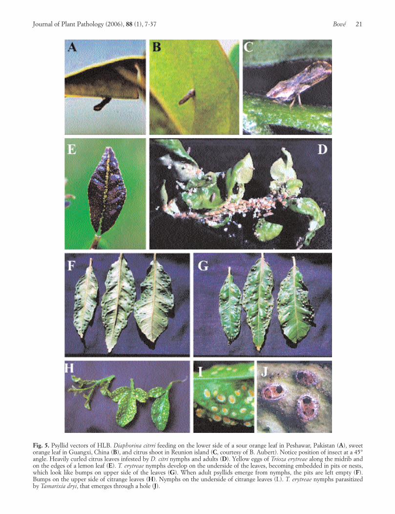
Fig. 5. Psyllid vectors of HLB. Diaphorina citrri feeding on the lower side of a sour orange leaf in Peshawar, Pakistan (A), sweetorange leaf in Guangxi, China (B), and citrus shoot in Reunion island (C, courtesy of B. Aubert). Notice position of insect at a 45°angle. Heavily curled citrus leaves infested by D. citri nymphs and adults (D). Yellow eggs of Trioza erytreae along the midrib andon the edges of a lemon leaf (E). T. erytreae nymphs develop on the underside of the leaves, becoming embedded in pits or nests,which look like bumps on upper side of the leaves (G). When adult psyllids emerge from nymphs, the pits are left empty (F).Bumps on the upper side of citrange leaves (H). Nymphs on the underside of citrange leaves (I.). T. erytreae nymphs parasitizedby Tamarixia dryi, that emerges through a hole (J).
along the Red Sea region from Mecca to Najran, close tothe Yemeni border. The vector is D. citri. HLB occurs inhot oases, and is, for that reason, of the heat-tolerantform. In Yemen, the vector is T. erytreae, and HLB is on-ly seen in cool highlands. Therefore, Yemeni HLB is ofthe heat-sensitive form (Bové and Garnier, 1984).
North of the Saudi/Yemeni border, in the Abha-Khamis Mushayt area, both vectors have been found(Bové and Garnier, 1984), and the two forms of HLB areprobably present. This situation can be explained in thefollowing way. In Yemen, African HLB and T. erytreaehave undoubtedly been introduced from nearbyEthiopia, across the Southern Red Sea, through the nar-row “Bab al Mandab” strait, and have moved north-wards. In Saudi Arabia, Asian HLB and D. citri have en-tered the Arabian Peninsula, probably with pilgrims toMecca, and have moved southwards. Eventually, the twovectors, as well as the African and Asian HLB, have metin the Abha-Khamis Mushayt region. Further movementof T. erytreae up North will be hindered by hot climate,but movement of D. citri southwards towards Yemenwould not be impaired by climatic conditions.
Indian Ocean islands: Reunion and Mauritius. Thetwo vectors and the two forms of HLB occur in Re-union and Mauritius (Garnier et al., 1996). Distributionof the two psyllid vectors according to temperature/alti-tude is remarkable. D. citri is present on the coastal ar-eas up to an altitude of ~500 m., while T. erytreae isprevalent above this altitude. D. citri has also been de-tected in Rodrigues island, East of Mauritius (J.Bovéand M. Garnier, unpublished). Both Ca. L. africanusand Ca. L. asiaticus are present. Some trees proved tobe simultaneously infected with the two liberibacters, asituation also observed in São Paulo State with Ca. L.americanus and Ca. L. asiaticus.
These islands are crossroads, and have been populat-ed by immigrants from both Africa and India. In Re-union, French, African, Malagasy, Chinese, Pakistaniand Indian ethnic groups are present. In Mauritius, In-do-Mauritians represent 68%, Creoles, 27%, and Sino-Mauritians, 3%. African HLB has probably been intro-duced from Eastern Africa and/or Madagascar, andAsian HLB, from the Indian subcontinent.
South America. SPS in Brazil has been the firstAmerican region to report HLB, in 2004. The vector isD. citri, present in SPS for almost 70 years. Two liberib-acters occur. The major HLB agent is the new liberibac-ter species, Ca. L. americanus, present in 92% of trees.Ca. L. asiaticus affects 6% of trees, and the two liberib-acters occur in 2% of trees (see above).
North America. Florida, U.S.A., has been the secondAmerican region to report HLB, in 2005. D. citri wasrecorded 7 years earlier. The liberibacter is Ca. L. asiaticus.
CITRUS REGIONS WITH PSYLLID VECTORS BUT NOHLB
The examples of SPS and especially Florida showthat once a psyllid vector is present, HLB is not very far.Therefore, the following regions and countries must beseriously on the alert, and scout for HLB, with the hopeto catch it early enough to try eradication. In SPS andFlorida, several years probably elapsed between the mo-ment the disease was correctly recognized and properlydiagnosed, and its first occurrence.
Atlantic ocean islands: Saint Helena, Madeira, andTenerife. The three islands harbor T. erytreae. The insectwas reported in 1994 from Madeira, and in 2002 fromTenerife (Canary islands). Madeira is located North ofTenerife, both islands being West of Morocco. It is be-lieved that the African citrus psyllid was introducedfrom Madeira into the Canary island by the dominantNorth-South trade winds (Gonzales Hernandez, 2003).
Middle East: Iran. A very small number of D. citripsyllids were seen in December 1997 in Iran, close tothe border with Pakistan (Bové et al., 2000a). By 2005,large populations of the insect occurred in the majorlime [Citrus aurantifolia (Christm.) Swing] growing re-gion, North of Bandar Abbas (Alimorad Sarafrazi, per-sonal communication).
Americas. Halbert and Nuñez (2004) have describedthe distribution of D. citri in South, Central and NorthAmerica, and the Caribbean basin.
South America: Brazil, Argentina, Venezuela. InBrazil, as of November 2005, 79 municipalities in SPSand 1 adjacent municipality in Minas Gerais are contam-inated with HLB, the vector being D. citri for Ca. L. asi-aticus, and probably also for Ca. L. americanus. OtherBrazilian regions grow citrus, but they do not (yet) haveHLB, even though D. citri is present. These regions areat great danger of becoming contaminated with HLB. In1997, D. citri was found on citrus in Corrientes, Argenti-na. The infestation was minor, the populations beingprobably controlled by local natural ennemies. D. citriwas reported for the first time in Venezuela in 1999 in the“Peninsula de Paraguana” on C. aurantifolia, C. reticu-lata, C. latifolia, and M. paniculata.
Central America, West Indies, Caribbeans. D. citriwas found in Guadeloupe, Bahamas, Cayman islands,Cuba and Dominican Republic, and Puerto Rico in 1998,1999, 2000, 2001, and 2002, respectively (Halbert andNuñez, 2004). D. citri was intercepted on citrus from Be-lize in a baggage at Houston, Texas, in October 2002.
North America: Texas, Yucatan. D. citri is present in
22 Huanglongbing of Citrus Journal of Plant Pathology (2006), 88 (1), 7-37
Journal of Plant Pathology (2006), 88 (1), 7-37 Bové 23
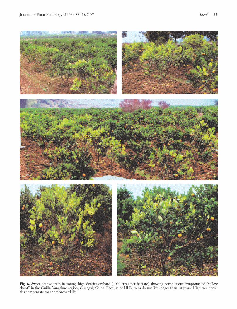
Fig. 6. Sweet orange trees in young, high density orchard (1000 trees per hectare) showing conspicuous symptoms of “yellowshoot” in the Guilin-Yangshuo region, Guangxi, China. Because of HLB, trees do not live longer than 10 years. High tree densi-ties compensate for short orchard life.

the Rio Grande Valley of Texas, U.S.A. (French et al.,2001). It was accidentally introduced in the spring of2001 on potted Murraya plants from Florida. D. citriwas reported in April 2002 in Cancun, Mexico, andsamples were collected in November 2003 (Halbert andNuñez, 2004).
CITRUS REGIONS FREE OF PSYLLIDS AND HLB
The Mediterranean basin, most of Western Asia(Near and Middle East), Australia, and North- andSouth-Pacific islands are still free of HLB as well as ofthe psyllid vectors of the disease. However, for these re-gions, HLB is a serious threat. The Mediterranean basincould become contaminated with T. erytreae, well estab-lished in the Portuguese Madeira island and the SpanishCanary islands, not very far from the Moroccan coast. Itmust be remembered that the most efficient aphid vectorof citrus tristeza, Toxoptera citricida, reported in Madeiraisland together with T. erytreae in 1994, is now present inNorthern Portugal and Spain. (It is however not firmlyestablished that Madeira was the source of contamina-tion of the Iberian Peninsula with the tristeza vector).
The Near and Middle East regions are endangeredby D. citri, which is well established in South-easternIran and in southern Saudi Arabia along the Red Sea. T.erytreae is in Yemen. Both Saudi Arabia and Yemen,but not yet Iran, host HLB in addition to the psyllidvectors.
Australia has two northern neighbours contaminatedwith both D. citri and HLB: East Timor and Papua NewGuinea.
The North- and South-Pacific islands have not re-ported HLB nor psyllid vectors. They must be aware ofthe situation in Papua New Guinea. During a recentsurvey for HLB in French Polynesia, no evidence of itspresence was found (Bové and Duran-Vila, unpublishedobservations). Similarly, no evidence was found for thepresence of HLB in four Pacific island countries (Cookislands, Fiji islands, Samoa and Tonga) (Davis et al.,2005b).
ORIGIN OF LIBERIBACTERS
Hitherto, Ca. L. africanus has only been found inAfrica, and Ca. L. asiaticus, only in Asia (Garnier andBové, 1996). This is probably not due to the fact thatthe psyllid vector T. erytreae is only present in Africa,and the psyllid vector, D. citri, only in Asia, becauseboth insects have the ability to transmit both liberibac-ters, at least under experimental greenhouse conditions(Massonié et al., 1976; Lallemand et al., 1986). There-fore, it seems as if Ca. L. africanus originated in Africa,and Ca. L. asiaticus in Asia. After having spread initially
within these continents, disease and vector have spreadlater to neighbouring regions. In the SPS in Brazil, thesituation is more complex. The Asian psyllid, Diaphori-na citri, has been present, undisturbed, since the 1940s.In these early years, the insect was probably free of anHLB agent, as HLB occurs in SPS only since about1995. The presence, in SPS, of Ca. L. asiaticus, a veryminor component of Brazilian HLB, is probably the re-sult of a relatively recent, uncontrolled introductionfrom Asia.
If, as suggested above, Ca. L. africanus and Ca. L. asi-aticus have originated in their respective continents, thesame could apply to Ca. L. americanus, supposed to hav-ing arisen in SPS. Ca. L. americanus is not a recent “mu-tation” of Ca. L. asiaticus, because the sequence differ-ences in the 16S rDNA and the RIR of the two liberibac-ters are far too great, and because there is very little se-quence variation between isolates of Ca. L. asiaticus, in-cluding the SPS isolate. The sequence of the 16S rDNAof Ca. L. americanus seems to be quite stable too.
The hypothesis proposing that different liberibacterspecies have evolved in different continents implies thatCa. L. americanus is native to SPS, and has not been in-troduced from known HLB-affected regions. Thecountless PCR assays made in Africa and Asia for detec-tion of Ca. L. asiaticus and Ca. L. africanus, have givenno indications of the occurrence, in these regions, of aliberibacter different of Ca. L. asiaticus and Ca. L.africanus. Several hundreds of PCR reactions have beencarried out in Africa and Asia, and have always detectedeither Ca. L. asiaticus or Ca. L. africanus. However, thenumber of these PCR assays might not have been suffi-ciently large to draw conclusions. Hence, in order toconfirm or infirm the above hypothesis, search for Ca.L. americanus, in Asia in particular, should be contin-ued and intensified, especially since PCR tools for spe-cific detection of the various liberibacters are available.
It has recently been suggested that the liberibactershave Gondwanan origins (Beattie et al., 2005). Indeed,until 160 million years ago, Australia, Africa, SouthAmerica, Madagascar, India, and Antartica were part ofthe single “super continent” Gondwana. Speciation ofthe putative Gondwanan liberibacter ancestor couldhave occurred after dislocation and fractionation of thesuper continent, isolating the African liberibacter line-age within Africa, the American lineage within SouthAmerica, and the Asian lineage within India, with sub-sequent eastward spread to China. A similar hypothesismight account for the distribution of the Diaphorineae.However, if so, it is difficult to explain why D. citri isapparently not native to South America, and seems tohave been introduced recently. In any case, the Gond-wanan hypothesis opens an interesting avenue for aca-demic and applied research.
24 Huanglongbing of Citrus Journal of Plant Pathology (2006), 88 (1), 7-37

RECOMMENDATIONS FOR HLB IDENTIFICATION
These recommendations apply to small HLB out-breaks, with eradication of the disease as the objective,as well as to situations were HLB is established, the goalbeing to live with the disease. They are meant to facili-tate quick diagnosis and confirmation of HLB.
Search and monitoring for citrus psyllid vectors.Halbert and Manjunath (2004) have recently reviewedthe literature on D. citri. Catling (1969a, b, c; 1970a, b)has carried out pioneering work on T. erytreae.
The psyllids can feed on many citrus species, but pre-ferred hosts of D. citri are M. paniculata (Orange jasmin,mock orange), M. exotica (Chinese box), and C. auran-tifolia, followed by Bergera koenigii (Curry leaf), andClausena spp.. These plants should be favoured formonitoring the insects. Lemon (Citrus limon) is a goodhost of T. erytreae. Psyllids need young, actively-grow-ing foliage (flush) for development, and their popula-tions peak at flush periods. In southern Florida, peaksof D. citri occur in May, August, and October throughDecember, and this coincides with the new flush growthof M. paniculata. This citrus relative has a more continu-ous flushing pattern, and serves as an alternative hostwhen citrus is not in flush. Psyllid numbers go down af-ter heavy rains. In regions with clear-cut dry and humidseasons, psyllids will peak at their highest level after thefirst growth flushes of the new wet season following dryweather (Davis et al., 2005a).
Eggs of T. erytreae are seen mostly along edges ofyoung leaves (Fig. 5 E). Those of D. citri occur on tips ofgrowing shoots, on and between unfurling leaves. Simi-larly, T. erytreae nymphs develop on the underside of leafblades, while D. citri nymphs are found on young stemsand petioles. The major difference between the two psyl-lids concerns precisely the nymphs. The T. erytreaenymphs are embedded in pits or nests on the undersideof the leaves (Fig. 5 F) which look like bumps on the up-per side (Fig. 5 G, H). After an adult has emerged froma nymph, the nest is empty, but the bump remains.Hence, the presence of even one single bump on onesingle leaf is proof of T. erytreae occurrence. No suchbumps are produced by D. citri. Leaves with bumps maybe severely distorted. With D. citri, leaves are heavilycurled (Fig. 5 D), and may be covered with honeydew.Characteristically, D. citri adults form a 45° angle withthe plant tissue on which they feed (Fig. 5 A, B, C).
Symptoms of HLB. It is generally thought that it isdifficult to diagnose HLB on the basis of symptoms, asnone of them is specific, and citrus trees are often af-fected by additional problems. However, some symp-toms are highly characteristic, and those will be empha-sized here. They apply to most species of citrus, sincenearly all citrus seedling trees and scion-roostock com-binations are sensitive.
Yellow shoots and blotchy mottle leaves. A tree,which begins to show symptoms in an orchard in earlystages of infection, can be identified, among still manysymptomless neighbouring trees, by the presence of oneor several yellow shoots. These shoots stand out againstthe green canopy of the tree (Fig. 6). These conspicuous”yellow shoots”, or “huang long” in Chinese, gave thedisease its name. A “yellow” shoot is seen as resultingfrom a shoot where psyllids have made a successful in-fection, as, indeed, HLB liberibacters can easily beendetected in the symptomatic leaves of these shoots. Ayellow shoot is thus an early symptom of the disease.On some trees, only one yellow shoot is present, and,with time, this affected shoot grows into a larger yellowbranch, or “yellow dragon”, since “long” means also“dragon”. This yellow shoot or yellow branch is a verycharacteristic aspect of HLB. Often, more than one yel-low dragon “wrap” themselves up around a tree. Even-tually, in late stages of the disease, the yellow brancheshave taken over and canopy the whole tree, which hasnow become totally infected.
Under cool weather, new flushes may be palegreen/yellowish, and zinc deficiency also induces yellowflushes. In these cases, practically all trees in an orchardshow the problem. With HLB outbreaks, only sometrees carry symptomatic shoots.
The yellow shoots on HLB affected trees look yellowbecause their leaves are partly yellow, partly green, withseveral shades of yellow, pale green and dark green,blending into each other, with no sharp limits betweenthe various shades of colour (Fig. 7, 8). Such leavesshow what is called “blotchy mottle”. This symptom wasdescribed in particular by Lin (1956) in China (Fig. 7A), and McClean and Schwarz (1970) in South Africa(Fig. 7 B, C). It is the most characteristic symptom ofHLB wherever the disease occurs, be it in Asia, Africaor America, and whatever the citrus species affected. Al-so, as seen on Fig. 7 and 8, the blotchy mottle pattern onone side of the leaf midrib is not symmetrical to that onthe other side. This asymmetry distinguishes blotchymottle from zinc (Fig. 8 D), manganese, magnesium, cal-cium, and iron deficiency symptoms. With time, thewhole leaf blade may ultimately turn uniformily yellow.In addition to blotchy mottle and yellowing, the leavesmay become thicker, and leathery. Midribs and lateralveins are sometimes enlarged, swollen, and corky. In ear-ly stages of infection, blotchy mottle might be the onlyleaf pattern to be seen. In later stages, foliar symptomsof zinc deficiency will eventually develop, and suchleaves have in general an upright growth, making a closeangle with the shoot on which they are attached. Even-tually, defoliation and dieback occur. With trees in earlystages of disease, as observed in SPS, blotchy mottle isseen to affect large, well developed leaves. The sametrees, one year later, had produced new mottled leaves,but smaller in size than those from the previous year.
Journal of Plant Pathology (2006), 88 (1), 7-37 Bové 25
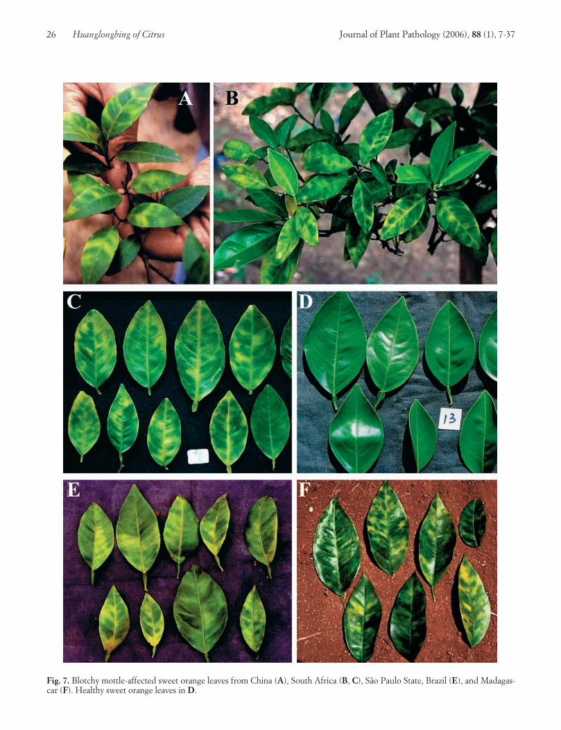
26 Huanglongbing of Citrus Journal of Plant Pathology (2006), 88 (1), 7-37
Fig. 7. Blotchy mottle-affected sweet orange leaves from China (A), South Africa (B, C), São Paulo State, Brazil (E), and Madagas-car (F). Healthy sweet orange leaves in D.
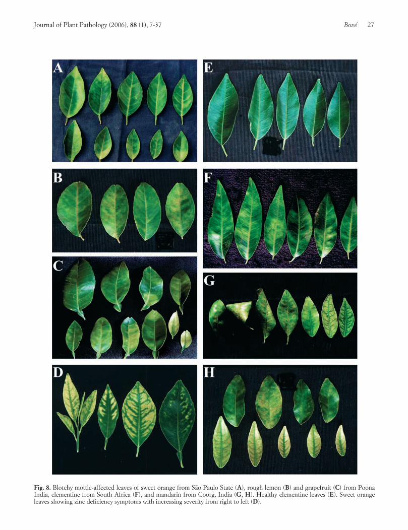
Journal of Plant Pathology (2006), 88 (1), 7-37 Bové 27
Fig. 8. Blotchy mottle-affected leaves of sweet orange from São Paulo State (A), rough lemon (B) and grapefruit (C) from PoonaIndia, clementine from South Africa (F), and mandarin from Coorg, India (G, H). Healthy clementine leaves (E). Sweet orangeleaves showing zinc deficiency symptoms with increasing severity from right to left (D).

In certain situations, blotchy mottle is difficult tofind. In 1996 in North Bali, Indonesia, HLB was severe-ly affecting almost 100% of young, 4-years-old Tejakulamandarin trees in many orchards of the coastal plain.The trees were uniformly yellow. Some leaves showedzinc deficiency symptoms, but leaves with clear-cutblotchy mottle were rare. These trees had practically nonew flushes. PCR tests did however confirm HLB as thecause of decline in all these orchards. At the same timein some orchards at higher altitudes, HLB was juststarting, yellow shoots occurred, and leaves had strongblotchy mottle.
Blotchy mottle is probably best observed on sweet or-ange trees, but most citrus species and varieties show it,including some mandarin varieties such as Tejakula inBali and clementine in South Africa (Fig. 8 F) Othermandarin varieties, such as the Coorg mandarin in India,do not present good, classic blotchy mottle. The colourpatches of yellow, dark green and pale green are not aslarge as on sweet orange leaves, and rather produce amosaic pattern (Fig. 8 G, H). Also, in the orchard, af-fected mandarin leaves tend to be more curled thansweet orange leaves (Fig. 8 G). However, in the green-house, leaves of affected sweet orange trees are frequent-ly curled.
Fruit symptoms. HLB induces also very characteris-tic fruit symptoms, seen especially well on sweet orangesand mandarins, and sometimes pummelos. Again, thesesymptoms are non specific, as similar symptoms are as-sociated with stubborn disease. Fruits with HLB symp-toms should be looked for on yellow shoots or branch-es. Symptomatic fruits are small, asymmetric, lopsided,with a bent fruit axis. At the time when the fruitchanges colour, from green to yellow/orange, HLB-af-fected fruit shows colour inversion: the peduncular endof the fruit turns yellow/orange, while the stylar end isstill green, whereas on normal fruit the coloration startsfirst at the stylar end, and moves only later to the pe-duncular end (Fig. 9 A, D, F to J). In addition, whenthe peduncle of a fruit with colour inversion is carefullyremoved, the resulting circular scar is stained orange,while on a normal fruit the scar is pale green. Colour in-version can be very pronounced in China, where it iscalled “red nose”. In South Africa, HLB-affected fruitcolours also unevenly (Fig. 9 C), and this symptom wasresponsible for calling the disease “greening”. Some-times, when one presses such fruit with the thumb, a sil-very “finger mark” results (Fig. 9 E).
To further observe the symptoms, fruits should becut in half. When the sectioning is made perpendicularto the fruit axis, it is easy to see the small, brownish/black aborted seeds, highly characteristic of HLB, butalso present in stubborn affected sweet oranges. Cuttinga lopsided fruit through the fruit axis, reveals its asym-metry, and some aborted seeds can also be seen (Fig. 10
A, B, C). In addition, the vascular bundles within thefruit axis at the peduncular end have a strong brownishstain (Fig. 10 B). As in stubborn-affected fruit, the albe-do is sometimes thicker at the peduncular end than atthe stylar end (Fig. 10 B).
In summary, when a tree shows one or several yellowshoots with blotchy mottled leaves, and small, lopsidedfruits with colour inversion and aborted seeds, it ishighly likely that the tree is affected by HLB. To con-firm the HLB diagnosis, PCR detection of the HLBliberibacters should be made.
PCR confirmation of HLB diagnosis. For PCR de-tection of the HLB liberibacters blotchy mottled leavesshould be used. Such leaves almost never fail to givestrong, positive PCR reactions when the symptoms aredue to HLB. Therefore it is untrue to state, as it issometimes written, that it is difficult to detect liberibac-ters. For detection of Ca. L. africanus and Ca. L. asiati-cus two sets of primers can be used: primers f-OA1 andr-OI2c for amplification of 16S rDNA, and primers f-rplA2 and r-rplJ5 for amplification of ribosomal proteingenes (rpl). In the case that the presence of both Africanand Asian liberibacters is suspected, the primer pair f-(OA1+OI1) / r- OI2c is recommended. Ca. L. ameri-canus is detected with PCR primers f-GB1 and r-GB3.Simultaneous detection of the three liberibacters bymultiplex PCR has been developed with the followingtwo primer pairs in the same reaction mixture: (i) f-rplA2 / r-rpl J5 and (ii) f-GB1 / r-GB3.
As mentioned above, initially in Brazil, most PCR re-actions gave negative results, even though leaves withstrong symptoms of blotchy mottle were used. This waslater shown to be due to the fact that a new liberibacterspecies, Ca. L. americanus, was involved (Teixeira et al.,2005a, b, c). If a similar situation is encountered else-where, search for a new liberibacter should be sought.
In some cases, when HLB symptoms are not immedi-ately recognized, and are attributed to some other caus-es, such as mineral deficiencies, the relevant trees mightbe in relatively late stages of the disease, and mottledleaves may be lacking. In such cases, leaves with zinc-deficiency symptoms should be used.
For PCR detection, HLB-affected leaves, infectedwith either one of the three liberibacters, should beavailable as positive controls. If not, DNA preparationsfrom such leaves, obtainable from several laboratories,can be used. Healthy citrus leaves are used as negativecontrols.
CONTROL OF HLB
There are no curative methods to control HLB.When HLB was found to be a bacterial, rather than a vi-ral disease, antibiotic control by trunk injections of tetra-
28 Huanglongbing of Citrus Journal of Plant Pathology (2006), 88 (1), 7-37

cyclines was tried in several countries, including SouthAfrica, Indonesia, and Taiwan, but soon abandoned, be-cause of ecological reasons, but essentially because tetra-cycline is bacteriostatic, rather than bactericidal, and thetreatment had to be repeated each year. Thus, control isby preventing trees from becoming infected. If HLB isnot yet present, quarantine measures should be enforcedto keep it out. The most serious situation is when HLBhas entered a region previously HLB-free. The first im-mediate measure is to survey and determine the extentof the outbreak. The next objective is to prevent as manytrees as possible from becoming infected. This can onlybe done: (i) by eliminating as much as possible liberibac-ter inoculum, i. e. by removing infected trees, and (ii) bykeeping psyllid populations as low as possible. Withoutthese control measures, HLB evolution in an affected or-chard is fast. In China, it has been stated repeatedly that,without control, it takes only about five years for an or-chard to reach 100% infection. Similar situations seemto occur in SPS. In the Reunion island, the time neededto reach 100% infection was estimated to be 13 years.Average time is eight years.
Quarantine measures. If HLB is still absent from agiven region, do not introduce it! The biggest quaran-tine threat is transport, by people, of citrus and citrusrelatives. These plants may be infected with liberibac-ters and may carry psyllid eggs and/or nymphs. M. pani-culata, an ornemental citrus relative, is a preferred hostof D. citri, and if further work confirms that it is also ahost of the liberibacters, it deserves special quarantineattention. Also, it must be remembered that the liberib-acters cannot be detected in symptomless leaves, eventhough their detection in mottled leaves is straightfor-ward. Symptomless leaves are either healthy, and willnever give positive PCR reactions, or they are alreadyinfected, but the liberibacter titer is still too low to bedetected by the PCR methods available today. For thesereasons it is meaningless to use conventional HLB PCRtechniques with symptomless plant material.
Eliminating inoculum by removal of infected trees.If all infected trees were symptomatic, it would be easyto spot them, on the basis of their symptoms, and re-move them. Unfortunately, there is a latency period dur-ing which recently infected trees do not show symp-toms. This period extends from the moment the tree be-comes infected by the psyllids up to the moment it ex-presses symptoms. The length of this period may varyfrom tree to tree, but is generally assumed to be 6 to 12months long, if not longer. Therefore, removal of allsymptomatic trees will not result in removal of all infect-ed trees. In practice, several surveys are required to re-move as many infected trees as possible. Some infectedtrees, which were still symptomless at survey # n, willshow symptoms at survey # n+1, and can be removed;
other trees will only be removed at survey # n+2. Also,within the time period between surveys n and n+1, orn+1 and n+2, psyllids (if not well controlled), may in-fect additional trees, and so on. Obviously, the time pe-riod between successive surveys is important. In partic-ular, if this period is too long, some symptomatic treeswill have become severely affected before they are re-moved, and serve as new sources of inoculum. In SPS,experience seems to show that a period of about threemonths is a good compromise.
Surveys must be carried out carefully. All trees in anorchard, however large it is, should be examined one byone, and enough time, say a few minutes, must be spentat each tree. Experience gained in a large grove (onemillion trees) in southern China suggests that the scoutsin charge of the surveys should work in pairs, so thateach tree is examined by the two scouts of a pair, onescout on each side of the row. In the case of orchardswith large, adult trees, it is essential to examine the topof the trees. In SPS, relatively high towers have beenbuilt onto tractors to permit efficient observation of treetops. Finally, once affected trees have been identified,they should be removed as quickly as possible.
In SPS, recommendation is that all symptomatic treesbe removed, whatever their age or the severity of theirinfection (Ayres et al., 2005). Removing HLB-affectedbranches by pruning, instead of removing the wholetree, has been discarded because the many sprouts thatdevelop at the site of pruning turn into “yellow shoots”and favour psyllid development. In South Africa, it hasbeen recommended (Buitendag and von Broembsen,1993) that HLB-affected trees up to 5 years of age beremoved. Trees from 6 to 10-year-old, when severely af-fected, are also removed. With less affected trees, andwith trees older than 10 years, only the affected branch-es are removed.
In SPS, citrus canker is another disease of citrus, thecontrol of which also involves removal of trees. In thiscase, the infected tree as well as all surrounding trees,within a distance of 15 m, are removed. From the epi-demiology studies of HLB (Gottwald, 2005; Bassanezi,2005), it seems that this strategy is probably inadequatefor HLB. Indeed, when HLB vectors move, either natu-rally in search of new feeding opportunities or whendisturbed, they not only move to neighbouring trees(primary foci), but also to trees located at distances of25 to 50 m (secondary foci). In addition, psyllids canmove a distance of at least 1.5 km with wind, and prob-ably much longer distances with hurricanes or tropicalstorms, such as those in Florida.
When an orchard is too severely affected, with a highpercentage of affected trees, it might be best to removethe whole stand. The following figures might help tomake the decision. It can be roughly estimated that withAsian HLB, D. citri being the vector, an orchard with10% symptomatic (Sy) trees, has a total of 20% infect-
Journal of Plant Pathology (2006), 88 (1), 7-37 Bové 29
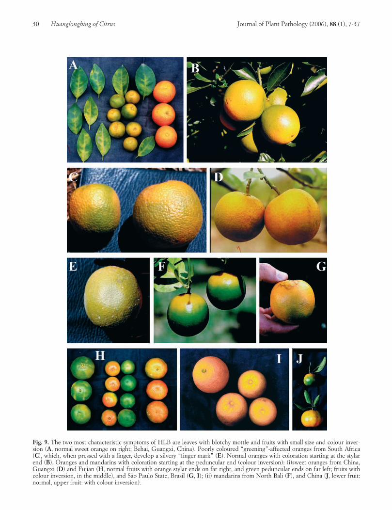
30 Huanglongbing of Citrus Journal of Plant Pathology (2006), 88 (1), 7-37
Fig. 9. The two most characteristic symptoms of HLB are leaves with blotchy mottle and fruits with small size and colour inver-sion (A, normal sweet orange on right; Behai, Guangxi, China). Poorly coloured “greening”-affected oranges from South Africa(C), which, when pressed with a finger, develop a silvery “finger mark” (E). Normal oranges with coloration starting at the stylarend (B). Oranges and mandarins with coloration starting at the peduncular end (colour inversion): (i)sweet oranges from China,Guangxi (D) and Fujian (H, normal fruits with orange stylar ends on far right, and green peduncular ends on far left; fruits withcolour inversion, in the middle), and São Paulo State, Brasil (G, I); (ii) mandarins from North Bali (F), and China (J, lower fruit:normal, upper fruit: with colour inversion).

ed trees, i.e., symptomatic trees plus asymptomatic trees(Sy+Asy). For 20%, 30% and 50% Sy trees, there willbe 36%, 50%, and 70% (Sy+Asy) trees. In other words,in an orchard with 30% symptomatic trees, half of thetrees are infected, and will have to be pulled out sooneror later. Is it worthwhile to keep such an orchard? Prob-ably not!
When affected trees have been pulled out, they canbe replaced by young, healthy trees. In SPS, by law, allnursery trees have to be produced in covered, insectproof screenhouses, and this obligation guarantees thatthey are HLB-free.
Even though in SPS removal of HLB-affected trees iscompulsory by law, great efforts have been madethrough the media (articles in newspapers, programs onradio and TV, training courses for growers and techni-cians) to explain the situation, and convince the growersto remove the affected trees on a voluntary base (Ayres
et al., 2005). Most of the growers cooperate, and 350thousand symptomatic trees have been voluntarily elim-inated, as of October 2005. Cooperation of the growersis also required for vector control.
Psyllid vector control. Key features of the psyllids,which need to be taken into account in the formulationof control strategies have been reviewed (Buitendag andvon Broembsen, 1993; Halbert and Manjunath, 2004;Hall, 2005).
Treatments with contact and systemic insecticides re-duce psyllid populations, and thus HLB spread. The ef-ficiency of the treatments is easier to estimate with T.erytreae than with D. citri, because of the foliar“bumps” produced by T. erytreae nymphs. In the large,South African citrus estates, control of T. erytreae canbe very efficient, as “bumps” are hardly seen. Systemicinsecticides, aimed specifically at psyllids (monocro-
Journal of Plant Pathology (2006), 88 (1), 7-37 Bové 31
Fig. 10. A. Far left and far right, normal sweet orange halves showing normal seeds; middle, two halves from a lopsided HLB-af-fected sweet orange from China. Notice asymmetry of the fruit section, and the brownish-black seeds. B. Sectioned lopsided sweetorange from São Paulo State, with brownish-black seeds, thick albedo at the peduncular end, and vascular bundles stained or-ange-brown at the peduncular end of fruit axis. C. Lopsided pummelo showing vascular bundles stained orange-brown.
tophos), have been applied directly to the tree trunks,and have been very effective. In Asia, a large range ofinsecticides, mostly organophosphates and pyretroids,are used in very intensive spray programs to kill eggsand nymphs on flush growth. Horticultural and agricul-tural mineral oils are being developed as alternativetreatments, and these are much less damaging to the en-vironment and less disruptive to biocontrol of otherpests (Davis et al., 2005a).
Reunion island is the only region where biologicalcontrol of both T. erytreae and D. citri has been effective(Etienne and Aubert, 1980; Aubert et al., 1980). Thesuccess is due to the fact that, initially, the two psyllidsoccurred in the island in the absence of parasites, andthus, hyperparasites. Therefore parasites of the twopsyllids were introduced into the island, care being tak-en not to introduce hyperparasites at the same time.Tamarixia dryi, a parasite of T. erytreae, and Tamarixiaradiata, a parasite of D. citri, were collected, respectivelyin South Africa and India, reared in Reunion and re-leased in abandoned citrus orchard having received noinsecticide treatments for quite some time. A secondreason for success is the fact that T. dryi multiplied notonly on T. erytreae, but also on an additional psyllid, Tri-oza eastopi, not a citrus psyllid, but multiplying on awidely distributed weed, Litsea chinensis. Releases ofPsyllaephagous pulvinatus, introduced from SouthAfrica, were also made, but the insect, much less vigor-ous than T. dryi, disappeared from the island.
In Florida, T. radiata has been introduced from Tai-wan and Vietnam, and is being established in Florida.In China, studies are conducted on Campylomma chi-nensis Schuh, as a potential biocontrol predator of D.citri (Wu, 2005).
The Brazilian experience. In SPS, encouraging re-sults have been obtained in the control of HLB by treeremoval and insecticide treatments against psyllids(Ayres et al., 2005). For instance, in one large citrus or-chard (71,000 trees), 10 surveys for identification ofHLB-affected trees were carried out from September2004 until September 2005. The first survey identified7% affected trees. On the 2nd survey, in December2004, the percentage went down to 0.01%, but in-creased again to 1.1% after the 6th survey in May 2005.From there on, the percentages decreased regularly to0.5% (7th survey, June 2005), 0.15% (8th survey, July2005), 0.05% (9th survey, August 2005), and 0.03 (10th
survey, September 2005). It is hoped that the percent-ages of affected trees will remain at the level of 0.05%,even with less frequent surveys and tree removals. HLBwill not be eradicated, but it will be possible to live withthe disease.
To achieve this goal, SPS has a number of favourablefactors. Brazil, contrary to Florida, is not affected, yearafter year, by hurricanes and tropical storms, the role of
which in spreading the disease and its vector, might bemore significant than thought. A task force of 800 peo-ple from both the private sector (Fundecitrus) and thepublic sector (Government, public research organiza-tions) is involved in HLB control and helps the growers.A good interface has been built between growers, Fun-decitrus and the government. Sixty million trees have al-ready been surveyed, 70 million more trees have still tobe inspected. The first wide range survey will be com-pleted in February 2006. The surveys for HLB benefitfrom the year long experience of Fundecitrus in thecontrol of citrus canker. The nursery trees required toreplace the removed HLB-affected trees are producedfrom healthy budwood in covered, insect proof nurs-eries, a procedure that guarantees that they are free ofHLB and all other graft-transmissible diseases (exceptCTV strains used for cross protection). Even thoughHLB has been present for several years before it wasrecognized, most orchards in the affected municipalitieshave still a low disease incidence.
Very few countries have been able to control AsianHLB. SPS might be one of the first to be successful, andlive with the disease until genetically engineered citruscultivars, resistant to HLB, will eventually become avail-able (Hartung et al., 2005b).
ACKNOWLEDGMENTS
This review has benefited from my recent visits toBrasil, China, and Florida to discuss yellow dragon dis-ease and be involved in control strategies. I am verygrateful to my colleagues from Fundecitrus and themany private citrus estates in São Paulo State for sharingtheir information with me, and making it possible toproduce new results. China is afflicted with huanglong-bing since more than a century, and we have taken greatadvantage of the experience gained by our colleaguesfrom Guangxi, Guangdong, Fujian, and Sichuanprovinces. In Florida, the dragon is emerging, and amost interesting workshop was held recently to ex-change information and put the beast on the leash. I amindebted to Andrew Beattie for having provided me withrare reprints and for interesting discussions over the net.Finally, I thank my laboratory colleagues for their years-long indulgence to me.
REFERENCES
Anonymous, 2004. Estudos indicam que a nova doença tem re-lação com o greening. Fundecitrus. http://www.fundecitrus.com
Asana R.D., 1958. The citrus dieback problem in relation tocultivation of citrus fruits in India. Indian Journal of Horti-culture 15: 283-286.
32 Huanglongbing of Citrus Journal of Plant Pathology (2006), 88 (1), 7-37
Aubert B., Bové J.M., 1980. Effect of penicillin or tetracyclineinjections of citrus trees affected by greening disease underfield conditions in Reunion island. Proceedings of 8th Con-ference of IOCV, IOCV, Riverside 1980, 103-108.
Aubert B., Bové J.M., Etienne J., 1980. La lutte contre la mal-adie du «greening» des agrumes à l’île de la Réunion. Ré-sultats et perspectives. Fruits 35: 605-624.
Aubert B., Garnier M., Guillaumin D., Herbagyandono B.,Setiobudi L., Nurhadi F., 1985. Le greening, une sérieusemenace pour les productions agrumicoles de l’archipel in-donésien. Fruits 40: 549-563.
Aubert B., Garnier M., Cassin J.C., Bertin Y., 1988. Citrusgreening disease survey in East and West African countriesSouth of Sahara. Proceedings of 10th Conference of IOCV,IOCV, Riverside 1988, 231-237.
Ayres A.J., Massari C.A., Lopes S.A., Bassanezi R.B., BelasqueJunior J., Yamamoto P.T., Teixeira D.C., Wulff N.A.,Gimenes-Fernandes N., Bergamaschi O.A., 2005. Proceedingsof 2nd International Citrus Canker and Huanglongbing Re-search Workshop, Florida Citrus Mutual, Orlando 2005, H-4.
Bassanezi R.B., Busato L.A., Bergamin Filho A., Amorim L.,Gottwald T.R., 2005. Spatial distribution of huanglongbingsymptomatic trees in citrus groves in São Paulo, Brasil.Proceedings 2nd Inernational Citrus Canker and Huanglong-bing Research Workshop, Florida Citrus Mutual, Orlando2005, H-7.
Bastianel C., Garnier-Semancik M., Renaudin J., Bové J.M.,Eveillard S., 2005. Diversity of “Candidatus Liberibacterasiaticus” based on the omp gene sequence. Applied Envi-ronmental Microbiology 71: 6473-6478.
Beattie G.A.C., Mabberley D.J., Holford P., Broadbent P., DeBarro P., 2005. Huanglongbing: its possible origins, collab-orative research in Southeast Asia, and developing incur-sion management plans for Australia. Proceedings of 2nd In-ternational Citrus Canker and Huanglongbing ResearchWorkshop, Florida Citrus Mutual, Orlando 2005, H-5.
Bonavia E., 1888. The cultivated oranges and lemons of Indiaand Ceylon. W.H. Allen & Co., London.
Bové J.M., Garnier M., 1984. Citrus greening and psylla vec-tors of the disease in the Arabian Peninsula. Proceedings of9th Conference IOCV, IOCV, Riverside 1984, 109-114.
Bové J.M., Saglio P., 1974. Stubborn and Greening: a review,1969-1972. Proceedings of 6th Conference IOCV, IOCV,Riverside 1974, 1-11.
Bové J.M., Calavan E.C., Capoor S.P., Cortez R.E., SchwarzR.E., 1974. Influence of température on symptoms of Cali-fornia stubborn, South African greening, Indian citrus de-cline and Philippines leaf mottling diseases. Proceedings of6th Conference IOCV, IOCV, Riverside 1974, 12-15.
Bové J.M., Bonnet P., Garnier M., Aubert B., 1980. Penicillinand tetracycline treatments of greening disease-affectedcitrus plants in the glasshouse, and the bacterial nature ofthe procaryote associated with greening. Proceedings of 8th
Conference IOCV, IOCV, Riverside 1980, 91-102.
Bové J.M., Garnier M., Ahlawat Y.S., Chakraborty N.K., Var-ma A., 1993. Detection of the Asian strains of the greeningBLO by DNA-DNA hybridisation in Indian orchard trees
and Malaysian Diaphorina citri psyllids. Proceedings of 12th
Conference IOCV, IOCV, Riverside 1993, 258-263.
Bové J.M., Chau N.M., Ha Minh Trung, Bourdeaut J., Gar-nier M., 1996. Huanglongbing (greening) in Vietnam: de-tection of Liberobacter asiaticum by DNA hybridisationwith probe In 2.6 and PCR amplification of 16S ribosomalDNA. Proceedings of 13th Conference IOCV, IOCV, River-side 1996, 258-266.
Bové J.M., Danet J.L., Bananej K., Hasanzadeh N., Salehi M.,Garnier M., 2000a. Witches’ broom disease of lime (WB-DL) in Iran. Proceedings of 14th Conference IOCV, IOCV,Riverside 2000, 207-212.
Bové J.M., Erti Dwiastuti M., Triviratno A., Supriyanto A.,Nasli E., Becu P., Garnier M., 2000b. Incidence of huang-longbing and citrus rehabilitation in North Bali, Indonesia.Proceedings of 14th Conference IOCV, IOCV, Riverside2000, 200-206.
Bruton B.D., Fletcher J., Pair S.D., Shaw M., Sittertz-BhatkarH., 1998. Association of a phloem-limited bacterium withyellow vine disease in Cucurbits. Plant Disease 82: 512-520.
Buitendag C.H., von Broembsen A., 1993. Living with citrusgreening in South Africa. Proceedings of 12th ConferenceIOCV, IOCV, Riverside 1993, 269-273.
Capoor S.P., 1963. Decline of citrus trees in India. BulletinNational Institute of Science India 24: 48-64.
Capoor S.P., Rao D.G., Viswanath S.M., 1967. Diaphorina cit-ri Kuway., a vector of the greening disease of citrus in In-dia. Indian Journal of Agricultural Science 37: 572-576.
Catling H.D., 1969a. The bionomics of the South African cit-rus psylla, Trioza erytreae (Del Guercio) (Homoptera:Psyllidae). I. The influence of the flushing rhythm of citrusand factors which regulate flushing. Journal of EntomologySociety South Africa 32: 191-208.
Catling H.D., 1969b. The bionomics of the South African cit-rus psylla, Trioza erytreae (Del Guercio) (Homoptera:Psyllidae). II. The influence of parasites and notes on thespecies involved. Journal of Entomology Society SouthAfrica 32: 209-223.
Catling H.D., 1969c. The bionomics of the South African cit-rus psylla, Trioza erytreae (Del Guercio) (Homoptera:Psyllidae). III. The influence of extremes of weather onsurvival. Journal of Entomology Society South Africa 32:273-290.
Catling H.D., 1970a. The bionomics of the South African cit-rus psylla, Trioza erytreae (Del Guercio) (Homoptera:Psyllidae). IV. The influence of predators. Journal of Ento-mology Society South Africa 33: 341-348.
Catling H.D., 1970b. Distribution of the psyllid vectors of cit-rus greening disease, with notes on the biology and bio-nomics of Diaphorina citri. FAO Plant Protection Bulletin18: 8-15.
Catling H.D., M. Garnier, J.M. Bové, 1978. Presence of citrusgreening disease in Bangladesh and a new method for rap-id diagnosis. FAO Plant Protection Bulletin 26: 16-18.
Chen Chi-bao, 1943. A report of a study on yellow shoot ofcitrus in Chaoshan. Quaterly Bulletin of New Agriculture 3:142-175.
Journal of Plant Pathology (2006), 88 (1), 7-37 Bové 33

Chen M.-H., Miyakawa T., Matsui C., 1971. Mycoplasmalikebodies associated with likubin-diseased Ponkan citrus.Phytopathology 61: 598.
Chen M.-H., Miyakawa T., Matsui C., 1973. Citrus likubinpathogens in the salivary glands of Diaphorina citri. Phy-topathology 63: 194-195.
Chen T.Y., Shen C.Y., Tao S.C., Kung T.H., Chao H.Y., Chi-ang Y.H., Chiu C.S., 1979a. Mycoplasma-like organismsassociated with huanglongping in Kwangxi. Acta Biochimi-ca et Biophysica Sinica 11: 389-391.
Chen T.Y., Shen C.Y., Tao S.C., Kung T.H., Chen H.Y., TaiY.M., 1979b. Studies on the pathogens of huanglongping(citrus yellow shoot disease) 3. Mycoplasma-like organismsassociated with huanglongping in Kwangtung. ActaBiochimica et Biophysica Sinica 11: 191-192.
Coletta-Filho H.D., Targon M.L.P.N., Takita M.A., De NegriJ.D., Pompeu J.J., Machado M.A., 2004. First Report ofthe Causal Agent of Huanglongbing (“Candidatus Liberib-acter asiaticus”) in Brazil. Plant Disease 88: 1382.
Da Graça J.V., 1991. Citrus greening disease. Annual Reviewof Phytopathology 29: 109-136.
Da Graça J.V., Korsten L., 2004. Citrus huanglongbing: review,present status and future strategies. In: S.A.M.H. Naqvi(Ed.). Diseases of Fruits and Vegetables, pp. 229-245. Kluw-er Academic Publishers, Dordrecht, The Netherlands.
Davis M.J., Kramer J.B., Ferwerda H., Brunner B.R., 1996.Association of a bacterium and not a phytoplasma with pa-paya bunchy top disease. Phytopathology 86: 102-109.
Davis R., Gunua T., Bellis G., Grimshaw J., 2005a. Huang-longbing disease of citrus trees. Pest advisory leaflet no.45. Plant Protection Service. Secretariat of the PacificCommunity. ISSN 1017-6276.
Davis R., Gunua T., Kame M., Tenakanai D., Ruabete T.,2005b. Spread of citrus huanglongbing (greening disease)following incursion into Papua New Guinea. AustralianPlant Pathology 34: 517-524.
Ding Fang, Yi Ganjun, Wang Guoping, 2004. Research on thePCR and nested-PCR detection of citrus huanglongbingpathogen. Acta Horticulturae Sinica 31: 803-806.
Ding Fang, Yi Ganjun, Wang Guoping, Yun Zhong, 2006.Huanglongbing (Candidatus Liberibacter asiaticus) infect-ing wampee and lemon in China by microscopy, PCR andnested PCR analysis. Plant Science (in press).
Doe Doe, Namgay Om, Chencho Dorji, Thinlay, Garnier M.,Jagoueix-Eveillard S., Bové J.M., 2003. First report of Can-didatus Liberibacter asiaticus, the agent of citrus huang-longbing (rx-greening), in Bhutan. Plant Disease 87: 448.
Doi Y., Teranaka M., Yora K., Asuyama H., 1967. Mycoplasmaor PLT group-like microorganisms found in the phloem ele-ments of plants infected with mulberry dwarf, potato witch-es’ broom, aster yellows, or Paulownia witches’ broom. An-nual of Phytopathology Society Japan 33: 259-266.
Etienne J., Aubert B., 1980. Biological control of psyllid vec-tors of greening disease on Reunion Island. Proceedings of8th Conference IOCV, IOCV, Riverside 1980, 118-121.
Fraser L.R., Singh D., 1968. Citrus dieback in India – the con-
tribution of greening virus. Proceedings of 4th ConferenceIOCV, IOCV, Riverside 1968, 141-144.
Fraser L.R., Singh D., Capoor S.P., Nariani T.K., 1966.Greening virus, the likely cause of citrus dieback in India.FAO Plant Protection Bulletin 14: 127-130.
French J.V., Kahlke C., da Graça J.V., 2001. Asian psyllidfound on Texas citrus. Citrus Center 19: 1.
Gao S., Garnier M., Bové J.M., 1993. Production of mono-clonal antibodies recognizing most strains of the greeningBLO by in vitro immunization with an antigenic proteinpurified from the BLO. Proceedings of 12th ConferenceIOCV, IOCV, Riverside 1993, 244-249.
Garnier M., Bové J.M., 1977. Structure trilamellaire des deuxmembranes qui entourent les organismes procaryotes asso-ciés à la maladie du “greening” des agrumes. Fruits 32:749-752.
Garnier M., Bové J.M., 1983. Transmission of the organismassociated with citrus greening disease from sweet orangeto periwinkle by dodder. Phytopathology 73: 1358-1363.
Garnier M., Bové J.M., 1993. Citrus greening disease and thegreening bacterium. Proceedings of 12th Conference IOCV,IOCV, Riverside 1993, 212-219.
Garnier M., Bové J.M., 1996. Distribution of the huanglong-bing (greening) liberobacter species in fifteen African ansAsian countries. Proceedings of 13th Conference IOCV,IOCV, Riverside 1996, 388-391.
Garnier M., Bové J.M., 2000. Huanglongbing in Cambodia,Laos, and Myanmar. Proceedings of 14th Conference IOCV,IOCV, Riverside 2000, 378-380.
Garnier M., Latrille J., Bové J.M., 1976. Spiroplasma citri andthe organism associated with likubin : comparison of theirenvelope systems. Proceedings of 7th Conference IOCV,IOCV, Riverside 1976, 13-17.
Garnier M., Danel N., Bové J.M., 1984a. Aetiology of CitrusGreening Disease. Annual of Microbiology (Institute Pas-teur) 135A: 169-179.
Garnier M., Danel N., Bové J.M., 1984b. The greening organ-ism is a Gram negative bacterium. Proceedings of 9th Con-ference IOCV, IOCV, Riverside 1984, 115-124.
Garnier M., Gao S.J., He Y., Villechanoux S., Gandar J., BovéJ.M.., 1991. Study of the greening organism (GO) withmonoclonal antibodies: serological identification, mor-phology serotypes and purification of the GO. Proceedingsof 11th Conference IOCV, IOCV, Riverside 1991, 428-435.
Garnier M., Jagoueix S., Toorawa P., Grisoni M., MallessardR., Dookun A., Saumtally S., Autrey J.C., Bové J.M., 1996.Both huanglongbing (greening) liberobacter species arepresent in Mauritius and Reunion. Proceedings of 13th Con-ference IOCV, IOCV, Riverside 1996, 392-394.
Garnier M., Bové J.M., Cronje C.P.R., Sanders G.M., KorstenL., Le Roux H.F., 2000a. Presence of “Candidatus Liberib-acter africanus” in the Western Cape province of SouthAfrica. Proceedings of 14th Conference IOCV, IOCV, River-side 2000, 369-372.
Garnier M., Jagoueix-Eveillard S., Cronje P., Le Roux H., BovéJ.M., 2000b Genomic characterisation of a liberibacter pres-ent in an ornamental Rutaceous tree, Calodendrum capense,
34 Huanglongbing of Citrus Journal of Plant Pathology (2006), 88 (1), 7-37
in the Western Cape province of South Africa. Proposal fora “Candidatus Liberibacter africanus subsp. capensis”. In-ternational Journal of Systematic Evolution Microbiology 50:2119-2125.
Gatineau F., Jacob N., Vautrin S., Larue J., Lherminier J.,Richard-Molard M., Boudon-Padieu E., 2002. Associationwith the syndrome «Basses richesses» of sugar beet of aphytoplasma and a bacterium-like organism transmitted bya Pentastiridius sp. Phytopathology 92: 384-392.
Gonzalez Hernandez A., 2003. Trioza erytreae (Del Guercio1918): nueva plaga de los citricos en Canarias. Phytoma153: 112-118.
Ghosh S.C., Giannotti J., Louis C., 1977. Multiplication in-tensive des prokaryotes associés aux maladies de type“greening” des agrumes dans les cellules criblées de cus-cute. Annals of Phytopathology 9: 525-530.
Gottwald T.R., 2005. Huanglongbing epidemiology: trackingthe dragon through time and space. Proceedings of 2nd In-ternational Citrus Canker And Huanglongbing ResearchWorkshop, Florida Citrus Mutual, Orlando 2005, H-6.
Halbert S.E., 1998. Entomology section. Triology 37: 6-7.
Halbert S.E., 2005. The discovery of huanglongbing in Flori-da. Proceedings Of 2nd International Citrus Canker AndHuanglongbing Research Workshop, Florida Citrus Mutual,Orlando 2005, H-3.
Halbert S.E., Manjunath K.L., 2004. Asian citrus psyllids(Sternorrhycha: Psyllidae) and greening disease of citrus: aliterature review and assessment of risk in Florida. FloridaEntomologist 87: 330-353.
Halbert S.E., Nuñez C.A., 2004. Distribution of the Asian cit-rus psyllid, Diaphorina citri Kuwayama (Rhynchota: Psylli-dae) in the Caribbean basin. Florida Entomologist 87: 401-402.
Halbert S.E., Niblett C.L., Manjunath K.L., Lee R.F., BrownL.G., 2003. Establishment of two new vectors of citruspathogens in Florida. In: Proceedings of IX Congress Inter-national Society Citriculture, ASHS Press, Alexandria,1016-1017.
Hall D.G., 2005. Overview of the Asian citrus psyllid. Pro-ceedings of 2nd International citrus canker and huanglong-bing research workshop, Florida Citrus Mutual, Orlando2003, H-12.
Hartung J.S., Li W., Levy L.E., 2005a. Comparison of meth-ods for the detection of Candidatus Liberibacter asiaticusin plant samples. Proceedings of 2nd International CitrusCanker And Huanglongbing Research Workshop, FloridaCitrus Mutual, Orlando 2005, H-9.
Hartung J.S., Folimonov A.S., Foliminov S.Y., Dawson W.O.,2005b. Screening of anti-bacterial peptides in citrus treesfor activity against Candidatus Liberibacter asiaticus. Pro-ceedings of 2nd International Citrus Canker And Huanglong-bing Research Workshop, Florida Citrus Mutual, Orlando2005, H-16.
Hocquellet A., Bové J.M., Garnier M., 1997. Production andevaluation of non radioactive probes for the detection ofthe two “Candidatus Liberobacter” species associated withcitrus huanglongbing (greening). Molecular Cellular Probes11: 433-438.
Hocquellet A., Toorawa P., Bové J.M., Garnier M., 1999. De-tection and identification of the two “Candidatus Liber-obacter sp.” Associated with citrus huanglongbing by PCRamplification of ribosomal protein genes of the beta oper-on. Molecular Cellular Probes 13: 373-379.
Hung T.H., Wu M.L., Su H.J., 2000. Identification of alterna-tive hosts of the fastidious bacterium causing citrus green-ing disease. Journal of Phytopathology 148: 231.
Igwegbe E.C.K., Calavan E.C., 1970. Occurrence of my-coplasma-like bodies in phloem of stubborn-infected citrusseedlings. Phytopathology 60: 1525-1526.
Jagoueix S., Bové J.M., Garnier M., 1994. The phloem-limitedbacterium of greening disease of citrus is a member of theα subdivision of the Proteobacteria. International Journal ofSystematic Bacteriology 44: 397-386.
Jagoueix S., Bové J.M., Garnier M., 1996. PCR detection of thetwo ‘Candidatus’ liberobacter species associated with green-ing disease of citrus. Molecular Cellular Probes 10: 43-50.
Jagoueix S., Bové J.M., Garnier M., 1997. Comparison of the16S/23S Ribosomal Intergenic Regions of “CandidatusLiberobacter asiaticum” and “Candidatus Liberobacterafricanum”, the two species associated with Citrus Huang-longbing (Greening) Disease. International Journal of Sys-tematic Bacteriology 47: 224-227.
Ke C., Lin X.J., Chen H., Chen Y.C., Chang L.J., 1979. Pre-liminary study on the relation betweena rickettsia-like or-ganism and a filamentous virusto citrus yellow shoot dis-ease. Kuxue Tongbao 10: 463-466.
Korsten L., Jagoueix S., Bové J.M., Garnier M., 1996. Huang-longbing (greening) detection in South Africa. Proceedingsof 13th Conference IOCV, IOCV, Riverside 1996, 395-398.
Laflèche D., Bové J.M., 1970a. Structures de type my-coplasme dans les feuilles d’orangers atteints de la maladiedu greening. Comptes Rendus de l’Académie des Sciences,Paris, 270: 1915-1917.
Laflèche D., Bové J.M., 1970b. Mycoplasmes dans lesagrumes atteints de “greening”, de “stubborn” ou de mal-adies similaires. Fruits 25 : 455-465.
Lallemand J., Fos A., Bové J.M., 1986. Transmission de labactérie associée à la forme africaine de la maladie du«greening» par le psylle asiatique Diaphorina citri Kuwaya-ma. Fruits 41: 341-343.
Lee H.A., 1921. The relation of stocks to mottled leaf of cit-rus leaves. Philippine Journal of Science 18: 85-95.
Li T., Ke C., 2002. Detection of bearing rate of Liberobacterasiaticum in citrus psylla and its host plant Murraya panicu-lata by nested PCR. Acta Phytopathologica Sinica 29: 31-35.
Li W., Hartung J.S., Levy L., 2005. Development of multiplexreal-time PCR fdor detection and identification of Candi-datus Liberibacter species associated with citrus huang-longbing. Proceedings of 2nd International Citrus CankerAnd Huanglongbing Research Workshop, Florida CitrusMutual, Orlando 2005, H-10.
Lima A.C., 1942. Insetos do Brasil, Homopteros. Serie didati-ca 4 Escola Nacional de Agronomia 3: 327.
Lin Kung Hsiang, 1956. Yellow shoot of citrus. Symptomatol-ogy. Investigations in the cause of huanglongbing. Natural
Journal of Plant Pathology (2006), 88 (1), 7-37 Bové 35

transmission and spread. General conclusions. Acta Phy-topathologica Sinica 2: 1-42.
Lin Kung Shun, Roistacher C.N., da Graça J.V., 1996. Dedi-cated to Lin Kungxiang. Proceedings of 13th ConferenceIOCV, IOCV, Riverside 1996.
Lopes S.A., Martins E.C., Frare G.F., 2005. Deteção de Can-didatus Liberibacter americanus em Murraya paniculata.Summa Phytopathologica 31: 48-49.
Martinez A.L., Wallace J.M., 1967. Citrus leaf mottle-yellowsdisease in the Philippines and transmission of the causalvirus by a psyllid, Diaphorina citri. Plant Disease Reporter51: 692-695.
Martinez A.L., Wallace J.M., 1968. Studies on leaf-mottle-yel-lows disease of citrus in the Philippines. In: Proceedings of4th Conference IOCV, University Florida Press, Gainesville1968, 167-176.
Massonié G., Garnier M., Bové J.M., 1976. Transmission ofIndian citrus decline by Trioza erytreae (Del Guercio), thevector of South African greening. In: Proceedings of 7th
Conference IOCV, IOCV, Riverside 1976, 18-20.
McClean A.P.D., Oberholzer P.C.J., 1965a. Greening diseaseof sweet orange: evidence that it is caused by a transmissi-ble virus. South Africa Journal of Agricultural Science 8:253-276.
McClean A.P.D., Oberholzer P.C.J., 1965b. Citrus psylla, avector of the greening disease of sweet orange. South AfricaJournal of Agricultural Science 8: 297-298.
McClean A.P.D., Schwarz R.E., 1970. Greening or blotchy-mottle disease of citrus. Phytophylactica 2: 177-194.
Miyakawa T., Tsuno K., 1989. Occurrence of citrus greeningin the southern islands of Japan. Annual PhytopathologySociety Japan 66: 667-670.
Moll J.N., Martin M.M., 1973. Electron microscope evidencethat citrus psylla (Trioza erytreae) is a vector of greeningdisease in South Africa. Phytophylactica 5: 41-44.
Moll J.N., Martin M.M., 1974. Comparison of the organismcausing greening disease with several plant pathogenicGram negative bacteria, rickettsia-like organisms and my-coplasma-like organisms. Proceedings of Conference LesMycoplasmes/Mycoplasmas, INSERM 33 : 89-96.
Murray R.G.E., Schleifer K.H., 1994. Taxonomic notes: a pro-posal for recording the properties of putative taxa ofprokaryotes. International Journal of Systemic Bacteriology44: 174-176.
Oberholzer P.C.J., von Standen D.F.A., Basson W.J., 1965.Greening disease of sweet orange in South Africa. In: Pro-ceedings of 3rd Conference IOCV, IOCV, University FloridaPress, Gainesville 1965, 213- 219.
Planet P., Jagoueix S., Bové J.M., Garnier M., 1995. Detectionand characterization of the African citrus greening liber-obacter by amplification, cloning and sequencing of therplKAJL-rpoBC operon. Current Microbiology 30: 137-141.
Regmi C., Garnier M., Bové J.M., 1996. Detection of theAsian huanglongbing (greening) liberibacter in Nepal byDNA-DNA hybridisation. In: Proceedings of 14th Confer-ence IOCV, IOCV, Riverside 1996, 267-270.
Reinking O.A., 1919. Diseases of economic plants in southernChina. Philippine Agricultural 8: 109-135.
Roistacher C.N., 1996. The economics of living with citrusdiseases : huanglongbing (greening) in Thailand. In: Pro-ceedings of 13th Conference IOCV, IOCV, Riverside 1996,279-285.
Saglio P., Laflèche D., Bonissol C., Bové J.M., 1971. Isole-ment, culture et observation au microscope électroniquedes structures de type mycoplasme associé à la maladie dustubborn des agrumes et leur comparaison avec les struc-tures observées dans le ces de la maladie du greening desagrumes. Physiologie Végétale 9: 569-582.
Saglio P., Lhospital M., Laflèche D., Dupont G., Bové J.M.,Tully J.G., Freundt E.A., 1973. Spiroplasma citri gen. andsp. nov.: a mycoplasma-like organism associated with Stub-born disease of citrus. Intern Journal of Systematic Bacteri-ology 23: 191-204.
Salibe A.A., Cortez R.E., 1968. Leaf mottling, a serious virusdisease of citrus in the Philippines. In: University Florida(ed.). Proceedings of 4th Conference IOCV, Gainesville 1968,131-136.
Schneider H., 1968. Anatomy of greening-diseased sweet or-ange shoots. Phytopathology 58: 1155-1160.
Schwarz R.E., Knorr L.C., Prommintara M., 1973. Presenceof citrus greening and its psylla vector in Thailand. FAOPlant Protection Bulletin 21: 132-138.
Subandiyah S., Iwanami T., Tsuyumu S., Ikki H., 2000. Com-parison of 16S RNA and 16S23S intergenic region se-quences among citrus greening organisms in Asia. PlantDisease 84: 15-18.
Sutton B.D., Duan Y-P., Halbert S., Sun Xiao-an, Schubert T.,Dixon W., 2005. Proceedings of 2nd International CitrusCanker And Huanglongbing Research Workshop, FloridaCitrus Mutual, Orlando, H-11.
Tanaka S., Doi Y., 1973. Mycoplasma-like organism found incitrus plants infected with likubin and leaf mottling. In: In-ternational Citrus Congress, Murcia-Valencia, pp. 352-353.
Tanaka F.A.O., Kitajima E.W., de Jesus Junio W.C., AyresA.J., Gimenes-Fernandes N., Bové J.M., 2004. First reporton electron micrographs of liberibacter-like structures incitrus in Brazil. In: 16th Conference IOCV, Instituto Nation-al de Investigaciones Forestales, Agricolas y Pecuarias deMexico, Monterrey, Mexico, p. 177.
Texeira D.C., Ayres A.J., Kitajima E.W., Tanaka F.A.O., DanetJ.L., Jagoueix-Eveillard S., Saillard C., Bové J.M., 2005a.First Report of a Huanglongbing-like Disease of Citrus inSao Paulo State, Brazil, and Association of a New liberib-acter Species, “Candidatus Liberibacter americanus”, withthe Disease. Plant Disease 89: 107.
Teixeira D. do Carmo, Dane J.L., Eveillard S., Martins E.C.,Jesus Junior W.C.de, Yamamoto P.T., Lopes S.A., Bas-sanezi R.B., Ayres A.J., Saillard C., Bové J.M., 2005b. Cit-rus huanglongbing in São Paulo State, Brazil: PCR detec-tion of the “Candidatus” Liberibacter species associatedwith the disease. Molecular Cellular Probes 19: 173-179.
Teixeira D. do Carmo, Saillard C., Eveillard S., Danet J.L.,Ayres A.J., Bové J.M., 2005c “Candidatus Liberibacter
36 Huanglongbing of Citrus Journal of Plant Pathology (2006), 88 (1), 7-37
americanus”, associated with citrus huanglongbing (green-ing disease) in São Paulo State, Brazil. International Jour-nal of Systematic Evolution Microbiology 55: 1857-1862.
Tirtawidjaja S., Hadewidjaja T., Lasheen A.M., 1965. Citrusvein phloem degeneration virus, a possible cause of citruschlorosis in Java. Proceedings of the American Society ofHorticultural Science 86: 235-243.
Van der Merwe A.J., Andersen F.G., 1937. Chromium andmanganese toxicity. Is it important in Transvaal citrusgreening? Farming South Africa 12: 439-440.
Varma A, Ahlawat Y.S., Chakraborty N.K., Garnier M., BovéJ.M., 1993. Detection of the greening BLO by electron mi-croscopy, DNA hybridization and ELISA in citrus leaveswith and without mottle from various regions in India. In:Proceedings of 12th Conference IOCV, IOCV, Riverside1993, 280-286.
Vasudeva R.S., Capoor S.P., 1958. Citrus decline in BombayStates. FAO Plant Protection Bulletin 6: 91.
Villechanoux S., Garnier M., Bové J.M., 1990. Purification ofthe bacterium-like organism associated with greening dis-ease of citrus by immunoaffinity chromatography andmonoclonal antibodies. Current Microbiology 21: 175-180.
Villechanoux S., Garnier M., Renaudin J., Bové J.M., 1992.Detection of several strains of the bacterium-like organism
of citrus greening disease by DNA probes. Current Micro-biology 24: 89-95.
Villechanoux S., Garnier M., Laigret F., Renaudin J., BovéJ.M., 1993. The genome of the non-cultured, bacterial-likeorganism associated with citrus greening disease containsthe nusG-rplKAJL-rpoBC gene cluster and the gene for abacteriophage type DNA polymerase. Current Microbiolo-gy 26: 161-166.
Weisburg W.G., Barns S.M., Pelletier D.A., Lane D.J., 1991.16S ribosomal DNA amplification for phylogenetic study.Journal of Bacteriology 173: 697-703.
Wu Z., 2005. Studies on Campylomma chinensis Schuh, a po-tential biocontrol predator of Diaphorina citri. Kuwayama.Proceedings of 2nd International Citrus Canker And Huang-longbing Research Workshop, Florida Citrus Mutual, Orlan-do 2005, H-13.
Zhao Xue-Yuan, 1981. Citrus yellow shoot disease (huang-longbing) in China - a review. Proceedings of the Interna-tional Society of Citriculture 1: 466-469.
Zreik L., Bové J.M., Garnier M., 1998. Phylogenetic charac-terization of the bacterium-like organism associated withmarginal chlorosis of strawberry and proposition of a Can-didatus taxon for the organism Candidatus Phlomobacterfragariae. International Journal of Systematic Bacteriology48: 257-261.
Journal of Plant Pathology (2006), 88 (1), 7-37 Bové 37
Received 1 December 2005Accepted 31 January 2006
