investigations of pathological conditions and circulation
TRANSCRIPT

Investigations of pathological conditions and circulation
during oncological reconstructive surgeries
PhD thesis
Laura Petrovics MD.
Supervisors: Gábor Jancsó, MD., Med. Habil.
Ildikó Takács, MD., PhD
Leader of program: Gábor Jancsó, MD., Med.Habil
Leader of doctoral school: Prof. Gábor L. Kovács MD, DSc
University of Pécs, Faculty of Medicine Department of Surgical Research and Techniques
Pécs, 2018

2
TABLE OF CONTENTS
TABLE OF CONTENTS ....................................................................................................................... 2
ABBREVIATIONS ................................................................................................................................ 4
1 INTRODUCTION .......................................................................................................................... 7
1.1 ISCHEMIA-REPERFUSION INJURY (IRI) .......................................................... 9
1.1.1 Trimetazidine..................................................................................................... 11
2 AIMS ............................................................................................................................................. 12
3 THE ROLE OF BFSP1 PROTEIN, IN PREDICTION OF (BREAST) CANCER ............... 14
3.1 INTRODUCTION ..................................................................................................... 14
3.2 MATERIALS AND METHODS ............................................................................. 16
3.2.1 Protocol .............................................................................................................. 16
3.2.2 Primary antibodies ............................................................................................ 17
3.2.3 Lysis and Homogenization ............................................................................... 17
3.2.4 Magnetic purification, lyophilization .............................................................. 18
3.2.5 Western blotting ................................................................................................ 19
3.2.6 Immunohistochemistry ..................................................................................... 19
3.3 RESULTS .................................................................................................................. 21
3.3.1 Western blotting ................................................................................................ 21
3.3.2 Immunohistochemistry ..................................................................................... 22
3.4 DISCUSSION ............................................................................................................ 25
3.5 CONCLUSION .......................................................................................................... 27
4 THE EFFECT OF TRIMETAZIDINE IN REDUCING THE ISCHEMIA-
REPERFUSION INJURY IN RAT EPIGASTRIC SKIN FLAP .................................................... 28
4.1 INTRODUCTION ..................................................................................................... 28
4.2 MATERIALS AND METHODS: ............................................................................ 29
4.2.1 Animal model ..................................................................................................... 29
4.2.2 Experimental protocol ...................................................................................... 29
4.2.3 Surgical procedure ............................................................................................ 30
4.2.4 Biochemical analysis ......................................................................................... 31
4.2.5 Histopathological analysis ................................................................................ 31
4.2.6 Hemorheological analysis ................................................................................. 32
4.2.7 Statistical analysis ............................................................................................. 32
4.3 RESULTS: ................................................................................................................. 33
4.3.1 Changes of oxidative stress parameters in blood samples ............................. 33

3
4.3.2 Changes of TNF-α level in skin samples.......................................................... 36
4.3.3 Histopathological results .................................................................................. 37
4.3.4 Changes in hemorheological parameters ........................................................ 39
4.4 DISCUSSION ............................................................................................................ 40
4.5 CONCLUSION .......................................................................................................... 43
5 COMPARISON OF THE EFFECT OF TRIMETAZIDINE WITH ISCHEMIC PRE-
AND POSTCONDITIONING IN REDUCING THE ISCHEMIA-REPERFUSION
INJURY IN RAT SMALL INTESTINE ............................................................................................ 44
5.1 INTRODUCTION ..................................................................................................... 44
5.2 MATERIALS AND METHODS ............................................................................. 45
5.2.1 Animal model ..................................................................................................... 45
5.2.2 Experimental protocol ...................................................................................... 46
5.2.3 Surgical procedure ............................................................................................ 48
5.2.4 Biochemical analysis ......................................................................................... 49
5.2.5 Histopathological analysis ................................................................................ 49
5.2.6 Statistical analysis ............................................................................................. 50
5.3 RESULTS .................................................................................................................. 51
5.3.1 Changes of biochemical parameters in blood ................................................. 51
5.4 DISCUSSION ............................................................................................................ 62
5.5 CONCLUSION .......................................................................................................... 66
6 NOVEL FINDINGS ..................................................................................................................... 67
7 ACKNOWLEDGEMENT ........................................................................................................... 68
8 LIST OF PUBLICATIONS AND PRESENTATIONS ............................................................ 69
8.1 Scientific publications/presentations related to the topic of this PhD .................. 69
8.2 Other scientific presentations ................................................................................... 70
9 REFERENCES ............................................................................................................................. 73

4
ABBREVIATIONS
Akt (PKB) Protein kinase B
ANOVA One-way analysis of variance
ATP Adenosine triphosphate
BC Breast cancer
BFSP1 Beaded Filament Structural Protein 1
BSA-TBS Bovine serum albumin-TRIS buffered
saline
cAMP Cyclic adenosine monophosphate
DC Dendritic cells
dH2O Distilled water
DNA Deoxyribonucleotic acid
DP Deltopectoral flap
DTT Dithiothreitol
ECG Electrocardiograph
EDTA Ethylene diamine tetra acetic acid
EI Elongation index
ELISA Enzyme linked immunosorbent assay
eNOS Endothelic nitric oxide synthase
ER Endoplasmatic reticulum
ERec Estrogen receptor
GSH Reduced glutathione
HE Hematoxylin and eosin
HO- Hydroxyl anion

5
I/R Ischemia-reperfusion
IFs Intermediate filaments
IL-1 Interleukine-1
IL-6 Interleukine-6
IL-8 Interleukin 8
iNOS Inducible nitric oxide synthase
IPostC Ischemic postconditioning
IPreC Ischemic preconditioning
IRI Ischemia-reperfusion injury
JNK C-Jun N-terminal kinase
MAPK Mitogen activated protein kinase
MCP-1 Monocyte chemoattractantprotein 1
MDA Malondialdehyde
MHC Major histocompatibility complex
mPTP Mitochondrial permeability transition pore
MS Mass spectrometry
MSch Medical School
NFkB Nuclear factor kappa B
NK Natural killer
NO Nitric oxide
O2- Superoxide anion
PBS Phosphate-buffered saline
PCR Polymerase chain reaction
PI3-kinase Phosphatidylinositol 3 kinase

6
PKC Protein kinase C
PKG Protein kinase G
PMMC Pectoral major myocutaneous flap
PMN cells Polymorphonuclear cells
PR Progesteron receptor
PRRs Pattern recognition receptors
RBCs Red blodd cells
RISK Reperfusion injury salvage kinase
ROI Reactive oxygen intermediers
ROS Reactive oxygen species
SD Standard deviation
SDS Sodium dodecyl sulfate
SEM Standard error of mean
SH - Thiol group
SOD Superoxide dismutase
SS Shear stress
TMZ Trimetazidine
TNF-alpha Tumor necrosis factor-alpha
TRAM Transversus rectus abdominis muscle flap
TRIS Tris (hydroxymethyl) aminomethane
TUNEL Terminal deoxynucleotidyl transferase
dUTP nick end labelling
UP University of Pécs, Hungary

7
1 INTRODUCTION
Cancer has a major impact on society across the world. In 2012, an estimated 14.1
million new cases of cancer occurred worldwide, of these 7.4 million cases were in men
and 6.7 million in women. This number is expected to increase to 24 million by 2035.
The global burden of cancer increases continuously and largely, because of the aging
and growth of the world population. In addition, an increasing adoption of cancer-
causing behaviors, particularly smoking in economically developing countries, also
contributes to elevating the numbers of cancer diseases. The four most common cancers
occurring worldwide are lung, female breast, colorectal and prostate cancer. These four
accounts for around 4 in 10 of all cancers diagnosed worldwide1.
In our study, we mainly focused on breast and head/neck (oesophagus,
hypopharynx) cancers. Breast cancer is the leading cause of cancer death among
females, and it is also the most frequently diagnosed cancer, accounting for 23% of the
total cancer cases and 14% of the cancer deaths. Oesophageal cancer is the eighth most
common cancer worldwide (3,2% of the total), and the sixth most common cause of
death from cancer (4,9% of the total) 2
.
The early diagnosis would be essential in all cases, to prevent further
complications and the development of metastases. Unfortunately, in most of the cases,
the specific symptoms occur only at the advanced stage, so the role of the screening
programs and of the suitable tumour markers are high. The importance of the tumour
markers should be also emphasized in the postoperative period, for early detection or
exclusion of the recurrence of cancer or for the detection of a second tumour. There is a
lot of attempts to find new markers, but still, it is very important to do researches on this
field and improve the diagnostic tool for cancers.
Besides the early diagnosis and adequate therapy, reconstruction of the defects
after oncological ablative surgeries is also a big challenge for the plastic-reconstructive
surgeons. The purpose of these operations is mainly to improve the quality of life and
restore the body image, without affecting the prognosis or detection of cancer’s
recurrence. For example, the most important consequence of mastectomy is the
psychosocial effect of the physical and aesthetic deformity, which can include anxiety,
depression, and negative effects on body image and on sexual function3,4
. Studies

8
suggest that breast reconstruction restores body image; improves vitality, femininity,
and positively affects the patient's sense of well-being and quality of life 5,6
.
The first reconstructive surgery in Europe was described in the 15th century. A
Sicilian family of surgeons developed methods to repair wounds to ears and lips and to
reconstruct nose defects7. Later in Bologna, a surgeon called Gaspare Tagliacozzi, who
wrote the first book devoted to plastic surgery, described the first delayed flap for nasal
reconstruction, and he experimented with cutting flaps of skin, called pedicles, from one
part of the body and sewing them to another. Although, the procedure was not
performed regularly until the 1800s8. The first successful skin graft was attributed to Sir
Astley Cooper in 1817. Since then the technique improved a lot, and there are different
opportunities for the reconstruction, depending on the affected area, and type of the
tumour. One optional procedure is the reconstruction with autologous tissues, when the
own tissue of the body is used for reconstruction. In these cases different flaps can be
chosen: local flap, regional flap or free tissue transfer. Local flaps are created by freeing
a layer of tissue and then stretching the freed layer to fill a defect. Regional or
interpolation flaps are not immediately adjacent to the defect. Instead, the freed tissue,
"island" is moved over or underneath normal tissue, to reach the defect, with the blood
supply still connected to the donor site via a pedicle 9. This pedicle can be removed later
on after new blood supply has formed, e.g: PMMC (pectoralis major myocutaneous),
DP (deltopectoral) flaps for head and neck defects, TRAM (transverse rectus abdominis
muscle) for breast reconstruction10
. Free tissue transfer is defined as the vascular
dissection and detachment of an isolated and specific region of the body (eg, skin, fat,
muscle, bone) which is transferred to another region of the body. For this purpose, an
anastomosis of the divided artery and vein to a separate artery and vein located at the
site of the defect is required. The microanastomosis ensures the perfusion, drainage, and
the survival of the flap. This ability to transplant living tissue from one region of the
body to another has greatly facilitated the reconstruction of complex defects.
Free tissue transfer has become a routine procedure in many centers around the
world. The numerous advantages of this technique include stable wound coverage,
improved aesthetic and functional outcomes, minimal donor site morbidity, and the
ability to utilize vascularized tissue from remote parts of the body that are outside the
zone of injury (trauma, malignancy, infection, irradiation, etc). Since the introduction of
free tissue transfer in the 1960s, the success rate has improved substantially, currently
being 90-95% among experienced surgeons. Although the success rates of these

9
surgeries are high, there are still some cases, where the insufficient microcirculation,
caused by ischemia-reperfusion injury (IRI), leads to partial flap loss and results in the
reoperation of the patient. In addition, the flap/limb can become irremediable because
its poor circulation, and it may make the reconstruction more difficult or
impossible11,12,13,14,15
. For these reasons the detection of biochemical changes and
microcirculatory disorders in flaps during ischemia-reperfusion (I/R), are of high
importance16,17
.
1.1 ISCHEMIA-REPERFUSION INJURY (IRI)
Ischemia-reperfusion injury is a cascade of pathophysiological events, that can
occur after the reperfusion of the tissues, exposed to prolonged ischemia and results in
tissue damage. Regarding with free flaps, it is mainly responsible for the damages of the
distal microcirculation and parenchyma of the flap and can lead to partial flap loss18
.
Metabolic alterations such as capillary narrowing, leukocyte sequestration, neutrophil
infiltration, dysfunction of endothelium, end-organ membrane dysfunction and
enzymatic defects of mediators, generation of free oxygen radicals, activation and
triggering of cytokines and chemokines, the role of the complement system and the
involvement of the mitochondria can influence the severity of the IRI19,20,21
. During
ischemia, the metabolism shifts towards the anaerobic, which results in a decrease in
cell pH. To buffer this accumulation of H+
ions, the Na+/H
+ exchanger excretes excess
hydrogen ions, which leads to a large influx of sodium ions22
. Ischemia also depletes
cellular adenosine-tri-phosphate (ATP), which inactivates ATPases (e.g., Na+/K
+
ATPase, Ca2+
ATPase), reduces active Ca2+
efflux, and limits the reuptake of calcium
by the endoplasmatic reticulum (ER), thereby producing calcium overload in the cell.
These changes are accompanied by opening of the mitochondrial permeability transition
(mPTP) pore, which dissipates mitochondrial membrane potential. This can result in
further depletion of the ATP, irreversible oxidation of proteins, lipids, DNA, and can
trigger cell-death pathways23,24
. Although prompt reperfusion restores the delivery of
oxygen and substrates required for aerobic ATP generation and normalizes extracellular
pH by washing out accumulated H+, reperfusion itself appears to have detrimental
consequences as well. The mechanism underlying reperfusion injury are complex,
multifactorial and involve: (1) generation of reactive oxygen species (ROS) that is
fueled by reintroduction of molecular oxygen when the blood flow is reestablished, (2)

10
calcium overload, (3) opening of the mPTP pore, (4) endothelial dysfunction, (5)
appearance of a prothrombogenic phenotype, and pronounced inflammatory
responses25
. The inflammation, induced by I/R, trough the release of endogenous
molecules from necrotic and injured cells, typically occurs in the absence of
microorganisms, so it has been termed as a sterile inflammation. Inflammation is an
important process required for tissue repair and regeneration through the clearance of
dead cells and the release of growth factors and chemokines that induce cell
proliferation and angiogenesis. In IRI, inflammation becomes excessive and the injury
and repair become unbalanced with innate immune cells playing a critical role in
mediating injury responses. All cells of the innate immune system, including
neutrophils, monocytes, macrophages, dendritic cells (DC), and natural killer (NK) cells
express specific receptors (pattern recognition receptors (PRRs)) and therefore
contribute to a pro-inflammatory environment that is established following reperfusion.
Additionally, non-immune cells such as endothelial and epithelial cells also express
PRRs. Ligation of PRRs results in the induction of nuclear factor kappa B (NFκB) and
mitogen-activated protein kinases (MAPK) pathways. As a result, pro-inflammatory
cytokines and chemokines, including interleukin 1 (IL-1), IL-6, tumor necrosis factor α
(TNFα), monocyte chemoattractant protein 1 (MCP-1) and IL-8 are induced. Major
histocompatibility complex (MHC) and costimulatory molecules are also upregulated
and promote the recruitment and activation of neutrophils in postischemic tissues.
Neutrophil infiltration promotes leukocyte adhesion to postcapillary venules and
subsequent emigration of the tissues, inducing microvascular barrier dysfunction
through the release of oxidants and hydrolytic enzymes26,27
.
Fig.1: Major pathological events
contributing to ischemic and re-
perfusion components of tissue
injury.Modified from Kalogeries
et al.28

11
1.1.1 Trimetazidine
Trimetazidine (TMZ), is a well known anti-ischemic drug, which so far, clinically
used only in cardiology, as an anti-anginal treatment. In the second and third study we
used trimetazidine (10 mg/kg) against ischemia-reperfusion injury since it has many
properties which can be effective against it:
- decreases fatty acid oxidation and stimulates glucose utilization (via the
inhibition of the mitochondrial long-chain 3 ketoacyl CoA thiolase) leading to
the production of adenosine triphosphate (ATP) with less oxygen
consumption29,30
- limits intracellular acidosis, reduces sodium and calcium accumulation into
cells31
- inhibits the production of deleterious lipid metabolites
- inhibits mitochondrial permeability transition pore opening and protects tissues
from prolonged ischemia-reperfusion injury32
.
- decreases cytolysis and membrane injury caused by oxygen free radicals33
- attenuates the inflammatory response and reduces the rate of apoptosis
expression34
Furthermore, Devynck et al. investigated the effect of TMZ on the membrane in
human platelets and found that TMZ reduced cAMP content and aggregation responses
to collagen and ADP35
. TMZ is accepted as an agent without any hemodynamic
activities, and mainly minor side effects (episodes of a headache) were mentioned in a
few cases36
.
Fig. 2: Mechanism of mitochondrial anti-ischemic effects of trimetazidine

12
2 AIMS
We planned to perform three major investigations. In the first study, we focus on a
new diagnostic opportunity for breast cancers. In the second and third study, the
possibilities of the reduction of ischemia-reperfusion injury during reconstructive free
flap surgeries, are in the centre of interest.
1. In the first study, we aimed to investigate the role of BFSP1 protein, in human
breast cancers. First of all, we would like to prove that BFSP1 proteins can
appear not just in the eye lens, but also in the tissues of human breast cancers.
We would like to examine, whether it is any difference between the normal and
the tumorous breast tissue, in the contents of BFSP 1 protein or not, so we plan
to perform Western-blot analysis and immunohistochemistry examination. We
also would like to determine, whether it is any difference in the BFSP1 content
in tissue samples of patients, who received different treatment, or not.
(Preliminary study to create a reliable diagnostic kit for breast cancers).
2. The first aim of the second study is to demonstrate, that measurable injury
caused by ischemia-reperfusion, occurring in the flaps before macroscopically
visible changes (e.g.: tissue necrosis) have developed. Furthermore, our main
aim is to investigate the effects of Trimetazidine on oxidative stress,
inflammation, and histopathological alterations, using the epigastric skin flap
model in rats. To determine the efficacy of TMZ, we would like to measure
different oxidative stress parameters, such as the levels of blood
malondialdehyde (MDA), reduced glutathione (GSH) and plasma thiol groups
(SH-). To evaluate the degree of the inflammation we also would like to
determine the tissue TNF-alpha levels. Histopathology, immunohistochemistry
and hemorheological examinations are planned to carry out to confirm the
results of the biochemical analysis. Furthermore, in this study, we would like to
examine two different administration route of the drug (preischemic and
postischemic), to determine which one is more effective in reducing ischemia-
reperfusion injury in skin flaps.

13
3. In the third study, we aimed to investigate the effect of Trimetazidine in rat
small intestine. Compared to the skin, the jejunum is much more sensitive for
the ischemic insult. We decided to administer the same dose of Trimetazidine as
we administer in the skin flaps and evaluate the effect. Moreover, we would like
to compare the effect of Trimetazidine with the effect of ischemic pre- and
postconditioning (IPre; IPostC) in reducing the ischemia-reperfusion injury. We
also would like to investigate, whether there is an additive effect of the
pharmacological (with TMZ) and the ischemic pre- and postconditioning, or not.
The level of the oxidative stress will be follow up with the determination of the
malondialdehyde (MDA), reduced glutathione (GSH) and thiol group (-SH)
plasma levels and of the superoxide dismutase (SOD) enzyme activity. From the
inflammatory cytokines, the level of TNF-alpha and IL-6 will be measured. To
evaluate the visible changes in jejunum, in the investigated groups,
histopathological (HE, TUNEL) investigations will be performed as well.

14
3 THE ROLE OF BFSP1 PROTEIN, IN PREDICTION OF
(BREAST) CANCER
3.1 INTRODUCTION
The global importance of cancer is unquestionable, considered the second cause
of death worldwide. Breast cancer (BC) is the second most common cancer overall and
the most frequent type of cancer in women worldwide37
.
BFSP1 (Beaded Filament Structural Protein 1, or Filensin) is an eye lens specific
cytoskeletal protein, forms intermediate filaments (IFs) with its assembly partner
(BFSP2; phakinin).
Intermediate filaments are major structural elements of cells that maintain the
shape of cells and nuclei and regulate cell motility and adhesion, which in the context of
the primate lens will include lens accommodation38
. A wide range of inherited diseases
is caused by mutations in intermediate filament proteins or their associated proteins as
IFs are closely linked into intracellular scaffolding and transport machinery as well as
important signaling pathways that determine cell survival and cell death39
. There are
around 70 different gene products attributed to the intermediate filament protein family.
IF proteins are divided into six types based on their amino acid sequences 40,41
.
Intermediate filaments are dynamic cytoskeletal structures that are involved in a wide
range of cellular processes during all life stages, from development to ageing, and, in
processes involving both stress and homeostasis. Furthermore, IFs form an extensive
and elaborate network which connects the cell cortex to intracellular organelles and
likely contributes its biophysical properties to the mechanical and motile properties of
the cell. By playing an extensive role in cell migration IFs are responsible for tumour
spreading where cancer cells invade adjacent tissues and form metastases42,43
.
The expression pattern of IF proteins are tissue-specific and developmentally
regulated. The expression of specific subsets of IF proteins classically serve as
biomarkers to identify the tissue origin of the tumours. IF typing distinguishes the major
tumor groups: carcinomas are characterized by cytokeratins, sarcomas of muscle cells
by desmin, nonmuscle sarcomas by vimentin, and gliomas by glial fibrillary acidic
protein. Therefore, the use of antibodies which are specific for one type of intermediate
filaments can determine the histogenesis of tumours in certain cases, that are difficult to
diagnose by conventional techniques44
.

15
BFSP1 has been known as a cytoskeletal intermediate filament expressed
exclusively in the eye lens so far. Although the biological functions of filensin and
phakinin are not clear, evidence indicates that they play an important role in
maintaining lens transparency during fetal development and fiber cell
differentiation45,46
.
Antal Tapodi, Daniel M. Clemens and co-workers examined the original role of
the BFSP1 in the lens and have discovered that BFSP1 is expressed unexpectedly in
human breast adenocarcinoma cell line (MCF7) as well as in human cervix carcinoma
cells (HeLa) (under review). The appearance of BFSP1 in cancer cells seems very
surprising and it indicates a new exciting approach in the field of tumour biology
The gene of filensin is located on chromosome 20: 17,493,905-17,569,220.
Alternative splicing of BFSP1 results in multiple transcript variants. Four splice variants
of BFSP1 were detected so far, out of which the splice variant No. 1 is the eye lens
specific. Compared to the splice variant No. 1, the splice variants No. 2, 3 and 4 are
shorter lacking even longer part of the N-terminal head-domain respectively.
Fig. 3.: Schematic representation of BFSP1 splice variants. Splice variant number 2 and 3 are
overlapping, truncated versions of splice variant number 1, while number 4 has a different amino acid
sequence at the N-terminal end, indicated with pink color on the figure. 433 and 549 are caspase cleavage
sites on the tail domain of the protein.

16
3.2 MATERIALS AND METHODS
Preliminary experiments of Antal Tapodi (PhD, Department of Biochemistry
and Medical Chemistry, Medical School (MSch), University of Pécs (UP)) and his
coworkers showed that BFSP1 is present in human-derived in vitro cultured tumorous
cell lines, hence raising a question, if filensin can be found in human tumour tissue as
well. In order, to ascertain our theory, we tested ex vivo human breast carcinomas. Our
research was approved by the Regional Research Ethics Committee of the Medical
Center, Pécs. (Document number: 6446-PTE 2017/2018).
3.2.1 Protocol
We started our study in April 2017, in cooperation with the Surgery Clinic, the
Department of Pathology and Department of Biochemistry and Medical Chemistry.
Since then, so far 25 patients were involved in this research. The only criteria for
participating in the experiment were the existence of a diagnosed tumour and the signed
declaration of agreement, regardless of gender, age, and type of carcinoma.
In this study, our research group examined particularly breast cancer-derived
tumour samples, after mastectomies. The patients, who were involved in this study
signed a declaration of agreement of this study at the Surgery Clinic (MSch, UP). After
the surgery, the completely removed breast side was sent to the Department of
Pathology (MSch; UP), where the pathologists first performed the histopathological
examinations for the sake of the proper diagnosis and further treatment. Then, the
sampling was performed for our study. A small amount of the breast tissue (both from
the tumor and tumor-free area) was sent in RNA later solution to the Department of
Biochemistry and Medical Chemistry (MSch, UP) for further investigations, such as
mass spectrometry (MS), RNA examination and Western-blot analysis. Remained part
of the breast tissue was evaluated by the same pathologist, under the microscope at the
Department of Pathology. He performed slices from the tumour, and from tumour-free
area. After the adequate preparations of the slices, they were incubated overnight at 4
°C in the presence of the primary antibody. (The exact method is written down below:
3.2.6: „Immunohistochemistry”)
In this study, only the results of the Western blot analysis and
immunohistochemistry are involved. For the Western blot analysis, we examined

17
tumorous and a non-tumorous (from behind the nipples) breast tissue parallelly, to make
sure if the filensin is present only in tumour cells. In the Department of Biochemistry
(MSch, UP), the samples were homogenized and immunoprecipitated to eliminate
unnecessary contamination, and then, the eluted samples were lyophilized and
examined with Western Blot.
3.2.2 Primary antibodies
We used two different primary rabbit polyclonal antibodies which were raised
against various parts of the BFSP1 protein to allow us the detection of the different
proteolytic fragments of BFSP1 as well. The S38 antibody is anti-BFSP1 (HPA042038
Sigma) antibody, which is capable to recognize both major proteolytic fragments. The
S48 is the anti-BFSP1 antibody (HPA040748 Sigma) raised against the N-terminus
proteolytic fragment of BFSP1.
Fig. 4: The full-length sequences of the BFSP1 protein. With orange color, the recognition sequence of
the S48, with blue color the recognition sequence of the S38 antibody are marked.
3.2.3 Lysis and Homogenization
The frozen tumour and non-tumour samples were thawed, then cut into pieces
(approx. 5mm) with scissors. Afterward, the tissue was ground with tissue grinder (IKA
ULTRA-TURRAX Homogenizer T-18) in a lysis buffer (8M urea, 20mM TRIS-HCl
pH=8.0, 1mM EDTA, 150 mM NaCl, 0,5% Triton® X-100, 1 tablet of protease inhibitor
18
cocktail, dH2O up to final volume), then sonicated with ultrasound. After centrifugation
(16.000 x g) the supernatant was separated from the pellet and the occasional lipid layer
and transferred to clean tubes. In order to remove urea and gain clean proteins, we
performed a chloroform-methanol total protein precipitation. This was achieved by
mixing 1-part sample with 4-part 100% methanol, 1-part 100% chloroform and 3-part
dH2O. After centrifugation (16.000 x g) the supernatant had been removed, the pellet
was washed with 7-part 100% methanol to remove the chloroform below the pellet.
Following a centrifugation step (16.000 x g) the pellet was resuspended and re-
sonicated in lysis buffer for further examination.
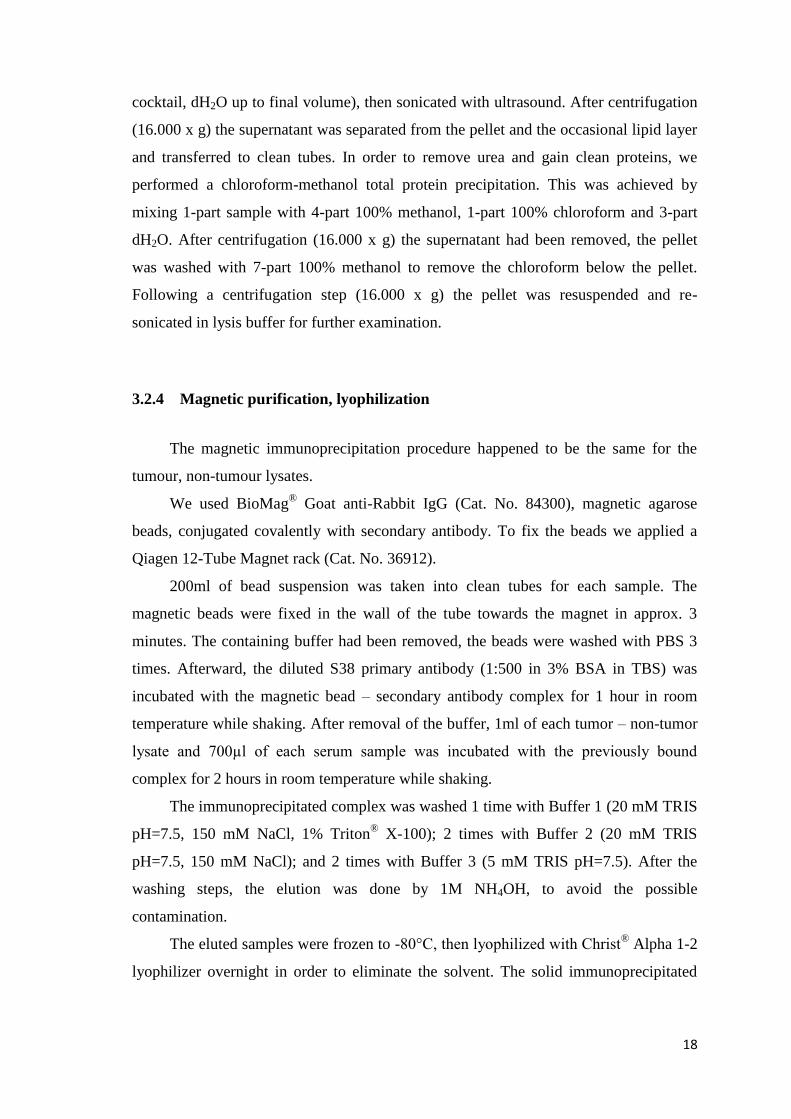
3.2.4 Magnetic purification, lyophilization
The magnetic immunoprecipitation procedure happened to be the same for the
tumour, non-tumour lysates.
We used BioMag® Goat anti-Rabbit IgG (Cat. No. 84300), magnetic agarose
beads, conjugated covalently with secondary antibody. To fix the beads we applied a
Qiagen 12-Tube Magnet rack (Cat. No. 36912).
200ml of bead suspension was taken into clean tubes for each sample. The
magnetic beads were fixed in the wall of the tube towards the magnet in approx. 3
minutes. The containing buffer had been removed, the beads were washed with PBS 3
times. Afterward, the diluted S38 primary antibody (1:500 in 3% BSA in TBS) was
incubated with the magnetic bead – secondary antibody complex for 1 hour in room
temperature while shaking. After removal of the buffer, 1ml of each tumor – non-tumor
lysate and 700µl of each serum sample was incubated with the previously bound
complex for 2 hours in room temperature while shaking.
The immunoprecipitated complex was washed 1 time with Buffer 1 (20 mM TRIS
pH=7.5, 150 mM NaCl, 1% Triton® X-100); 2 times with Buffer 2 (20 mM TRIS
pH=7.5, 150 mM NaCl); and 2 times with Buffer 3 (5 mM TRIS pH=7.5). After the
washing steps, the elution was done by 1M NH4OH, to avoid the possible
contamination.
The eluted samples were frozen to -80°C, then lyophilized with Christ® Alpha 1-2
lyophilizer overnight in order to eliminate the solvent. The solid immunoprecipitated

19
samples were re-dissolved in 5x Sample buffer [0,25 M Tris-HCl, pH: 6.8; 0,5 M DTT;
10% SDS; 50% Glycerol; 0,5% bromophenol blue].
3.2.5 Western blotting
Sodium dodecyl sulfate-polyacrylamide gel electrophoresis was performed in
10% gel, with Bio-Rad® Mini-PROTEAN Tetra cell with the current of 120 mV in 1X
Running Buffer (250 mM Tris, 1.92 M Glycine, 1% SDS). The run proteins were then
transferred to nitrocellulose membrane for blotting using semi-dry method. After the
visualization with Ponceau dye, the remaining membrane surface was blocked by 10%
milk powder and 6% BSA in TBS at room temperature, for 2 hours. The membranes
then were welded into plastic bags, together with 1 ml of the primary antibody, S38 or
S48, respectively, diluted in 1:500 ratio in 3% BSA – TBS for overnight at 4°C.
Afterward, the nitrocellulose membranes were placed into secondary antibody (EIA
Grande Anti-Rabbit IgG Horseradish Peroxidase Conjugate, Cat. No. 172-1019) diluted
in 3% BSA – TBS, incubated for 2 hours in room temperature. Finally, the membranes
were visualized with ImmunoCruz Western Blotting Luminol Reagent, at the
Szentágothai Research Center with a Fujifilm LAS 4001 multipurpose CCD camera
system. The detected images were merged with the molecular weight marker with
Adobe Photoshop CC 2018.
3.2.6 Immunohistochemistry
Human breast tumour tissues were fixed in 4% buffered formaldehyde.
Dehydration was performed with Leica automatic instrument. Tissues were embedded
in paraffin, slices were cut with a thickness of 3-4 m and put onto silanized slides.
Deparaffination was performed three times for 5 minutes in xylene, following three
times 5 minutes washing with ethanol in decreasing concentration (90%-70%-50%).
Then the samples were washed in distilled water for 3 minutes. Antigen retrieval was
performed in citrate buffer pH=6.0 three times for 5 minutes. Samples were
microwaved for one minute at 750 W. Then the samples were washed in 0.5 M Tris
buffer pH=7.6 three times for 2 minutes. Inhibition of endogen peroxide was achieved
in 3% hydrogen peroxide for 10 minutes at room temperature. Samples were washed in
0.5 M Tris buffer pH=7.6 three times for 2 minutes and then were blocked in 1 % horse

20
sera diluted in 0.5 M Tris buffer pH=7.6. Slices were dried on air and were incubated
overnight at 4 °C in the presence of primary antibody. (S38 and S48, 1:200 dilution)
diluted in 1% horse sera in 0.5 M Tris buffer pH=7.6. Visualization was made with
EnVision TM
FLEX, High pH (Dako Autostainer/Autostainer Plus). Background
staining was according to Mayer’s Hematoxylin staining for 3 minutes. Rehydration
was performed in increasing concentration of ethanol (50%-70%-90%), then samples
were washed in xylene three times for 5 minutes. Finally, the slices were covered with
Pertex Mounting Media (Leica Biosystem).

21
3.3 RESULTS
3.3.1 Western blotting
Preliminary data has shown that filensin appears in “in vitro” cultured human
tumour cell lines. Following this logic, we examined human ex vivo clinical samples,
precisely breast cancer tumour samples from cancer patients.
In this study, we have proved with immunoblotting, that BFSP1 is present in
tumour samples (T1, T2, T3). The non-tumour tissue was used from the same person
(mainly from behind the nipple-areola complex) as a control. We proved the absence of
filensin within the normal tissue with both types of antibodies. (N1, N2, N3).
Fig. 5.: Western Blot analysis of human ex vivo breast cancer samples performed by two primary
antibodies, S38 and S48, respectively. In the case of S48, the top arrow shows the native BFSP1, while
the bottom arrow shows a proteolytic fragment of the protein, similarly to the S38 membrane.
As a pilot experiment, we performed Western blot from lyophilized tumour and serum
samples as well, from the same individuals, hence proving that the BFSP1, which we
identified, is the same in both types of samples. This provides a good background for
our further investigations with serum.

22
Fig. 6.: This western blot image represents lyophilized tumour and serum samples run simultaneously,
proving that BFSP1 is present in both samples, and shows similar electrophoretic behavior.
3.3.2 Immunohistochemistry
For the identification of BFSP1 protein, immunohistochemistry was performed with the
S38 and S48 anti-BFSP1 antibody. We are presenting our results from three patients
according to their therapy. Samples with the same letter belong together and come from
one patient. Letter A means: the patient did not receive neoadjuvant therapy and the
tumour was presented. Letter B samples are from a patient, who received neoadjuvant
therapy before the surgery but the tumour is still presented. Letter C represents a
patient who received neoadjuvant therapy and the tumour was regressed.
Fig.7. shows those results which come from the tumour-free area of the different treated
patients. Here the structures are kept, we can identify the followings: adipose tissue,
lobules and the stroma (interlobular/intralobular). Here only the ducts, ductules and
acini were stained positively with the antibody, but there were no positive cells in the
surrounding tissues. The different treatment did not have an effect on the number of
positive staining cells in this area. The results are the same with the S38 and S48
antibody.
Fig.8. shows those areas where the tumour was found or where the tumour was
described before the neoadjuvant therapy. In the case of the S38 antibody strongly
positive staining cells can be seen on the first and second pictures, where the tumour
was presented. (Fig. 8 /A, B). The positivity is not well-defined and more extensive than
in Fig. 7. Picture C on Fig. 8, shows significantly less positive staining cells than the
other pictures. This patient got neoadjuvant therapy, and after this, the macroscopical
tumour could not be detected. This can be the explanations of the reduced amount of
100
70
140
T1 T2 T3 S1 S2

23
the positive cells. According to our results so far, BFSP1 protein can be a sensitive
marker in the case of ductal carcinomas.
Fig.7.: Immunohistochemistry with S38 and S48 anti-BFSP1 antibodies. (10x). These pictures are from
the tumour-free area (behind the nipple). Every picture represents another treatment method. (Description
is in the text). Here only the ducts are stained positively with the antibody.
Fig.8.: Immunohistochemistry with S38 and S48 antibodies. (5x) These pictures are from the tumour or
from that area where the tumour was. Every picture represents another treatment method, and the pictures
on Fig.7. and on Fig. 8. with the same letters are from the same patient.

24
Comparing the results of the S38 and S48 antibodies, we found stronger positivity in
those cases where the S38 antibody was used. One possible explanation for this, is the
different binding and recogniton sites, the S48 antibody is bound to the membrane and
recognise only one proteolytic fragment of the protein, while the S38 shows up in the
cytoplasm (Fig.9) and recognise both proteolytic fragments. Since the positiviy in the
tumour cells was stronger with S38 antibody, it suggests that the proteolytic fragment
after the D433 caspase cleavage site has stronger relation with the presence of the
tumour cells.
Fig. 9: These pictures show the results at greater magnification (20x). It is clearly visible that the S38
antibody shows up in the cytoplasm (Picture A), while the S48 antibody is bound to the membrane
(Picture B).

25
3.4 DISCUSSION
The global importance of cancer is unquestionable, considered the second cause
of death worldwide. Breast cancer (BC) is the second most common cancer overall and
the most frequent type of cancer in women worldwide37
. For routine surveillance and
for staging mammography and ultrasonography are commonly used. There are also
different BC markers which can help to predict the prognosis or to select the suitable
therapy. The most common BC markers are the estrogen receptor (ER) and the
progesterone receptor (PR) status. The absence of these receptors is a predictor of a
poor prognosis. Furthermore, today, they are also used and suitable to select hormone
therapy47
. There are several well-established serum markers; as the cancer antigen (CA)
15-3 (MUC-1 antigen) and carcinoembryonic antigen (CEA) levels, which are
determined at diagnosis of systemic recurrence. However, they do not increase in all
patients; a recent study showed increased CA 15-3 and CEA levels in only 55.6 and
36.0% patients at diagnosis of systemic recurrence, respectively48
. Moreover, these
markers are used to help for the detection of distant metastases, however, they have
limited value in diagnosing micrometastases or locoregional recurrences. Several
benign diseases, as well as chemotherapy, seem to influence their levels so they also
suffer from a lack of cancer specificity49
.
Therefore, the identification of markers that could predict tumor behavior is
particularly important in breast cancer, since the determination of tumor markers is a
useful tool for the clinical management of cancer patients, assisting in diagnostic
procedures, staging, evaluation of therapeutic response, detection of recurrence, distant
metastasis and prognosis, helping in the development of new treatment modalities.
The cytoskeleton comprises three major filament systems — microfilaments,
microtubules, and intermediate filaments (IFs), and collectively, these provisions and
maintain cell shape and structure, and are key to important cellular events, including
cell division, movement, and vesicular transport. IFs can be formed from 40 different
subunit proteins. The different types of IFs can be distinguished according to their
localization and protein composition. Intermediate filaments are expressed in various
cells with determined specificity. Due to this phenomenon, IFs can be used as indicators
determining the origin of protein based on the tissue-specific expression pattern in such
cells. IF typing is also a method in cancer diagnosis because of the above-mentioned
properties.

26
Beaded Filament Structural Proteins (BFSPs) belong to the family of
intermediate filament proteins (IF). BFSP1 or filensin is expressed in lens fiber cells
after differentiation has begun. Although their role in lens biology has still not been
clearly defined, these intermediate filament (IF) proteins are essential to the optical
properties of the lens50
. They are also important to its biomechanical properties, to the
shape of the lens fiber cells, and to the organization and function of the plasma
membrane51
. The critical role of these proteins is mainly emphasized by the presence
of cataracts52,53,54
.
Antal Tapodi and coworkers previously examined the biological role of BFSP1 in
the eye lens. Originally, they were about to determine the caspase cleavage events of the
endogenous filensin protein. Achieving this, they cloned and expressed recombinant
BFSP1 in human, commercially available cell lines, namely MCF7, a breast carcinoma
derived- and HeLa, cervical cancer-derived, widely used cell lines. While visualizing
their results via western blot, however, an extra band was observed in the untransfected,
negative control cell lines as well. This surprising discovery raised many further
questions since BFSP1 was only known as an eye lens specific intermediate filament
protein. In 2014 they also analyzed six commercially available cell lines with western
blotting, namely: U-118 MG, U-251 MG (glioblastoma cell lines), A-549 (human lung
cancer), T24/83 (human bladder carcinoma), HeLa and HepG2 (human liver cancer),
and have proven that BFSP1 is present in each of them.
In this study, we continued an ongoing project examining the unexpected
presence of BFSP1 protein in tumour cells. This is the first study, where the expression
of BFSP1 was demonstrated in ex vivo tumour samples and serum as well (Figure 4, 5).
Furthermore, I would like to emphasize, that with Western-blot analysis this protein was
presented only in the tumour samples, and we proved the absence of filensin within the
normal tissue.
With immunohistochemistry, using the S38 antibody, we could confirm that there
is a significant difference in the contents of BFSP1 according to the presence of the
tumour. In those patients, who received/did not receive neoadjuvant therapy but/and the
tumour was presented macroscopically, the number of the positive staining cells
increased considerably, compared with the tissue samples of those patients who
received neoadjuvant therapy and the tumour was regressed. In the case of the S48
antibody our results were not so convincing. The explanation of this can be the different
recognition sequence of the two antibodies. Based on our results it seems that certain

27
part of the BFSP1 protein can be sensitive enough to indicate the tumour cells in the
case of ductal breast carcinomas, but to understand the role of the protein further
investigations are needed.
It is very important to note that, this study is part of a greater project, therefore
our research group has numerous plans according to the future. We are planning to
continue examining the human ex vivo clinical samples, including benign breast
tumours, other types of breast cancers, and other types of cancers, like melanoma and
colorectal cancers. Furthermore, we would like to perform a quantitative real-time PCR
analysis from ex vivo clinical samples as well as mass spectrometry. Investigation of the
pre- and postoperatively (on the 1st and 3
rd week) collected serum, to assess the possible
quantitative changes of the BFSP1 protein after the surgery, is also included in our
future plans.
On the other hand, the biological role of BFSP1 in tumour cells is yet unknown.
One of the major goals of our research team is, to identify the possible interacting
proteins co-precipitated with native BFSP1. Antal Tapodi and his co-workers proved
previously different intracellular distribution of the proteolytic fragments of BFSP1, so
we suppose that BFSP1 fragments might play a different role in tumour cells. Finally,
we can say that the major goal of our ongoing research is to create an affordable
diagnostic tool, which could be used in daily medicine, helping cancer patients in a time
of need.
3.5 CONCLUSION
As a conclusion, so far we can say, that BFSP1 protein is expressed not just in the eye
lens but also in human breast cancers. We examined 25 patients with ductal carcinomas
in this study. With immunohistochemistry, we proved that BFSP1 protein shows
sensitivity for the tumour cells, independently that the patients received neoadjuvant
therapy or not. Furthermore, the same type of the protein is presented in the serum as in
the tissue samples. This study provides a good base for further investigations which can
specify the exact role and the type of splice variant of the BFSP1 protein involved in
cancers, and which can study the presence of this protein, in a different type of cancers.
In the case of ductal carcinomas, the protein can have an important role after the surgery
in the follow-up period of the patients, and it could also be able to provide an adequate
information about the recurrence of cancer from the serum of the patients, although
additional studies are also required in this field.

28
4 THE EFFECT OF TRIMETAZIDINE IN REDUCING THE
ISCHEMIA-REPERFUSION INJURY IN RAT EPIGASTRIC
SKIN FLAP
4.1 INTRODUCTION
Ischemia-reperfusion injury (IRI) can cause considerable problems in various
fields of the surgery, like in reconstructive plastic surgery, vascular surgery,
traumatology or cardiac surgery. IRI is a cascade of pathophysiological events, that can
occur after the reperfusion of the tissues, exposed to prolonged ischemia and results in
tissue damage55,56
. Unfortunately, this condition is unavoidable during free flap surgery
or during replantation. Free tissue transfer has become a routine procedure to cure tissue
defects after oncological ablative surgery or trauma. In the last decade, the technique of
the free flap surgeries improved a lot and it has reached the 90-95% success rate.
Although the success rates of these surgeries are high, there are still some cases, where
the insufficient microcirculation, caused by IRI, leads to partial flap loss and results in
the reoperation of the patient. In addition, the flap/limb can become irremediable
because its poor circulation, and it may make the reconstruction more difficult or
impossible11-15
. For these reasons, the detection of biochemical changes and
microcirculatory disorders in flaps during I/R, are of high importance16-17
.
Even though many drugs and methods have shown promising results
experimentally, there hasn’t got any existing consensus treatment in the clinical
practice, because of their unfavourable systemic side effects, excess toxicity, limited
efficacy, invasive administration or because of the time-consuming technique57,58,59
,60,61,62.
Trimetazidine (TMZ, water-soluble form: trimetazidine-dihydrochloride) is a
widely used anti-anginal drug worldwide. It is a potent anti-ischemic agent and a free
radical scavenger. It has been used in many studies63,64,65
to protect different organs
(myocardium, intestine, liver, and kidney) from the ischemia-reperfusion injury.
Numerous evidence exists, which shows that the reperfusion injury could be decreased
by TMZ-preconditioning in animals. It was found that TMZ conserves ATP production,
maintains cellular homeostasis and reduces the intracellular acidosis. Moreover, it
decreases the oxidative damage to the mitochondria and protects the organ from tissue
damage, induced by IRI31,66
.

29
According to the previous studies, we believe that a single shot of TMZ will be
protective against IRI also in our study. This study aimed to investigate the effect of
trimetazidine on oxidative stress, inflammation, and histopathological alterations
(before visible changes (e.g. tissue necrosis) occur), using the epigastric skin flap
model. To determine the efficacy of TMZ, levels of blood malondialdehyde (MDA),
reduced glutathione (GSH), and plasma thiol groups (SH-) and tissue TNF-alpha were
measured, histopathology and immunohistochemistry were performed.
4.2 MATERIALS AND METHODS:
4.2.1 Animal model
Forty male Wistar rats of the same age, weighing between 350 to 400 g, were
used for this study. The rats were housed in separate cages, under standard conditions
(temperature: 25±2 °C, and air filtered room), with 12/12-hour light-dark regimen and
were fed with standard rat chow and water ad libitum. Food was withdrawn 12 hours
prior to the experiment. The study protocol was approved by the National Scientific
Ethical Committee on Animal Experimentation. (number: ZOHU0104L 16)
4.2.2 Experimental protocol
The animals were divided randomly into four groups (10 rats in each group). The
first group was the non-ischemic control group. Although the control flaps did not
undergo ischemic insult, flap harvest may produce some temporary ischemia. In the
other groups (groups 2 through 4) ischemia was induced by placing a single
microvascular clamp across the epigastric superficial artery and vein. In the second
group (I/R) the superficial epigastric vessels were clamped for 6 hours, followed by 24
hours of reperfusion. The third (Preisch.TMZ + I/R) and fourth (I/R+Postisch.TMZ)
groups were the trimetazidine treated groups. In the third group, the TMZ was
administered 30 minutes prior to the ischemic period. In the last group, animals received
the drug at the onset of the reperfusion (Fig. 10.). To standardize the study, all
procedures were performed at similar time points in all groups. Animals, in the treated
groups, received 10 mg/kg trimetazidine (trimetazidine-dihydrochloride, Sigma-
Aldrich, St. Louis, Missouri, USA) intraperitoneally (i.p) depending on the groups, 30

30
minutes prior to ischemia (Preisch.TMZ+I/R) or at the onset of the reperfusion
(I/R+Postisch.TMZ). The drug was freshly solved into 0,9 % NaCl solution before the
administration.
Fig. 10.: Investigation groups: Group 1: Control, Group 2: Ischemia-reperfusion (I/R), Group 3: Preisch.
TMZ + I/R, Group 4: I/R + Postisch. TMZ
4.2.3 Surgical procedure
The rats have perioperatively anesthetized with an intraperitoneal (i.p) application
of a mixture consisting of ketamine hydrochloride (5 mg/100g) and diazepam (0,5
mg/100g). The ratio was 1:1. The skin of the abdomen was depilated using an animal
depilatory agent. During the operation, the animals were placed on a heated pad and
ECG monitoring was also used. The carotid artery was catheterized (22 gauge) for
blood pressure measurement. (Siemens Sirecust 1260, Düsseldorf, Germany). The skin
of the abdomen was scrubbed with betadine and then 3x6 cm flap was created on both
sides of the abdomen. In our study, the epigastric flap was chosen, because it simulates
microsurgical free tissue transfer closely. This model was first described in 1967 by
Strauch and Murray and has been widely used in various experimental animal
researches on IRI and skin flap survival19,67,68
. The flaps include the area within the
boundaries of the costal arch as an upper limit, the inguinal ligament as a lower limit

31
and both axillary lines as lateral borders. The medial borders were on both sides of the
midline structures (the xiphoid and pubis). The vascular supply of the flap is provided
by the medial and lateral branches of the superficial epigastric artery and accompanying
veins, based on the superficial epigastric vascular pedicle. After 6 hours of ischemia, the
microvascular clamp was released, and the blood flow was confirmed by arterial
pulsation, flap colour, and vascular patency tests was also performed to ensure that the
blood flow is recovered successfully. Flaps, where we could not detect any flow, were
not included in this study. After checking the blood flow, the skin was sutured back to
its original place with interrupted stitches (5-0, Prolene®
(Ethicon), 30 stitches on both
flaps). After the operation, the animals got a collar neck to prevent the automutilation.
On the next day, before the sampling, animals were re-anesthetized.
Skin samples (3x1 cm) were taken from the most distal end of the flaps, after 24
hours of reperfusion, for biochemical examination. The samples were stored
immediately at -80 °C within individual containers.
4.2.4 Biochemical analysis
MDA, GSH, SH levels were measured from the blood. MDA is a marker for the
quantification of membrane lipid peroxidation. MDA levels were detected using a
photometric method of Placer, Cushman and Johnson69
. GSH and plasma SH levels
were determined in anticoagulated whole blood by Ellman’s reagent, according to the
method of Sedlak and Lindsay70
. Both indicate the antioxidant status of the body.
To measure the TNF-alpha levels, samples were taken from the central part of the
flap. Tissue TNF-α (one of the indicators of the inflammatory response) levels were
studied by using the Rat TNF-α ELISA Kit (Abcam, Cambridge, UK) following the
manufacturer’s protocol.
4.2.5 Histopathological analysis
A histopathological study of the samples was carried out by the same pathologist.
The tissue samples were fixed in 4% neutral buffered formaldehyde solution and
embedded in paraffin. Three-micron-thick (Microtome: Thermo Scientific Microm Hm
325) histological sections were cut, mounted on glass slides, stained with hematoxylin-
eosin (HE) and evaluated by light microscope to quantify foreign body giant cells,

32
polymorphonuclear, and mono-nuclear reactive cells. For detection of apoptosis,
TUNEL (Terminal deoxynucleotidyl transferase dUTP nick end labeling) was also
performed.
4.2.6 Hemorheological analysis
4.2.6.1 Red blood cell deformability
In our study, we measured red blood cell deformability with LORCA
ektacytometer. The blood sample was suspended in a high viscosity (28-32 mPas)
polyvinylpyrrolidone solution and injected into the gap between the two cylinders. A
laser beam transversing the suspension creates a diffraction pattern on a diaphragm that
is recorded and analyzed by a video camera and a computer controlled ellipse fitting. At
rest, red blood cells show circular diffraction pattern and parallel to the applied shear
stress it becomes elliptical as cells deform and elongate.
For analysis, elongation index (EI) was calculated as the (length - width) / (length
+ width) of the pattern for each shear stress (SS) at 9 different shear stresses (from 0.3
to 30 Pa). For data analysis, the Lineweaver-Burke nonlinear curve fitting technique
was used to calculate the maximal EI (EImax) value at extrapolated infinite shear and
the shear stress value required for half of EImax (SS1/2). EI ranges from 0 to 1, 0 refers
to an undeformed, randomly oriented RBC, and EI increases with cell deformation.
Higher EImax refers to higher deformation ability, while RBCs with higher SS1/2 are
harder to deform. Measurements were performed at 37°C 71,72,73
. This micro-rheological
parameter is an important determinant of the microcirculatory pattern.
Blood samplings were performed from lateral tail veins, and hemorheological
examination was carried out before the surgery and on the 1st, 4
th and 7
th postoperative
days, under anesthesia.
4.2.7 Statistical analysis
For statistical evaluation, one-way analysis of variance (ANOVA) was used,
followed by adequate post hoc tests (Dunnett’s, Sidak) for multiple comparisons. All
data are represented as the mean ± SEM. The difference was considered statistically
significant when the p-value was less than 0.05.

33
4.3 RESULTS:
4.3.1 Changes of oxidative stress parameters in blood samples
4.3.1.1 Malondialdehyde
The statistical analysis of the MDA levels showed reduced values both in pre-and
postischemic trimetazidine treated groups compared to the I/R group, however, a
significant decrease was shown only in that group where the TMZ was administered
prior to the ischemia (59,84±2,8 vs. 75,3±6,4; p=0,0145). These results refer to smaller
lipid peroxidation in the treated groups. MDA levels were considerably higher only in
the I/R group, compared to the control (75,3±6,4 vs. 50,85±1,4; p<0,0001). The results
of the treated groups were nearly as good as in the control group. (Fig.11)
Fig. 11.: Malondialdehyde concentrations in the experimental groups. MDA serves as a marker of the
lipid peroxidation. ˙: p<0,05 vs. control; : p<0,05 between the signed groups; error bars: SEM

34
4.3.1.2 Reduced Gluthatione
GSH levels were reduced in all groups comparing to the control (Control:
1116±38,09). Significantly higher GSH levels were measured both in pre-and
postischemic trimetazidine treated groups (preisch. TMZ: 965,5±6,3, p=0,0035;
postisch. TMZ: 1002±38,6, p= 0,0002 vs. 820,9±13,5) compared to the I/R group,
which supported the antioxidant effect of the drug. (Fig.12)
Fig. 12.: Plasma concentration of reduced glutathione in the investigated groups. GSH serves as a marker
of the antioxidant status. : p<0,05 vs. control; : p<0,05 among the signed groups; error bars: SEM

35
4.3.1.3 Sulfhydryl group (SH-)
There were no significant differences in the SH- levels among the groups (control:
94,03±8,584; I/R: 74,3±3,763; preisch.TMZ: 98,62±11,4; postisch.TMZ: 91,65±6,5).
(Fig.13)
Fig.13.: Concentrations of SH- groups in the plasma. The levels of SH- refer to the antioxidant status.
Error bars: SEM

36
4.3.2 Changes of TNF-α level in skin samples
Comparing to the control group, except the Preisch. TMZ+ I/R group,
significantly higher values were measured (Control: 35093±4640). Considerable
decrease of TNF- α levels in the treated groups were noticed, compared to the I/R group
(preisch. TMZ: 41243±2183 p=0,0001; postisch. TMZ: 54025±5924 p=0,0437 vs.
73331±5762), which can prove the anti-inflammatory effect of the drug. (Fig.14)
Fig. 14.: TNF-alpha concentrations show the grade of the inflammatory response in the investigated
groups. : p<0,05 vs. control; : p<0,05 among the signed groups; error bars: SEM

37
4.3.3 Histopathological results
4.3.3.1 Hematoxylin-eosin
Our histopathological findings correlate with the biochemical results. Four zones
are identified in all tissue samples. (Fig.15, Control) In the control group, the basic
tissue structures mainly kept, oedema, necrosis or significant inflammation cannot be
detected.
In the I/R group (Fig. 15, I/R) many changes can be noticed: oedema was
occurring in the fatty zone and in the submuscular zone. A large number of
polymorphonuclear (PMN) cells could be seen under the muscle. The muscle fibres
were swollen and irregular-shaped.
In both TMZ treated groups significantly fewer tissue changes were seen than in
the I/R group. The muscle fibres were approximately normal shaped, oedema and PMN-
cells were barely detected in the different zones. (Fig 15; Preisch. TMZ+I/R,
I/R+Postisch. TMZ)
Fig. 15.: Staining: HE, magnification: 5x. In the control group, the four zones can be clearly identified:
A: epidermal-dermal zone; B: fatty zone; C: muscular zone (B+C=panniculus carnosus), D: submuscular
zone. In the I/R group oedema can be seen in the submuscular and fatty zone and the muscle fibres are
swollen and irregular-shaped in the zone C. The protective function of the TMZ is well demonstrated in
both (Preisch. TMZ+I/R and I/R+Postisch. TMZ) groups, showing less changes in the tissue samples:
muscle fibres are approximately normal shaped, oedema and PMN-cells are barely detected in the
different zones.

38
4.3.3.2 TUNEL- staining
The good influence of the drug is also supported by TUNEL staining (Fig 16).
TUNEL-positive nuclei were stained brown. In the control group, the high number of
positive cells are physiological, because they are showing up only in the follicle of the
skin and these are holocrine glands. (Fig.16; Control). In the I/R group (Fig.16, I/R)
many apoptotic cells were found in every zone of the flap. This confirms that I/R also
promotes the apoptosis. The TMZ management of skin flaps clearly decreased the
quantity of the apoptotic cells. Apart from the epidermal-dermal zone, where apoptotic
cells can be found physiologically, the number of the positive cells were considerably
fewer in the treated groups, compared to the I/R group. (Fig 16; Preisch. TMZ+I/R,
I/R+Postisch. TMZ)
Fig.16.: Staining: TUNEL, magnification 10x: TUNEL staining demonstrates the apoptotic nuclei. 1. In
the control group the high number of positive cells, showing up only in the follicle, are physiological -
since these are holocrine glands. 2. The homogeneous positivity in the I/R group is the evidence to
demonstrate the damage in the tissue, caused by the ischemia/reperfusion. 3. The protective function of
the TMZ is well demonstrated in both (Preisch. TMZ+I/R and I/R+Postisch. TMZ) groups, showing
barely positivity in all investigated zones.

39
4.3.4 Changes in hemorheological parameters
Figure 17. illustrates the red blood cell deformability changes. The curve shows
the elongation index (EI) of the red blood cells in the function of shear stress (SS). The
preoperative and 7th postoperative days parameters did not differ; at most of the shear
stress values the parameters were overlapping. However, on the 1st and mainly on the 4
th
postoperative day, the red blood cell deformability values were markedly worsened,
dominantly in the I/R group.
Fig. 17.: Deformability of red blood cells in different groups preoperatively (Base) and on the 1st, 4
th, and
7th
postoperative days. The graph shows the elongation index (EI) of the red blood cells in the function of
shear stress (SS)

40
4.4 DISCUSSION
The use of microvascular flap tissue transfer is very popular to reconstruct the
defects of the whole body. It is known, that the success rate of the microsurgical
vascular anastomosis, even with experienced surgeons is 90 to 95 percent, however,
some severe problem such as IRI and the inadequate blood perfusion may still impede
the complete success. IRI can cause severe problems in the microcirculation and it may
lead to patient’s morbidity and prolonged hospitalization. The intracellular biochemical
changes that occur during the ischemic period can cause cellular dysfunction, cellular
and interstitial oedema and finally can lead to cell death. The severity of these changes
depends on the length of the ischemic time since it is well known that brief ischemic
condition can be protective against the negative alterations74
. During reperfusion,
following the ischemic period, reactive oxygen species are produced, which include
oxygen ions, free radicals, and peroxides, all of which worsen ischemia-reperfusion
damage75,76
, impact on red blood cells micro-rheological parameters and may result in
considerable disturbance of blood flow77,78,79
. In the pathogenesis of I/R injury
inflammation is also considered to be a critical element80,81
.
In our study, we chose the superficial epigastric skin flap model, because it was
suitable to simulate a clinical situation, that occurs when microsurgical tissue transfer is
made. As Yoshida and Campos suggested the model could also simulate a vascular
pedicle thrombosis, where the procedure from the diagnosis to the restoration of blood
supply could reach or exceed 6 hours, or it also can simulate a traumatic situation when
replantation of amputated fingers is made82
. In these types of models, flaps contain the
epidermal-dermal zone, fatty zone, muscular zone (panniculus carnosus) and
submuscular zone with a vascular pedicle of the superficial inferior epigastric artery and
vein. There are controversies related to the position of the microvascular clamp. They
could be used both on the artery and on the vein, or separately on the vein or on the
artery to simulate different situations, which can occur in the clinical practice. Our
experimental model based on superficial inferior epigastric artery and veins to reach a
higher level of I/R injury. The extension of the flaps was 6,0 x 3,0 cm bilaterally.
The length of the ischemic time was based on the literature83
; ÇetIn et al.78
subjected the rats to 6 hours and 10 hours of ischemia, because these time points have
been reported to produce consistent biochemical, histopathological and macroscopic
findings84
.
#

41
TMZ is a potent anti-ischemic drug, which decreases fatty acid oxidation and
stimulates glucose utilization via the inhibition of the mitochondrial long-chain 3
ketoacyl-CoA thiolase, leading to the production of adenosine triphosphate (ATP) with
less oxygen consumption. It limits intracellular acidosis, decreases sodium and calcium
accumulation into cells, inhibits the extracellular leakage of potassium during cellular
ischemia and reduces cytolysis and membrane injury caused by oxygen free radicals. In
addition, TMZ conserves mitochondrial function and energy metabolism and it is
capable of inhibiting platelet adhesion-aggregation and neutrophil infiltration 29,85,86
.
Because it does not have a negative alteration on the hemodynamic status, besides the
cardiology, probably it can also be useful in other areas of the clinical practice.
Previously, the effect of the TMZ on the survival of skin flaps was already studied
and the agent was proved to be effective. Nieto et al. investigated various
pharmacological agents on the survival of skin flaps in rats. All treated groups showed a
significantly greater survival of the flap than the control group. One of the best
outcomes was shown in those groups receiving trimetazidine and hydralazine87
. Kara et
al. studied the effect of trimetazidine on the survival of rat island skin flaps. They
compared the pre-ischemic and post-ischemic effect of the drug, and both ways seemed
to be effective to improve flap survival88
.
However, this is the first study where, before the visible tissue changes, the
histological and biochemical alterations were investigated after pre-and postischemic
TMZ treatment in skin flaps. Blood MDA, GSH, and SH- levels and tissue TNF-α
levels were evaluated for biochemical analysis. MDA is a stable product of
polyunsaturated lipid peroxidation in cells, that is generated after free radical damage.
GSH is one of the major endogenous antioxidants produced by the cells, participating
directly in the neutralization of free radicals and reactive oxygen compounds. The
serum levels of protein -SH in the body, can indicate antioxidant status. TNF-α is a
polypeptide compound and it is an important member of the cytokine family, which
plays a significant role in the regulation of the systemic inflammatory response.
The micro-rheological parameters, such as red blood cell deformability is
influenced by the effect of ischemia-reperfusion. Red blood cell deformability is a
pivotal ability of the cells to pass the capillary system which is required for sufficient
tissue oxygenation. Deformability is determined by the internal viscosity of the cell, the
membrane viscoelasticity, the surface-volume ratio and the morphology of erythrocytes.
Mostly on the 1st – 4
th postoperative days changes in red blood cell deformability are

42
related to the inflammatory reactions, hemodynamic alteration, induction of free-
radicals and mediators, acute phase reactions and changes in the coagulation state. In
the early hours of reperfusion metabolic and free radical alterations are more dominant.
All these factors can further aggravate the postoperative complication of microvascular
tissue transfer. Pathologically altered red blood cells show a reduction in their
deformability and may lead to capillary occlusion and decreased oxygen supply for the
tissues. Most likely, most of these reactions (metabolic disturbance and induction of
free radicals) are passed off by the 7th postoperative day74
; thereby we did not find any
definitive difference on this day.
In the literature, there are controversies in the administration routes and doses of
the TMZ89,90,91
. In our study 10 mg /kg dose was chosen and the drug was administered
intraperitoneally, based on some previous studies where this dose was proved to be
effective 74,92
. The timing was also different in many studies. For example, Khan and
colleagues90
published that TMZ was cardioprotective (via the activation of p38
mitogen-activated protein kinase and Akt signaling pathway) when administered at the
beginning of the reperfusion period. Elimadi et al.92
investigated the effect of TMZ on
hepatic warm I/R injury, administered as an intramuscular injection with different doses
(5 mg, 10 mg, 20 mg). They demonstrated that 10 mg/kg/day for 7 days before the
induction of ischemia was the optimal dose, that gave the maximal protective effects at
both the cellular and mitochondrial level. All these observed differences among the
studies could be a consequence of different animal models, examined organs and I/R
protocols. Further investigations are required to determine the optimal time and dose of
administration of TMZ and to have more insight into clinical application.
In our study, we hypothesized that a single shot of TMZ will be preventive against
I/R injury in epigastric skin flaps. Since in the previous studies the timing of the
administration of TMZ was different, we investigated both pre- and postischemic TMZ
treatment. Our data confirm the earlier findings, that TMZ has anti-inflammatory and
anti-ischemic effects, independently of the timing. It could be a useful drug in the
surgical practice to increase the survival time of the tissues, not just given before a
planned ischemic period but also after an unexpected trauma where a reconstructive
surgery is required.

43
4.5 CONCLUSION
The harmful effect of I/R can occur in the skin flaps without macroscopically
visible changes (e.g.: tissue necrosis).
According to our results, TMZ is shown to be protective, against I/R injury and it
is also suitable to decrease the inflammatory response. The administration of TMZ was
effective independently of the timing: there is no unambiguous difference between the
preischemic and postischemic TMZ administration. The beneficial effect of the
postischemic administration can be especially important, because it can protect the
tissues from ischemia-reperfusion injury, even after an unexpected ischemic insult.
Furthermore, TMZ is a clinically applied and nontoxic agent, which may increase
the ischemic tolerance of the tissues and it is a promising drug to decrease the negative
consequences of I/R in the surgical practice during free tissue transfer, replantation or
even during revascularization procedures.

44
5 COMPARISON OF THE EFFECT OF TRIMETAZIDINE WITH
ISCHEMIC PRE- AND POSTCONDITIONING IN REDUCING
THE ISCHEMIA-REPERFUSION INJURY IN RAT SMALL
INTESTINE
5.1 INTRODUCTION
Since 1959, when the first free jejunal flap was used by Seidenberg93
to
reconstruct a cervical oesophageal defect, jejunal flaps have commonly been used for
the reconstruction of oesophagus after cancer resections. Their main advantages are the
followings: tubular structure similar to the oesophagus, available length, production of
mucus, lack of functional gastrointestinal complication after removal, peristaltic activity
similar to the pharyngoesophagus and the ease of preoperative preparation12,94
. Besides
the oesophagus, the jejunal flap is also suitable for vaginal reconstruction (in the case of
congenital absence of the vagina, sex assessment surgery, surgical resection of tumors),
because it provides a durable, stable coverage and a tube passage for sexual intercourse.
In these cases, jejunum transfer is a primary choice contrary to other intestinal transfers,
particularly because it causes fewer defecation problems95,96
. However, the use of
jejunal flaps is challenging, because of their sensitivity to ischemia and other technical
details related to the operation. During the preparation of flaps, segments of the jejunum
are removed and exposed to a period of warm ischemia until revascularisation.
Although short ischemia times are associated with minimal damage, the injury from
warm ischemia progresses even after revascularization. Ischemia-reperfusion injury
(IRI) is a complex, multifactorial cascade of pathophysiological events, which include
the depletion of oxygen to cells, low adenosine-triphosphate (ATP) stores, accumulation
of toxic metabolites, reactive oxygen species, various cytokines, and other mediators,
leading to further cellular dysfunctions. This structural injury might then endanger the
functional outcome of the flap. Consequently, I/R induced tissue injuries are significant
problems that might lead to different complications such as, segmental stenosis, fistula
formation, peristalsis dysfunction, anastomosis leakage, and partial or complete flap
failure97,98,99
. In spite of several suggested strategies/methods, so far effective, widely
used method is not clinically available as a solution to this problem.
Ischemic preconditioning (IPreC) has been proved to produce resistance to the
loss of blood supply and this method is able to improve the survival of the tissues,

45
subject to global ischemia. During IPreC brief period of ischemia followed by
reperfusion is used, which increase the ischemic tolerance of the tissues against the
detrimental effects of subsequent prolonged ischemia. First described by Murry100
and
colleagues for myocardial tissue, since then, IPreC has been shown to increase the
survival of a variety of flaps, subjected to ischemia101,102,103
. Beside the IPreC, ischemic
postconditioning (IPostC) was also proved to be as effective as IPreC to improve the
viability of the different tissues and organs/flaps after a prolonged ischemic insult. The
main advantage of this method, that it is also able to reduce the degree of the damage
even after an unexpected event.
Trimetazidine (TMZ) is a potent anti-ischemic drug and free radical scavenger,
which besides the heart, has been protective in different organs against ischemia-
reperfusion injury104,105
. Many studies have suggested that TMZ is able to reduce the
intracellular acidosis, preserves the ATP production, limits the inflammatory reaction,
the generation of reactive oxygen species and prevents the calcium overload of the cells,
which can lead to further damage.29,106
Our aim was to investigate the results of chemical preconditioning and
postconditioning with trimetazidine, in a jejunal-flap model in the rat, compared its
efficacy with ischemic preconditioning and ischemic postconditioning in reducing the
oxidative stress and inflammation. Furthermore, the presence of any additive effects of
simultaneous IPrec and TMZ; or IPostC and TMZ administration in improving the level
of ischemic protection was also evaluated in this study.
5.2 MATERIALS AND METHODS
5.2.1 Animal model
Eighty male Wistar rats of the same age, weighing between 400 to 450 g, were
used for this study. The rats were housed in separate cages, under standard conditions
(temperature: 25±2◦C, and air filtered room), with 12/12-hour light-dark regimen and
were fed with standard rat chow and water ad libitum. Food was withdrawn 12 hours
prior to the experiment. The study protocol was approved by the National Scientific
Ethical Committee on Animal Experimentation (number: ZOHU0104L 16).

46
5.2.2 Experimental protocol
In the first part of this experiment (A), we compared the effect of IPreC with
TMZ preconditioning. The animals were divided randomly into five groups (10 rats in
each group). The first group (Group 1) was the nonischemic control group. Although
the control flaps did not undergo ischemic insult, laparotomy was performed. In the
other groups (groups 2 through 5) ischemia was induced by placing a single
microvascular clamp across the superior mesenteric artery (SMA). In the second group
(I/R; Group 2) the SMA was clamped for 40 minutes, followed by 1 hour of
reperfusion. The third (Preisch.TMZ+I/R; Group 3) group was the trimetazidine treated
group. In this group, the TMZ was administered 30 minutes prior to the ischemic
period. In the fourth group (IPreC+ I/R; Group 4) ischemic preconditioning was used:
2x5 min. (2 cycles of 5 minutes ischemia then 5 minutes of reperfusion) before the
onset of ischemia. In the last group (TMZ, IPreC+ I/R; Group 5), animals received the
TMZ 30 minutes prior to the ischemia and ischemic preconditioning was also
performed, as mentioned above. (Fig.: 18)
Fig. 18.: Investigation groups for the comparison of TMZ and IPreC (first part of the study, A)

47
In the second part of the study (B), we compared the effect of IPostC with the
TMZ postconditioning, and the additive effect of these two methods was also evaluated.
Group 1 (nonischemic control); Group 2 (I/R) were the same as in the part A. However,
the other groups were different: Group 3 (I/R + TMZ) was the TMZ treated group, but
in this case, it was administered at the onset of reperfusion. In the fourth group (I/R+
IPostC) ischemic postconditioning was used: 3x30 sec (3 cycles of 30 secundum
reperfusion, followed by 30-sec ischemia). In the last group (I/R+TMZ, IPostC) animals
received TMZ at the onset of reperfusion and ischemic postconditioning was also
performed, as mention above. (Fig.: 19)
Fig. 19.: Investigation groups of the comparison of TMZ and IPostC. (second part of the study, B)
To standardize the study, all procedures were performed at similar time points in all
groups. Animals, in the treated groups, received 10 mg/kg trimetazidine (trimetazidine-
dihydrochloride, Sigma-Aldrich, St. Louis, Missouri, USA) intravenously (i.v), through
the jugular vein. The drug was freshly solved into 0,9 % NaCl solution before the
administration.

48
5.2.3 Surgical procedure
The rats have perioperatively anesthetized with an intraperitoneal (i.p) application
of a mixture consisting of ketamine hydrochloride (5 mg/100 g) and diazepam (0,5
mg/100 g). The ratio was 1:1. During the operation, the animals were placed on a heated
pad and ECG monitoring was also used. The carotid artery was catheterized (22 gauge)
for blood pressure measurement. (Siemens Sirecust 1260, Düsseldorf, Germany). The
skin of the abdomen was scrubbed with betadine and then laparotomy was performed.
Then, the superior mesenteric artery was explored. Collaterals from the right colic and
jejunal arteries were ligated as described by Megison et al107
(Fig. 20/A). Then
according to the different groups, except in the control group, IRI was induced by
placing a clip on the superior mesenteric artery for 40 minutes and trimetazidine,
ischemic preconditioning/ischemic postconditioning or both were used. During the
ischemic period the bowels were placed back into the abdominal cavity and the skin
incision was temporarily closed.
After 40 minutes of ischemia, the microvascular clamp was released and the blood
flow was confirmed by arterial pulsation, jejunal colour, and vascular patency tests were
also performed to ensure that the blood flow is recovered successfully. The jejunal
segment was reperfused for 60 minutes. At the end of the reperfusion period,
approximately 10 cm segment of the jejunum (15 cm proximal to the ileocecal valve)
was harvested. The resected jejunal segment was soaked in saline and then it was
divided into 2 parts. One half was fixed in 4% neutral formaldehyde for
histopathological examination and evaluation of apoptosis. The other half was instantly
frozen in liquid nitrogen and stored at -80 °C for further biochemical examinations.
Fig. 20.: On picture A, the schematic illustration of the model can be seen, described by Megison et al.
Picture “B” and “C” are intraoperative pictures of the bowels before (B) and after (C) the clip application.
Picture “B” represents a bowel with normal blood supply and normal color. Picture “C” shows bluish/pale
discoloration of the bowel, because of the clamp.

49
5.2.4 Biochemical analysis
Jejunal segments and blood were taken for biochemical and histopathological
examinations after 1 hour of reperfusion. The samples for the biochemical purpose were
stored immediately at –80◦C within individual containers. The tissue samples were
stored in formalin, until the histopathological evaluations.
5.2.4.1 Oxidative stress parameters
MDA, GSH, SOD, and SH- levels were measured from the serum. MDA is a
marker for the quantification of membrane lipid peroxidation. MDA levels were
detected using a photometric method of Placer, Cushman, and Johnson69
. GSH and
plasma SH levels were determined in anticoagulated whole blood by Ellman’s reagent,
according to the method of Sedlak and Lindsay70
. Both indicate the antioxidant status of
the body.
For the measurement of superoxide dismutase enzyme (SOD) activity from
serum, the OxiSelect™ Superoxide Dismutase Activity Assay was used, following the
manufacturer’s protocol. (Cell Biolabs Inc., STA-340)
5.2.4.2 Inflammatory cytokines
The inflammatory cytokine levels (TNF-α, IL-6) were studied by using the
enzyme-binding immunosorbent assay (ELISA) method, following the manufacturer’s
protocol (Assay Rat TNF-α ELISA kit, #AB46070; Assay Rat IL-6 ELISA kit,
#AB119548, Abcam, Cambridge, UK).
5.2.5 Histopathological analysis
The histopathological study of the samples was carried out by the same
pathologist. The tissue samples were fixed in 4% neutral buffered formaldehyde
solution and embedded in paraffin. Three-micron-thick (Microtome: Thermo Scientific
Microm Hm, 325) histological sections were cut, mounted on glass slides, stained with

50
hematoxylin-eosin (HE) and evaluated by light microscope to quantify intestinal
mucosal injury, inflammation, necrosis or ulceration. Mucosal damage was graded from
0 to 5, based on the histologic injury scale, determined by Chiu et al108
.
According to this: grade 0: Normal mucosal villi,
grade 1: Development of subepithelial Gruenhagen’s space at the
apex,
grade 2: Moderate lifting of the epithelial layer at the apex of
villi/Extension of subepithelial space;
grade 3: Massive epithelial lifting down the side of villi;
grade 4: Denuded villi and dilated capillaries/increased cellularity
of lamina propria;
grade 5: Disintegration of lamina propria/ Hemorrhage and
ulceration.
For the detection of apoptotic cells, TUNEL (Terminal deoxynucleotidyl transferase
dUTP nick end labeling) staining was also performed.
5.2.6 Statistical analysis
For statistical evaluation, one-way analysis of variance (ANOVA) was used,
followed by adequate post hoc tests (Dunnett’s, Sidak) for multiple comparisons. All
data are represented as the mean±SEM. The difference was considered statistically
significant when the p value was less than 0.05.
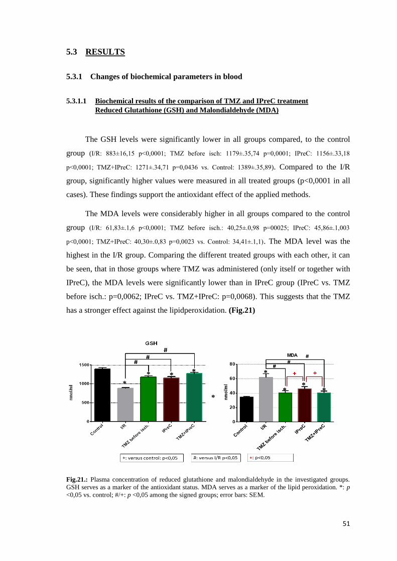
51
5.3 RESULTS
5.3.1 Changes of biochemical parameters in blood
5.3.1.1 Biochemical results of the comparison of TMZ and IPreC treatment
Reduced Glutathione (GSH) and Malondialdehyde (MDA)
The GSH levels were significantly lower in all groups compared, to the control
group (I/R: 883±16,15 p<0,0001; TMZ before isch: 1179±.35,74 p=0,0001; IPreC: 1156±.33,18
p<0,0001; TMZ+IPreC: 1271±.34,71 p=0,0436 vs. Control: 1389±.35,89). Compared to the I/R
group, significantly higher values were measured in all treated groups (p<0,0001 in all
cases). These findings support the antioxidant effect of the applied methods.
The MDA levels were considerably higher in all groups compared to the control
group (I/R: 61,83±.1,6 p<0,0001; TMZ before isch.: 40,25±.0,98 p=00025; IPreC: 45,86±.1,003
p<0,0001; TMZ+IPreC: 40,30±.0,83 p=0,0023 vs. Control: 34,41±.1,1). The MDA level was the
highest in the I/R group. Comparing the different treated groups with each other, it can
be seen, that in those groups where TMZ was administered (only itself or together with
IPreC), the MDA levels were significantly lower than in IPreC group (IPreC vs. TMZ
before isch.: p=0,0062; IPreC vs. TMZ+IPreC: p=0,0068). This suggests that the TMZ
has a stronger effect against the lipidperoxidation. (Fig.21)
Fig.21.: Plasma concentration of reduced glutathione and malondialdehyde in the investigated groups.
GSH serves as a marker of the antioxidant status. MDA serves as a marker of the lipid peroxidation. *: p
<0,05 vs. control; #/+: p <0,05 among the signed groups; error bars: SEM.

52
Sulphhydryl groups (SH-) and superoxide dismutase enzyme activity (SOD)
SH-levels were also reduced in all groups comparing to the control (I/R: 39,45±.0,95
p<0,0001; TMZ before isch.: 48,07±.1,81 p<0,0001; IPreC: 41,51±.1,08 p<0,0001; TMZ+IPreC:
46,54±.1,07 vs. Control: 66,83±.1,43). In those groups where TMZ was administered the SH-
levels were significantly higher than in I/R group (TMZ before isch vs I/R: p=0,0002.;
TMZ+IPreC vs. I/R: p=0,0023). Furthermore, in the IPreC group, the SH- levels were
considerably decreased compared to that group, which received the TMZ before the
ischemia (p=0,0055).
The SOD enzyme activity was also increased in all treated groups comparing to
the I/R group, although the IPreC group was less elevated than the TMZ treated groups.
The best result was in that group, where the TMZ and IPreC were also applied. (Fig. 22)
Both findings support our previous results, that TMZ has stronger antioxidant property
than the IPreC.
Fig. 22.: Concentrations of SH- groups, and SOD enzyme activity in the plasma. The levels of SH- and
SOD refer to the antioxidant status. Error bars: SEM.

53
Inflammatory cytokines: TNF-alfa, IL-6
On the first diagram, the results of TNF-alfa measurement can be seen. The TNF-
alfa levels were elevated in all groups compared to the control (I/R: 27,49±0,65; TMZ before
isch.: 20,33±0,55; IPreC: 21,05±0,54; TMZ+IPreC: 18,36±0,60 p<0,0001 in all cases vs. Control:
13,57±0,85). We measured significantly decreased values in all treated groups compared
to the I/R group (p<0,0001 in all cases). Among the treated groups considerable changes
were seen only between the IPreC and TMZ+IPreC groups (p=0,0387).
The results of the IL-6 were similar to the TNF-alfa level, except that the last
group (combined treated) did not show significant elevation compared to the control
(I/R: 132,5±3,37 p<0,0001; TMZ before isch.: 92,86±0,26 p=0,0063; IPreC: 95,62±0,42 p=0,0002;
TMZ+IPreC: 90,32±0,79 p=0,0971 vs. Control: 85,28±0,47). Considerable differences were not
seen among the treated groups, but compared to the I/R group, all treatment caused
significant drop in the IL-6 levels.
All kind of treatment has anti-inflammatory properties to some extent, but the
combination of TMZ and IPreC seems to be the most effective method. (Fig. 23.)
Fig.: 23.: TNF-alpha and IL-6 concentrations show the grade of the inflammatory response in the
investigated groups. : p<0,05 vs. control; /+: p<0,05 among the signed groups; error bars: SEM

54
5.3.1.2 Biochemical results of the comparison of TMZ and IPostC treatment
Reduced Glutathion (GSH) and Malondialdehyde (MDA)
The GSH levels were significantly lower in all groups, except the last one,
compared, to the control group (I/R: 882,6±16,15; TMZ before rep.: 1112±33,96; IPostC:
1047±26,16 p<0,0001 in all cases; TMZ+IPostC: 1351±54,54 p=0,7031 vs Control: 1400±30,8).
Compared to the I/R group, significantly higher values were measured in all treated
groups (I/R vs. TMZ before rep.: p=0,0002; vs. IPostC: p=0,01; vs. TMZ+IPostC:
p<0,0001). The best results were found in the last group, which received also TMZ and
IPostC.
The MDA levels were considerably higher in all groups, except the last, compared
to the control group (I/R: 61,83±1,6 p<0,0001; TMZ before rep.: 41,36±0,95 p=0,0003; IPostC:
42,55±0,68 p<0,0001; TMZ+IPostC: 35,73±1,1 p=0,8207 vs. Control: 34,41±1,076). The MDA level
was the highest in the I/R group. Comparing the different treated groups with each
other, the best result was found in the combined treated group, where the MDA levels
were significantly lower than in the other two treated groups (vs. TMZ before rep.: p=0,0055;
vs. IPostC: p=0,0005). (Fig. 24)
These findings support the antioxidant effect of the applied methods, and suggest that
TMZ can improve the effect of IPostC, and the best results occur when the two methods
are combined with each other.
Fig.24.: Plasma concentration of reduced glutathione and malondialdehyde in the investigated groups.
GSH serves as a marker of the antioxidant status. MDA serves as a marker of the lipid peroxidation. *: p
<0,05 vs. control; #/+: p <0,05 among the signed groups; error bars: SEM.

55
Sulphhydryl groups (SH-) and superoxide dismutase enzyme activity (SOD)
SH-levels were also reduced in all groups comparing to the control (I/R: 39,45±0,95;
TMZ before rep.: 50,25±2,03; IPostC: 39,82±0,95; TMZ+IPostC: 48,37±1,98 p<0,0001 in all cases). In
those groups where TMZ was administered (TMZ before rep.: p<0,0001.; TMZ+IPostC p=0,001
vs. I/R) the SH- levels were significantly higher than in I/R group. Furthermore, in the
IPostC group, the SH- levels were considerably decreased compared to those groups,
which received the TMZ (TMZ before rep.: p=0,0001 and TMZ+IPostC: p=0,0018).
The SOD enzyme activity was significantly lower in all groups compared to the
control (I/R: 281,5±20,24; TMZ before rep.: 528,6±5,33; IPostC: 377±11,39 p<0,0001 in all cases;
TMZ+IPostC: 612,9±1,75 p=0,0009 vs. Control: 663,1±7,75), and significantly higher in all
treated groups comparing to the I/R group (p<0,0001 in all groups), although the values of
IPreC group was less elevated than the TMZ treated groups (IPostC vs. TMZ before
rep./TMZ+IPostC p<0,0001). The best result was in that group, where the TMZ and IPostC
were also applied (TMZ+IPostC vs. TMZ before rep.: p<0,0001). (Fig. 25.)
Both findings support that TMZ itself has stronger antioxidant property than the IPostC,
however, the combination of TMZ and IPostC provided the best results.
Fig.: 25.: Concentrations of SH- groups, and SOD enzyme activity in the plasma. The levels of SH- and
SOD refer to the antioxidant status. *: p <0,05 vs. control; #/+: p <0,05 among the signed groups; Error
bars: SEM.

56
Inflammatory cytokines: TNF-alfa, IL-6
On the first diagram, the results of TNF-alfa measurement can be seen. The TNF-
alfa levels were elevated in all groups compared to the control (I/R: 27,49±0,65 p<0,0001;
TMZ before rep.: 17,72±0,97 p=0,0028; IPostC: 21,46±0,82 p<0,0001; TMZ+IPostC: 17,86±0,52
p=0,002 vs. Control: 13,57±0,855). We measured significantly decreased values in all treated
groups compared to the I/R group (p<0,0001 in all cases). Considerable changes were seen
among TMZ treated groups (TMZ before rep.: p=0,0116 and TMZ+IPostC p=0,0162 vs. IPostC.)
and IPostC group.
The results of the IL-6 were similar to the TNF-alfa level, regarding the control
(I/R: 132,5±3,37; TMZ before rep.: 97,81±0,51; IPostC: 106,5±0,39; TMZ+IPostC: 100,2±1,17 p<0,0001
in all groups) and I/R groups (p<0,0001 in all cases). Among the treated groups, a significant
difference was measured only between the TMZ before rep. group and the IPostC group
(p=0,0038).
All kind of treatment has anti-inflammatory properties to some extent, however, the
TMZ itself and, the combination of TMZ and IPostC seems to be more effective
methods, than the IPostC itself. (Fig. 26)
Fig. 26.: TNF-alpha and IL-6 concentrations show the grade of the inflammatory response in the
investigated groups. : p<0,05 vs. control; /+: p<0,05 among the signed groups; error bars: SEM

57
Histopathological results
5.3.1.3 Hematoxylin-eosin staining
For the determination of intestinal mucosal injury: inflammation, necrosis or
ulceration, HE-staining was performed. For the histopathological evaluation, we used
the Chiu score which distinguishes 6 grades from each other, from grade 0-grade 5.
Grade 0 represents the normal mucosal villi, and in grade 5 disintegration of lamina
propria, hemorrhage and ulceration can occur. In the control group, we can see normal
mucosal villi, without any signs of the mucosal injury (Fig.27.). In our study, the
nontreated ischemic-reperfusion group (I/R) was between grade 4 and 5 with denuded
villi and disintegration of lamina propria (Fig.28.). Administration of TMZ before
ischemia was more effective than the administration of the drug before the reperfusion
period (Fig.29-30.). According to our histopathological findings IPreC itself is more
effective than the IPostC, and its effectiveness is similar to TMZ when it was given
before the reperfusion period (Fig.31-32.). Based on our histopathological results, the
best outcome was in the last group where both TMZ and IPostC were used.
Furthermore, the additive effect of TMZ and IPostC was stronger than TMZ and IPreC
(Fig.33-34.).
Fig. 28.: In the I/R group denuded villi
and disintegration of lamina propria
were found. (HE, 5x)
(Grade 4-5)
Fig. 27.: In the control group, the
structure is kept, normal mucosal villi
can be seen. (HE, 5x)
(Grade 0)

58
Fig.29.: Development of Gruenhagen’s space
(arrows) and moderate lifting of the epithelial
layer at the apex of villi can be seen in the
group of TMZ before ischemia. (HE, 7,3x)
(Grade 1-2)
Fig.30.: In the group of TMZ before
reperfusion, moderate lifting of the
epithelial layer at the apex of villi is
demonstrated. (HE, 7,3x)
(Grade 2)
Fig.31.: In IPreC group extension of
subepithelial space is presented. (HE, 7,3x)
(Grade 2)
Fig.32.: In IPostC group epithelial lifting
down the side of the villi, extension of
subepithelial space and dilated capillaries
were found. (HE, 7,3x)
(Grade 3)

59
Fig.33.: In TMZ+IPreC group development
of subepithelial Gruenhagen’s space
(arrows) at the apex and moderate lifting of
the epithelial layer at the apex of villi can
be seen. (HE, 7,3x)
(Grade 1-2)
Fig.34.: In TMZ+IPostC group only
development of subepithelial Gruenhagen’s
space (arrows) at the apex is presented (HE,
7,3x)
(Grade 1)

60
5.3.2.2. TUNEL-staining
In the control group, positive cells (staining brown) were barely seen in the crypts.
However, compared with the control group, in the I/R group, the amounts of the
positive cells were significantly higher. Within the treated groups, we can see a
tendency according to the treatments: the worse results were shown in the IPostC group,
followed by the IPreC, TMZ before isch., TMZ before rep.; TMZ+IPreC and TMZ +
IPostC groups. The same pathologist, who performed the TUNEL-staining valued the
results according to the number of the positive cells in 8 crypts (from a representative
area). The similar treated groups (e.g: IPreC-IPostC; TMZ before isch-TMZ before rep.;
TMZ+IPreC- TMZ+IPostC) did not show any considerable differences. Nevertheless,
the drop in the number of positive cells was significant between the combined treated
groups (TMZ+IPreC; TMZ+IPostC) and the ischemic pre/ or postconditioned groups.
Compare the number of the positive cells in the treated groups to the I/R group, a
considerable decrease was detected in all cases, however, the most significant reduction
was found in the combined treated groups.
Fig.35.: TUNEL-staining, magnification 10x: TUNEL staining demonstrates the apoptotic nuclei. There
is a significant difference in the number of apoptotic cells between the control (first picture) and the I/R
group (second picture): a high number of TUNEL-positive nuclei were detected in the crypts after I/R.

61
Fig. 36.: TUNEL-staining, magnification 10x: Comparison of the effect of the different treatments. The
best outcome was found in the combined treated groups (TMZ+IPreC; TMZ+IPostC), and the most
apoptotic areas were demonstrated in the ischemic pre-and postconditioned groups. These findings also
support that the TMZ has a stronger protective effect than the IPreC/IPostC against apoptosis, induced by
I/R.

62
5.4 DISCUSSION
The jejunum is one of the most frequently used free flap in the head and neck
region for pharynx and cervical oesophagus reconstruction, but it is also a suitable flap
for the reconstruction of the vagina. However, the jejunum is one of the most sensitive
tissues to ischemia-reperfusion injury in the body, so the success of intestinal
transplantation is highly influenced by the susceptibility of the small bowel to IRI,
which inescapably affects the graft. This property of jejunal flaps is also an important
stress factor for the surgeons during the operation (vessel anastomosis) and in the
postoperative phase. Although the success rates are mainly high (around 90-95 percent),
ischemic injury has been implicated as one of the most important etiologic factors in the
occurrence of postoperative complications like fistula formation and stenosis.
During the free jejunal flap transfer, the ischemic period is unavoidable, which
together with the subsequent reperfusion, can lead to serious mucosal injury. Structural
damage can be established microscopically, already several minutes after the onset of
the ischemic insult109
. The underlying mechanism of the rapid destruction is not
unambiguous yet, although acid provoked disruption of lysosomal membranes,
depletion of cellular energy stores and accumulation of toxic metabolites might be
involved110
. The sudden oxygen influx during reperfusion of the ischemic tissues
aggravates the damage via the generation of oxygen free radicals, apoptosis and the
production of inflammatory mediators. Inflammatory response involves the increased
expression of endothelial cell adhesion molecules, complement activation, endothelial
barrier dysfunction, increased recruitment of leucocytes and macrophages, nuclear
transcription factor ĸB activation and consequent overexpression of pro-inflammatory
cytokines, including TNF-alfa, IL-1 β, IL-6, IL-8. Inflammatory changes of the small
bowel mucosa, caused by IRI, can result in increased enterocyte apoptosis, villous
ulceration, epithelial sloughing and leukocyte and platelet adhesion to intestinal
microcirculation111,112,113,114,115
.
In the present study, we explored the effect of the Trimetazidine (TMZ) treatment,
compared to the ischemic preconditioning (IPreC) or ischemic postconditioning
(IPostC), on immunological and morphological changes, induced in the small bowel
mucosa by I/R.
There is a significant amount of data from animal studies indicating the safety and
efficacy of ischemic preconditioning and ischemic postconditioning. The common of

63
the two latter methods is the usage of short sublethal cycles of I/R prior, or right after
the prolonged ischemic insult, providing protection against IRI. There is a lot of studies,
which proved the effectiveness and the ability of the two methods to increase the
ischemic tolerance of different organs and tissues, like the myocardium, brain, liver,
kidney, jejunum, skin and muscle flaps116,117,118,119
.
Ischemic preconditioning was first described by Murry100
et al., and since then
this topic is still in the public eye. Either local (applied on a target organ) and remote
IPreC (applied on remote organs/tissues) are well known. The molecular mechanisms
for this effect include modification in adenosine triphosphate metabolism120
, decrease of
neutrophil-mediated reperfusion injury, activation of KATP- channels and therefore
prevention of intracellular Ca2+
-overload. Decreased glycogen breakdown is also an
important part, hereby IPreC preserves energy substrate. Protein kinase C (PKC),
phosphatidylinositol 3-kinase (PI3-kinase), Akt, nitric oxide synthase (NOS), guanylyl
cyclase, protein kinase G (PKG) and other molecular intermediates are also involved121
.
First Zhao et al.122
described that the ischemic postconditioning is similar to the
ischemic preconditioning, and it is able to reduce the degree of myocardial IRI equally.
The mechanism in the background of ischemic postconditioning is still remaining
unclear, probably the signaling pathway is similar between the IPreC and IPostC. It has
been proved that IPostC is able to protect the mitochondrial integrity through the
regulation of mitochondrial permeability transition pores (mPTPs), it can reduce the
sensitivity to the increased intracellular calcium overload and the restoration of nitric
oxide-mediated vasorelaxation is also an important effect of the method123,124
.
Trimetazidine (TMZ), 1-(2,3,4 trimetoxibenzyl)-piperazine dihydrochloride, is
an effective, well-tolerated drug mainly used in angina pectoris. The favourable effect
of TMZ treatment in patients with ischemic heart disease and heart failure is well
documented and the anti-ischemic, anti-inflammatory effects of the drug are reported on
other ischemic organs as well. Among the possible mechanisms, that could be
responsible for these effects were suggested the prevention of the intracellular decrease
of ATP levels and the intracellular acidosis, reduction in ROS toxicity, decrease in the
inflammatory reaction, reduction in the utilization of fatty acids, prevention of Ca2+
-
induced mPTP opening
125,126,127,128. It is also proved that TMZ has no alteration in
hemodynamic parameters and besides the fact that TMZ can worsen the symptoms of
Parkinson’s disease and gait disorders, only episodes of a headache were mentioned as a
side effect36,129
.

64
For the determination of the tissue damage and the level of the oxidative stress,
the following parameters were evaluated: MDA, GSH, SH, SOD. To gain information
about the inflammatory response IL-6 and TNF-α were measured. MDA is one of the
final products of polyunsaturated fatty acids peroxidation in the cells. An increase in
free radicals causes overproduction of MDA. GSH, SH, and SOD are the members of
the antioxidant systems, therefore an increase in the number of reactive oxygen species
leads to the depletion of these parameters. That explains our results, namely the highest
MDA levels and the lowest GSH, SH levels and the lowest SOD enzyme activity were
measured in the I/R group. In all treated groups, especially where TMZ was used, a
reduced oxidative stress with a smaller elevation of MDA (p<0,05), and a less depletion
of antioxidant systems (SOD, GSH, SH) were detected.
The current study also showed that I/R induced inflammatory response, which
was demonstrated by a significant elevation in the proinflammatory cytokines levels
including TNF-α, IL-6. Our different treatments were also able to decrease the
inflammation.
During our histopathological examinations, we used a scoring method, created by
Chiu et al108
, which describes the morphological changes and mucosal damages,
associated with IRI in the intestine. According to them the Grade 0 is the normal
intestine, Grade 1 means the development of subepithelial Gruenhagen’s spaces at the
apex. These spaces develop as enterocytes lift off the basement membrane. As the
extent of IRI increases moderate lifting of the epithelial layer at the apex of villi or
extension of the subepithelial space can be noticed (Grade 2). At an advanced stage,
massive epithelial lifting occurs down the side of the villi (Grade 3) or denuded villi,
dilated capillaries and increased cellularity of lamina propria can be seen (Grade 4). The
signs of the final stage are the followings: the disintegration of lamina propria,
hemorrhage, and ulceration (Grade 5). In our study Grade 0- Grade 4 appeared. After
the hematoxylin-eosin staining, TUNEL-staining was also applied in all groups. This
method is able to establish the level of apoptosis. In our case, the most involved area
was in the I/R group, where the number of apoptotic cells was the highest. The best
results were found in the control group, however, the results of TMZ+IPreC and
TMZ+IPostC were nearly as good as in the control.
Earlier Tetik et al85
. also investigated the cytoprotective effect of TMZ in the rats
against the intestinal ischemia-reperfusion injury. They administered 3 mg/kg TMZ
intravenously 10 minutes before the induction of ischemia. The ischemic period was 60

65
minutes which was followed by 120 minutes of reperfusion. In this study only the
measurement of MDA, myeloperoxidase levels were evaluated and HE-staining was
assessed. According to their results, TMZ pretreatment attenuated but did not prevent
histological damage from I/R by inhibiting lipid peroxidation and neutrophil infiltration
in the mucosal tissue. In contrast with this study, we applied only 40 minutes of
ischemia, followed by 60 minutes of reperfusion and the dosis of TMZ was 10 mg/kg,
based on previous studies. We chose the 40 minutes of ischemia because this was
described as a critical ischemia time in the small bowel in a transplantation model, even
if the intestinal damage occurs already within 15 minutes of ischemia. Interestingly,
during the reperfusion, the small bowel has the ability to start a self-repairing
mechanism, so the previous mucosal damage may disappear, if the reperfusion period is
long enough. Therefore, we preferred to use only 60 minutes of reperfusion in our
study.
In another study Yalcin et al.130
investigated the effect of trimetazidine on burn-
induced intestinal mucosal injury and kidney damage in rats. They used 3mg/kg TMZ,
and they found that TMZ decreased MPO levels, but there was no effect on GSH/GSSG
and MDA levels. The explanation of the different results in MDA and GSH levels
between our study and theirs, can be caused by the different dose of the drug, and also
the severity of the established ischemia. In our experiment, the length of the ischemic
pre- and postconditioning was established, according to earlier studies, where the most
optimal time for ischemic preconditioning in bowel was 2 x 5 minutes and the best
option for ischemic postconditioning was 3x 30 sec131,132
.
In this study, our aim was to compare the effect of TMZ with ischemic pre- and
postconditioning, and to evaluate the effect if the TMZ and IPreC/IpostC are used in a
combination.
Based on our study, the treatment with TMZ is promising, and it could be a useful
drug during free jejunal flap surgeries or in any case, where intestinal I/R injury occurs.
Undoubtedly, further studies are required to find the optimal usage of the drug in human
surgeries.
66
5.5 CONCLUSION
All types of conditioning alone, or in combination, decreased the oxidative stress
and the inflammation and improved histopathological appearance. However, according
to our results, the pharmacological preconditioning/postconditioning with TMZ alone,
seemed to be more effective in the jejunum, than the ischemic pre/-or postconditioning.
Furthermore, TMZ was able to increase the efficacy of both above-mentioned methods.
As a conclusion, TMZ is a promising drug to increase the ischemic tolerance of the
tissues, and it may have an important role not just in cardiology but also in the surgical
field.

67
6 NOVEL FINDINGS
In our first study, we investigated the role of BFSP1 protein, in human breast cancers.
Based on our results we can confirm the followings:
a) BFSP1 is presented not only in the eye lens but also in ex vivo human breast
cancer.
b) The same variants of BFSP1 protein occur in the tumour samples and also in the
serum.
c) There is a difference in the contents of BFSP1 according to that, the tissue
samples are from a tumour or from a tumour-free area.
d) BFSP1 protein may become a new diagnostic tool for breast cancers
(histopathological type: carcinoma ductale infiltrans) and can be useful during
the diagnostic period and also in the follow-up phase.
In the second and third study, we demonstrated the effect of trimetazidine (TMZ)
against ischemia-reperfusion injury in skin flaps and, in the jejunum. According to these
studies we can conclude that:
a) The harmful effect of ischemia-reperfusion can occur in the flaps without
macroscopically visible changes
b) TMZ is able to decrease the level of oxidative stress parameters (MDA, GSH,
SH-, SOD) and also inflammatory response (TNF-alfa, IL-6, histopathology).
c) TMZ can reduce the number of apoptotic cells.
d) There is no unambiguous difference between the preischemic and postischemic
TMZ administration, however, both of them were effective methods to increase
the ischemic tolerance of the distal parts of skin flaps.
e) The same dose of TMZ (10 mg/kg), we used in skin flaps was also effective in
the jejunum.
f) TMZ alone is more effective (independently the time of the administration: even
it is given before ischemia or at the onset of reperfusion period) than the
ischemic pre (IPreC)- or postconditioning (IPostC) itself.
g) TMZ is able to improve the anti-ischemic effect of ischemic pre-and
postconditioning. This effect is stronger, when the TMZ is applied together with
IPostC.

68
7 ACKNOWLEDGEMENT
I would like to take this opportunity to express my thanks for the overwhelming
support I have received from my supervisors Gábor Jancsó MD, and Ildikó Takács MD.
in completing this work.
I would also like to acknowledge the help and assistance of Tibor Nagy MD, Péter
Hardi MD, Dr. János Lantos and of all the staff (Ágnes Pázmándy, Adrienn Jakabovics,
Erika Átol, Gábor Mák, Nikolett Buza) at the Department of Surgical Research and
Technique of Pécs University to carrying out the investigations and giving me the
inward support over the years.
I am grateful for Gábor Pavlovics MD, and György Tizedes MD. for the
professional support what they provided me in the field of reconstructive surgeries.
I am thankful for Antal Tapodi MD, Endre Kálmán MD and Gyula Kovács MD,
for the common work and for the indispensable help with the study of BFSP1, in the
Department of Biochemistry and Medical Chemistry, Pathology Department and at the
Surgery Clinic.
I would like to express my thanks to Péter Kenyeres MD., and to Dóra Praksch
MD., from the Hemorheological Laboratory of 1st Department of Medicine.
Finally, I would like to give thanks to my parents and my family for their patience
and love, and I appreciate that they supported me all the time.
This work could not be prepared without the financial support which was
provided by „EFOP 3.6.1-16-2016-00004”.

69
8 LIST OF PUBLICATIONS AND PRESENTATIONS
8.1 Scientific publications/presentations related to the topic of this PhD
Petrovics L, Nagy T, Hardi P, Bognar L, Pavlovics G, Tizedes Gy, Takacs I,
Jancso G: The effect of trimetazidine in reducing the ischemia-reperfusion
injury in rat epigastric skin flaps. CLINICAL HEMORHEOLOGY AND
MICROCIRCULATION &: p. &. (2018)
(IF (2018): 1,914)
Nagy T, Hardi P, Takacs I, Toth M, Petrovics L, Jancso G, Sinay L, Fazekas G,
Pinter O, Arato E: Pentoxifylline attenuates the local and systemic inflammatory
response after infrarenal abdominal aortic ischemia-reperfusion. CLINICAL
HEMORHEOLOGY AND MICROCIRCULATION 65:(3) pp. 229-240. (2017)
(IF (2017): 1,679)
L. Petrovics, I. Takacs, G. Jancso, T. Nagy, P. Hardi, F. Németh, Z. Trojnar, L.
Bognar, P. Varga, B. Gasz, G. Pavlovics: Comparison the effect of chemical
preconditioning with ischemic preconditioning, in reducing the ischemia-
reperfusion injury in rat small intestine;
53rd Congress of the European Society for Surgical Research (ESSR 2018),
Madrid, Spain; 05.30-06.02.2018. (Poster section 1. price)
G. Jancso, I. Takacs, P. Hardi, B. Gasz, T. Nagy, L. Bognar, L. Petrovics, P.
Varga, E. Arato, L. Sinay: Ischemic postconditioning decreased inflammatory
response and oxidative stress in reperfusion injury evoked by aorto-bifemoral
bypass surgery. 53rd Congress of the European Society for Surgical
Research (ESSR 2018) Madrid, Spain; 05.30-06.02.2018 (Poster)
Petrovics Laura, Nagy Tibor, Hardi Péter, Németh Franciska, Trojnár Zoltán,
Takács Ildikó, Pavlovics Gábor, Jancsó Gábor: Iszkémia-reperfúziós
károsodások csökkentése ismert anti-iszkémiás szerrel, patkány bőrlebenyekben,

70
A Magyar Sebész Társaság Kísérletes Sebészeti Szekciójának XXIV.
Kongresszusa, Herceghalom, 2017. szeptember.29
L. Petrovics, F. Németh, Z. Trojnár, T. Nagy, P. Hardi, G. Pavlovics, I. Takács,
G. Jancsó Ischaemia-Reperfusion Injury Is Reduced by an Anti-Ischaemic Agent
in Skin Flaps, 52nd Congress of the European Society for Surgical Research
(ESSR 2017), Amsterdam, The Netherlands; June 2017
Eur Surg Res 2017;58(suppl 2):1–69 51 DOI: 10.1159/000479831 Published
online: September 21, 2017
Németh F, Petrovics L.: The role of trimetazidine in reducing ischemia-
reperfusion injury in rat epigastric skin flap model, HMAA, Balatonfüred,
Hungary 25-26. 08.2017.
Németh F., Trojnár Z, Petrovics L.: Trimetazidin szerepe az iszkémia-
reperfúziós károsodások csökkentésében, patkány bőrlebenyek esetén, Grastyán
Konferencia, Pécs, 2017.03.29-31
8.2 Other scientific presentations
Veres Gyöngyvér Tünde, Takács Ildikó, Nagy Tibor, Hardi Péter, Kondor
Ariella, Jávor Szaniszló, Sárvári Katalin, Petrovics Laura, Baracs József,
Lantos János, Jancsó Gábor, Wéber György, Vereczkei András:
Prekondicionálással csökkenthető-e a laparoszkópos epeműtét során kialakult
oxidatív stressz? MAGYAR SEBÉSZET 68:(3) pp. 126-127. (2016) A Magyar
Sebész Társaság Kísérletes Sebészeti Szekciójának XXV. Kongresszusa, Pécs,
Magyarország: 2015.05.14-2015.05.15
Sárvári Katalin, Veres Gyöngyvér Tünde, Petrovics Laura, Nagy Ágnes,
Takács Ildikó, Hardi Péter, Nagy Tibor, Vereczkei András, Jancsó Gábor;
PPAR-gamma-agonista adásával csökkenthető a pneumoperitoneum hatására
létrejövő reperfúziós károsodás mértéke patkányokban; MAGYAR
SEBÉSZET 68:(3) p. 125. (2015) A Magyar Sebész Társaság Kísérletes

71
Sebészeti Szekciójának XXV. Kongresszusa, Pécs, Magyarország: 2015.05.14-
2015.05.15
T Gy Veres, K Sárvári, L Petrovics, I Takács, T Nagy, P Hardi, A Kondor, J
Baracs, Gy Wéber, G Jancsó, A Vereczkei: Ischemic preconditioning may
reduce oxidative stress following laparoscopic cholecystectomies – Clinical trial,
EUROPEAN SURGICAL RESEARCH 55: p. &. (2015) ESSR. Liverpool,
United Kingdom/ England: 06.2015
K. Sárvári, L. Petrovics, Á. Nagy, I. Takács, T. Nagy, P. Hardi, G. Jancsó, A.
Vereczkei, T. Gy. Veres, Injection of PPAR-gamma agonist may reduce
ischaemia-reperfusion injuries caused by pneumoperitoneum in rats, ESSR,
(poster presentation), Lissabon, Portugal, 2015
Sárvári K., Petrovics L., Veres Gy. T.: Role of pre-and postconditioning to
avoid noxious oxidative stress induced by pneumoperitoneum, Ninth YES-
Young European Scientist- Meeting, September 2014
Petrovics L., Sárvári K., Veres Gy. T.: Pre-és posztkondícionálás szerepe a
pneumoperitoneum által kiváltott ischaemia reperfúzió indukálta káros oxidatív
stressz kivédésében, Tudomány-Tudás-Disszemináció II. Minősítő Konferencia,
Pécs, 2014. szeptember
Katalin Sárvári, Laura Petrovics, Tünde Gyöngyvér Veres; A prekondícionálás
szerepe a pneumoperitoneum okozta káros oxidatív hatások kivédésében/ Role
of preconditioning to avoid noxious oxidative stress induced by
pneumoperitoneum, HMAA (Balatonfüred), 2014. augusztus
Sárvári Katalin, Petrovics Laura, Nagy Tibor, Kovács Viktória, Hardi Péter,
Mathé Ervin, Jancsó Gábor, Wéber György, Vereczkei András, Veres
Gyöngyvér Tünde, A prekondícionálás és posztkondícionálás szerepének
összehasonlítása a pneumoperitoneum okozta káros oxidatív hatások
kivédésében; A Magyar Sebész Társaság 62. Kongresszusa, Győr, 2014. június

72
Veres Gyöngyvér Tünde, Nagy Tibor, Sárvári Katalin, Petrovics Laura, Hardi
Péter, Kovács Viktória, Takács Ildikó, Lantos János, Kondor Ariella, Baracs
József, Máthé Ervin, Wéber György, Jancsó Gábor, Vereczkei András:
Széndioxiddal készített pneumoperitoneum okozta káros hatások vizsgálata és
csökkentésének lehetősége - Klinikai vizsgálat; MAGYAR
SEBÉSZET 67:(3) pp. 221-222. (2014)
T Gy Veres, K Sárvári, L Petrovics, G Jancsó, I Takács, T Nagy, P Hardi, A
Vereczkei: The role of pre- and postconditioning to avoid the ischaemia-
reperfusion injury caused by pneumoperitoneum; EUROPEAN SURGICAL
RESEARCH 52:p. 93.(2014),ESSR.Budapest,Magyarország:2014.05.21-
2014.05.24
Veres Gyöngyvér Tünde, Katalin Sárvári, Laura Petrovics, Tibor Nagy, Péter
Hardi, Ildikó Takács, Ervin Máthé, György Wéber, András Vereczkei, Gábor
Jancsó: The role of pre- and postconditioning to avoid the ischaemia-reperfusion
injury caused by pneumoperitoneum; EUROPEAN SURGICAL
RESEARCH 52:(3-4) p. 110. (2014)
Petrovics. L, Sárvári K, Veres Gy. T; Posztkondícionálás szerepe a
pneumoperitoneum által kiváltott ischaemia reperfúzió indukálta oxidatív stressz
kivédésében, VI. Nemzetközi XII. Országos Interdiszciplináris Grastyán
Konferencia, Pécs, 2014. március,
Petrovics L., Veres Gy. T.: Posztkondícionálás szerepe a pneumoperitoneum
által kiváltott ischaemia reperfúzió indukálta oxidatív stressz kivédésében, 10th
János Szentágothat Transdisciplinary Conference, Pécs, 2013. november

73
9 REFERENCES 1 http://www.cancerresearchuk.org/health-professional/cancer-statistics/worldwide-cancer#heading-One
2 Source: Ferlay J, Soerjomataram I, Ervik M, Dikshit R, Eser S, Mathers C, Rebelo M, Parkin DM,
Forman D, Bray, F. GLOBOCAN 2012 v1.1, Cancer Incidence and Mortality Worldwide: IARC
CancerBase No. 11 [Internet]. Lyon, France: International Agency for Research on Cancer; 2014.
Available from: http://globocan.iarc.fr,
3 Rowland JH, Desmond KA, Meyerowitz BE, et al. Role of breast reconstructive surgery in physical and
emotional outcomes among breast cancer survivors. J Natl Cancer Inst 2000;92:1422-1429[Erratum, J
Natl Cancer Inst 2001;93:68.]
4 Parker PA, Youssef A, Walker S, et al. Short-term and long-term psychosocial adjustment and quality of
life in women undergoing different surgical procedures for breast cancer. Ann Surg Oncol. 2007;14:3078-
3089
5 Dean C, Chetty U, Forrest AP. Effects of immediate breast reconstruction on psychosocial morbidity
after mastectomy. Lancet 1983;1:459-462
6 Metcalfe KA, Semple JL, Narod SA. Satisfaction with breast reconstruction in women with bilateral
prophylactic mastectomy: a descriptive study. Plast Reconstr Surg 2004;114:360-366
7 Greco M, Ciriaco AG, Vonella M.et al. The Primacy of the Vianeo Family in the Invention of Nasal
Reconstruction Technique. Ann Plast Surg 2010;64: 702–705
8 Marc S. Zimbler, Gaspare Tagliacozzi (1545-1599)Renaissance Surgeon. Arch Facial Plast
Surg. 2001;3(4):283-284
9 Mellette JR, Ho DQ. Interpolation flaps. Dermatol Clin 2005; 23(1):87-112
10
Guyuron, Bahman; Elof Eriksson; John A. Persing (2009). "Chapter 8: Flaps". Plastic Surgery:
Indications and Practice. 1. Saunders Elsevier. ISBN 978-1-4160-4081-1.
11 Gusenoff JA, Vega SJ, Jiang S, et al. Free tissue transfer: comparison of outcomes between university
hospitals and community hospitals. Plast Reconstr Surg 2006;118:671– 675
12
Hidalgo DA, Disa JJ, Cordeiro PG, et al. A review of 716 consecutive free flaps for oncologic surgical
defects: refinement in donor-site selection and technique. Plast Reconstr Surg 1998;102:722–732;
discussion 733–724
13
Scholz T, Evans GRD: Flap Microcirculation: Effects of Tissue Manipulation and In Situ Preparation
during Soft Tissue Harvest. J Reconstr Microsurg 2008;24:277–284.
14
Kroll SS, Schusterman MA, Reece GP et al. Timing of pedicle thrombosis and flap loss after free-tissue
transfer. Plast Reconstr Surg 1996;98:1230–1233
15
Khalil AA, Aziz FA, Hall JC. Reperfusion injury. Plast Reconstr Surg 2006;117:1024–1033
16
Mueller S, Wendl CM, Ettl T,et al. Contrast-enhanced ultrasonography as a new method for assessing
autonomization of pedicled and microvascular free flaps in head and neck reconstructive surgery. Clin
Hemorheol Microcirc. 2017;65(4):317-325
17
Geis S, Prantl L, Schoeneich M, et al. Contrast enhanced ultrasound (CEUS) - an unique monitoring
technique to assess microvascularization after buried flap transplantation. Clin Hemorheol Microcirc.
2016;62(3):205-14
18
Carroll WR, Esclamado RM. Ischemia/reperfusion injury in microvascular surgery. Head Neck
2000;22:700-13.

74
19
Kayiran O, Cuzdan SS, Uysal A,et al. Tadalafil significantly reduces ischemia reperfusion injury in
skin island flaps. Indian J Plast Surg 2013;46:75-81
20
al-Qattan MM. Ischaemia-reperfusion injury. Implications for the hand surgeon. J Hand Surg Br
1998;23:570-3.
21
Siemionow M, Arslan E. Ischemia/reperfusion injury: a review in relation to free tissue transfers.
Microsurgery 2004;24:468–475
22
Sanada S, Komuro I, Kitakaze M. Pathophysiology of myocardial reperfusion injury: preconditioning,
postconditioning and translational aspects of protective measures. Am. J. Physiol. 2011; 301:H1723-
H1741
23
Nicoud IB, Knox CD, Jones CM, et al. 2-APB protects against liver ischemia-reperfusion injury by
reducing cellular and mitochondrial calcium uptake. Am J Physiol Gastrointest Liver Physiol
2007;293:G623–G630
24
Argaud L, Gateau-Roesch O, Augeul L, et al. Increased mitochondrial calcium coexists with decreased
reperfusion injury in postconditioned (but not preconditioned) hearts. Am J Physiol Heart Circ Physiol
2008;294:H386–H391
25
Yellon DM, Hausenloy DJ. Myocardial reperfusion injury. N. Engl. J. Med. 2007; 357:1121-1135
26 Slegtenhorst BR, ,JMF Dor F,Rodriguez H. et al, Ischemia/reperfusion Injury and its Consequences on
Immunity and Inflammation. Curr Transplant Rep. 2014 Sep 1; 1(3): 147–154.
27
Kvietys PR, Granger DN. Role of reactive oxygen and nitrogen species in the vascular responses to
inflammation. Free Radic. Biol. Med. 2012; 52:556-592
28
Kalogeries T, Baines C, Krenz M et al.: Cell Biology of Ischemia/Reperfusion Injury. Int Rev Cell Mol
Biol. 2012 ; 298 :229-317
29 Kantor PF, Lucien A, Kozak R et al. The antianginal drug trimetazidine shifts cardiac energy
metabolism from fatty acid oxidation to glucose oxidation by inhibiting mitochondrial long-chain 3-
ketoacyl coenzyme A thiolase. Circ Res. 2000;86:580–588. doi:
10.1161/01.RES.86.5.580. [PubMed] [Cross Ref]
30 Stanley WC, Marzilli M: Metabolic therapy in the treatment of ischaemic heart disease: the
pharmacotherapy of trimetazidine. Fundamental Clinical Pharmacol, 2003, 17:133–145
31
Morin D, Hauet T, Spedding M, et al. Mitochondria as target for anti-ischemic drugs. Adv Drug Deliv
Res 2001;49:151-74.
32
Argaud L, Gomez L, Gateau-Roesch O et al. Trimetazidine inhibits mitochondrial permeability
transition pore opening and prevents lethal ischemia-reperfusion injury. J Mol Cell Cardiol 2005 ;
39 :893-899
33
Monteiro P, Duarte Al, Goncalves LM, et al.: Protective effect of trimetazidine on myocardial
mitochondrial function in an ex-vivo model of global myocardial ischemia. Eur J Pharmacol 503: 123–
128, 2004
34
P. Parang, B. Singh, R. Arora, Metabolic modulators for chronic cardiac ischemia, J. Cardiovasc
Pharmacol. Ther. 10, 2005, 217-223.
35
Devynck MA, Le Quan Sang KH, Joulin Y, et al. Acute membrane effects of trimetazidine in human
platelets. Eur J Pharmacol. 1993 Apr 15;245(2):105-10.

75
36
Barré J, Ledudal P, Oosterhuis B et al. Pharmacokinetic profile of a modified release formulation
of trimetazidine (TMZ MR 35 mg) in the elderly and patients with renal failure. Biopharm Drug Dispos.
2003 May;24(4):159-64.
37
Ferlay J, Shin H-R, Bray F, Forman D, Mathers C, Parkin DM: Estimates of worldwide burden of
cancer in 2008: GLOBOCAN 2008. Int J Cancer 2010; 127: 2893–2917.
38
Guo M, Ehrlicher AJ, Mahammad S et al. Role of Vimentin Intermediate Filaments in Cortical and
Cytoplasmic Mechanics, Biophysical Journal. - 2013. - pp. 1562–1568.
39
Song S, Landsbury A, Dahm R, et al.: Functions of the intermediate filament cytoskeleton in the eye
lens. The Journal of clinical investigation 2009, 119(7): 1837-48.
40 Lodish H, Berk A, Zipursky SL et al.: Molecular Cell Biology 2000, W. H. Freeman, 4th edition
41
Herrmann H, Strelkov SV, Burkhard P, et al.: Intermediate filaments: primary determinants of cell
architecture and plasticity. The Journal of clinical investigation, 2009, 119(7):1772-83.
42 Leduc C, Etienne-Manneville S: Intermediate filaments in cell migration and invasion: the unusual
suspects, Current Opinion in Cell Biology. - 2015. - pp. 102-112.
43
Hyder CL, Isoniemi KO, Torvaldson ES et al. : Insights into intermediate filament regulation from
development to ageing, Journal of Cell Science. 2011; 1363-1372.
44
Osborn M, Weber K.: Tumor diagnosis by intermediate filament typing: a novel tool for surgical
pathology. Laboratory investigation; a journal of technical methods and pathology, 1983; 48(4):372-94.
45 Alizadeh A, Clark J, Seeberger T, Hess J, Blankenship T, FitzGerald PG. Targeted deletion of the lens
fiber cell-specific intermediate filament protein filensin. Invest Ophthalmol Vis Sci. 2003; 44:5252–8.
46
Alizadeh A, Clark JI, Seeberger T, Hess J, Blankenship T, Spicer A, FitzGerald PG. Targeted genomic
deletion of the lens-specific intermediate filament protein CP49. Invest Ophthalmol Vis Sci. 2002;
43:3722–7
47
Duffy MJ: Predictive markers in breast and other cancers: a review. Clin Chem 2005; 51: 494–503.
48
Lee JS, Park S, Park JM, Cho JH, Kim SI, Park B-W: Elevated levels of serum tumor markers CA 15-3
and CEA are prognostic factors for diagnosis of metastatic breast cancers. Breast Cancer Res Treat 2013;
141: 477–484.
49
Duffy MJ: Serum tumor markers in breast cancer: are they of clinical value? Clin Chem 2006; 52: 345–
351.
50
Blankenship TN, Hess JF, FitzGerald PG: Development- and differentiation-dependent reorganization
of intermediate filaments in fiber cells. Invest Ophtalmol Vis Sci, 201, 42:735-742
51
Jarrin M., Young L.,Wu W.,et al.: Methods in enzymology, 2016, volume 568, p:581-603
52 Alizadeh A, Clark J, Seeberger T. et al.: Targeted deletion of the lens fiber cell-specific intermediate
filament protein filensin. Invest Ophtalmol Vis Sci, 2003, 44:5252-5258
53
Conley YP, Erturk D, Keverline A, Mah TS, Keravala A, Barnes LR, Bruchis A, Hess JF, FitzGerald
PG, Weeks DE, Ferrell RE, Gorin MB (2000) A juvenile-onset, progressive cataract locus on
chromosome 3q21-q22 is associated with a missense mutation in the beaded filament structural protein-2.
Am J Hum Genet 66:1426–1431
54
Zhang Q, Guo X, Xiao X et al.: Clinical description and genome wide linkage study of Y-sutural
cataract and myopia in Chinese family. Mol Vis, 2004; 10:890-900

76
55
Van den Heuvel MG, Buurman WA, Bast A, et al. Review: Ischaemia-reperfusion injury in flap
surgery. J Plast Reconstr Aesthet Surg 2009;62(6):721–726
56
Kerrigan CL, Stotland MA. Ischemia reperfusion injury: a review. Microsurgery 1993;14(3):165–175
57
Wang WZ, Baynosa RC, Zambori WA. Update on ischemiareperfusion injury for the plastic surgeon:
2011. Plast Reconstr Surg 2011; 128:685.
58
Ghali S, Butler PE, Tepper OM, et al. Vascular delay revisited. Plast Reconstr Surg 2007
May;119(6):1735-44.
59
Giunta RE, Holzbach T, Taskov C, et al. AdVEGF165 gene transfer increases survival in
overdimensioned skin flaps. J Gene Med 2005 Mar;7(3):297-306.
60
Harder Y, Amon M, Laschke MW, et al. An old dream revitalised: preconditioning strategies to protect
surgical flaps from critical ischaemia and ischaemia-reperfusion injury. J Plast Reconstr Aesthet
Surg2008;61(5):503-11.
61
Kuntscher MV, Hartmann B, Germann G. Remote ischemic preconditioning of flaps: a review.
Microsurgery 2005;25(4):346-52.
62
Wasserberg N, Pileggi A, Salgar SK, et al. Heme oxygenase-1 upregulation protects against intestinal
ischemia/reperfusion injury: a laboratory based study. Int J Surg 2007 Aug;5(4):216-24.
63
Hauet T, Goujon JM, Vandewalle A. et al. Trimetazidine reduces renal dyfunction by limiting the cold
ischemia/reperfusion injury in autotransplanted pig kidneys. J Am Soc Nephrol 2000; 11:138-148
64
Inci I, Dutly A, Inci D et al. Recipient treatment with trimetazidine improves graft function and protects
energy status after lung transplantation. J Heart Lung Transplant 2001; 20:1115-1122
65
Kaya Y, Coskun T, Aral E et al. The effect of trimetazidine on liver regeneration after partial
hepatectomy under hepatic blood inflow occlusion. Hepatogastroenterology 2003; 50:651-655
66 Guarnieri C, Muscari C. Effect of trimetazidine on mitochondrial function and oxidative damage during
reperfusion of ischemic hypertrophied rat myocardium. Pharmacology. 1993;46:324–331.
67 Eskitascioglu T, Karaci S, Canoz O, et al. The impact of lidocaine on flap survival following
reperfusion injury. J Surg Res 2011;167:323-8.
68
Uygur F, Noyan N, Hahaolu A. The effect of simvastatin on the survival of ischaemic skin flap: An
experimental study in rats. J Plast Reconstr Aesthet Surg 2010;63:1723-32.
69
Placer ZA, Cushman LL, Johnson BC: Estimation of product of lipid peroxidation (malonyl
dialdehyde) in biochemical systems, Anal Biochem 16 (2)(1966), 359-364
70
Sedlak J, Lindsay RH, Estimation of total, protein-bound, and nonprotein sulfhydril groups in tissue
with Ellman’s reagent, Anal Biochem 25(1) (1968), 192-205
71
Baskurt OK, Boynard M, Cokelet GC et al.: New guidelines for hemorheological laboratory
techniques. Clin Hemorheol Microcirc 2009; 42:75–97
72
Hardeman MR, Goedhart TP, Schu NH: Laser-assisted optical rotational cell analyser (LORCA). II:
Red blood cell deformability: elongation index versus cell transit time. Clin Hemorheol 1994; 14:619-
630.
73
Kenyeres P, Rabai M, Toth A, et al. Methods to simplify, correct and compare ektacytometric results.
13th International Congress of Biorheology and 6th International Conference on Clinical Hemorheology
State College, PA, USA 9-13 July 2008, Biorheol 2008; 45:138

77
74
Kolbenschlag J, Sogorski A, Timmermann C, et al. Ten minutes of ischemia is superior to shorter
intervals for the remote ischemic conditioning of human microcirculation. Clin Hemorheol Microcirc.
2017;66(3):239-248.
75
Aydogan H, Gurlek A, Parlakpinar H, et al. Beneficial effects of caffeic acid phenethyl ester (CAPE)
on the ischaemia-reperfusion injury in rat skin flaps. J Plast Reconstr Aesthet Surg 2007; 60(5):563–568
76
Wang Y, Orbay H, Huang CH, et al. Preclinical Efficacy of Slow-Release bFGF in Ischemia-
Reperfusion Injury in a Dorsal Island Skin Flap Model, J Reconstr Microsurg 2013;29:341–346
77
Klarik Z, Tamas R, Toth E, et al., Intra and postoperative evaluations of microcirculation and micro-
rheological parameters in a rat model of musculocutaneous flap ishemia-reperfusion. Acta Cir. Bras.
2015;30:551-560
78 Magyar Z, Molnar A, Nachmias DB, et al. Impact of groin flap ischemia-reperfusion on red blood cell
micro-rheological parameters in a follow-up study on rats. Clin. Hemorheol. Microcirc. 2017 May 24.
doi: 10.3233/CH-170277
79
Grau M, Kollikowski A, Bloch W. Remote ischemia preconditioning increases red blood cell
deformability through red blood cell-nitric oxide synthase activation. Clin Hemorheol Microcirc. 2016
Sep 12;63(3):185-97.
80
Zamboni WA, Stephenson LL, Roth AC, et al. Ischemia–reperfusion injury in skeletal muscle: CD-18
dependent neutrophil–endothelial adhesion and arteriolar vasoconstriction. Plast Reconstr Surg
1997;99:2002–2007
81
ÇetIn C, Köse AA, Aral E, et al. Protective effect of Fucoidin (a neutrophil rolling inhibitor) on
ischemia reperfusion injury: experimental study in rat epigastric island flaps. Ann Plast Surg
2001;47:540–546
82
Yoshida WB, Campos EBP. Ischemia and reperfusion in skin flaps: effects of mannitol and vitamin C
in reducing necrosis area in a rat experimental model. Acta Cir Bras. [serial on the Internet] 2005 Sept-
Oct;20(5).
83
Zaccaria A, Weinzweig N, Yoshitake M et al. Vitamin C reduces ischemia-reperfusion injury in a rat
epigastric island skin flap model. Ann Plast Surg.1994;33:620-3.
84
Cordeiro PG, Mastorakos DP, Hu Q, et al. The protective effect of L-arginine on ischemia–reperfusion
injury in rat skin flaps. Plast Reconstr Surg 1997;100:1227–1233
85
Tetik C, Ozden A, Calli N, et al. Cytoprotective effect of trimetazidine on 60 minutes of intestinal
ischemia-repetfusion injury in rats, Transpl. Int (1999) 12: 108-112
86
Dehina L, Vaillant F, Tabib A et al. Trimetazidine demonstrated cardioprotective effects through
mitochondrial pathway in a model of acute coronary ischemia, Naunyn-Schmiedeberg's Arch Pharmacol
(2013) 386:205–215
87
Suárez Nieto C, Suárez García MJ, Barthe García P. A comparative study on the effect of various
pharmacological agents on the survival of skinflaps in the rat. Br J Plast Surg. 1992 Feb-Mar;45(2):113-6.
88
Kara IG, Kara CO, Ozden A et al. The effect of trimetazidine on the survival of rat island skin flaps
subjected to ischaemia-reperfusion-injury Ann Plast Surg. 2001 Aug;47(2):168-71.
89
Iskesen I, Saribulbul O, Cerrahoglu M et al. Trimetazidine reduces oxidative stress in cardiac surgery.
Circ J 2006; 70: 1169–73.2010; 333: 421–9.

78
90
Khan M, Meduru S, Mostafa M et al. Trimetazidine administered at the onset of reperfusion,
ameliorates myocardial dysfunction and injury by activation of p38 Mitogen-Activated Protein Kinase
and Akt signaling. J Pharmacol Exp Ther
91
Tsimoyiannis EC, Moutesidou KJ, Moschos CM et al. Trimetazidine for prevention of hepatic injury
induced by ischaemia and reperfusion in rats. Eur J Surg 1993; 159: 89–93.
92
Elimadi A, Settaf A, Morin D et al. Trimetazidine counteracts the hepatic injury associated with
ischemia-reperfusion by preserving mitochondrial function. J Pharmacol Exp Ther 1998; 286: 23–8.
93
Seidenberg B, Rosenak SS, Hurwitt ES, et al. Immediate reconstruction of the cervical esophagus by a
revascularized isolated jejunal segment. Ann. Surg 1959; 149: 162-71
94
Chen HC, Tang YB. Microsurgical reconstruction of the esophagus. Semin Surg Oncol
2000;19(3):235-45
95
Omer Ozkan, Ozenen Ozkan, Gamze Bektas et al. The relationship between ischemia time and mucous
secretion in vaginal reconstruction with the jejunal free flap. Annals of Plastic Surgery 2015;75:98-101
96 Bistoni G,Alamouti R,
Alcaide SM et al. Versatility of the free jejunum flap in vaginal reconstruction.
Clin Case Rep. 2016 Mar; 4(3): 240–243.
97
Granger DN, Rutili G, McCord JM. Superoxide radicals in feline intestinal ischemia. Gastroenterology
1981;81:22-9
98
Tamion F, Richard V, Lyoumi S, et al. Gut ischemia and mesenteric synthesis of inflammatory
cytokines after hemorrhagic or endotoxic shock. Am J Physiol 1997;273:314-21
99
Feng GM, Yang WG, Chen SHT et al. Periodic alterations of jejunal mucosa morphology following
free microvascular transfer for pharyngoesophageal reconstruction. JPRAS, 2006;59; 1312-1317
100
Murry CE, Jennings RB, Reimer KA. Preconditioning with ischemia: a delay of lethal cell injury in
ischemic myocardium. Circulation. 1986 Nov; 74(5):1124-36.
101
Marian CF, Jiga LP, Ionac M. Ischemic preconditioning of free muscle flaps: an experimental study.
Microsurgery 2005;
25:524–531
102 Shah AA, Arias JE, Thomson JG. The effect of ischemic preconditioning on secondary ischemia in
myocutaneous flaps. J Reconstr Microsurg. 2009 Nov;25(9):527-31
103
Liu SZ, He XM, Zhang X et al. Ischemic Preconditioning-Induced SOCS-1 Protects Rat Intestinal
Ischemia Reperfusion Injury via Degradation of TRAF6. Dig Dis Sci (2017) 62:105–114
104 Mahfoudh Boussaid A, Selmi R, Bejaoui M
et al.Effectiveness of a single versus repeated
administration of trimetazidine in the protection against warm ischemia/reperfusion injury of rat liver.
Turk J Med Sci. 2016 Jun 23;46(4):1258-64
105 Ayyildiz SN, Ayyildiz A, Benli E et al. The effect of trimetazidine on renal oxidative stress in partial
and complete ureteral obstructions in a rat model. Turk J Med Sci. 2017 Jun 12;47(3):1012-1018
106
El Banani H, Bernard M, Baetz D et al. Changes in intracellular sodium and pH during ischemia-
reperfusion are attenuated by trimetazidine. Comparison between low-and zero-flow ischemia.
Cardiovasc Res 2000;47:688-96.

79
107
Megison S.M, Horton J.W, Chao H et al. A new model for intestinal ischemia in the rat. J Surg. Res.
49: 168, 1990
108
Chiu C.J, McArdle A.H, Brown R et al. Intestinal mucodal lesions in low-flow states: I. A
morphological, hemodynamic, and metabolic reappraisal. Arch. Surg. 101:478, 1970.
109
Brown RA, Chiu CJ, Scott HJ et al. Ultrastructural changes in the canine ileal mucosal cell after
mesenteric arterial occlusion. Arch Surg 1970;101(2):290-7
110
Robinson JW, Mirkovitch V, Winistorfer B et al. Response of the intestinal mucosa to ischemia. Gut
1981;22(6):512-27
111
Carden DL, Granger DN. Pathophisiology of ischaemia-reperfusion injury. J Pathol. 2000;190:255-
266
112
Boros P, Bromberg JS. New cellular and molecular immune pathways in ischemia/reperfusion injury.
Am J Transplant 2006; 6:652-658
113
Gourdin MJ, Bree B, De Kock M. The impact of ischaemia-reperfusion ont he blood vessel. Eur J
Anaesthesiol 2009;26: 537-547
114
Morini S, Elias G, Brown M et al. Chronic morpho-functional damage as a consequence of transient
ischemia/reperfusion injury of the small bowel. Histol Histopathol 2010; 25:277-286
115
Chen Y, Lui VC, Rooijen NV et al. Depletion of intestinal resident macrophages prevents ischaemia
reperfusion injury in gu. Gut 2004; 53:1772-1780
116
Santos CH, Gomes OM, Pontes JC et al. The ischemic preconditioning and postconditioning effect on
the intestinal mucosa of rats undergoing mesenteric ischemia/reperfusion procedure.Acta Cir Bras. 2008
Jan-Feb; 23(1):22-8.
117
Jia R.P., Xie J.J., Luo F.Y. et al. Ischemic preconditioning improves rat kidney allograft function after
ischemia/reperfusion injury role tumor necrosis factor-alpha. Transplant Proc. 2008;40:3316–3320.
118
Szwarc S., Soullier N., Gayrard N et al. Ischemic postconditioning prevents ischemic acute renal
failure. Transplant Proc. 2007;39:2554.
119
Yang M, Angel MF, Pang Y et al. Expression of inducible nitric oxide synthase in muscle flaps treated
with ischemicpostconditioning. Hand (N Y). 2012 Sep;7(3):297-302
120 Steenbergen C, Perlman ME, London RE et al. Mechanism of preconditioning. Ionic alterations. Circ
Res 1993;72:112–125
121
Downey JM, Davis AM, Cohen MV. Signaling pathways in ischemic preconditioning. Heart Fail
Rev. 2007 Dec;12(3-4):181-8.
122 Zhao ZQ, Corvera JS, Halkos ME, et al. Inhibition of myocardial injury by ischemic postconditioning
during reperfusion: comparison with ischemic preconditioning. Am J Physiol Heart Circ Physiol 2003;
285: 579.
123
Argaud L, Gateau-Roesch O, Raisky O et al. Postconditioning inhibits mitochondrial permeability
transition. Circulation 2005; 111:194.
124
Penna C, Cappello S, Mancardi D, et al. Post-conditioning reduces infarct size in the isolated rat heart:
role of coronary flow and pressure and the nitric oxide/cGMP pathway. Basic Res Cardiol 2006;101:168.

80
125
Asma Mahfoudh-Boussaid, Kaouther Hadj Ayed Tka, Mohamed Amine Zaouali et al. Effects of
trimetazidine on the Akt/ eNOS signaling pathway and oxidative stress in an in vivo rat model of renal
ischemia-reperfusion, Renal Failure, 2014; 36:9, 1436-1442
126
Aygen B, Celiker H, Dogukan A et al. The effects of trimetazidine on lipid peroxidation in patients
with end-stage renal disease. Methods Find Exp Clin Pharmacol. 2008 Dec;30(10):757-60
127 Elena N. Dedkova, Lea K. Seidlmayer, Lothar A. Blatter Mitochondria-mediated cardioprotection by
trimetazidine in rabbit heart failure. J Mol Cell Cardiol. 2013 June ; 59: 41–54
128
Kaur H, Padi SSV, Chopra K. Attenuation of renal ischemia-reperfusion injury by trimetazidine:
evidence of an in vivo antioxidant effect. Methods and Findings in Experimental and Clinical
Pharmacology. 2003;25(10):803–809
129
Martí Massó JF, Martí I, Carrera N et al. Trimetazidine induces parkinsonism, gait disorders and
tremor. Therapie, 2005; 60:419-422
130 Yalcin AD, Bisgin A, Erbay RH et al. Trimetazidine effect on burn-induced intestinal mucosal injury
and kidney damage in rats. Int J Burns Trauma. 2012; 2(2): 110–117.
131
Unal S, Demirkan F, Arslan E. et al. Comparison of ischemic and chemical preconditioning in jejunal
flaps in rat. Plast Reconstr Surg. 2003 Sep 15;112(4):1024-31.
132
Chu W, Li S, Wang S. et al. Ischemic postconditioning provides protection against ischemia-
reperfusion injury in intestines of rats. Int J Clin Exp Pathol 2015; 8(6):6474-6481

Correc
ted P
roof
Clinical Hemorheology and Microcirculation xx (20xx) x–xxDOI 10.3233/CH-170335IOS Press
1
The effect of trimetazidine in reducingthe ischemia-reperfusion injury in ratepigastric skin flaps
Laura Petrovicsa,*, Tibor Nagya, Peter Hardia, Laura Bognara, Gabor Pavlovicsb,Gyorgy Tizedesc, Ildiko Takacsa and Gabor Jancsoa
aDepartment of Surgical Research and Techniques, Medical School, University of Pecs, HungarybSurgery Clinic, Medical School, University of Pecs, HungarycDaVinci Private Hospital, Pecs, Hungary
Abstract.BACKGROUND: Ischemia-reperfusion injury may lead to insufficient microcirculation and results in partial flap lossduring the free flap surgeries.OBJECTIVE: This study aimed to investigate the effect of trimetazidine (TMZ) on oxidative stress, inflammation andhistopathological changes, using the epigastric skin flap model in rats.METHODS: 40 male Wistar rats were used, that were divided into four groups. Control group, non-treated ischemic (I/R)-group and two trimetazidine treated groups (preischemically, postischemically) were established. To create ischemia in theskin flap, the superficial epigastric vessels were clamped for six hours, followed by twenty-four hours of reperfusion. Bloodsamples and biopsies from skin flaps were collected at the end of the reperfusion period. The inflammatory response, thedegree of oxidative stress (by measuring the plasma level of malondialdehyde (MDA), reduced glutathione (GSH); sulfhydryl(–SH) groups) and histopathological changes were evaluated.RESULTS: Inflammatory response, and oxidative stress were significantly attenuated in the trimetazidine treated groups,compared to the non-treated ischemic group. Histopathological findings were also correlated with the biochemical results.CONCLUSION: In our study trimetazidine could reduce the ischaemia-reperfusion injury, even after an unexpected ischemicperiod, so it is a promising drug during free tissue transfer, replantation or during revascularization procedures in the future.
Keywords: Ischaemia-reperfusion injury, free flaps, reconstruction, trimetazidine, inflammation, oxidative stress
1. Introduction
Ischemia-reperfusion (I/R) injury can cause considerable problems in various fields of the surgery,like in reconstructive plastic surgery, vascular surgery, traumatology or cardiac surgery. Ischemia-reperfusion injury is a cascade of pathophysiological events, that can occur after the reperfusionof the tissues, exposed to prolonged ischemia and results in tissue damage [1, 2]. Unfortunately, thiscondition is unavoidable during free flap surgery or during replantation. Free tissue transfer has becomea routine procedure to cure tissue defects after oncological ablative surgery or trauma. In the last decade,the technique of the free flap surgeries improved a lot and it has reached the 90–95% success rate.Although, the success rates of these surgeries are high, there are still some cases, where the insufficientmicrocirculation, caused by I/R injury, leads to partial flap loss and results in the reoperation of thepatient. In addition, the flap/limb can become irremediable because its poor circulation, and it may make
∗Corresponding author: Laura Petrovics, M.D., Department of Surgical Research and Techniques, University of PecsFaculty of Medicine, Szigeti ut 12., 7624 Pecs, Hungary. Tel.: +36 20 579 6939; E-mail: [email protected].
1386-0291/17/$35.00 © 2017 – IOS Press and the authors. All rights reserved
Correc
ted P
roof
2 L. Petrovics et al. / The effect of trimetazidine in reducing the ischemia-reperfusion
the reconstruction more difficult or impossible [3–7]. For these reasons the detection of biochemicalchanges and microcirculatory disorders in flaps during I/R, are of high importance [8, 9].
Even though many drugs and methods have shown promising results experimentally, there is notan existing consensus treatment in the clinical practice, because of their unfavourable systemic sideeffects, excess toxicity, limited efficacy, invasive administration or because of the time-consumingtechnique [10–15].
Trimetazidine (TMZ, water soluble form: trimetazidine-dihydrochloride) is a widely used anti-anginal drug worldwide. It is a potent anti-ischemic agent and a free radical scavenger. It has beenused in many studies to protect different organs (myocardium, the intestine, liver, and kidney) from theischemia-reperfusion injury. Numerous evidence exists, which shows that the reperfusion injury couldbe decreased by TMZ-preconditioning in animals. It was found that TMZ conserves ATP production,maintains cellular homeostasis and reduces the intracellular acidosis [16]. Moreover, it decreases theoxidative damage to the mitochondria and protects the organ from tissue damage, induced by I/Rinjury [17, 18]. Furthermore, Devynck et al. investigated the effect of TMZ on membrane in humanplatelets and found that TMZ reduced cAMP content and aggregation responses to collagen and ADP[17]. TMZ is accepted as an agent without any hemodynamic activities, and only minor side effects(episodes of a headache) were mentioned in a few cases [19].
We hypothesised that a single shot of TMZ will be protective against I/R injury. This study aimedto investigate the effect of trimetazidine on oxidative stress, inflammation, and histopathological alter-ations (before visible changes (e.g. tissue necrosis) occur), using the epigastric skin flap model.To determine the efficacy of TMZ, levels of blood malondialdehyde (MDA), reduced glutathione(GSH), and plasma thiol groups (SH-) and tissue TNF-alpha were measured, histopathology andimmunohistochemistry were performed.
2. Material and methods
2.1. Animal model
Forty male Wistar rats of the same age, weighing between 350 to 400 g, were used for this study. Therats were housed in separate cages, under standard conditions (temperature: 25 ± 2◦C, and air filteredroom), with 12/12-hour light-dark regimen and were fed with standard rat chow, and water ad libitum.Food was withdrawn 12 hours prior to experiment. The study protocol was approved by the NationalScientific Ethical Committee on Animal Experimentation (number: ZOHU0104L 16).
2.2. Experimental protocol
The animals were divided randomly into four groups (10 rats in each group). The first group wasthe non-ischemic control group. Although the control flaps did not undergo ischemic insult, flapharvest produced some temporary ischemia. In the other groups (groups 2 through 4) ischemia wasinduced by placing a single microvascular clamp across the epigastric superficial artery and vein. Inthe second group (I/R) the superficial epigastric vessels were clamped for 6 hours, followed by 24hours of reperfusion. The third (Preisch.TMZ+I/R) and fourth (I/R+Postisch.TMZ) groups were thetrimetazidine treated groups. In the third group, the TMZ was administered 30 minutes prior to theischemic period. In the last group, animals received the drug at the onset of the reperfusion (Fig. 1).To standardize the study, all procedures were performed at similar time points in all groups. Animals,in the treated groups, received 10 mg/kg trimetazidine (trimetazidine-dihydrochloride, Sigma-Aldrich,St. Louis, Missouri, USA) intraperitoneally (i.p) depending on the groups, 30 minutes prior to ischemia

Correc
ted P
roof
L. Petrovics et al. / The effect of trimetazidine in reducing the ischemia-reperfusion 3
Fig. 1. Investigation groups: I: ischemia, R: reperfusion, TMZ: trimetazidine.
(Preisch.TMZ+I/R) or at the onset of the reperfusion (I/R+Postisch.TMZ). The drug was freshly solvedinto 0,9 % NaCl solution before the administration.
2.3. Surgical procedure
The rats were perioperatively anesthetized with an intraperitoneal (i.p) application of a mixtureconsisting of ketamine hydrochloride (5 mg/100 g) and diazepam (0,5 mg/100 g). The ratio was 1:1. Theskin of the abdomen was depilated using an animal depilatory agent. During the operation, the animalswere placed on a heated pad and ECG monitoring was also used. The carotid artery was catheterized(22 gauge) for blood pressure measurement. (Siemens Sirecust 1260, Dusseldorf, Germany). The skinof the abdomen was scrubbed with betadine and then 3 × 6 cm flap was created on both sides of theabdomen. In our study, the epigastric flap was chosen, because it simulates microsurgical free tissuetransfer closely. This model was first described in 1967 by Strauch and Murray and has been widelyused in various experimental animal researches on I/R injury and skin flap survival [20–23]. The flapsinclude the area within the boundaries of costal arch as an upper limit, the inguinal ligament as a lowerlimit and both axillary lines as lateral borders. The medial borders were on both sides of the midlinestructures (the xiphoid and pubis). The vascular supply of the flap is provided by the medial and lateralbranches of the superficial epigastric artery and accompanying veins, based on the superficial epigastricvascular pedicle. After 6 hours of ischemia, the microvascular clamp was released and the blood flowwas confirmed by arterial pulsation, flap colour, and the vascular patency test was also performed toensure that the blood flow is recovered successfully. Flaps, where we could not detect any flow, werenot included in this study. After checking the blood flow, the skin was sutured back to its original placewith interrupted stitches (5–0, Prolene (Ethicon), 30 stitches on both flaps). After the operation, theanimals got a collar neck to prevent the automutilation. On the next day, before the sampling, animalswere re-anesthetized.
Skin samples (3 × 1 cm) were taken from the most distal end of the flaps, after 24 hours of reperfu-sion, for biochemical examination. The samples were stored immediately at –80◦C within individual

Correc
ted P
roof
4 L. Petrovics et al. / The effect of trimetazidine in reducing the ischemia-reperfusion
containers. MDA, GSH, SH levels were measured from these skin samples. MDA is a marker for thequantification of membrane lipid peroxidation. MDA levels were detected using a photometric methodof Placer, Cushman and Johnson [24]. GSH and plasma SH levels were determined in anticoagulatedwhole blood by Ellman’s reagent, according to the method of Sedlak and Lindsay [25]. Both indicatethe antioxidant status of the body. To measure the TNF-alpha levels and to perform histopathologicalanalysis, samples were taken from the central part of the flap. Tissue TNF-� (one of the indicators ofthe inflammatory response) levels were studied by using the Rat TNF-� ELISA Kit (Abcam, Cam-bridge, UK) following the manufacturer’s protocol. A histopathological study of the samples wascarried out by the same pathologist. The tissue samples were fixed in 10% neutral buffered formalde-hyde solution and embedded in paraffin. Three-micron-thick (Microtome: Thermo Scientific MicromHm 325) histological sections were cut, mounted on glass slides, stained with haematoxylin-eosin(HE) and evaluated by light microscope to quantify foreign body giant cells, polymorphonuclear, andmono-nuclear reactive cells. For detection of apoptosis, TUNEL was also performed.
For statistical evaluation, one-way analysis of variance (ANOVA) was used, followed by adequatepost hoc tests (Dunnett’s, Sidak) for multiple comparisons. All data are represented as the mean ± SEM.The difference was considered statistically significant when p value was less than 0.05.
3. Results
The statistical analysis of the MDA levels showed significantly reduced values in the pre-ischemictrimetazidine treated group compared to the I/R group (59,84 ± 2,8 vs. 75,3 ± 6,4; p = 0,0145), whichrefers to smaller lipid peroxidation (Fig. 2).
Significantly higher GSH levels, both in pre-and postischemic trimetazidine treated groups (preisch.TMZ: 965,5 ± 6,3, p = 0,0035; postisch. TMZ: 1002 ± 38,6, p = 0,0002 vs. 820,9 ± 13,5) also sup-ported an antioxidant effect of the drug (Fig. 3).
There were no significant differences in the SH- levels among the groups (control: 94,03 ± 8,584;I/R: 74,3 ± 3,763; preisch.TMZ: 98,62 ± 11,4; postisch.TMZ: 91,65 ± 6,5) (Fig. 4).
Fig. 2. Malondialdehyde concentrations in the experimental groups. MDA serves as a marker of the lipid peroxidation.*: p < 0,05 vs. control; #: p < 0,05 between the signed groups; error bars: SEM.

Correc
ted P
roof
L. Petrovics et al. / The effect of trimetazidine in reducing the ischemia-reperfusion 5
Fig. 3. Plasma concentration of reduced glutathione in the investigated groups. GSH serves as a marker of the antioxidantstatus. *: p < 0,05 vs. control; #: p < 0,05 among the signed groups; error bars: SEM.
Fig. 4. Concentrations of SH- groups in the plasma. The levels of SH- refer to the antioxidant status. Error bars: SEM.
The considerable decrease of TNF- � levels in the treated groups compared to the I/R group (preisch.TMZ: 41243 ± 2183 p = 0,0001; postisch. TMZ: 54025 ± 5924 p = 0,0437 vs. 73331 ± 5762) can provethe anti-inflammatory effect of the drug (Fig. 5).
Our histopathological findings correlate with the biochemical results. Four zones are identified in alltissue samples (Fig. 6, Control). In the control group, the basic tissue structures mainly kept, oedema,necrosis or significant inflammation cannot be detected.
In the I/R group (Fig. 6, I/R) many changes can be noticed: oedema was occurring in the fatty zoneand in the submuscular zone. A large number of polymorphonuclear (PMN) cells could be seen underthe muscle. The muscle fibres were swollen and irregular-shaped.

Correc
ted P
roof
6 L. Petrovics et al. / The effect of trimetazidine in reducing the ischemia-reperfusion
Fig. 5. TNF-alpha concentrations show the grade of the inflammatory response in the investigated groups. *: p < 0,05 vs.control; #: p < 0,05 among the signed groups; error bars: SEM.
Fig. 6. Staining: HE, magnification: 5x. In the control group, the four zones can be clearly identified: A: epidermal-dermalzone; B: fatty zone; C: muscular zone, D: submuscular zone. In the I/R group oedema can be seen in the submuscular andfatty zone and the muscle fibres are swollen and irregular-shaped in the zone C. The protective function of the TMZ is welldemonstrated in both (Preisch. TMZ+I/R and I/R+Postisch. TMZ) groups, showing less changes in the tissue samples: musclefibres are approximately normal shaped, oedema and PMN-cells are barely detected in the different zones.

Correc
ted P
roof
L. Petrovics et al. / The effect of trimetazidine in reducing the ischemia-reperfusion 7
Fig. 7. Staining: TUNEL, magnification 10x: TUNEL staining demonstrates the apoptotic nuclei. 1. In the control group thehigh number of positive cells, showing up only in the follicle, are physiological - since these are holocrine glands. 2. The homo-geneous positivity in the I/R group is the evidence to demonstrate the damage in the tissue, caused by the ischemia/reperfusion.3. The protective function of the TMZ is well demonstrated in both (Preisch. TMZ+I/R and I/R+Postisch. TMZ) groups,showing barely positivity in all investigated zones.
In both TMZ treated groups tissue changes were seen to a lesser degree than in the I/R group. Themuscle fibres were approximately normal shaped, oedema and PMN-cells were barely detected in thedifferent zones (Fig. 6; Preisch. TMZ+I/R, I/R+Postisch. TMZ).
The good influence of the drug is also supported by TUNEL staining (Fig. 7). In the control groupthe high number of positive cells, showing up only in the follicle, are physiological - since these areholocrine glands (Fig. 7; Control). In the I/R group (Fig. 7, I/R) many apoptotic cells were found inevery zone of the flap. TUNEL-positive nuclei were stained brown. This confirms that I/R also promotesthe apoptosis. The TMZ management of skin flaps clearly decreased the quantity of the apoptotic cells.Apart from the epidermal-dermal zone, where apoptotic cells can be found physiologically, the numberof the positive cells were considerably less in the treated groups, compared to the I/R group (Fig. 7;Preisch. TMZ+I/R, I/R+Postisch. TMZ).
4. Discussion
The use of microvascular flap transfer is very popular for defects of the whole body. It is known, thatthe success rate of the microsurgical vascular anastomosis, even with experienced surgeons is 90 to 95percent, however, some severe problem such as I/R injury or the inadequate blood perfusion may stillimpede the complete success. I/R injury can cause severe problems in the microcirculation and it maylead to patient’s morbidity and prolonged hospitalization. The intracellular biochemical changes that

Correc
ted P
roof
8 L. Petrovics et al. / The effect of trimetazidine in reducing the ischemia-reperfusion
occur during the ischemic period can cause cellular dysfunction, cellular and interstitial oedema andfinally can lead to cell death. Severity of these changes depend on the length of the ischemic time, sinceit is well known that brief ischemic condition can be protective against the negative alterations [26].During reperfusion, following the ischemic period, reactive oxygen species are produced, which includeoxygen ions, free radicals, and peroxides, all of which worsen ischemia-reperfusion damage [27, 28],impact on red blood cells micro-rheological parameters and may result in considerable disturbance ofblood flow [29–31]. In the pathogenesis of I/R injury inflammation is also considered to be a criticalelement [32, 33].
In our study, we chose the superficial epigastric skin flap model, because it was suitable to simulatea clinical situation, that occurs when microsurgical tissue transfer is made. As Yoshida and Campossuggested the model could also simulate a vascular pedicle thrombosis, where the procedure fromthe diagnosis to the restoration of vascular supply could reach or exceed 6 hours, or it also cansimulate a traumatic situation when replantation of amputated fingers is made [34]. In these type ofmodels, flaps contain the epidermal-dermal zone, fatty zone, muscular zone (panniculus carnosus) andsubmuscular zone with a vascular pedicle of the superficial inferior epigastric artery and vein. Thereare controversies related to the position of the microvascular clamp. They could be used on both theartery and on the vein, or separately on the vein or on the artery to simulate different situations, whichcan occur in the clinical practice. Our experimental model based on superficial inferior epigastricartery and veins to reach a higher level of I/R injury and the extension of the flaps were 6,0 × 3,0 cmbilaterally.
The length of the ischemic time was based on the literature [35]; CetIn et al. [33]. subjected therats to 6 hours and 10 hours of ischemia, because these time points have been reported to produceconsistent biochemical, histopathological and macroscopic findings [36].
TMZ is a potent anti-ischemic drug, which decreases fatty acid oxidation and stimulates glucoseutilization via the inhibition of the mitochondrial long chain 3 ketoacyl-CoA thiolase, leading tothe production of adenosine triphosphate (ATP) with less oxygen consumption. It limits intracellularacidosis, decreases sodium and calcium accumulation into cells, inhibits the extracellular leakage ofpotassium during cellular ischemia and reduces cytolysis and membrane injury caused by oxygen freeradicals. In addition, TMZ conserves mitochondrial function and energy metabolism and it is capableof inhibiting platelet adhesion-aggregation and neutrophil infiltration [19, 37, 38]. Because it does nothave a negative alteration on the hemodynamic status, besides the cardiology, it also can be useful inother areas of the clinical practice.
Previously, the effect of the TMZ on the survival of skin flaps was already studied and the agent wasproved to be effective. Nieto et al. investigated various pharmacological agents on the survival of skinflaps in rats. All treated groups showed a significantly greater survival of the flap than the control group.One of the best outcomes was shown in those groups receiving trimetazidine and hydralazine [39].Kara et al. studied the effect of trimetazidine on the survival of rat island skin flaps. They compared thepre-ischemic and post-ischemic effect of the drug, and both ways seemed to be effective to improveflap survival [40].
However, this is the first study where, before the visible tissue changes, the histological and bio-chemical alterations were investigated after pre-and postischemic TMZ treatment in skin flaps. BloodMDA, GSH, and SH- levels and tissue TNF-� levels were evaluated for biochemical analysis. MDAis a stable product of polyunsaturated lipid peroxidation in cells, that is generated after free radicaldamage. GSH is one of the major endogenous antioxidants produced by the cells, participating directlyin the neutralization of free radicals and reactive oxygen compounds. The serum levels of protein -SHin the body, can indicate antioxidant status. TNF-� is a polypeptide compound and it is an impor-tant member of the cytokine family, which plays a significant role in the regulation of the systemicinflammatory response.

Correc
ted P
roof
L. Petrovics et al. / The effect of trimetazidine in reducing the ischemia-reperfusion 9
In the literature, there are controversies in the administration routes and doses of this antioxidantagent [41–43]. In our study 10 mg /kg dose was chosen and the drug was administered intraperitoneally,based on some previous studies where this dose was proved to be effective [34, 44]. The timingwas also different in many studies. For example, Khan and colleagues [42] published that TMZ wascardioprotective (via the activation of p38 mitogen-activated protein kinase and Akt signalling pathway)when administered at the beginning of the reperfusion period. Elimadi et al. [44] investigated the effectof TMZ on hepatic warm I/R injury, administered as an intramuscular injection with different doses(5 mg, 10 mg, 20 mg). They demonstrated that 10 mg/kg/day for 7 days before the induction of ischemiawas the optimal dosage, that gave the maximal protective effects at both cellular and mitochondriallevel. All these observed differences among the studies could be a consequence of different animalmodels, examined organs and I/R protocols. Further investigations are required to determine the optimaltime and dose of administration of TMZ and to have more insight into clinical application.
In our study, we hypothesised that a single shot of TMZ will be preventive against I/R injuryin epigastric skin flaps. Since in the previous studies the timing of the administration of TMZ wasdifferent, we investigated both pre- and postischemic TMZ treatment. Our data confirm the earlierfindings, that TMZ has anti-inflammatory and anti-ischemic effects, independently of the timing. Itcould be a useful drug in the surgical practice to increase the survival time of the tissue, not just givenbefore a planned ischemic period but also after an unexpected trauma where a reconstructive surgeryis required.
5. Conclusion
TMZ is a clinically applicable and non-toxic agent, which may increase the ischemic tolerance ofthe tissues and can protect them from ischemia-reperfusion injury, even after an unexpected ischemicinsult.
References
[1] Van den Heuvel MG, Buurman WA, Bast A, van der Hulst RR. Review: Ischaemia-reperfusion injury in flap surgery. JPlast Reconstr Aesthet Surg. 2009;62(6):721-26.
[2] Kerrigan CL, Stotland MA. Ischemia reperfusion injury: A review. Microsurgery. 1993;14(3):165-75.[3] Gusenoff JA, Vega SJ, Jiang S, Behnam AB, Sbitany H, Herrera HR, et al. Free tissue transfer: Comparison of outcomes
between university hospitals and community hospitals. Plast Reconstr Surg. 2006;118:671-75.[4] Hidalgo DA, Disa JJ, Cordeiro PG, Cordeiro PG, Hu QY. A review of 716 consecutive free flaps for oncologic surgical
defects: Refinement in donor-site selection and technique. Plast Reconstr Surg. 1998;102:722-32; discussion 724-733.[5] Scholz T, Evans GRD: Flap Microcirculation: Effects of Tissue Manipulation and In Situ Preparation during Soft Tissue
Harvest. J Reconstr Microsurg. 2008;24:277-84.[6] Kroll SS, Schusterman MA, Reece GP, Miller MJ, Evans GRD, Robb GL, et al. Timing of pedicle thrombosis and flap
loss after free-tissue transfer. Plast Reconstr Surg. 1996;98:1230-33.[7] Khalil AA, Aziz FA, Hall JC. Reperfusion injury. Plast Reconstr Surg. 2006;117:1024-33.[8] Mueller S, Wendl CM, Ettl T, Klingelhoffer C, Geis S, Prantl L, Reichert TE, Jung EM. Contrast-enhanced ultra-
sonography as a new method for assessing autonomization of pedicled and microvascular free flaps in head and neckreconstructive surgery. Clin Hemorheol Microcirc. 2017;65(4):317-25.
[9] Geis S, Prantl L, Schoeneich M, Lamby P, Klein S, Dolderer J, Mueller S, Jung EM. Contrast enhanced ultra-sound (CEUS) - an unique monitoring technique to assess microvascularization after buried flap transplantation. ClinHemorheol Microcirc. 2016;62(3):205-14.
[10] Wang WZ, Baynosa RC, Zambori WA. Update on ischemiareperfusion injury for the plastic surgeon: 2011. PlastReconstr Surg. 2011;128:685.
[11] Ghali S, Butler PE, Tepper OM, Gurtner GC. Vascular delay revisited. Plast Reconstr Surg. 2007;119(6):1735-44.

Correc
ted P
roof
10 L. Petrovics et al. / The effect of trimetazidine in reducing the ischemia-reperfusion
[12] Giunta RE, Holzbach T, Taskov C, Holm PS, Konerding MA, Schams D, et al. AdVEGF165 gene transfer increasessurvival in overdimensioned skin flaps. J Gene Med. 2005;7(3):297-306.
[13] Harder Y, Amon M, Laschke MW, Schramm R, Rucker M, Wettstein R, et al. An old dream revitalised: Preconditioningstrategies to protect surgical flaps from critical ischaemia and ischaemia-reperfusion injury. J Plast Reconstr AesthetSurg. 2008;61(5):503-11.
[14] Kuntscher MV, Hartmann B, Germann G. Remote ischemic preconditioning of flaps: A review. Microsurgery.2005;25(4):346-52.
[15] Wasserberg N, Pileggi A, Salgar SK, Ruiz P, Ricordi C, Inverardi L, et al. Heme oxygenase-1 upregulation protectsagainst intestinal ischemia/reperfusion injury: A laboratory based study. Int J Surg. 2007;5(4):216-24.
[16] Kantor PF, Lucien A, Kozak R, Lopaschuk GD. The antianginal drug trimetazidine shifts cardiac energy metabolismfrom fatty acid oxidation to glucose oxidation by inhibiting mitochondrial long-chain 3-ketoacyl coenzyme A thiolase.Circ Res. 2000;86:580-88.
[17] Guarnieri C, Muscari C. Effect of trimetazidine on mitochondrial function and oxidative damage during reperfusion ofischemic hypertrophied rat myocardium. Pharmacology. 1993;46:324-31.
[18] Morin D, Hauet T, Spedding M, Tillement J. Mitochondria as target for antiischemic drugs. Adv Drug Deliv Rev.2001;49:151-74.
[19] Devynck MA, Le Quan Sang KH, Joulin Y, Mazeaud M. Acute membrane effects of trimetazidine in human platelets.Eur J Pharmacol. 1993;15:105-10.
[20] Barre J, Ledudal P, Oosterhuis B, Brakenhoff JP, Wilkens G, Sollie FA, et al. Pharmacokinetic profile of a modifiedrelease formulation of trimetazidine (TMZ MR 35mg) in the elderly and patients with renal failure. Biopharm DrugDispos. 2003;24(4):159-64.
[21] Eskitascioglu T, Karaci S, Canoz O, Kilic E, Gunay GK. The impact of lidocaine on flap survival following reperfusioninjury. J Surg Res. 2011;167:323-8.
[22] Uygur F, Noyan N, Hahaolu A. The effect of simvastatin on the survival of ischaemic skin flap: An experimental studyin rats. J Plast Reconstr Aesthet Surg. 2010;63:1723-32.
[23] Kayiran O, Cuzdan SS, Uysal A, Kocer U. Tadalafil significantly reduces ischemia reperfusion injury in skin islandflaps. Indian J Plast Surg. 2013;46:75-81.
[24] Placer ZA, Cushman LL, Johnson BC. Estimation of product of lipid peroxidation (malonyl dialdehyde) in biochemicalsystems. Anal Biochem. 1966;16(2):359-64.
[25] Sedlak J, Lindsay RH. Estimation of total, protein-bound, and nonprotein sulfhydril groups in tissue with Ellman’sreagent. Anal Biochem. 1968;25(1):192-205.
[26] Kolbenschlag J, Sogorski A, Timmermann C, Harati K, Daigeler A, Hirsch T, et al. Ten minutes of ischemia is supe-rior to shorter intervals for the remote ischemic conditioning of human microcirculation. Clin Hemorheol Microcirc.2017;66(3):239-48.
[27] Aydogan H, Gurlek A, Parlakpinar H, Askar I, Bay-Karabulut A, Aydogan N, et al. Beneficial effects of caffeicacid phenethyl ester (CAPE) on the ischaemia-reperfusion injury in rat skin flaps. J Plast Reconstr Aesthet Surg.2007;60(5):563-8.
[28] Wang Y, Orbay H, Huang CH, Tobita M, Hyakusoku H, Myamoto M, et al. Preclinical Efficacy of Slow-Release bFGFin Ischemia-Reperfusion Injury in a Dorsal Island Skin Flap Model. J Reconstr Microsurg. 2013;29:341-46.
[29] Klarik Z, Tamas R, Toth E, Kiss F, Kovacs LE, Jackel M, et al. Intra and postoperative evaluations of microcirculationand micro-rheological parameters in a rat model of musculocutaneous flap ishemia-reperfusion. Acta Cir Bras. 2015;30:551-60.
[30] Magyar Z, Molnar A, Nachmias DB, Mann D, Sogor V, Mester A, et al. Impact of groin flap ischemia-reperfusionon red blood cell micro-rheological parameters in a follow-up study on rats. Clin Hemorheol Microcirc. 2017. doi:10.3233/CH-170277
[31] Grau M, Kollikowski A, Bloch W. Remote ischemia preconditioning increases red blood cell deformability through redblood cell-nitric oxide synthase activation. Clin Hemorheol Microcirc. 2016;63(3):185-97.
[32] Zamboni WA, Stephenson LL, Roth AC, Sushy H, Russel RC. Ischemia–reperfusion injury in skeletal muscle: CD-18dependent neutrophil–endothelial adhesion and arteriolar vasoconstriction. Plast Reconstr Surg. 1997;99:2002-2007.
[33] CetIn C, Kose AA, Aral E, Colak O, Ercel C, Karabagli Y, et al. Protective effect of Fucoidin (a neutrophil rollinginhibitor) on ischemia reperfusion injury: Experimental study in rat epigastric island flaps. Ann Plast Surg. 2001;47:540-6.
[34] Yoshida WB, Campos EBP. Ischemia and reperfusion in skin flaps: Effects of mannitol and vitamin C in reducingnecrosis area in a rat experimental model. Acta Cir Bras. [serial on the Internet] 2005;20(5).
[35] Zaccaria A, Weinzweig N, Yoshitake M, Matsuda T, Cohen M. Vitamin C reduces ischemia-reperfusion injury in a ratepigastric island skin flap model. Ann Plast Surg. 1994;33:620-3.

Correc
ted P
roof
L. Petrovics et al. / The effect of trimetazidine in reducing the ischemia-reperfusion 11
[36] Cordeiro PG, Mastorakos DP, Hu Q, et al. The protective effect of L-arginine on ischemia–reperfusion injury in rat skinflaps. Plast Reconstr Surg. 1997;100:1227-33.
[37] Tetik C, Ozden A, Calli N, Bilgihan A, Bostanci B, YiS O, et al. Cytoprotective effect of trimetazidine on 60 minutesof intestinal ischemia-repetfusion injury in rats. Transpl Int. 1999;12:108-12.
[38] Dehina L, Vaillant F, Tabib A, Bui-Xuan B, Chevalier PH, Dizerens N, et al. Trimetazidine demonstrated cardioprotectiveeffects through mitochondrial pathway in a model of acute coronary ischemia. Naunyn-Schmiedeberg’s Arch Pharmacol.2013;386:205-15.
[39] Suarez Nieto C, Suarez Garcıa MJ, Barthe Garcıa P. A comparative study on the effect of various pharmacologicalagents on the survival of skinflaps in the rat. Br J Plast Surg. 1992;45(2):113-6.
[40] Kara IG, Kara CO, Ozden A, Ocsel H. The effect of trimetazidine on the survival of rat island skin flaps subjected toischaemia-reperfusion-injury. Ann Plast Surg. 2001;47(2):168-71.
[41] Iskesen I, Saribulbul O, Cerrahoglu M, Var A, Nazli Y, Sirin H. Trimetazidine reduces oxidative stress in cardiac surgery.Circ J. 2006;70:1169-73.
[42] Khan M, Meduru S, Mostafa M, Khan S, Hideg K, Kuppusamy P. Trimetazidine administered at the onset of reperfusion,ameliorates myocardial dysfunction and injury by activation of p38 Mitogen-Activated Protein Kinase and Akt signaling.J Pharmacol Exp Ther. 2010;333:421-9.
[43] Tsimoyiannis EC, Moutesidou KJ, Moschos CM, Karayianni M, Karkabounas S, Kotoulas OB. Trimetazidine forprevention of hepatic injury induced by ischaemia and reperfusion in rats. Eur J Surg. 1993;159:89-93.
[44] Elimadi A, Settaf A, Morin D, Sapena R, Lamchouri F, Cherrah Y, et al. Trimetazidine counteracts the hepatic injuryassociated with ischemia-reperfusion by preserving mitochondrial function. J Pharmacol Exp Ther. 1998;286:23-8.

Clinical Hemorheology and Microcirculation 65 (2017) 229–240DOI 10.3233/CH-16169IOS Press
229
Pentoxifylline attenuates the localand systemic inflammatory responseafter infrarenal abdominal aorticischemia-reperfusion
Tibor Nagya,∗, Peter Hardia, Ildiko Takacsa, Monika Totha, Laura Petrovicsa, Gabor Jancsoa,Laszlo Sınayb, Gabor Fazekasb, Ors Pinterc and Endre Aratob
aDepartment of Surgical Research and Techniques, Medical School, University of Pecs, HungarybDepartment of Vascular Surgery, Medical School, University of Pecs, HungarycHeart Institute, Medical School, University of Pecs, Hungary
Abstract.AIMS: We studied the new anti-inflammatory effects of non-specific phosphodiesterase (PDE) inhibitor pentoxifylline (PTX)on ischaemia-reperfusion injury and postconditioning of the lower extremities. We aimed to examine the oxidative stressparameters (OSP), the inflammatory response and the changes in structure of skeletal muscle after revascularization surgery.METHODS: 50 Wistar rats in five groups underwent a 60 min infrarenal aortic cross clamping. After the ischaemia in IR+PCgroup ischemic postconditioning was performed, intermittent 15 seconds reperfusion, 15 seconds ischaemic periods wereapplied four times. The ischemic phase was followed by a 120 min of reperfusion. In IR+PTX group the animals were treatedwith PTX. In IR+PC+PTX group both ischemic postconditioning and PTX treatment were performed. Blood samples andbiopsy from quadriceps muscle were collected. Plasma malondialdehyde, reduced glutathione, -SH-groups, TNF-alpha, IL-6concentrations and superoxide dismutase enzyme activity were measured.RESULTS: The levels of OSP and the inflammatory proteins were significantly higher in the IR group. PTX treatment andPC could significantly decrease the levels of OSP and inflammatory proteins. When the animals were co-treated with PTXand PC the results were even better.CONCLUSIONS: Inhibition of PDE by PTX could markedly decrease the inflammatory response and moderate theischaemia-reperfusion damages after lower limb ischemia and reperfusion. Administration of PTX could potentiate thebeneficial effects of PC.
Keywords: Postconditioning, reperfusion injury, vascular surgery, reactive oxygen species, TNF-alpha, leukocyte,inflammatory response, pentoxifylline (PTX)
1. Introduction
Despite significant research efforts and aggressive treatment strategies, in case of acute ischemia theextent of ischemia reperfusion injuries after revascularization surgery remains high. The severity ofthese injuries depends on the ischemic time, the collateral circulation of the affected limb, the localiza-tion of the occlusion and the general state of affected tissues. In reperfusion injury the developing local
∗Corresponding author: Tibor Nagy, M.D., PhD, Department of Surgical Research and Techniques, Medical School,University of Pecs, 7624 Pecs, Szigeti ut 12, Hungary. Tel.: +36 72 536 330; E-mail: [email protected].
1386-0291/17/$35.00 © 2017 – IOS Press and the authors. All rights reserved

230 T. Nagy et al. / Pentoxifylline attenuates the local and systemic inflammatory
than systemic inflammatory response plays a crucial role in severe tissue injury and organ dysfunctionand may develop into multiple organ dysfunction syndrome-MODS. In the early reperfusion, whenthe molecular oxygen appears in the cell, the – xanthine oxidase catalyzed – hypoxanthine-xanthineconversion will produce a mass of superoxide radicals. Rapid generation of ROS by activated endothe-lial cells, neutrophils (NADPH oxidase, myeloperoxidase-MPO), lipid mediators (platelet activatingfactor-PAF, leukotriene B4-LTB4) are main pathways in the process of inflammatory response. Duringreperfusion the superoxide radicals neutralize the nitrogen monoxide-NO produced by endothelialcells. Reduced NO availability leads to augmented expression of cellular adhesion molecules, vaso-constriction, formation of micro-thrombi, induction of local inflammation, leukocyte infiltration. Thenuclear factor kappa B (NFkB) is a transcription factor which determines an up-regulation of thegenes responsible of the production of molecules of cellular adhesion [22]. These molecules favourthe adhesion of leukocytes to the endothelium and possibly the migration within the cells [4]. Thesemechanisms can lead to the so-called “no-reflow phenomenon” [21].
Pentoxifyllin (1-[5-oxohexyl]-3,7-dimethylxanthine, PTX) a xanthine-derived non-specific phos-phodiesterase (PDE) inhibitor, has been used for the treatment of intermittent claudication in patientssuffering from peripheral and cerebrovascular disease [20]. Through its hemorheological properties,PTX can modify the conformation of red blood cells and improve the microcirculatory blood flowin chronic arterial insufficiency. PTX decreases platelet and cell aggregation and lowers the plasmaviscosity. On the other hand PTX has been used in the attenuation of the inflammatory responsetoo. Recent studies have focused on the anti-inflammatory effects of PTX, more specifically, theneutrophils. This drug improves leukocyte deformability and chemotaxis, depresses neutrophil degran-ulation, decreases endothelial leukocyte adhesion and lowers the sensitivity of leukocytes to cytokines.It has been reported that PTX can inhibit the production of inflammatory cytokines, and thus, reducesadhesion of neutrophils to endothelial cells and lowers the production of free radicals.
We hypothesized that single-shot, increased dose of PTX treatment in conjunction with its knownhemorheological effects decreases the developing ischemia-reperfusion injury and can attenuate thelocal and systemic inflammatory response.
2. Materials and methods
2.1. Animal model
50 male albino Wistar rats, weighed between 200–250 g were used in the present study from CharlesRiver Breeding Laboratories (Hungary, Isaszeg). The animals were housed in individual cages in atemperature (25 ± 2◦C), light controlled (12 hours of light-dark cycle) and air-filtered room with freeaccess to food and water. Food was withdrawn 12 hours prior to experiment. The present study conformsto the Guide for the Care and Use of Laboratory Animals published by the US National Institutesof Health (NIH Publication No. 85-23, revised 1996) and was approved by the local institutionalCommittee on Animal Research of Pecs University (BA02/2000-29/2001).
2.2. Aortic ischemia reperfusion model
The animals were anaesthetized with an intraperitoneal injection of ketamine hydrochloride(500 mg/10 ml) and diazepam (10 mg/2 ml). The ratio was 1:1 (0.2 ml/100 g = 5 mg ketamine + 0.5 mgdiazepam/100 g) and the animals were placed on a heated pad. ECG was placed and the carotid arterywas catheterized (22 gauge) for blood pressure measurement (Siemens Sirecust 1260, Dusseldorf,Germany). The skin was disinfected and a midline laparotomy was performed. 2 ml of warm saline

T. Nagy et al. / Pentoxifylline attenuates the local and systemic inflammatory 231
was injected into the abdominal cavity to help maintain the fluid balance. The inferior mesenteric veinwas catheterized for collecting blood samples, fluid equilibration and supplemental anesthetic. Theabdominal aorta was exposed by gently deflecting the intestine loops to the left. After fine isolation ofthe infrarenal segment, an atraumatic microvascular clamp was placed on the aorta for 60 minutes. Theabdomen was then closed and the wound was covered with warm, wet compress to minimize heat andfluid losses. The microvascular clamp was then removed and the infrarenal abdominal aorta was reper-fused for 120 minutes. Aortic occlusion and reperfusion was confirmed by the loss and reappearanceof satisfactory pulsation in the distal aorta.
2.3. Administration of pentoxifyllin
Animals in the treated groups received intravenous bolus of PTX (50 mg/kg) half an hour beforethe reperfusion. Control animals received only normal saline solution. The dosage based on data fromliterature in conjunction with a new anti-inflammatory effects of PTX.
2.4. Protocol of ischemic postconditioning
Those groups wherein the animals underwent ischemic postconditioning, after the ischemic phaseintermittent 15 seconds reperfusion – 15 seconds ischemic periods were applied four times.
2.5. Experimental groups
Rats were divided into five groups (10 rats in each group). In the control group a midline laparotomywas performed for three hours. Normal saline solution was administered to the animals intravenously30 minutes before the reperfusion phase (control). The infrarenal abdominal aorta in the second groupwas closed for 60 minutes and then 120 minutes of reperfusion followed (IR). Rats in the third groupunderwent a 60 minutes of ischemia, after the ischemic phase postconditioning was performed followedby a 120 minutes of reperfusion phase (IR+PC). In the fourth group 60 minutes of ischemia wasperformed, 30 minutes before the reperfusion PTX was administered to the animals and then 120minutes of reperfusion is followed (IR+PTX). Rats in the fifth group underwent a 60 minutes ofischemia, 30 minutes before the reperfusion PTX was administered to the animals, after the ischemicphase postconditioning was performed followed by 120 minutes of reperfusion (IR+PC+PTX) (Fig. 1).
During the experiments there was 1 exitus in control group and 1 in IR group. In other groups therewas no exitus. Peripheral blood samples and biopsy from quadriceps muscle were collected from theanimals at the end of the reperfusion phase. The serum and tissue samples were harvested and storedat minus 78◦C until biochemical assays.
2.6. Analysis of oxidative stress parameters
Measurement of MDA: Malondialdehyde is a marker for the quantification of lipid peroxidationin cell membranes. MDA was determined in anticoagulated whole blood, by photometric method ofPlacer, Cushman and Johnson [18].
Measurement of reduced glutathione and plasma thiol-groups: Reduced glutathione is the predom-inant low-molecular-weight thiol in cells. Because of the cysteine residue GSH is readily oxidizednonenzymatically to glutathione disulfide by electrophilic substances. GSH concentrations reducemarkedly in response to protein malnutrition and oxidative stress [13].
GSH and plasma SH levels were determined in anticoagulated whole blood EDTA by Ellman’sreagent according to the method of Sedlak and Lindsay [23].

232 T. Nagy et al. / Pentoxifylline attenuates the local and systemic inflammatory
Fig. 1. Investigation groups. I: ischemia, R: reperfusion, PC: postconditioning, PTX: pentoxifylline.
For measuring of SOD activity in serum we used Superoxide Dismutase Assay Kit (TrevigenInc., Gaithersburg, USA), following the manufacturers protocol. This method determines the freei.e. biological active SOD activity.
2.7. Serum TNF-alpha and IL-6 quantification
For measuring TNF-alpha and IL-6 concentration in serum we used Rat TNF-alpha and Rat IL-6ELISA kit (R&D Systems, Inc., Minneapolis, USA), following the manufacturers protocol. Thesemethods determine the free i.e. biological active TNF-alpha and IL-6 concentrations.
2.8. Histological examinations
The animals were terminated at the end of the experiment and biopsy was taken from quadricepsfemoris muscle. The fragments of muscle did not contain well-identified fascia. The definite aim ofthe biopsy was to register the qualitative differences in changes between the animal groups, firstly thetransformations in the striated muscular tissue. 5-6 paraffin-embedded blocks were made from striatedmuscle-pieces, and sample slices were prepared staining by hematoxylin and eosin.
The biopsies were made with the following method:The fresh tissue was fixed in 10% neutral buffered formalin. Sample preparation was performed with
a tissue processor equipment (Thermo Shandon Path centre, Thermo Fisher Scientific Inc., Waltham,MA, USA). Sectioning was performed with a sledge microtome (5 �m, Reichert Optische Werke AG,Vienne, Austria) from the paraffin-embedded blocks, and staining was carried out with a carousel-type slide stainer (Thermo Varistain 24-4, Thermo Fisher Scientific Inc., Waltham, MA, USA) withhematoxylin and eosin at the Medical School University of Pecs, Department of Pathology, Pecs,Hungary. To evaluate the histological slices we used the Pannoramic Viewer software (3DHistec Ltd.)and 200x magnification.

T. Nagy et al. / Pentoxifylline attenuates the local and systemic inflammatory 233
2.9. Statistical analysis
All values are expressed as means ± SEM. Differences between the variances of the groups wereassessed with one-way analysis of variance (ANOVA) and when the results were significant we usedadequate post-hoc tests for multiple comparisons. For comparing the treated groups to the control groupwe performed in case of each investigated parameters Dunnett’s test. We used Sidak post-hoc test forcomparisons across multiple different groups. Multiple comparisons tests resulted in adjusted p-values,each p-value is adjusted to account for multiple comparisons. We performed five-five comparisons(Dunnett’s and Sidak) per investigated parameter. T-tests were performed independently to show thedifferences between the investigated groups. Data were considered significant when p-value was lessthan 0.05.
3. Results
3.1. Plasma malondialdehyde levels
We measured in an in vivo animal model the values of malondialdehyde plasma-level indi-cating membrane damage and lipid peroxidation. MDA concentration was significantly higher inall groups (IR, IR+PC, IR+PTX, IR+PC+PTX) comparing to the control group (79.39 ± 0.64;68.16 ± 0.62; 70.97 ± 1.23; 65.52 ± 0.98 nmol/ml vs. 61.12 ± 1.75 nmol/ml/p < 0.0001; p = 0.002;p < 0.0001; p = 0.0285). Our data showed significantly lower MDA concentrations in IR+PC, IR+PTXand IR+PC+PTX groups comparing to the IR group (68.16 ± 0.62; 70.97 ± 1.23; 65.52 ± 0.98 nmol/mlvs. 79.39 ± 0.64 nmol/ml/p < 0.0001; p < 0.0001; p < 0.0001). In the IR+PC+PTX group we found sig-nificantly lower MDA concentrations than in IR+PTX group (65.52 ± 0.98 nmol/ml vs. 70.97 ± 1.23nmol/ml/p = 0.0065) (Fig. 2).
3.2. Reduced glutathione levels (GSH)
The values of reduced glutathione levels were significantly lower in two groups (IR,IR+PTX) comparing to the control group (725.1 ± 11.26; 808.6 ± 14.72 nmol/ml vs. 877.1 ± 20.7nmol/ml/p < 0.0001; p = 0.033). Our data showed significantly higher concentrations in IR+PC,
Fig. 2. Malondialdehyde concentrations in the experimental groups. MDA signs the severity of lipidperoxidation. ∗:p < 0.05vs. control; #:p < 0.05 between the signed groups; error bars: SD.

234 T. Nagy et al. / Pentoxifylline attenuates the local and systemic inflammatory
IR+PTX and IR+PC+PTX groups comparing to IR group (822.8 ± 23.13; 808.6 ± 14.72; 830.6 ± 17.3nmol/ml vs. 725.1 ± 11.26 nmol/ml/p = 0.0018; p = 0.0097; p = 0.0007) (Fig. 3).
3.3. Plasma thiol groups (–SH)
We detected in the IR group significantly lower level of –SH comparing to control group(42.09 ± 2.15 nmol/ml vs. 54.02 ± 2.68 nmol/ml/p = 0.003). There was no significant difference in–SH level between other groups (Fig. 4).
3.4. Enzyme activity of superoxide dismutase (SOD)
We have detected in two investigated groups significantly elevated (IR+PC, IR+PC+PTX) and in onegroup significantly lower (IR) SOD activity comparing to the control group (1088 ± 42.1; 1113 ± 52.8U/l vs. 893.7 ± 32.6 U/l/p = 0.0026; p = 0.0006; 533.8 ± 17.4 U/l vs. 893.7 ± 32.6 U/l/p < 0.0001).
Fig. 3. Plasma concentrations of reduced glutathione in the investigated groups. ∗:p < 0.05 vs. control; #:p < 0.05 between thesigned groups; error bars: SD.
Fig. 4. Concentrations of –SH groups in the plasma. ∗:p < 0.05 vs. control; error bars: SD.

T. Nagy et al. / Pentoxifylline attenuates the local and systemic inflammatory 235
In IR+PC, IR+PTX and IR+PC+PTX groups we have detected significantly elevated SOD activitycomparing to IR group (1088 ± 42.1; 952.4 ± 34.1; 1113 ± 52.8 U/l vs. 533.8 ± 17.4 U/l/p < 0.0001in all three comparisons). In IR+PC+PTX group we found significantly elevated SOD activity than inIR+PTX group (1113 ± 52.8 U/l vs. 952.4 ± 34.1 U/l/p = 0.02) (Fig. 5).
3.5. Serum TNF-α levels
In the study we measured the TNF-� levels in the groups. The values were significantly higher in IRgroup than in the control group (21.9 ± 0.49 pg/ml vs. 18.4 ± 0.3 pg/ml/p < 0.0001). In IR+PC, IR+PTXand IR+PC+PTX groups we detected significantly lower values comparing to the IR group (19.7 ± 0.3;18.6 ± 0.4; 19.05 ± 0.3 pg/ml vs. 21.9 ± 0.5 pg/ml/p = 0.0002; p < 0.0001; p < 0.0001) (Fig. 6).
3.6. Serum interleukin-6 (IL-6)
We investigated the serum IL-6 levels in our groups. The values were significantly higher in IRgroup, than in the control group (144.3 ± 4.2 pg/ml vs. 109.3 ± 1.9 pg/ml/p = 0.002). We have found
Fig. 5. Enzyme-activity of superoxide dismutase in the investigated groups. ∗:p < 0.05 vs. control; #:p < 0.05 between thesigned groups; error bars: SD.
Fig. 6. TNF-alpha concentrations shows the grade of inflammatory response in the groups. ∗:p < 0.05 vs. control; #:p < 0.05between the signed groups; error bars: SD.

236 T. Nagy et al. / Pentoxifylline attenuates the local and systemic inflammatory
Fig. 7. IL-6 plasma-concentrations shows the grade of inflammatoric response in the groups. ∗:p < 0.05 vs. control; #:p < 0.05between the signed groups; error bars: SD.
significantly lower concentrations in IR+PTX, IR+PC and IR+PC+PTX groups than in IR group(112.9 ± 2.1; 119.9 ± 3; 115.9 ± 2.7 pg/ml vs. 144.3 ± 4.2 pg/ml/p < 0.0001; p < 0.0001; p < 0.0001)(Fig. 7).
3.7. Histological results (Fig. 8)
In the control group of animals the basic tissue structure is mainly kept in the striated muscle tissue,there are no fibrosis and necrosis cannot be defined with absolute certainty and neither significantinflammation cannot be observed (C).
In the IR group the muscle fibres are swelled, irregular-shaped and the interstitial space between thefibres is pressed, decreased. Focal atrophy and necrosis were seen in the picture as well (IR).
Fig. 8. Quadriceps muscle slices, HE, 200x. Control group: Healthy muscle tissue. IR group: Muscle fibres are swelled. Focalatrophy and necrosis can be seen. IR+PC group: Healthy muscle structure, the fibers are gently swelled and the interstitiumis splayed. IR+PTX group: Healthy muscle structure, the fibers are gently swelled. IR+PC+PTX group: The muscle structureis kept, healthy.

T. Nagy et al. / Pentoxifylline attenuates the local and systemic inflammatory 237
In the IR+PC group the basic muscle structure is mainly kept. Muscle fibres are gently swelled butinterstitial edema or necrosis cannot be defined (IR+PC).
In the slice of the IR+PTX group the muscle structure is undamaged, healthy, there is no necrosisor atrophy in the fibres (IR+PTX).
In the IR+PC+PTX group potentially healthy muscle structure can be seen. Edema or necrosis cannotbe defined (IR+PC+PTX).
4. Discussion
After revascularization procedures we always have to face with severe or less reperfusion injury.Numerous factors can modulate the extent of reperfusion injury including inflammatory response.Among the outcomes of reperfusion injury are included: (I.) endothelial and vascular dysfunctionand the sequels of impaired arterial flow, which may concur with the ‘no-reflow phenomenon’; (II.)metabolic and contractile dysfunction; (III.) arrhythmias in case of myocardial I/R; (IV.) cellular deathby cellular swelling, and apoptosis. These processes lead to changes of hemorheological environmentand these changes may be harmful for red blood cells, impairing their deformability and influencingtheir aggregation behavior [16]. Ischemic pre-, postconditioning and remote conditioning are wellknown methods for reducing ischemia-reperfusion injury. These methods can initiate pathways whichlead to attenuation of superoxide anion generation by activation of neutrophils and endothelial cells,and activation of mitochondrial KATP channels via adenosinergic G protein-coupled receptor activa-tion. Better endothelial function increases NO release by endothelial cells, which further attenuatessuperoxide anion levels and both neutrophil activation and adherence to the endothelial cells. Postcon-ditioning decreases the intracellular buildup of oxidants and calcium in cardiomyocytes, which inhibitsmPTP opening, thereby inhibiting both apoptosis and necrosis [25]. Recently Grau et al. reported thatremote preconditioning increases red blood cell deformability through red blood cell-nitric oxide syn-thase activation [9]. Nemeth el al. investigated simultaneously the hemodynamic, microcirculatory andarterio-venous micro-rheological parameters in infrarenal or suprarenal aortic cross-clamping modelin the rat [17]. Recently reported examinations investigated the beneficial effects of improvement ofmicrocirculatory system (contractile recovery) after reperfused acute myocardial infarction [2] andin experimental intestinal ischemia/reperfusion [28]. So these processes cannot be separable fromeach other. Change in microcirculatory system and hemorheological environment are parts of defenseagainst reperfusion injury.
PTX a xanthine-derived non-specific PDE inhibitor, has been used for the treatment of intermit-tent claudication in patients suffering from peripheral and cerebrovascular disease [19]. Throughits hemorheological properties, PTX can modify the conformation of red blood cells and improvethe microcirculatory blood flow in chronic arterial insufficiency. PTX decreases platelet and cellaggregation and lowers the plasma viscosity. PTX can improve the microcirculatory parameters incerulean-induced acute pancreatitis in rat [29].
On the other hand, recently PTX has been used in the attenuation of the inflammatory responsetoo. PTX can decrease the inflammatory process after cardiopulmonary bypass in open-heart surgery,sepsis, and acute respiratory distress syndrome (ARDS) in neonates. PTX exerts multiple benefi-cial effects on the inflammatory cascade by increasing intracellular cyclic adenosine monophosphate(cAMP) and decreasing TNF-alpha and IL-6 synthesis [5, 24]. An increase in cAMP levels inmuscle fibers results in the activation of protein kinase-A-PKA and facilitates synaptic transmis-sion in the mammalian neuromuscular junction (NMJ). Blocking the production of TNF-alpha byPTX takes place by activation of adenyl cyclase and increased levels of intracellular cAMP. Thisin turn decreases the amount of arachidonic acid that undergoes peroxidation. The overall effect

238 T. Nagy et al. / Pentoxifylline attenuates the local and systemic inflammatory
is a decrease in systemic and local concentrations of inflammatory agents such as cyclooxygenase[1, 15].
NFkB is a transcription factor which plays a double edged sword role in tissue processes. Activa-tion of NFkB is essential for late preconditioning, in which NFkB is involved in the up-regulation ifinducible NO synthase (iNOS) and cyclooxygenase-2 (COX-2) genes. NFkB is also important in reper-fusion injury. It contributes to exacerbation of the tissues’ lesions sustaining inflammatory reactions.The activation of NFkB is induced by inter alia hydrogen peroxide.
The relationship between transcription factors and PTX has yet to be determined. PTX dose-dependently reduced NFkB subunit nuclear translocation when given lipopolysaccharide (LPS) [10].PTX also diminishes NFkB translocation in activated T lymphocytes [27]. These results suggest thatPTX is involved in a common signaling pathway, however, further experimentation is necessary.
In our study we hypothesized that single shot, increased dose of PTX treatment in conjunction withits known hemorheological effects decreases the extent of developing ischemia-reperfusion injuryand can attenuate the local and systemic inflammatory response. A recent study has demonstratedthat PTX attenuates ischemia reperfusion injury in skeletal muscle and other tissues by decreasingneutrophil adhesion to endothelial cells, ROS production, and platelet activation (PAF) [12]. Duringinvestigation of oxidative parameters we have found that postconditioning and PTX administrationdecreased significantly the plasma levels of MDA comparing to the IR group which further decreasedin the “co-treated” group. GSH is an endogenous antioxidant. Postconditioning and administration ofPTX could significantly moderate the decrease of GSH level in the groups. Enzyme activity of SOD wassignificantly higher both postconditioned and PTX-administered groups comparing to IR group. Besidethe hemorheological effects, the additive beneficial pathway of PTX can be the anti-inflammatory effect.Recently El-Ghoneimi et al. [6] reported significantly lower levels of serum TNF-alpha and a lowernecrotic area in liver tissue in the PTX group. PTX has been shown to downregulate the synthesisof proinflammatory mediators like IL-6, improve microvascular hepatic and intestinal blood flowafter hemorrhagic shock [7, 11, 14, 26]. During investigation of inflammatory response we performedTNF-alpha and IL-6 ELISA. We found that administration of PTX could decrease significantly boththe TNF-alpha and the IL-6 concentrations in plasma. The degree of these decreases could beyondthe decrease observed in the postconditioned groups. Our data seems to be confirmed the recentfindings, that PTX has anti-inflammatory effects through inhibition of TNF-alpha and IL-6 formationand attenuation of neutrophil adhesion to endothelial cells and platelet activation. As TNF-alpha is aninducer of the inflammatory cascade, it also acts as trigger to the extrinsic pathway of apoptosis [3, 8].So decreased TNF-alpha concentration can lead to attenuation of apoptosis as well.
5. Conclusion
Our results showed that administration of PTX can decrease the extent of ischemia reperfusioninjuries including the inflammatory response through its hemorheological- and recently described anti-inflammatory effects. In our study the administration of PTX could reach almost the same protectionlike ischemic postconditioning. The results of the investigated inflammatory mediators could supportthe finding, that PTX has anti-inflammatory or immunmodulating effects as well. So the clinicalimportance of this investigation is the possible beneficial effects of PTX on ischemia-reperfusioninjury due to its hemorheological and anti-inflammatory effects.
Acknowledgments
This work was supported by the Hungarian Science Research Fund OTKA-K108596.

T. Nagy et al. / Pentoxifylline attenuates the local and systemic inflammatory 239
References
[1] O.M. Abdel-Salam, A.R. Baiuomy, S.M. El-Shenawy and M.S. Arbid, The anti-inflammatory effects of the phospho-diesterase inhibitor pentoxifylline in the rat, Pharmacol Res 47(4) (2003), 331–340.
[2] N. Abegunewardene, K.F. Kreitner, K. Oberholzer, M. Vosseler, K.H. Schmidt, E. Wimmer, A. Elsaßer, T. Gori, C. Duber,T. Munzel and G. Horstick, Serial assessments of microvascular obstruction by contrast-enhanced magnetic resonancepredict contractile recovery and clinical outcome after reperfused acute myocardial infarction, Clin Hemorheol Microcirc62(4) (2015), 345–357.
[3] S. An, Y. Hishikawa, J. Liu and T. Koji, Lung injury after ischemia-reperfusion of small intestine in rats involvesapoptosis of type II alveolar epithelial cells mediated by TNF-alpha and activation of Bid pathway, Apoptosis 12(11)(2007), 1989–2001. doi: 10.1007/s10495-007-0125-1
[4] A.S. Baldwin, The transcription factor NFkB and human disease, J Clin Invest 107 (2001), 3–6.[5] S. Dunzendorfer, P. Schratzberger, N. Reinisch, C.M. Kahler and C.J. Wiedermann, Pentoxifylline differentially regu-
lates migration and respiratory burst activity of the neutrophil, ANN NY Acad Sci 15 (1997), 330–340.[6] A. El-Ghoneimi, R. Cursio, A. Schmid-Alliana, M. Tovey, A. Lasfar, J.F. Michiels, et al., Pentoxifylline inhibits liver
expression of tumor necrosis factor alpha mRNA following normothermic ischemia-reperfusion, HPB (Oxford) 9 (2007),112–119.
[7] W.J. Flynn, H.G. Cryer and R.N. Garrison, Pentoxifylline restores in-testinal microvascular blood flow during resusci-tated hemor-rhagic shock, Surgery 110 (1991), 350–356.
[8] S. Fulda and K.M. Debatin, Extrinsic versus intrinsic apoptosis pathways in anticancer chemotherapy, Oncogene 25(34)(2006), 4798–811. doi: 10.1038/sj.onc.1209608
[9] M. Grau, A. Kollikowski and W. Bloch, Remote ischemia preconditioning increases red blood cell deformability throughred blood cell-nitric oxide synthase activation, Clin Hemorheol Micricirc 63(3) (2016), 185–197.
[10] J.J. Haddad, S.C. Land, W.O. Tarnow-Mordi, M. Zembala, D. Kowalczyk and R. Lauterbach, Immunopharmacologicalpotential of selective phosphodiesterase inhibition: II – evidence for the involvement of an inhibitory-kappaB/nuclearfactor kappaB-sensitive pathway in alveolar epithelial cells, J Pharmacol Exp Ther 300 (2002), 567–576.
[11] E. Hernandez, L. Bucio, V. Souza, M.C. Escobar, L.E. Gomez-Quiroz, B. Farfan, et al., Pentoxifylline downregulatesalpha (I) col-lagen expression by the inhibition of Ikappabalpha degrada-tion in liver stellate cells, Cell Biol Toxicol 24(2008), 303–314.
[12] M. Kishi, H. Tanaka, A. Seiyama, M. Takaoka, T. Matsuoka, T. Yoshioka, et al., Pentoxifylline attenuates reperfusioninjury in skeletal muscle after partial ischemia, Am J Physiol 274 (1998), H1435–H1442.
[13] S.C. Lu, Regulation of glutathione synthesis, Curr Top Cell Regul 36 (2000), 95–116.[14] I. Marzi, M. Maier, C. Herzog and M. Bauer, Influence of pentoxifyl-line and albifylline on liver microcirculation and
leukocyte adhe-sion after hemorrhagic shock in the rat, J Trauma 40 (1996), 90–96.[15] B. Modzelewski and A. Janiak, Pentoxiphilline as a cyclooxygenase (cox-2) inhibitor in experimental sepsis, Med Sci
Monit 10(7) (2004), BR233–BR237.[16] N. Nemeth, I. Furka and I. Miko, Hemorheological changes in ischemia-reperfusion: An overview on our experimental
surgical data, Clin Hemorheol Microcirc 57(3) (2014), 215–225.[17] N. Nemeth, F. Kiss, Z. Klarik, E. Toth, A. Mester, I. Furka and I. Miko, Simultaneous investigation of hemodynamic,
microcirculatory and arterio-venous micro-rheological parameters in infrarenal or suprarenal aortic cross-clampingmodel in the rat, Clin Hemorheol Microcirc 57(4) (2014), 339–353.
[18] Z.A. Placer, L.L. Cushman and B.C. Johnson, Estimation of product of lipid peroxidation (malonyl dialdehyde) inbiochemical systems, Anal Biochem 16(2) (1966), 359–364.
[19] J.M. Porter, B.S. Cutler, B.Y. Lee, et al., Pentoxifylline efficacy in the treatment of intermittent claudication: Multicentercontrolled double-blind trial with objective assessment of chronic occlusive arterial disease patients, Am Heart J 104(1982), 66–72.
[20] J.M. Porter, B.S. Cutler, B.Y. Lee, et al., Pentoxifylline efficacy in the treatment of intermittent claudication: Multicentercontrolled double-blind trial with objective assessment of chronic occlusive arterial disease patients, Am Heart J 104(1982), 66–72.
[21] T. Reffelmann and R.A. Kloner, The “no-reflow” phenomenon: Basic science and clinical correlates, Heart 87 (2002),162–168.
[22] R. Schreck, K. Albermann and P.A. Baeuerle, Nuclear factor kappa B: An oxidative stress-responsive transcriptionfactor of eukariotic cells, Free Radical Res Commun 17 (1992), 221–237.
[23] J. Sedlak and R.H. Lindsay, Estimation of total, protein-bound, and nonprotein sulfhydryl groups in tissue with Ellman’sreagent, Anal Biochem 25(1) (1968), 192–205.

240 T. Nagy et al. / Pentoxifylline attenuates the local and systemic inflammatory
[24] R.M. Strieter, D.G. Remick, P.A. Ward, et al., Cellular and molecular regulation of tumor necrosis factor-alpha productionby pentoxifylline, Biochem Biophys Res Commun 155 (1988), 1230–1236.
[25] A. Tsang, D.J. Hausenloy, M.M. Mocanu and D.M. Yellon, Postconditioning: A form of “modified reperfusion” protectsthe myocardium by activating the phosphatidylinositol 3-kinase-Akt pathway, Circ Res 95(3) (2004), 230–232.
[26] P. Wang, Z.F. Ba, M.H. Morrison, A. Ayala and I.H. Chaudry, Mecha-nism of the beneficial effects of pentoxifyllineon hepatocel-lular function after trauma hemorrhage and resuscitation, Surgery 112 (1992), 451–458.
[27] W. Wang, W.F. Tam, C.C.W. Hughes, S. Rath and R. Sen, c-Rel is a target of pentoxifylline-mediated inhibition og Tlymphocyte activation, Immunity 6 (1997), 165–174.
[28] J. Zhou, K. Zimmermann, T. Krieg, M. Soltow, D. Pavlovic, V. Cerny and C. Lehmann, Adenosine receptor activa-tion improves microcirculation in experimental intestinal ischemia/reperfusion, Clin Hemorheol Microcirc 59 (2015),257–265.
[29] S. Zsolt, K. Robert, K. Ferenc, K. Zoltan, P. Janos, F. Istvan, S. Peter, M. Iren, P. Katalin and N. Norbert, Effects ofvarious drugs (flunixin, pentoxifylline, enoxaparin) modulating micro-rheological changes in cerulein-induced acutepancreatitis in the rat, Clinical Hemorheology and Microcirculation 57(4), 303–314.

Copyright of Clinical Hemorheology & Microcirculation is the property of IOS Press and itscontent may not be copied or emailed to multiple sites or posted to a listserv without thecopyright holder's express written permission. However, users may print, download, or emailarticles for individual use.