investigation of toxic metabolites during drug development
TRANSCRIPT
www.elsevier.com/locate/ytaap
Toxicology and Applied Pharmac
Review
Investigation of toxic metabolites during drug development
Kevin Park*, Dominic P. Williams, Dean J. Naisbitt, Neil R. Kitteringham, Munir Pirmohamed
Department of Pharmacology and Therapeutics, Drug Safety Research Group, University of Liverpool, Sherrington Buildings,
Liverpool, Merseyside L69 3GE, UK
Received 15 July 2004; revised 1 February 2005; accepted 15 February 2005
Available online 5 July 2005
Abstract
Adverse drug reactions (ADRs) are a significant human health problem. Any organ system can be affected, including the liver, skin and
kidney. Drug-induced liver injury is the most frequent reason for the withdrawal of an approved drug from the market, and it also accounts for
up to 50% of cases of acute liver failure. The clinical picture is often diverse, even for the same drug. Mild, asymptomatic effects occur at a
relatively high frequency with a number of drugs. Idiosyncratic toxicity is rare but potentially life-threatening. Many serious ADRs that occur
in man are unpredictable from routine pathology and clinical chemistry in laboratory animals and are therefore poorly understood. The drug
metabolist can determine the propensity of a novel chemical entity to either accumulate in the hepatocyte or undergo bioactivation in numerous
model systems, from expressed enzymes, genetically engineered cells to whole animals. Bioactivation can be measured using trapping
experiments with model nucleophiles or by measurement of non-specific covalent binding. The chemistry of the process is defined and the
medicinal chemist can address the issue by seeking a metabolically stable pharmacophore to replace the potential toxicophore. However, we
require a more fundamental understanding of the role of drug chemistry and biochemistry in ADRs. This requires knowledge of the ultimate
toxin, signalling in cell defense and the sequence of molecular events, which ultimately lead to cell and tissue damage. It is imperative that such
studies have a clinical level, but then translated into laboratory-based molecular studies. This will provide a deeper understanding of potential
toxicophores for drug design and define candidate genes for pharmacogenomic approaches to individualized medicines.
D 2005 Elsevier Inc. All rights reserved.
Keywords: Drug metabolism; Bioactivation; Bioinactivation; Critical protein antigen
Contents
Introduction . . . . . . . . . . . . . . . . . . . . . . . . . . . . . . . . . . . . . . . . . . . . . . . . . . . . . . . . . . . S425
Drug metabolism and drug toxicity . . . . . . . . . . . . . . . . . . . . . . . . . . . . . . . . . . . . . . . . . . . . . . . S426
Chemical detection of bioactivation. . . . . . . . . . . . . . . . . . . . . . . . . . . . . . . . . . . . . . . . . . . . . . S428
Biochemical basis of bioactivation and toxicity. . . . . . . . . . . . . . . . . . . . . . . . . . . . . . . . . . . . . . . . S428
Molecular relationship between metabolism and toxicity . . . . . . . . . . . . . . . . . . . . . . . . . . . . . . . . . . . S429
Relationship between metabolism and T-lymphocyte activation . . . . . . . . . . . . . . . . . . . . . . . . . . . . . . . . . S430
Conclusion . . . . . . . . . . . . . . . . . . . . . . . . . . . . . . . . . . . . . . . . . . . . . . . . . . . . . . . . . . . . S433
Acknowledgments . . . . . . . . . . . . . . . . . . . . . . . . . . . . . . . . . . . . . . . . . . . . . . . . . . . . . . . . S433
References . . . . . . . . . . . . . . . . . . . . . . . . . . . . . . . . . . . . . . . . . . . . . . . . . . . . . . . . . . . . S433
0041-008X/$ - see front matter D 2005 Elsevier Inc. All rights reserved.
doi:10.1016/j.taap.2005.02.029
* Corresponding author. Fax: +44 151 794 5540.
E-mail address: [email protected] (K. Park).
Introduction
Adverse drug reactions (ADRs) are a significant health
problem, which contribute to both patient morbidity and
mortality (Pirmohamed et al., 2004). In addition, ADRs
represent a major concern for the pharmaceutical industry
ology 207 (2005) S425 – S434

K. Park et al. / Toxicology and Applied Pharmacology 207 (2005) S425–S434S426
because of drug withdrawal during development (attrition)
and after licensing, both of which represent a considerable
loss of investment in human effort and technical resource.
There are many different types of ADRs, affecting every
organ system in the body, including the liver, skin, and
immune system. Hematological, gastrointestinal and cardi-
ovascular toxicities show good concordance between ani-
mals and man, whereas those ADRs which involve the liver,
skin and immune system show much poorer concordance.
Indeed, drug-induced liver injury is the most frequent reason
for the withdrawal of an approved drug, and it also accounts
for more than 50% of cases of acute liver failure (Lee, 2003).
More than 600 drugs have been associated with hepatotox-
icity. The clinical picture is diverse, even for the same drug
when given to different patients. The manifestations range
from mild, asymptomatic changes in serum transaminases,
which are relatively common, to fulminant hepatic failure,
which although rare, is potentially life threatening and may
necessitate a liver transplant (Park et al., 1998).
All ADRs mimic natural disease, and therefore, lessons
learnt from the study of drug-induced toxicity should not
only enhance drug safety but may also provide new
pharmacological strategies for the treatment of autoimmune
disease and liver disease. The study of drug disposition
(metabolism, pharmacokinetics and toxicokinetics) is essen-
tial for understanding drug action and for drug development.
The definition of the active chemical entities and their
concentrations in plasma, and within cells, is required in the
assessment of benefit and risk (Park et al., 1998).
ADRs may be classified as follows (Park et al., 1998):
& Type A (augmented). ADRs that are predictable from the
known primary or secondary pharmacology of the drug.
Such reactions usually represent an exaggerated pharma-
cological effect and are dose dependent. They can
therefore usually be avoided by the selection of the
appropriate dose for the patient. Inter-individual variation
in pharmacokinetics and pharmacodynamics may be
confounding factors. Such reactions account for approx-
imately 80% of all ADRs.
& Type B (idiosyncratic). These ADRs are not predictable
from the basic pharmacology of the drug and exhibit
marked inter-individual susceptibility. They do not show
any simple dose–response relationship for the target
patient population, although dose may be important for
the susceptible individual. Such ADRs are feared by
physicians because of their unpredictable nature, and
they may be life-threatening. They are an important cause
of drug attrition. The mechanisms for reactions remain
poorly understood. Drug metabolism, immune respon-
siveness, environment, genetics and the chemical struc-
ture of the drug (metabolite) are all factors which can, in
theory, contribute to inter-individual susceptibility.
Major advances in molecular pharmacology and toxico-
logy over the last decade have provided a conceptual
framework for the mechanism of action of chemical toxins
at the chemical, biochemical, cellular and clinical levels. We
now have the opportunity to define the processes that link
drug metabolism and the formation of toxic metabolites to
changes in cell function and the evolution of a particular
ADR. In this review, we shall consider how the drug
metabolist can contribute to an understanding of drug ADRs
and at what stages such information might be used to assess
the progression through various stages of drug development.
Drug metabolism and drug toxicity
The biotransformation of lipophilic compounds into
water-soluble derivatives that are more readily excreted is
the physiological role of drug metabolism. The primary site
of drug metabolism is the liver. The liver is exposed to
xenobiotics immediately after their absorption from the
gastro-intestinal tract and has a high capacity for both phase
I and phase II biotransformations. Cytochrome P450
enzymes play a primary role in the metabolism of an
incredibly diverse range of foreign compounds, including
therapeutic agents. Such compounds may undergo concen-
tration in the liver by various processes, including active
transport systems, which extract foreign compounds from
the blood and contribute to the first pass elimination of
drugs.
Variability in the rate of drug metabolism can influence
both efficacy and safety. Factors which influence this
process and are of clinical relevance are well documented
(Park and Maggs, 1986; Park and Pirmohamed, 2001; Park
et al., 1992; Pirmohamed and Back, 2001; Pirmohamed and
Park, 2001a, 2001b) and include pharmacogenetics, enzyme
induction, enzyme inhibition, disease and age.
During the past 20 years, a range of test systems have
been developed in academia and industry to explore the
pharmacokinetic basis of human variation in drug response,
with respect to the patient variables, and include: human
liver banks, expression systems, cell lines, in silico
techniques, transgenic animals, volunteers (phenotyped
and genotyped) and patients. These test systems enable
the drug metabolist to relate molecule to man at various
stages in drug development and thus allow the medicinal
chemist to create molecules that the clinician can use safely
and effectively in the target patient population (Williams
and Naisbitt, 2002; Williams and Park, 2003; Williams et
al., 2002). A vast amount of human experience with respect
to drug metabolism by the cytochrome P450 superfamily
has been obtained over the past 20 years, which has been
assimilated in various international databases/web-sites.
Such information has been used retrospectively to improve
the safety/efficacy of established drugs through transfer of
information into clinical practice via the Specific Product
Characteristics for physicians and Patient information leaf-
lets. The same information has been used prospectively in
the design of new chemical entities, which do not possess

K. Park et al. / Toxicology and Applied Pharmacology 207 (2005) S425–S434 S427
the unwanted pharmacokinetic characteristics of past prod-
ucts. Overall, this represents a major contribution of drug
metabolism to human health.
Although the physiological role of drug metabolism is
detoxication and clearance, certain biotransformations can
act as an ‘‘intoxication’’ process. Thus, xenobiotics undergo
biotransformation to toxic metabolites that can interfere with
cellular functions and may have intrinsic chemical reactivity
towards certain types of cellular macromolecules. The
propensity of a molecule to form either toxic and/or
chemically reactive metabolites is simply a function of its
chemistry (Fig. 1). Studies with model chemical toxins, and
also a limited number of therapeutic agents, have provided
the medicinal chemist with a large number of well-defined
structural alerts. A number of drug-metabolizing enzymes,
and in particular the cytochromes P450, can generate toxic
metabolites. The versatility of the superfamily of P450
enzymes, together with the reactivity of the oxygen
intermediate, enables them to functionalize even relatively
inert substrates, but this may also lead to the direct
formation of diverse chemically reactive species. Such
metabolites are short-lived, with half-lives of generally less
than 1 min, and are not usually detectable in plasma. Their
intracellular formation can be inferred from in vitro studies
and animal experiments by either radiometric analysis or
trapping reactions with either endogenous nucleophiles or
chemical reagents in combination with highly sensitive and
specific bioanalytical techniques. However, none of these
experimental procedures is directly applicable to patients,
where the assessment of exposure is almost impossible.
The Millers, in their work on chemical carcinogenicity,
developed the concept that small organic molecules can
undergo bioactivation to electrophiles and free radicals and
elicit toxicity by chemical modification of cellular macro-
molecules (Miller and Miller, 1947, 1952). The application
of such concepts to human drug-induced toxicity was
established through the studies of Brodie, Gillette and
Fig. 1. The relationship between drug metabolism and drug toxicity. The figure d
drugs can elicit a toxic response. Examples of some reactive metabolites and tox
Mitchell (Brodie et al., 1971; Gillette et al., 1974) on the
hepatotoxicity of the widely used analgesic paracetamol
(acetaminophen), for which the mouse is an appropriate
animal. Such studies provided a focus on the measurement
of covalent modification of tissues macromolecules as either
a biomarker or actual cause of drug-induced toxicity.
Drug bioactivation does not necessarily lead to drug
toxicity. Indeed, in many cases, bioactivation and bioinacti-
vation operate in concert for the efficient physiological
removal of a drug. The best example of this is the
bioactivation of paracetamol by CYP2E1 to a reactive
metabolite, N-acetyl benzoquinone imine (NAPQI), and
immediate bioinactivation by conjugation to glutathione,
hence the lack of hepatotoxicity with therapeutic doses of
the drug. Important pathways of bioinactivation include
glutathione conjugation, epoxide hydrolysis and quinone
reduction. However, when a reactive metabolite is a poor
substrate for such enzymes, it can escape bioinactivation, or
when it is formed in such quantities that the bioinactivation
process is overwhelmed, there is damage to the cell. Toxic
metabolites have the potential to modify the function of
various critical cellular macromolecules and are therefore
able to cause a diverse range of drug toxicities, including
tissue necrosis, cellular apoptosis, chemical carcinogenesis,
hypersensitivity, reproductive toxicity and idiosyncratic
toxicity.
Clearly, the role of drug metabolism in these processes is
far more complex than that encountered in Type A ADRs.
Nevertheless, the need to develop predictive test systems is
just as great in order to improve the ability of the medicinal
chemist to develop safer drugs. The drug metabolist can
contribute to this process. At present, we are at the stage of
trying to understand and define the role of drug metabolism
in non-pharmacological or chemically based ADRs. Pre-
dictive test systems can only be developed once there is
sufficient conceptual knowledge and information available
in readily accessible databases.
etails the metabolically dependent and independent mechanisms by which
icophores are given.
K. Park et al. / Toxicology and Applied Pharmacology 207 (2005) S425–S434S428
Chemical detection of bioactivation
A number of bioanalytical techniques have been utilized
to detect chemically reactive metabolites at a very early
stage in drug development, because of the potential hazard
associated with such metabolites. These include the
detection of glutathione adducts, DNA adducts, proteins
and oxidative damage in systems of varying biological
integrity such as microsomes, expressed enzymes, hepato-
cytes, cell lines and animal models including transgenics.
Such an exercise provides a useful iteration between the
drug metabolist and the medicinal chemist, especially when
a novel series of chemical entities is under investigation.
However, such information can also be used to assess
human risk. The availability of radiolabeled compound is
critical for the determination of covalent binding of drug to
tissues and macromolecules. It is essential that such
analyses are interpreted in the context of the overall picture
of drug metabolism. Many drugs, which are perfectly safe in
man, will undergo bioactivation in the presence of micro-
somes and NADPH. This is simply a consequence of
repeated oxidation reactions (removal of electrons) making
an inert molecule electrophilic. This does provide a
chemical marker of potential hazard that can then be
assessed further in progressively more integrated biological
systems. Ultimately, the chemistry of the process must be
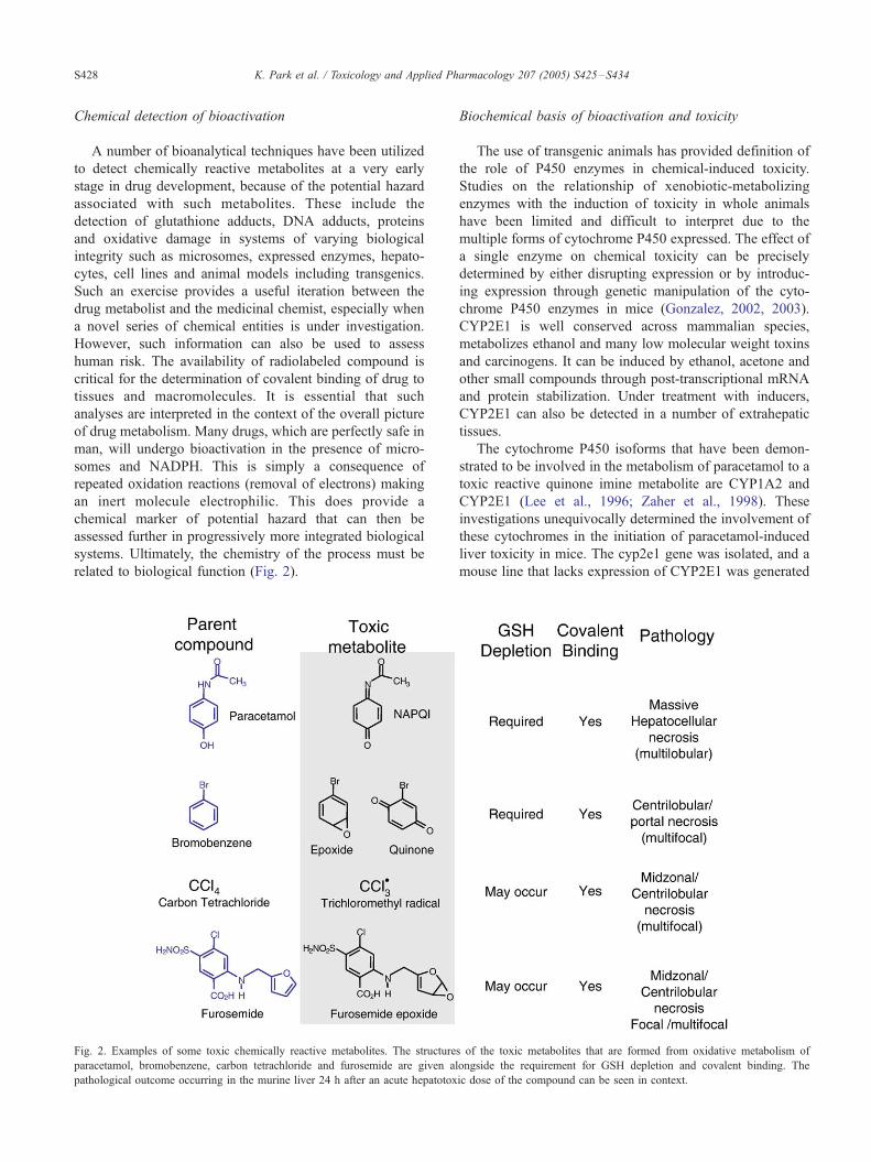
related to biological function (Fig. 2).
Fig. 2. Examples of some toxic chemically reactive metabolites. The structure
paracetamol, bromobenzene, carbon tetrachloride and furosemide are given al
pathological outcome occurring in the murine liver 24 h after an acute hepatotox
Biochemical basis of bioactivation and toxicity
The use of transgenic animals has provided definition of
the role of P450 enzymes in chemical-induced toxicity.
Studies on the relationship of xenobiotic-metabolizing
enzymes with the induction of toxicity in whole animals
have been limited and difficult to interpret due to the
multiple forms of cytochrome P450 expressed. The effect of
a single enzyme on chemical toxicity can be precisely
determined by either disrupting expression or by introduc-
ing expression through genetic manipulation of the cyto-
chrome P450 enzymes in mice (Gonzalez, 2002, 2003).
CYP2E1 is well conserved across mammalian species,
metabolizes ethanol and many low molecular weight toxins
and carcinogens. It can be induced by ethanol, acetone and
other small compounds through post-transcriptional mRNA
and protein stabilization. Under treatment with inducers,
CYP2E1 can also be detected in a number of extrahepatic
tissues.
The cytochrome P450 isoforms that have been demon-
strated to be involved in the metabolism of paracetamol to a
toxic reactive quinone imine metabolite are CYP1A2 and
CYP2E1 (Lee et al., 1996; Zaher et al., 1998). These
investigations unequivocally determined the involvement of
these cytochromes in the initiation of paracetamol-induced
liver toxicity in mice. The cyp2e1 gene was isolated, and a
mouse line that lacks expression of CYP2E1 was generated
s of the toxic metabolites that are formed from oxidative metabolism of
ongside the requirement for GSH depletion and covalent binding. The
ic dose of the compound can be seen in context.
K. Park et al. / Toxicology and Applied Pharmacology 207 (2005) S425–S434 S429
by homologous recombination in embryonic stem cells.
When cyp2e1 knockout mice were dosed, they were found
to be considerably less sensitive to its hepatotoxic effects
than wild-type animals, indicating that this P450 is the
principal enzyme responsible for the metabolic conversion
of the drug to its active hepatotoxic metabolite (Lee et al.,
1996). The observation that CYP1A2 and CYP 2E1 double
null mice were much less sensitive than either of the single
knockouts also suggested a major role for CYP1A2 in the
formation of the toxic metabolite of paracetamol. Doses of
1200 mg/kg to double null mice showed infrequent liver
lipidosis and mild kidney lesions, depletion of hepatic
glutathione was retarded, and covalent binding was not
observable (Zaher et al., 1998).
The sensitivity of CYP2E1-null animals to benzene has
also been seen analyzed (Valentine et al., 1996). Benzene
was administered at 200 ppm to null and wild-type mice.
Total urinary radioactivity and all radiolabeled individual
metabolites were reduced in the urine of cyp2e1-null mice
compared to wild-type controls. Also, more urinary radio-
activity could be accounted for as phenylsulfate conjugates
in cyp2e1-null mice compared to wild-type mice, indicating
the importance of CYP2E1 in the oxidation of phenol
following benzene exposure in normal mice. No benzene-
induced cytotoxicity or genotoxicity was observed in
cyp2e1-null mice. By contrast, benzene exposure resulted
in severe genotoxicity and cytotoxicity in wild-type mice.
This study indicates that CYP2E1 is the major determinant
of in vivo benzene metabolism and benzene-induced
myelotoxicity in mice.
Additional studies also conclusively demonstrated that
CYP2E1 is the major enzyme involved in the initiation of
toxicity due to cisplatin, acrylonitrile and chloroform (Con-
stan et al., 1999; Liu et al., 2000; Wang et al., 2002).
Cytochrome P450 1B1 knockout mice have been employed
to determine that CYP1B1 metabolizes 7,12-dimethylben-
z[a]anthracene to a DNA alkylating species, which corre-
lates with the formation of ovarian cancers in mice (Buters
et al., 1999).
Molecular relationship between metabolism and toxicity
Paracetamol is a useful tool with which to explore the
molecular interface between drug metabolism and drug
toxicity. It is also a highly relevant drug with respect to
clinical toxicology. The drug is a major cause of drug-related
morbidity and mortality in humans, producing massive
hepatic necrosis after a single toxic dose. Importantly, the
same pathology and clinical chemistry is observed in rodents.
Toxicity is essentially dose-dependent, although there can be
inter-species, intra-species and inter-individual variability in
susceptibility. For example, mice are generally more suscep-
tible than rats, while in man, alcoholics and patients on
enzyme-inducing drugs can have increased susceptibility. At
therapeutic doses, paracetamol undergoes metabolic clear-
ance by glucuronylation and sulphation to metabolites, which
are rapidly excreted in urine. However, a proportion of the
drug undergoes bioactivation to NAPQI by CYP2E1,
CYP1A2 and CYP3A4 (Raucy et al., 1989; Thummel et
al., 1993) (Figs. 1 and 2).
NAPQI is rapidly quenched by a spontaneous reaction
with hepatic glutathione after a therapeutic dose of para-
cetamol. After a toxic (over) dose, glutathione depletion
occurs, which is an obligatory step for covalent binding and
toxicity (Davis et al., 1974). The standard treatment for
paracetamol intoxication is N-acetylcysteine, which replaces
hepatic glutathione and prevents toxicity, and is most
effective when given within 16 h of the overdose.
Thus, the chemical and biochemical basis of paracetamol
toxicity is well established. More recently, the molecular
toxicology of the process has been explored by a number of
groups (Goldring et al., 2004; Kitteringham et al., 1999,
2000; Laskin and Pilaro, 1986; Laskin et al., 1986; Reilly et
al., 2001; Williams et al., 2004). We have been particularly
interested in the role of drug metabolism in cell defense and
cell destruction and have adopted an integrated approach in
which we have simultaneously explored:
& The chemistry of drug metabolism
& The biochemistry of drug metabolism
& Activation of transcription factors
& Gene expression (Williams et al., 2004)
& Analysis of the hepatoproteome and plasma proteome
& Proteomic analysis of modified proteins
& Conventional pathology and clinical chemistry
The massive chemical stress mediated by a single over-
dose leads to an immediate adaptive defense response in the
hepatocyte. This involves various mechanisms, including the
nuclear translocation of redox-sensitive transcription factors
such as Nrf-2, which ‘‘sense’’ chemical danger and orches-
trate cell defense. The Nrf-2 genes of immediate relevance
are those involved in glutathione synthesis such as g-
glutamylcysteine synthetase (g-GCS), glutathione-S-trans-
ferases (GSTs), glucuronyltransferases and heme oxygenase
(Goldring et al., 2004). Thus, the gene expression seems
designed, in a chemical and biochemical sense, to respond to
the toxic insult of the metabolite. Importantly, in terms of the
development of potential biomarkers that might be used in
drug development, it was shown that nuclear translocation of
Nrf-2 occurs at non-toxic doses of paracetamol and at time-
points before overt toxicity is observed. We have also
observed nuclear translocation of this redox sensitive tran-
scription with other compounds that cause hepatotoxicity
after bioactivation to a toxic metabolite, such as carbon
tetrachloride, furosemide and bromobenzene. We are cur-
rently exploring the usefulness of this transcription factor
and related downstream genes as markers of those forms of
chemicals that can lead to idiosyncratic drug-induced
hepatotoxicity in man (Fig. 2). It will also be important to
determine inter-individual variability in the responsiveness
of this system and its relevance to idiosyncratic drug toxicity.

K. Park et al. / Toxicology and Applied Pharmacology 207 (2005) S425–S434S430
Over the past few years, especially since the elucidation
of the human and murine genomes, numerous technologies
have been developed that are transforming pharmaceutical
research (Pennie et al., 2001; Waring et al., 2001). In
particular, the application of DNA micro-arrays and Gene
Chips to the quantitative comparison of the expression
levels of thousands of individual genes after exposure to
potential therapeutic candidates is termed toxicogenomics.
The comparison of expression profiles from known tox-
icants to those of novel compounds in a searchable data set
could elucidate underlying mechanisms and identify and
predict potential toxicities without the need to conduct
extensive, long-term animal or human clinical studies
(Bulera et al., 2001; Williams et al., 2004). Toxicogenomic
applications can be divided into mechanistic research and
predictive toxicology (Pennie et al., 2001). In mechanistic
research, the experimental model chosen should follow the
biological/toxicological process as closely as possible, with
similar clearly defined end-points.
Since the initial discovery that covalent binding of
paracetamol to hepatic proteins was associated with
hepatotoxicity, a number of techniques have been used to
identify the protein targets. Thus, radiolabeled drug and
Western blotting enable the detection and quantification of
protein adducts. This provides a non-specific biomarker of
drug bioactivation but does not provide any insight into
altered biological function and thus cannot be used in risk
assessment. More recently, proteomics has allowed the
simultaneous identification of several adducted proteins,
and also the determination of the amino acids modified
within the target protein. The question always asked with
respect to drug development is—Is there a particular protein
that will serve as biomarker of a particular form of drug
toxicity?
Before attempting to answer this question, we need to
define which protein modifications are important for cell
function, for both cell defense and cell destruction, and
separate these protein modifications that are of no functional
consequence and thus can be regarded as ‘‘white noise’’. At
least 17 liver enzymes that show a loss of activity ex vivo
after the administration of a toxic dose of paracetamol to a
rodent species have now been investigated. In addition,
another 14 liver enzymes are known to be adducted by
paracetamol in vivo and in vitro, but have yet to be shown to
be inhibited.
Modification of proteins can occur in most intracellular
compartments of the hepatocyte, including endoplasmic
reticulum, cytosol, mitochondria and plasma membrane, an
indication of the intracellular stability and mobility of the
reactive metabolite NAPQI. The loss of hepatocyte viability
is likely to be a consequence of the disruption of certain
critical proteins. Thus, the inhibition of g-GCS, glyceralde-
hyde 3-phosphate dehydrogenase (GAPDH) and Ca2+/Mg2+
ATPase will severely impair hepatocyte function by
uncoupling mitochondria, depleting glutathione and ATP,
and disturbing Ca2+ homeostasis which could lead to the
expression of TNFa and Fas receptors on cell membranes.
g-Glutamylcysteine synthetase catalyzes the rate-limiting
step in glutathione synthesis, the primary biochemical
defense of the hepatocyte against NAPQI. GAPDH, which,
as a component of the glycolytic pathway, contributes to
ATP production, is more than 80% inhibited at 2 h after a
toxic dose of paracetamol. On the basis of a reaction with
NAPQI in vitro, inhibition is thought to be due to the
modification of a critical cysteine (cys-149) residue within
the active site of the enzyme.
The rapid disruption of several proteins suggests that
cellular failure is a consequence of multiple parallel events
rather than a single protein target. It is well established that
one of the main events in isolated hepatocytes is overall
energy failure (Andersson et al., 1990; Burcham and
Harman, 1991), which is accompanied by the generation
of megamitochondria that are apparently ATP-depleted and
non-functional (Karbowski et al., 1999). The final destruc-
tion of hepatocytes involves interplay between hepatocyte
damage mediated by chemical stress and the activation of
non-parenchymal cells and the subsequent release of various
mediators. The role of Kupffer cells has been demonstrated
by the fact that mice treated with clodronate (dichlorome-
tyhylene bisphosphonate, DMDP), which depletes 99% of
macrophages from the liver, were protected against para-
cetamol (Goldin et al., 1996). Furthermore, infiltrating
macrophages are observed (Laskin and Pilaro, 1986).
Neutralization of Fas ligand (Zhang et al., 2000) and TNFa
(Blazka et al., 1996) affords a degree of protection against
the early apoptotic processes and the final overwhelming
necrosis, which is the overriding feature of paracetamol’s
hepatotoxicity. Thus, measurement of extracellular signal-
ling pathways may provide biomarkers of drug-induced
hepatotoxicity. This is extremely important in a clinical
context. Any observation in the plasma compartment that
reflects drug-induced organ toxicity, impending or actual,
would be of immense value in phase I and phase II studies.
From a physiological point of view, it is essential that
sensors in the cell defense system recognize the same
toxicophore in the metabolite that is responsible for damage
of critical macromolecules. The lack of an appropriate
response to chemical ‘‘danger’’, for chemical, biochemical
or genetic reasons, could lead to idiosyncratic drug toxicity.
Relationship between metabolism and T-lymphocyte
activation
Many idiosyncratic drug reactions have all the clinical
hallmarks of an immunological reaction. However, the
mechanism(s) of T-cell activation are not fully understood.
The majority of drugs associated with severe hypersensi-
tivity reactions also form chemically reactive metabolites.
Is this coincidence or consequence? There is good
circumstantial experimental evidence to relate the formation
of a reactive drug metabolite to immune activation.

K. Park et al. / Toxicology and Applied Pharmacology 207 (2005) S425–S434 S431
Halothane, a drug associated with immune-mediated hep-
atitis, is possibly the best example where there is direct
experimental data to relate metabolic activation and the
development of clinical symptoms of hypersensitivity.
Approximately 20–50% of halothane is converted by
CYP 2E1 to a reactive trifluoroacetyl chloride intermediate,
which binds covalently to protein. The structural modifica-
tion of halothane, which reduces the extent of metabolic
activation by over 95%, dramatically reduces the incidence
of severe hepatotoxicity (Park and Kitteringham, 1994).
Chemically reactive metabolites can stimulate T-cells
via two pathways. Pathway 1 is a classical hapten
mechanism (Fig. 3). The covalently modified protein
conjugate is taken up by antigen presenting cells,
processed into peptide fragments, which translocate to
the cell surface in the context of the major histocompat-
ibility complex (MHC) for presentation to T-cells (Naisbitt
et al., 2000). As yet, it is not clear whether T-cell receptor
activation requires the presence of a haptenated drug
bound to the peptide embedded in the MHC. Drug
metabolites also bind directly to MHC molecules
expressed on the surface of antigen presenting cells. This
pathway avoids the requirement of an antigen presenting
cell’s processing machinery (Schnyder et al., 2000). A
recent study has shown that acid elution of pre-existing
peptides on the MHC binding groove does not prevent
drug presentation to T-cells (Burkhart et al., 2002); thus,
drugs might ignore the peptide embedded in MHC binding
groove and bind directly to MHC itself.
Recently, Pichler has proposed an alternative pathway of
T-cell receptor activation by drugs. Based on findings from
in vitro experiments with T-cell clones generated from the
peripheral blood of hypersensitive patients, the authors
proposed that drugs might bind directly in the absence of
drug metabolism, covalent binding and antigen processing
Fig. 3. Hapten hypothesis of drug hypersensitivity. The scheme illustrates how d
antigen formation. Antigen formation is thought to initiate hypersensitive reactions
presenting cells in conjunction with a Fdanger_ signal.
to MHC molecules (Fig. 3). The resultant ‘‘pharmacolog-
ical’’ bridging interaction between MHC and the T-cell
receptor, although relatively weak in a chemical sense and
readily reversible, is sufficiently stable to stimulate all the
activation events of the T-cell receptor (Schnyder et al.,
1997).
To study the chemical mechanisms of drug hyper-
sensitivity reactions, we have used sulfamethoxazole as a
paradigm. Sulfamethoxazole is a sulphonamide-containing
compound that was first developed in the 1960’s as an
antibacterial agent and is still used today as a cost-effective
alternative to the new generation expanded spectrum
antibacterial agents to decrease or delay the development
of resistance. The drug has been associated with a range of
severe immunological reactions that include anaphylaxis
and Stevens–Johnson syndrome. The experimental evi-
dence for a role for metabolism in the toxicity associated
with this drug is as follows:
& Sulphamethoxazole undergoes bioactivation by both
CYP2C9 and myeloperoxidase to form protein-reactive
metabolites.
& Sulphamethoxazole undergoes metabolism to a hydrox-
ylamine and protein adduct in keratinocytes in vitro and
in animal models.
& The nitroso metabolite of sulphamethoxazole is an
extremely potent immunogen in rat, mouse and rabbit.
& The nitroso metabolite can stimulate IL-5 production in
an animal model.
& T-cells have been detected in hypersensitive patients that
recognize sulphamethoxazole and covalently bound
nitroso metabolite.
& The nitroso metabolite of sulpamethoxazole can stim-
ulate naı̈ve T-cells from healthy volunteers (not exposed
to drug).
rugs and/or their metabolites can interact with cellular proteins, leading to
via interaction with major histocompatibility complex (MHC) II on antigen

K. Park et al. / Toxicology and Applied Pharmacology 207 (2005) S425–S434S432
To generate a drug antigen, sulfamethoxazole is metab-
olized in the liver, blood cells and keratinocytes to a
hydroxylamine metabolite (Cribb and Spielberg, 1990;
Reilly et al., 2000). These reactions are catalyzed by
CYP2C9 and/or myeloperoxidase. Sulfamethoxazole
hydroxylamine is not chemically reactive, is sufficiently
stable to circulate in the periphery, and is excreted
unchanged in urine (Gill et al., 1997). Further auto-
oxidation of sulfamethoxazole hydroxylamine generates
nitroso sulfamethoxazole, which is chemically reactive and
has been shown to haptenate cellular protein (Naisbitt et al.,
1999). In further studies, we have used LC-MS and NMR
technology to investigate the stability of nitroso sulfame-
thoxazole (SMX). Here, we demonstrated that nitroso
sulfamethoxazole is rapidly degraded in solution; degrada-
tion yielded products of oxidation (nitro SMX), reduction
(SMX, SMX hydroxylamine) and dimerization (azo and
azoxy adducts) (Naisbitt et al., 1996, 2002). Despite this, we
have shown by flow cytometry, using a specific anti-
sulfamethoxazole antibody, that nitroso sulfamethoxazole
circulates in the periphery in rats and binds covalently to
epidermal keratinocytes (Naisbitt et al., 2001). These data
are somewhat paradoxical; however, it is our view that they
provide experimental evidence to support the hypothesis
that a futile redox cycle between hydroxylamine, nitroso and
nitro metabolites of sulfamethoxazole is established in vivo
in patients following the conversion of sulfamethoxazole to
sulfamethoxazole hydroxylamine.
To investigate the role of drug metabolism and covalent
binding in the immunogenicity of sulfamethoxazole, we
have developed an in vivo rat model. Splenocytes from rats
administered the nitroso metabolite, but not the hydroxyl-
amine or parent drug proliferated following in vitro
stimulation with nitroso sulfamethoxazaole. No proliferation
was seen following in vitro stimulation with sulfamethox-
azole. Antigen-specific T-cells proliferated in the presence
of nitroso sulfamethoxazole bound covalently to cellular,
but not serum protein, in an MHC restricted fashion
(Naisbitt et al., 2001). The antigenic threshold of nitroso
sulfamethoxazole for T-cell activation was estimated to be
between 0.5 and 1 AM, which is less than the concentration
of sulfamethoxazole metabolites found in human plasma
after the administration of a therapeutic dose of sulfame-
thoxazole (Gill et al., 1996). Interestingly, we have recently
observed similar results in mice and rabbits (Farrell et al.,
2003). The essential aspect of these animal models is that
there is a T-cell response to nitroso sulfamethoxazole but not
the parent drug.
Traditionalists believe that the immune system has
evolved to differentiate between self and non-self; the
presence of non-self stimulates an immune response.
Matzinger (1994) has recently proposed an alternative
‘‘danger’’ hypothesis. Matzinger’s theory states that the
primary signal controlling whether the presence of an
antigenic signal results in tolerance (ignorance) or immune
activation is the presence of danger, not non-self (Matzinger,
1994). Danger signals derive from damaged cells that
release intracellular molecules such as heat shock proteins
into extracellular matrix (Gallucci and Matzinger, 2001; Shi
et al., 2000). In terms of drug hypersensitivity, we have
recently shown that non-toxic and toxic concentrations of
nitroso sulfamethoxazole up- and down-regulate, respec-
tively, the expression of CD40 (cluster of differentiation 40),
a co-stimulatory receptor on antigen presenting cells. In on-
going experiments, we are studying the complex relation-
ship between covalent binding cell death and regulation of
stimulatory receptors expressed on antigen presenting cells.
Although certain aspects of Matzinger’s danger theory
remain contentious (Vance, 2000), the hypothesis might
explain why drug hypersensitivity reactions occur much
more frequently with concomitant infection.
It is important to note that results from the studies
described thus far derive from models of drug immunoge-
nicity and not hypersensitivity (i.e., there was no sign of
tissue damage). Thus, we have recently changed the focus of
our research to concentrate on the only currently available
model of drug hypersensitivity—lymphocytes isolated from
the peripheral blood of hypersensitive patients. Lympho-
cytes from patients hypersensitive to sulfamethoxazole
proliferated in the presence of nitroso sulfamethoxazole
(Burkhart et al., 2001; Farrell et al., 2003; Schnyder et al.,
2000). These data show that patients are exposed to nitroso
sulfamethoxazole at the time of the adverse drug reaction;
however, and in contrast to our animal models of
sulfamethoxazole immunogenicity, lymphocytes from
hypersensitive patients also proliferated in the presence of
sulfamethoxazole. It is possible that there may be ongoing
metabolism in human cells from hypersensitive patients,
which is below the limit of detection using available
analytical techniques. Since the number of antigen mole-
cules required to stimulate T-cells is incredibly low–Irvine
et al. (2002) estimated that T-cells can be stimulated when as
little as 10 antigenic ligands are present–it is not surprising
that the immune system is more sensitive than analytical
methodology (Irvine et al., 2002). It is also possible that
human T-cells may be hyper-responsive in a chemical sense
whereby non-covalently bound parent drug may bind in a
‘‘pharmacological-like’’ interaction with the MHC-restricted
T-cell receptor.
These recent advances in our understanding of how drugs
stimulate T-cells has provided a framework to develop in
vitro cell culture assays that might be useful for patient
diagnosis and drug evaluation. In this respect, we are
validating an in vitro induction protocol using lymphocytes
from drug naive volunteers to study the ability of
sulfamethoxazole and sulfamethoxazole metabolite to
induce a primary T-cell response using cells from drug
naive healthy volunteers. Lymphocytes from the vast
majority of individuals reacted in response to the reactive
metabolite nitroso sulfamethoxazole (Engler et al., 2004),
which suggests that T-cell recognition of nitroso sulfame-
thoxazole may not be MHC allele restricted. Following

K. Park et al. / Toxicology and Applied Pharmacology 207 (2005) S425–S434 S433
future development, we hope that this in vitro induction
protocol might be used to predict the potential of a new
chemical entity to cause T-cell-mediated reactions in man
(Engler et al., 2004).
Conclusion
Toxic metabolites can be formed from various functional
groups present in drugs by normal physiological biotrans-
formations. Constitutive and inducible systems exist for the
efficient bioinactivation of such reactive intermediates. Any
failure in cell defense to toxic metabolites can lead to
chemical and/or functional modification of critical cellular
macromolecules. The modification of critical macromole-
cules provides a credible explanation for certain types of
ADRs. This is now a major concern in drug development
and one that must be approached at various stages in the
process. Preclinical systems are available to detect chemical
hazard from metabolites and assess comparative (chemical)
risk. The prediction of individual (idiosyncratic) toxicity
induced by such metabolites is not yet possible, and will
require novel systems that incorporate individual suscepti-
bility factors.
Acknowledgments
The authors wish to acknowledge the support of The
Wellcome Trust and Pfizer PLC.
References
Andersson, B.S., Rundgren, M., Nelson, S.D., Harder, S., 1990. N-acetyl-p-
benzoquinone imine-induced changes in the energy metabolism in
hepatocytes. Chem. Biol. Interact. 75, 201–211.
Blazka, M.E., Elwell, M.R., Holladay, S.D., Wilson, R.E., Luster, M.I.,
1996. Histopathology of acetaminophen-induced liver changes—Role
of interleukin-1-alpha and tumor-necrosis-factor-alpha. Toxicol. Pathol.
24, 181–189.
Brodie, B.B., Reid, W.D., Cho, A.K., Sipes, G., Krishna, G., Gillette, J.R.,
1971. Possible mechanism of liver necrosis caused by aromatic organic
compounds. Proc. Natl. Acad. Sci. U.S.A. 68, 160–164.
Bulera, S.J., Eddy, S.M., Ferguson, E., Jatkoe, T.A., Reindel, J.F., Bleavins,
M.R., De La Iglesia, F.A., 2001. RNA expression in the early
characterization of hepatotoxicants in Wistar rats by high-density
DNA microarrays. Hepatology 33, 1239–1258.
Burcham, P.C., Harman, A.W., 1991. Acetaminophen toxicity results
in site-specific mitochondrial damage in isolated mouse hepato-
cytes. J. Biol. Chem. 266, 5049–5054.
Burkhart, C., von Greyerz, S., Depta, J.P., Naisbitt, D.J., Britschgi, M.,
Park, K.B., Pichler, W.J., 2001. Influence of reduced glutathione on the
proliferative response of sulfamethoxazole-specific and sulfamethox-
azole-metabolite-specific human CD4+ T-cells. Br. J. Pharmacol. 132,
623–630.
Burkhart, C., Britschgi, M., Strasser, I., Depta, J.P., von Greyerz, S.,
Barnaba, V., Pichler, W.J., 2002. Non-covalent presentation of sulfame-
thoxazole to human CD4+ T cells is independent of distinct human
leukocyte antigen-bound peptides. Clin. Exp. Allergy 32, 1635–1643.
Buters, J.T., Sakai, S., Richter, T., Pineau, T., Alexander, D.L., Savas, U.,
Doehmer, J., Ward, J.M., Jefcoate, C.R., Gonzalez, F.J., 1999.
Cytochrome P450 CYP1B1 determines susceptibility to 7, 12-dime-
thylbenz[a]anthracene-induced lymphomas. Proc. Natl. Acad. Sci.
U.S.A. 96, 1977–1982.
Constan, A.A., Sprankle, C.S., Peters, J.M., Kedderis, G.L., Everitt, J.I.,
Wong, B.A., Gonzalez, F.L., Butterworth, B.E., 1999. Metabolism of
chloroform by cytochrome P450 2E1 is required for induction of
toxicity in the liver, kidney, and nose of male mice. Toxicol. Appl.
Pharmacol. 160, 120–126.
Cribb, A.E., Spielberg, S.P., 1990. Hepatic microsomal metabolism of
sulfamethoxazole to the hydroxylamine. Drug Metab. Dispos. 18,
784–787.
Davis, D.C., Potter, W.Z., Jollow, D.J., Mitchell, J.R., 1974. Species
differences in hepatic glutathione depletion, covalent binding and
hepatic necrosis after acetaminophen. Life Sci. 14, 2099–2109.
Engler, O.B., Strasser, I., Naisbitt, D.J., Cerny, A., Pichler, W.J., 2004. A
chemically inert drug can stimulate T cells in vitro by their T cell
receptor in non-sensitised individuals. Toxicology 197, 47–56.
Farrell, J., Naisbitt, D.J., Drummond, N.S., Depta, J.P., Vilar, F.J.,
Pirmohamed, M., Park, B.K., 2003. Characterization of sulfamethox-
azole and sulfamethoxazole metabolite-specific T-cell responses in
animals and humans. J. Pharmacol. Exp. Ther. 306, 229–237.
Gallucci, S., Matzinger, P., 2001. Danger signals: SOS to the immune
system. Curr. Opin. Immunol. 13, 114–119.
Gill, H.J., Maggs, J.L., Madden, S., Pirmohamed, M., Park, B.K., 1996.
The effect of fluconazole and ketoconazole on the metabolism of
sulphamethoxazole. Br. J. Clin. Pharmacol. 42, 347–353.
Gill, H.J., Hough, S.J., Naisbitt, D.J., Maggs, J.L., Kitteringham,
N.R., Pirmohamed, M., Park, B.K., 1997. The relationship between
the disposition and immunogenicity of sulfamethoxazole in the rat.
J. Pharmacol. Exp. Ther. 282, 795–801.
Gillette, J.R., Mitchell, J.R., Brodie, B.B., 1974. Biochemical mechanisms
of drug toxicity. Annu. Rev. Pharmacol. 14, 271–288.
Goldin, R.D., Ratnayaka, I.D., Breach, C.S., Brown, I.N., Wickramasinghe,
S.N., 1996. Role of macrophages in acetaminophen (paracetamol)-
induced hepatotoxicity. J. Pathol. 179, 432–435.
Goldring, C.E., Kitteringham, N.R., Elsby, R., Randle, L.E., Clement, Y.N.,
Williams, D.P., McMahon, M., Hayes, J.D., Itoh, K., Yamamoto, M.,
Park, B.K., 2004. Activation of hepatic Nrf2 in vivo by acetaminophen
in CD-1 mice. Hepatology 39, 1267–1276.
Gonzalez, F.J., 2002. Transgenic models in xenobiotic metabolism and
toxicology. Toxicology 181–182, 237–239.
Gonzalez, F.J., 2003. Role of gene knockout and transgenic mice
in the study of xenobiotic metabolism. Drug Metab. Rev. 35,
319–335.
Irvine, D.J., Purbhoo, M.A., Krogsgaard, M., Davis, M.M., 2002. Direct
observation of ligand recognition by T cells. Nature 419, 845–849.
Karbowski, M., Kurono, C., Wozniak, M., Ostrowski, M., Teranishi, M.,
Nishizawa, Y., Usukura, J., Soji, T., Wakabayashi, T., 1999. Free
radical-induced megamitochondria formation and apoptosis. Free
Radical Biol. Med. 26, 396–409.
Kitteringham, N.R., Powell, H., Henderson, C.J., Wolf, C.R., Park, B.K.,
1999. Protection against paracetamol hepatotoxicity in glutathione S-
transferase pi knockout mice does not involve altered covalent binding.
Br. J. Clin. Pharmacol. 48, 887P–888P.
Kitteringham, N.R., Powell, H., Clement, Y.N., Dodd, C.C., Tettey, J.N.,
Pirmohamed, M., Smith, D.A., McLellan, L.I., Park, B.K., 2000.
Hepatocellular response to chemical stress in CD-1 mice: induction of
early genes and gamma-glutamylcysteine synthetase. Hepatology 32,
321–333.
Laskin, D.L., Pilaro, A.M., 1986. Potential role of activated macrophages in
acetaminophen hepatotoxicity: I. Isolation and characterization of
activated macrophages from rat liver. Toxicol. Appl. Pharmacol. 86,
204–215.
Laskin, D.L., Pilaro, A.M., Ji, S., 1986. Potential role of activated
macrophages in acetaminophen hepatotoxicity: II. Mechanism of

K. Park et al. / Toxicology and Applied Pharmacology 207 (2005) S425–S434S434
macrophage accumulation and activation. Toxicol. Appl. Pharmacol.
86, 216–226.
Lee, W.M., 2003. Drug-induced hepatotoxicity. N. Engl. J. Med. 349,
474–485.
Lee, S.S., Buters, J.T., Pineau, T., Fernandez-Salguero, P., Gonzalez, F.J.,
1996. Role of CYP2E1 in the hepatotoxicity of acetaminophen. J. Biol.
Chem. 271, 12063–12067.
Liu, J., Sendelbach, L.E., Parkinson, A., Klaassen, C.D., 2000. Endotoxin
pretreatment protects against the hepatotoxicity of acetaminophen and
carbon tetrachloride: role of cytochrome P450 suppression. Toxicology
147, 167–176.
Matzinger, P., 1994. Tolerance, danger, and the extended family. Annu.
Rev. Immunol. 12, 991–1045.
Miller, E., Miller, J., 1947. The presence and significance of bound
aminoazo dyes in the livers of rats fed p-dimethylaminobenzene.
Cancer Res. 7, 468–480.
Miller, E., Miller, J., 1952. In vivo combinations between carcinogens and
tissue constituents and their possible role in carcinogenesis. Cancer Res.
12, 547–556.
Naisbitt, D.J., O’Neill, P.M., Pirmohamed, M., Park, B.K., 1996. Synthesis
and reactions of nitroso sulphamethoxazole with biological nucleo-
philes: implications for immune mediated toxicity. Bioorg. Med. Chem.
Lett. 6, 1511–1516.
Naisbitt, D.J., Hough, S.J., Gill, H.J., Pirmohamed, M., Kitteringham, N.R.,
Park, B.K., 1999. Cellular disposition of sulphamethoxazole and its
metabolites: implications for hypersensitivity. Br. J. Pharmacol. 126,
1393–1407.
Naisbitt, D., Gordon, S., Pirmohamed, M., Park, B., 2000. Immuno-
logical principles of adverse drug reactions. The initiation and
propagation of immune responses elicited by drug treatment. Drug
Saf. 23, 483–507.
Naisbitt, D.J., Gordon, S.F., Pirmohamed, M., Burkhart, C., Cribb,
A.E., Pichler, W.J., Park, B.K., 2001. Antigenicity and immunoge-
nicity of sulphamethoxazole: demonstration of metabolism-dependent
haptenation and T-cell proliferation in vivo. Br. J. Pharmacol. 133,
295–305.
Naisbitt, D.J., Farrell, J., Gordon, S.F., Maggs, J.L., Burkhart, C., Pichler,
W.J., Pirmohamed, M., Park, B.K., 2002. Covalent binding of the
nitroso metabolite of sulfamethoxazole leads to toxicity and major
histocompatibility complex-restricted antigen presentation. Mol. Phar-
macol. 62, 628–637.
Park, B.K., Kitteringham, N.R., 1994. Effects of fluorine substitution on
drug metabolism: pharmacological and toxicological implications. Drug
Metab. Rev. 26, 605–643.
Park, B.K., Maggs, J.L., 1986. Inter-individual variation in the metabolism
of ethynylestradiol. Prog. Clin. Biol. Res. 214, 289–302.
Park, B.K., Pirmohamed, M., 2001. Toxicogenetics in drug development.
Toxicol. Lett. 120, 281–291.
Park, B.K., Pirmohamed, M., Kitteringham, N.R., 1992. Idiosyncratic drug
reactions: a mechanistic evaluation of risk factors. Br. J. Clin.
Pharmacol. 34, 377–395.
Park, B.K., Pirmohamed, M., Kitteringham, N.R., 1998. Role of drug
disposition in drug hypersensitivity: a chemical, molecular, and clinical
perspective. Chem. Res. Toxicol. 11, 969–988.
Pennie, W.D., Woodyatt, N.J., Aldridge, T.C., Orphanides, G., 2001.
Application of genomics to the definition of the molecular basis for
toxicity. Toxicol. Lett. 120, 353–358.
Pirmohamed, M., Back, D.J., 2001. The pharmacogenomics of HIV
therapy. Pharmacogenomics J. 1, 243–253.
Pirmohamed, M., Park, B.K., 2001a. Genetic susceptibility to adverse drug
reactions. Trends Pharmacol. Sci. 22, 298–305.
Pirmohamed, M., Park, B.K., 2001b. HIV and drug allergy. Curr. Opin.
Allergy. Clin. Immunol. 1, 311–316.
Pirmohamed, M., James, S., Meakin, S., Green, C., Scott, A.K., Walley,
T.J., Farrar, K., Park, B.K., Breckenridge, A.M., 2004. Adverse drug
reactions as cause of admission to hospital: prospective analysis of
18,820 patients. BMJ 329, 15–19.
Raucy, J.L., Lasker, J.M., Lieber, C.S., Black, M., 1989. Acetaminophen
activation by human liver cytochromes P450IIE1 and P450IA2. Arch.
Biochem. Biophys. 271, 270–283.
Reilly, T.P., Lash, L.H., Doll, M.A., Hein, D.W., Woster, P.M., Svensson,
C.K., 2000. A role for bioactivation and covalent binding within
epidermal keratinocytes in sulfonamide-induced cutaneous drug reac-
tions. J. Invest. Dermatol. 114, 1164–1173.
Reilly, T.P., Bourdi, M., Brady, J.N., Pise-Masison, C.A., Radonovich,
M.F., George, J.W., Pohl, L.R., 2001. Expression profiling of
acetaminophen liver toxicity in mice using microarray technology.
Biochem. Biophys. Res. Commun. 282, 321–328.
Schnyder, B., Mauri-Hellweg, D., Zanni, M., Bettens, F., Pichler, W.J.,
1997. Direct, MHC-dependent presentation of the drug sulfamethox-
azole to human alphabeta T cell clones. J. Clin. Invest. 100, 136–141.
Schnyder, B., Burkhart, C., Schnyder-Frutig, K., von Greyerz, S., Naisbitt,
D.J., Pirmohamed, M., Park, B.K., Pichler, W.J., 2000. Recognition of
sulfamethoxazole and its reactive metabolites by drug-specific CD4+ T
cells from allergic individuals. J. Immunol. 164, 6647–6654.
Shi, Y., Zheng, W., Rock, K.L., 2000. Cell injury releases endogenous
adjuvants that stimulate cytotoxic T cell responses. Proc. Natl. Acad.
Sci. U.S.A. 97, 14590–14595.
Thummel, K.E., Lee, C.A., Kunze, K.L., Nelson, S.D., Slattery, J.T., 1993.
Oxidation of acetaminophen to N-acetyl-p-aminobenzoquinone imine
by human CYP3A4. Biochem. Pharmacol. 45, 1563–1569.
Valentine, J.L., Lee, S.S., Seaton, M.J., Asgharian, B., Farris, G., Corton,
J.C., Gonzalez, F.J., Medinsky, M.A., 1996. Reduction of benzene
metabolism and toxicity in mice that lack CYP2E1 expression. Toxicol.
Appl. Pharmacol. 141, 205–213.
Vance, R.E., 2000. Cutting edge: cutting edge commentary: a
Copernican revolution? Doubts about the danger theory. J. Immunol.
165, 1725–1728.
Wang, H., Chanas, B., Ghanayem, B.I., 2002. Cytochrome P450 2E1
(CYP2E1) is essential for acrylonitrile metabolism to cyanide: com-
parative studies using CYP2E1-null and wild-type mice. Drug Metab.
Dispos. 30, 911–917.
Waring, J.F., Jolly, R.A., Ciurlionis, R., Lum, P.Y., Praestgaard, J.T.,
Morfitt, D.C., Buratto, B., Roberts, C., Schadt, E., Ulrich, R.G., 2001.
Clustering of hepatotoxins based on mechanism of toxicity using gene
expression profiles. Toxicol. Appl. Pharmacol. 175, 28–42.
Williams, D.P., Naisbitt, D.J., 2002. Toxicophores: groups and metabolic
routes associated with increased safety risk. Curr. Opin. Drug Discovery
Dev. 5, 104–115.
Williams, D.P., Park, B.K., 2003. Idiosyncratic toxicity: the role of
toxicophores and bioactivation. Drug Discovery Today 8, 1044–1050.
Williams, D.P., Kitteringham, N.R., Naisbitt, D.J., Pirmohamed, M., Smith,
D.A., Park, B.K., 2002. Are chemically reactive metabolites responsible
for adverse reactions to drugs? Curr. Drug Metab. 3, 351–366.
Williams, D.P., Garcia-Allan, C., Hanton, G., Lenet, J.L., Provost, J.P.,
Brain, P., Walsh, R., Johnston, G.I., Smith, D.A., Park, B.K., 2004.
Time course toxicogenomic profiles in CD-1 mice after nontoxic and
nonlethal hepatotoxic paracetamol administration. Chem. Res. Toxicol.
17, 1551–1561.
Zaher, H., Buters, J.T., Ward, J.M., Bruno, M.K., Lucas, A.M., Stern, S.T.,
Cohen, S.D., Gonzalez, F.J., 1998. Protection against acetaminophen
toxicity in CYP1A2 and CYP2E1 double-null mice. Toxicol. Appl.
Pharmacol. 152, 193–199.
Zhang, H., Cook, J., Nickel, J., Yu, R., Stecker, K., Myers, K., Dean, N.M.,
2000. Reduction of liver Fas expression by an antisense oligonucleotide
protects mice from fulminant hepatitis. Nat. Biotechnol. 18, 862–867.