investigation of the amycolatopsis sp. atcc 39116...
TRANSCRIPT

1
Investigation of the Amycolatopsis sp. ATCC 39116 vanillin 1
dehydrogenase (VDHATCC 39116) and its impact on the biotechnical 2
production of vanillin 3
4
Christian Fleige1 ● Gunda Hansen1 ● Jens Kroll1 ● Alexander Steinbüchel1, 2 5
6
1 Institut für Molekulare Mikrobiologie und Biotechnologie 7
Westfälische Wilhelms-Universität Münster 8
Corrensstrasse 3 9
D-48149 Münster 10
Germany 11
email: [email protected] 12
Phone: +49 (251) 8339821. 13
Fax: +49 (251) 8338388 14
2 Environmental Sciences Department, King Abdulazis University, Jeddah, Saudi Arabia 15
16
Keywords: vanillin, vanillic acid, vanillin dehydrogenase, ferulic acid, 17
Amycolatopsis, vdh, VDH 18
Abbreviations: AmpR, ampicillin resistance cassette; ATCC, American Type Culture 19
Collection; EC no., European Commission number; HPLC, high performance liquid 20
chromatography; KmR, kanamycin resistance cassette 21
Copyright © 2012, American Society for Microbiology. All Rights Reserved.Appl. Environ. Microbiol. doi:10.1128/AEM.02358-12 AEM Accepts, published online ahead of print on 12 October 2012
on June 4, 2018 by guesthttp://aem
.asm.org/
Dow
nloaded from

2
ABSTRACT 22
The actinomycete Amycolatopsis sp. ATCC 39116 is capable of synthesizing large 23
amounts of vanillin from ferulic acid, which is a natural cell wall component of higher plants. 24
The desired intermediate vanillin is subject to undesired catabolism caused by the 25
metabolic activity of a hitherto unknown vanillin dehydrogenase (VDHATCC 39116). In order to 26
prevent the oxidation of vanillin to vanillic acid and thereby to obtain higher yields and 27
concentrations of vanillin, the responsible vanillin dehydrogenase in Amycolatopsis sp. 28
ATCC 39116 was investigated for the first time using the data of our genome sequence 29
analysis and further bioinformatic approaches. The vdh gene was heterologously 30
expressed in E. coli, and the encoded vanillin dehydrogenase was characterized in detail. 31
VDHATCC 39116 was purified to apparent electrophoretic homogeneity and exhibited NAD+-32
dependent activity towards vanillin, coniferylaldehyde, cinnamaldehyde and benzaldehyde. 33
The enzyme showed its highest activity towards vanillin at a pH of 8.0 and a temperature 34
of 44°C. In a next step, a precise vdh-deletion mutant of Amycolatopsis sp. ATCC 39116 35
was generated. The mutant lost its ability to grow on vanillin and did not show vanillin 36
dehydrogenase activity. A 2.3 times higher vanillin concentration and a substantially 37
reduced amount of vanillic acid occurred in the mutant Amycolatopsis sp. ATCC 39116 38
∆vdh::KmR when ferulic acid was provided for biotransformation in a cultivation 39
experiment at the 2-L bioreactor scale. Based on these results and taking further metabolic 40
engineering into account, Amycolatopsis sp. ATCC 39116 ∆vdh::KmR represents an 41
optimized and industrial applicable platform for biotechnological production of natural 42
vanillin. 43
44
on June 4, 2018 by guesthttp://aem
.asm.org/
Dow
nloaded from

3
INTRODUCTION 45
Vanillin is one of the most important and widely used flavour compounds in the food and 46
fragrance industry. The extraction of natural vanillin from cured seed pods of the orchid 47
Vanilla planifolia is highly cost and labor intensive and does not satisfy the global demand 48
of the continuously increasing market. Therefore, new sources for the synthesis of vanillin 49
are of great interest for the industry. More than 99 % of the industrially produced vanillin is 50
chemically synthesized from petrochemicals or lignin derivatives (8). Due to the increasing 51
interest in ’natural‘ and ’healthy‘ ingredients, especially in food industry, alternative sources 52
for natural vanillin originating from biotechnical processes were intensively investigated 53
(37). Vanillin can be labeled as ’natural’ if it is extracted from natural sources or “obtained 54
by appropriate physical, enzymatic or microbiological processes from material of 55
vegetable, animal or microbiological origin” (11). 56
The most attractive approaches for microbial vanillin synthesis are the biotransformation 57
of cheap natural aromatic compounds, such as ferulic acid (3-(4-hydroxy-3-methoxy-58
phenyl)prop-2-enoic acid), or the de novo biosynthesis from primary metabolites like 59
glucose (17). Bioconversion of ferulic acid into vanillin was demonstrated for various 60
microorganisms, including bacteria like Pseudomonas and Rhodococcus (27, 31, 33, 34) 61
or fungi such as the basidiomycete Pycnoporus cinnabarinus (44). Additionally, several 62
genes encoding the responsible enzymes in some of these organisms were heterologously 63
expressed in E. coli and enabled the corresponding strains to convert ferulic acid into 64
vanillin (5, 32). However, high concentrations of vanillin are attended by serious problems 65
for the cells due to the toxicity of this highly reactive aromatic aldehyde, which is detoxified 66
in the course of further catabolism or leads to cell death. Therefore, most processes did 67
not achieve industrial applicability because of insufficient vanillin yields. 68
on June 4, 2018 by guesthttp://aem
.asm.org/
Dow
nloaded from

4
In order to overcome this drawback, screenings for vanillin tolerant bacterial strains 69
were performed, which are able to convert ferulic acid and thereby accumulate high 70
amounts of the biotransformation product vanillin (26, 38). Rabenhorst et al. (38) reached 71
a vanillin concentration of 11.5 g/L after biotransformation of 19.9 g/L ferulic acid using 72
Amycolatopsis sp. HR167, corresponding to a molar yield of 77.8%. With Streptomyces 73
setonii ATCC 39116, which meanwhile has been reclassified to Amycolatopsis sp. ATCC 74
39116, Muheim et al. (26) achieved a final vanillin concentration of 13.9 g/L with a molar 75
yield of 75%. In contrast to most other used strains, both approaches are applicable in 76
industrial processes, although they also face the problem of further vanillin degradation to 77
vanillic acid and other unwanted degradation products (1, 43). 78
For an improved vanillin production process using the actinomycete Amycolatopsis sp. 79
ATCC 39116, whose 16S-rDNA-sequence is identical to that of Amycolatopsis sp. HR167, 80
the whole genome was sequenced to identify enzymes involved in the vanillin catabolism, 81
as previously done for other vanillin-producing strains (9, 10, 23, 31, 34). In order to 82
promote a more efficient accumulation of vanillin in by preventing its oxidation to vanillic 83
acid, enzymes with homology to characterized vanillin dehydrogenases of other bacteria 84
were screened. After identifying a set of enzymes with homologies to vanillin and other 85
aldehyde dehydrogenases, the related genes were heterologously expressed in E. coli, 86
and the encoded enzymes were tested for their activity on vanillin. The enzyme exhibiting 87
VDH activity showed highest homology to an aldehyde dehydrogenase of 88
Nocardioidaceae bacterium Broad-1 (ZP_08197264) and was therefore designated 89
VDHATCC 39116. In a next step, a precise deletion mutant was generated and the impact of 90
VDHATCC 39116 on the catabolism of vanillin was examined. First attempts were made to 91
employ the deletion mutant in fermentation processes as novel production platform for 92
natural vanillin. 93
on June 4, 2018 by guesthttp://aem
.asm.org/
Dow
nloaded from

5
MATERIALS AND METHODS 94
95
Bacterial strains, plasmids and cultivation conditions. All bacterial strains and 96
plasmids used in this study are listed in Table 1. Cells of Escherichia coli were grown in 97
Lysogeny Broth- (LB-) medium at 30 °C (40). Cells of Amycolatopsis sp. ATCC 39116 were 98
grown at 42 °C in Caso-medium (Merck, Darmstadt, Germany) or in a modified mineral 99
salts medium (19). Carbon sources were provided from stock solutions, which were 100
sterilized by filtration before usage, as indicated in the text. For selection of plasmid 101
harboring strains, antibiotics were added to the medium at the following concentrations: 102
Kanamycin: 50 µg/ml for E. coli, 100 µg/ml for Amycolatopsis sp. ATCC 39116; Ampicillin: 103
100 µg/ml for E. coli. Cell growth was measured photometrically using a Klett-Summerson 104
photometer equipped with filter no. 54 (520-580 nm) or by measuring the optical density at 105
400 nm (Amycolatopsis sp. ATCC 39116) or 600 nm (E. coli). 106
107
Biotransformation experiments. For biotransformation experiments of E. coli or 108
Amycolatopsis sp. ATCC 39116, cells were harvested within the stationary growth phase, 109
washed twice with phosphate buffered saline (PBS; 137 mM NaCl, 2.7 mM KCl, 10 mM 110
Na2HPO4, 1.8 mM KH2HPO4, pH 7.5) and resuspended in an appropriate volume of the 111
same buffer or mineral salts medium (19). The experiments were carried out with 50 ml 112
mineral salts medium in 250 ml Erlenmeyer flasks (E. coli) or in a 2-L bioreactor with a 113
total volume of 1,000 ml PBS (Amycolatopsis sp. ATCC 39116). Ferulic acid or vanillin 114
were added from sterile stock solutions as indicated in the text. 115
116
Determination of metabolic intermediates. Excreted intermediates of the ferulic acid 117
metabolism were analyzed by high performance liquid chromatography using a “UltiMate 118
on June 4, 2018 by guesthttp://aem
.asm.org/
Dow
nloaded from

6
3000 HPLC System“ (Dionex, Idstein, Germany) without prior extraction. Culture 119
supernatants were obtained after centrifugation (5 min, 16,000 x g) and catalytic reactions 120
were stopped by acidification of the withdrawn samples to pH 2 by the addition of 121
trichloroacetic acid to a concentration of 6.1 mM. Intermediates were separated by 122
reverse-phase chromatography using an Acclaim® 120 C8 column (particle size: 5 µm; 123
column: 250 x 2.1 mm) with a gradient of 0.1% (vol/vol) formic acid (eluant A) and 124
acetonitrile (eluant B) in a range from 26 to 100% (vol/vol) eluant B. The run started with a 125
flow rate of 0.1 ml/min, which was raised to 0.3 ml/min after 16 min when eluent B reached 126
100 %. For quantification, all intermediates were calibrated with external standards. The 127
compounds were identified by their retention times and their absorption at a specific 128
wavelength determined using a multiple wavelength detector “MWD – 3000” (259 nm, 129
280 nm, 285 nm, 340 nm; Dionex, Idstein, Germany). Data analyses and quantifications 130
were performed with the associated software “Chromeleon 6.8 Chromatography Data 131
Systems“ (Dionex, Idstein, Germany). 132
133
DNA isolation and manipulation. Genomic DNA of Amycolatopsis sp. ATCC 39116 134
was isolated with “DNeasy Blood & Tissue Kit” (Qiagen GmbH, Hilden, Germany). Plasmid 135
DNA was isolated from E. coli using the “peqGOLD Plasmid MiniPrep Kit I” (PEQLAB 136
GmbH, Erlangen, Germany). Plasmid isolation from Amycolatopsis sp. ATCC 39116 was 137
performed using the “Qiagen Plasmid Purification Kit (Mini)“ (Qiagen GmbH, Hilden, 138
Germany). DNA was digested with restriction endonucleases (Fermentas GmbH, St. Leon-139
Rot, Germany) under the conditions described by the manufacturer. Phusion high-fidelity 140
DNA polymerase and other DNA manipulating enzymes (Fermentas GmbH, St. Leon-Rot, 141
Germany) were used according to the instructions of the manufacturer. Oligonucleotides 142
were purchased from Eurofins MWG Synthesis GmbH (Ebersberg, Germany). DNA 143
on June 4, 2018 by guesthttp://aem
.asm.org/
Dow
nloaded from

7
fragments were isolated from agarose gels or reaction mixtures using the “peqGOLD Gel 144
Extraction Kit” (PEQLAB GmbH, Erlangen, Germany). 145
146
Genome sequencing, gene prediction and annotation. Sequencing of the whole 147
genome of Amycolatopsis sp. ATCC 39116, prediction of open reading frames and 148
automatic annotation was performed as described by Hiessl et al. (18). Homologues of 149
putative VDHs were identified using BLAST analyses, and the corresponding genes were 150
ranked based on their homology to already characterized VDHs. 151
152
Transfer of DNA. Plasmids were transferred into E. coli by employing the CaCl2-153
method (40), whereas the transfer of plasmids and linear DNA fragments into 154
Amycolatopsis sp. ATCC 39116 was performed by direct mycelia transformation as 155
described by Priefert et al. (35). 156
157
Plasmid and mutant construction. The vdh gene of Amycolatopsis sp. ATCC 39116 158
was amplified with its own ribosomal binding site (RBS) via PCR from total chromosomal 159
DNA using the Phusion high-fidelity polymerase and oligonucleotides vdh_for_RBS_NdeI 160
and vdh_rev_XhoI (Tab. S1). For an additional C-terminal His-tag, vdh was also amplified 161
without its native stop codon using vdh_for_RBS_NdeI and vdh_rev_nostop_XhoI 162
(Tab. S1) as reverse primer. Both fragments were digested with NdeI/XhoI and ligated with 163
the NdeI/XhoI-linearized vector pET23a (+). After transformation of E. coli Mach-1 T1, the 164
resulting plasmids pet23::vdh and pet23::vdhHis were sequenced and subsequently 165
transferred to the expression host E. coli BL21(DE3). 166
A precise deletion of vdh was accomplished via homologous recombination by replacing 167
the native gene with a kanamycin resistance cassette. For this purpose, the flanking 168
on June 4, 2018 by guesthttp://aem
.asm.org/
Dow
nloaded from

8
regions upstream and downstream of vdh in the genome were amplified using the 169
following oligonucleotides (Tab. S1): upstream region: vdhLF_for3 and vdhLF_rev_KmR; 170
downstream region: vdhRF_for_PromKmR and vdhRF_rev3. Additionally, the kanamycin 171
resistance cassette of pRLE6 was amplified using KmR_rev_vdhLF and 172
PromKmR_for_vdhRF. The resulting fragments were purified and combined in a 173
subsequent fusion-PCR using primers vdhLF_for3 and vdhRF_rev3. The resulting 174
fragment vdhLF_KmR_vdhRF combined the upstream and downstream region of vdh with 175
the kanamycin resistance cassette and was transferred into Amycolatopsis sp. ATCC 176
39116 as described above. Transformants of Amycolatopsis sp. ATCC 39116 were 177
selected on solid Caso-medium containing 100 µg/ml kanamycin. Mutants were screened 178
by colony-PCR and gene replacement was finally analyzed via diagnostic PCR with 179
different primer combinations showing a specific integration of the kanamycin resistance 180
cassette into the vdh-locus. Furthermore, the resulting PCR-fragments were verified via 181
digestion and sequencing. 182
183
Preparation of crude cell extracts and soluble fractions. Cell pellets of E. coli were 184
resuspended in 3-4 ml of 100 mM potassium phosphate buffer (pH 7.1). Crude extracts 185
were obtained by three passages through a French pressure cell (120 MPa). For the 186
preparation of soluble fractions, cell debris was removed by centrifugation for 60 min at 187
20,000 x g at 4 °C. For subsequent enzyme assays, samples were stored on ice, while 188
samples for polyacrylamide gel electrophoresis were stored at -20 °C. His6-tagged proteins 189
were purified using “His SpinTrap“-columns (GE Healthcare Europe GmbH, Freiburg, 190
Germany) according to the instructions of the manufacturer. 191
192
on June 4, 2018 by guesthttp://aem
.asm.org/
Dow
nloaded from

9
Polyacrylamide gel electrophoresis and Western blot analysis. Samples from crude 193
cell extracts and soluble fractions were analyzed for their protein contents according to the 194
method of Bradford (7). Sodium dodecyl sulfate polyacrylamide gel electrophoresis (SDS-195
PAGE) was performed in 12.5% (wt/vol) gels as described by Laemmli (20). Amounts of 196
proteins were added as indicated in the text. Staining of proteins was performed with 197
Serva Blue G (47). Molecular weight reference proteins were purchased from GE 198
Healthcare Europe GmbH (Freiburg, Germany), “LMW SDS Markers“: Phosphorylase b, 199
rabbit muscle, 97,000 kDa; bovine serum albumin, 66,000 kDa; Ovalbumin, chicken egg 200
white, 45,000 kDa; Carbonic anhydrase, bovine erythrocyte, 30,000 kDa; Trypsin inhibitor, 201
soybean, 20,100 kDa; α-Lactalbumin, bovine milk, 14,400 kDa. 202
Proteins were blotted from SDS-polyacrylamide gels onto PVDF membrane “PVDF 203
Transfer Membrane Hybond-P“ (GE Healthcare Europe GmbH, Freiburg, Germany) 204
according to the method of Towbin et al. (45), using a “PeqLab Western Blotting Semi Dry 205
Sedec M“ apparatus (PEQLAB GmbH, Erlangen, Germany). Transfer of proteins was 206
accomplished for 120 min at a voltage of 14 V and a constant current of 5 mA/cm2. 207
Immuno detection of His6-tagged proteins was performed with a primary antibody 208
“Mouse anti histidine tag“ (AbD Serotec, MorphoSys AbD GmbH, Düsseldorf, Germany) 209
and a secondary antibody “AP-goat anti-mouse IgG“ (Zymed, Invitrogen GmbH, Karlsruhe, 210
Germany). Membranes with blotted proteins were incubated in skim milk (2.5 %, wt/vol, 211
Tris-buffered saline (TBS; total volume 1L: TRIS/HCl (pH 7.6), 2.42 g; NaCl, 8 g; Tween 20, 212
0.05 % (wt/vol)) at room temperature to block nonspecific binding. After washing with TBS, 213
the membrane was incubated with the primary antibody (1:2,000 in TBS-Tween (0.1 %), 214
200 µL/cm² membrane) overnight at room temperature. After threefold washing with TBS 215
for 10 min, the membrane was incubated with the secondary antibody (1:10,000 in TBS-216
Tween (0.1 %), 200 µL/cm² membrane) for 2 h at room temperature. After finally washing 217
on June 4, 2018 by guesthttp://aem
.asm.org/
Dow
nloaded from

10
threefold with TBS as described above, the antigen-antibody complexes were visualized 218
using “Western Blue stabilized substrate for AP“ (Promega GmbH, Mannheim, Germany). 219
220
VDH Enzyme assay. VDH enzyme activity was assayed according to Gasson et al. 221
(13). The reaction mixture contained in a total volume of 1 ml: 0.1 mM potassium 222
phosphate buffer (pH 7.1), 0.5 mM NAD+, 1.2 mM pyruvate (sodium salt), lactate 223
dehydrogenase (rabbit muscle, 1 U), 500 µg of the enzyme preparation of interest and 224
0.125 mM vanillin, benzaldehyde, coniferylaldehyde or cinnamaldehyde, respectively. 225
Oxidation of the substrates was measured at 30 °C and a suitable wavelength: vanillin 226
340 nm (ε= 11.492 cm2/µmol), cinnamaldehyde 340 nm (ε= 0.588 cm2/µmol), 227
benzaldehyde 293 nm (ε= 0.339 cm2/µmol) and coniferylaldehyde 410 nm 228
(ε= 3.452 cm2/µmol). Enzyme activity is stated in units (U). One unit is defined as the 229
amount of enzyme which converts 1 µmol vanillin per minute. 230
The temperature and pH optimum of VDH ATCC 39116 was determined in a discontinuous 231
assay by measuring the vanillic acid formation using HPLC, since the spectrophotometric 232
assay is additionally dependent on the activity of the lactate dehydrogenase. The reaction 233
mixture contained in a total volume of 1 ml: 0.1 mM potassium phosphate buffer (pH 7.1), 234
2 mM NAD+, 500 µg of the enzyme preparation of interest and 1.25 mM vanillin. The 235
reactions were started by the addition of NAD+, and the mixture was incubated for 15 min 236
before the reaction was stopped by heating to 85°C for 5 min. To test the temperature 237
preference, the assay was carried out at 15-65°C in steps of 5°C. After allocating the 238
optimum between 40 and 45°C, the temperature steps were refined to 1°C. The effect of 239
different pH values (5.0 – 9.5 in 0.5 steps) was observed at 30°C in Britton-Robinson-240
buffer consisting of 40 mM boric acid, 40 mM acetic acid and 40 mM phosphoric acid and 241
titrated to the desired pH with 200 mM NaOH. 242
on June 4, 2018 by guesthttp://aem
.asm.org/
Dow
nloaded from
11
243
Nucleotide sequence accession numbers. The genome sequence of Amycolatopsis 244
sp. ATCC 39116 has recently been deposited in GenBank under the accession no. 245
AFWY00000000 (9). Additionally, the nucleotide sequence of vdh is provided within the 246
patent PCT/EP2012/061600 (12). Moreover all five tested gene candidates are available in 247
GenBank under the accession numbers: JX292129 (vdh), JX292130, JX292131, 248
JX292132, JX292133 249
250
251
RESULTS 252
In silico analyses of the genome and identification of the Amycolatopsis sp. ATCC 253
39116 vanillin dehydrogenase (VDHATCC 39116). Based on the genome sequence of 254
Amycolatopsis sp. ATCC 39116 and an automatic annotation, five genes, coding for 255
putative aldehyde dehydrogenases were identified. After heterologous expression of these 256
gene candidates in E. coli, only one of them lead to vanillin dehydrogenase activity and 257
might therefore be involved in the catabolism of vanillin. The gene, designated vdh, 258
consists of 1461 bp and encodes a protein of 486 amino acids with a theoretical molecular 259
weight of 50.6 kDa. A comparison of the deduced amino acid sequence of VDHATCC 39116 260
with internet-based sequence data using “BLASTP 2.2.25” (2, 3) showed homology to 261
different (benz)aldehyde dehydrogenases of various sources. The highest homology (73%) 262
was determined for an aldehyde dehydrogenase of Nocardioidaceae bacterium Broad-1 263
(ZP_08197264). By using “ClustalX 1.83” (16), a multiple sequence alignment (MSA) with 264
various representative aldehyde dehydrogenases was done for assessing their 265
relationship to VDHATCC 39116 in a dendrogramm (Fig. 1). Additionally, a MSA using “BioEdit 266
on June 4, 2018 by guesthttp://aem
.asm.org/
Dow
nloaded from

12
Sequence Alignment Editor 7.0.5.3“(16) was performed to demonstrate the similarity on 267
the amino acid level of VDHATCC 39116 to different enzymes with already verified vanillin 268
dehydrogenase activity (Fig. S1). Further in silico analysis using “ScanProsite” (Expert 269
Protein Analysis System, ExPASy; (14)) revealed a characteristic pattern of aldehyde 270
dehydrogenases within the amino acids 251 to 258 including glutamic acid in the catalytic 271
center. 272
273
Characterization of VDHATCC 39116. Beside the in silico analyses, the specific vanillin 274
dehydrogenase activity in cells of Amycolatopsis sp. ATCC 39116 was investigated after 275
cultivating the strain with or without vanillin and structurally related compounds like ferulic 276
acid as carbon source, since former investigations with Pseudomonas showed an inducing 277
effect (4, 13). Amycolatopsis sp. ATCC 39116 was cultivated in mineral salts medium 278
containing 0.5 % (wt/vol) gluconate for 32 h at 42 °C. In addition, 0.1 % (wt/vol) of vanillin 279
or ferulic acid was added to one of the cultures. The cells were harvested after cultivation, 280
soluble fractions were prepared, and the specific activity of the vanillin dehydrogenase was 281
measured photometrically as described in materials and methods. The activity assays 282
revealed specific vanillin dehydrogenase activities of 25 ± 2 U/g protein if cells were 283
previously cultivated with vanillin as carbon source. In cells, which were cultivated only 284
with gluconate as sole carbon source, no VDHATCC 39116 activity was detectable. The 285
enhancing effect was also obvious during SDS-PAGE analysis of the soluble fractions 286
which revealed a prominent protein band at a size of approximately 54 kDa in case of the 287
vanillin-induced cells. A corresponding band was missing in samples of cells which were 288
not incubated with vanillin. Comparable results could be observed, if cells were cultivated 289
with ferulic acid as inducer (data not shown). 290
on June 4, 2018 by guesthttp://aem
.asm.org/
Dow
nloaded from
13
After demonstrating the activity of a vanillin dehydrogenase in cells of Amycolatopsis sp. 291
ATCC 39116 grown with vanillin and after identifying a putatively responsible gene, vdh 292
was cloned and heterologously expressed in E. coli. For this, the gene was inserted into 293
the vector pET23a (+) and expressed in E. coli BL21 (DE3) under the control of the strong 294
T7 promoter. In order to purify the heterologous enzyme for further investigations, vdh was 295
also expressed without its native stop-codon to append a C-terminal His6-tag provided by 296
the vector pET23a (+). SDS-PAGE and subsequent Western blot analysis were carried out 297
to verify the correct gene expression and purification of VDHATCC 39116 with the His6-tag 298
(Fig. 2). 299
Protein analyses by SDS-PAGE and Western blot revealed the presence of an 300
appropriate protein with an apparent molecular weight of about 54 kDa. The purification of 301
VDHHis using “His SpinTrap“-columns yielded an almost homogenous protein (lane 7). 302
Immuno detection of the C-terminal His6-tag additionally confirmed the assumption that the 303
occurring protein signal represents the heterologously expressed VDHHis. Further analysis 304
of vanillin dehydrogenase activity within the eluate showed a specific activity of 72 ± 2 U/g 305
protein, which was remarkably low due to activity loss during the purification, which we 306
could not overcome, although we changed the composition of all used buffers. No enzyme 307
activity was observed in the absence of the cofactor NAD+. Furthermore, no oxidation of 308
vanillin was detectable, if NADP+ was used as cofactor (data not shown). 309
The optimal condition for the oxidation of vanillin was determined by measuring the 310
formed vanillic acid using cell free extracts of the recombinant vdh-expressing E.coli strain. 311
Maximum activities were found at a pH of 8.0 and a temperature of 44°C. The enzyme was 312
active at a broad pH range from 5.0 to 9.0 and was able to catalyze the reaction at 313
temperatures between 15°C and 65°C. After storage at 4°C for 24h VDHATCC 39116 exhibited 314
86% of its former activity. 315
on June 4, 2018 by guesthttp://aem
.asm.org/
Dow
nloaded from
14
The most specific substrate was found to be vanillin, but VDHATCC 39116 also catalyzed 316
the oxidation of coniferylaldehyde, benzaldehyde and cinnamalaldehyde with specific 317
activities up to 66% (benzaldehyde) in comparison to vanillin. 318
In a next step, vanillin biotransformation experiments using the recombinant E. coli 319
strain expressing VDHATCC 39116 were performed. The cells were cultivated until they 320
reached the stationary growth phase, were then harvested and washed twice with 321
potassium phosphate buffer. After suspending the cells in 50 ml mineral salts medium, 14 322
mM vanillin was added, and the formation of vanillic acid was observed using HPLC 323
analysis of culture supernatants (Fig. 3). As a control experiment, E. coli BL21(DE3), 324
harboring the plain vector pET23a, was incubated under identical conditions. 325
The biotransformation experiments demonstrated that the VDHATCC 39116 expressing 326
strain is able to metabolize vanillin to vanillic acid (Fig. 3). Vanillic acid was synthesized in 327
equimolar amounts immediately after the addition of vanillin, while a transformation could 328
not be observed in the control experiment (data not shown). 329
After identifying the responsible gene and allocating vanillin dehydrogenase activity to 330
VDHATCC 39116, vdh was additionally overexpressed episomally in Amycolatopsis sp. 331
ATCC 39116 using a derivative of the Amycolatopsis – E. coli shuttle vector pRLE6 (35): 332
The homologous expression led to a 3.7-fold increase of VDHATCC 39116 activity in 333
comparison to the wild type. 334
335
Construction of a vdh-deletion mutant of Amycolatopsis sp. ATCC 39116. In order 336
to investigate the impact of VDHATCC 39116 on the catabolism of vanillin in Amycolatopsis sp. 337
ATCC 39116, a precise deletion mutant was generated by replacing the native gene with a 338
kanamycin resistance cassette. For this, the flanking regions located upstream and 339
downstream of vdh in the genome were amplified and combined with the resistance gene 340
on June 4, 2018 by guesthttp://aem
.asm.org/
Dow
nloaded from
15
in a fusion-PCR. Amycolatopsis sp. ATCC 39116 was transformed with the resulting linear 341
fragment, and the gene replacement in the resulting mutant Amycolatopsis sp. 342
ATCC 39116 ∆vdh::KmR was confirmed by a diagnostic PCR. In a first step, the integration 343
of the deletion construct into the genome was demonstrated by amplifying a 3.36 kb-344
fragment (Fig. 4, lane 2). This fragment distinguishes in size from the genomic 345
organization of the wild type as a consequence of the smaller size of the resistance gene 346
in comparison to that of vdh. A second PCR with two different primer combinations verified 347
the specific integration of the deletion fragment into the vdh-locus by amplifying segments 348
which started upstream (Fig. 4, lane 3) or downstream (Fig. 4, lane 4), respectively, of the 349
used flanking regions. Finally, the amplified fragments were sequenced to ensure the 350
correct replacement of vdh with the kanamycin resistance gene. 351
352
Characterization of Amycolatopsis sp. ATCC 39116 ∆vdh::KmR. The vdh-deletion 353
mutant was characterized to determine the impact of the enzyme on the catabolism of 354
vanillin in Amycolatopsis sp. ATCC 39116. For this purpose, biotransformation experiments 355
in a 2-L bioreactor scale were carried out to analyze the utilization of ferulic acid. Ferulic 356
acid is catabolized via vanillin and subsequently to vanillic acid and is therefore a potential 357
substrate for the biotechnical production of vanillin. The difference between the vdh-358
deletion mutant and the wild type Amycolatopsis sp. ATCC 39116 in terms of ferulic acid 359
utilization was demonstrated for ’resting cells‘, independently from growth. The cells were 360
grown in Caso-medium at 42 °C until they reached the stationary growth phase. 361
Afterwards, they were harvested, washed twice with PBS and resuspended in the same 362
buffer (1,000 ml, pH 7.5, 0.5 g/L cell dry weight). Ferulic acid was added at an initial 363
concentration of 2.0 mM (stock concentration: 210 mM, pH 8, NaOH) to the cell 364
suspension, and the occurring intermediates were analyzed by HPLC. During the 365
on June 4, 2018 by guesthttp://aem
.asm.org/
Dow
nloaded from

16
transformation process 0.75 mM/h ferulic acid were fed continuously. In order to promote 366
continuing vanillin synthesis, the feeding rate was readjusted to 3.1 mM/h for 3 hours, after 367
the cells had adapted to the conversion conditions and the substrate concentration was 368
decreasing (7h from start). Accordingly, feeding was stopped after 10 hours, and the 369
disappearance of ferulic acid and the occurrence of the products were observed for 370
additional 20 hours. 371
The biotransformation experiments revealed a significant difference between the wild 372
type Amycolatopsis sp. ATCC 39116 and the vdh-deletion mutant with regard to the 373
occurrence of catabolism products (Fig. 5). While Amycolatopsis sp. ATCC 39116 started 374
to accumulate vanillic acid at first, Amycolatopsis sp. ATCC 39116 ∆vdh::KmR 375
accumulated vanillin as the first over-flow product. Concerning the wild type experiment, 376
vanillin was not detectable in the supernatants until vanillic acid was formed at a 377
concentration of about 1 mM. Additional vanillic acid was further synthesized from vanillin, 378
which was meanwhile formed. The latter was completely catabolized after ferulic acid was 379
no longer available for the cells. In contrast, only a very low concentration of vanillic acid 380
was formed by the cells of the mutant strain, while cultures of these cells accumulated 381
vanillin continuously up to 6.8 mM (wild type 2.9 mM) as long as ferulic acid was present in 382
the experiment. Furthermore, in contrast to the wild type, degradation of the synthesized 383
vanillin occurred only at a very low rate, even during the last 10 hours, when ferulic acid 384
was completely degraded. Further catabolism intermediates like guaiacol were only 385
detected in trace amounts in cultures of the wild type. 386
Growth analysis on vanillin and ferulic acid in mineral salts medium with 0.1 % (wt/vol) 387
of the substrates as sole source of carbon were also carried out. While the wild type 388
exhibited slow growth on both substrates after a long lag-phase of about 30-40h, the 389
mutant did not grow at all on vanillin. During the experiment no utilization of vanillin was 390
on June 4, 2018 by guesthttp://aem
.asm.org/
Dow
nloaded from

17
detectable for the mutant strain. On ferulic acid Amycolatopsis sp. ATCC 39116 391
∆vdh::KmR showed a significantly reduced growth rate when compared to the wild type, 392
indicating that VDHATCC 39116 has a substantially impact on the metabolism of ferulic acid. 393
However, the experiment suggests that ferulic acid can serve as source of carbon and 394
energy despite of the knockout. VDH activity of both, the wild type strain and the mutant 395
was observed subsequently. While the wild type exhibited dehydrogenase activity after 396
growth with ferulic or vanillin as previously observed, no VDH activity was detected in the 397
mutant strain, even despite of induction. 398
399
DISCUSSION 400
401
The actinomycete Amycolatopsis sp. ATCC 39116 is capable of synthesizing large 402
amounts of vanillin from the natural substrate ferulic acid (26). This bacterium therefore 403
represents the most promising platform for the biotechnical production of natural vanillin. 404
However, significant amounts of the desired product are lost because of the inherent 405
vanillin catabolism via vanillic acid (43). The availability of the genome sequence of 406
Amycolatopsis sp. ATCC 39116 contributed to our understanding of the vanillin catabolism 407
in this strain. Furthermore, it offered the possibility to identify responsible genes as a target 408
for metabolic engineering of this production strain. Systematic gene deletions have been 409
intensively used for strain development of various production processes. In order to 410
improve industrial applications that approach the theoretical product yields, catabolic or 411
competing pathways of the desired product were disrupted. Prominent examples are the 412
production of amino acids, ethanol and succinate by Corynebacterium glutamicum, 413
S. cerevisiae and E. coli, respectively (6, 48). 414
on June 4, 2018 by guesthttp://aem
.asm.org/
Dow
nloaded from

18
In several vanillin-degrading bacteria, a vanillin dehydrogenase was identified to 415
catalyze the oxidation of vanillin to vanillic acid (13, 23, 34, 36, 46). The activity of a vanillin 416
dehydrogenase could now be demonstrated in Amycolatopsis sp. ATCC 39116, too. 417
Moreover, the expression of the corresponding gene seems to be inducible by the 418
enzyme’s substrate and structurally related compounds, such as ferulic acid. As already 419
shown for P. fluorescens AN103 (13, 21) enzyme activity could not be observed in cells 420
cultivated in the absence of these substance. 421
Based on the genome sequence of Amycolatopsis sp. ATCC 39116 and its annotation, a 422
set of putatively responsible genes was identified showing homology to previously 423
characterized enzymes with aldehyde dehydrogenase activity of other organisms. 424
Selected gene candidates were heterologously expressed in E. coli and VDH activity was 425
monitored. One of the encoded enzymes was capable of catalyzing the oxidation of vanillin 426
to vanillic acid and thus was designated as VDHATCC 39116. The enzyme exhibited homology 427
to several aldehyde dehydrogenases including also a characteristic motif with glutamic 428
acid as a central amino acid of the catalytic center (Fig. S1). Interestingly the protein does 429
not show a remarkably high homology to the group of enzymes with already verified 430
vanillin dehydrogenase activity (Fig. 1). However, all up to now characterized proteins 431
derive from a phylogenetically closely related group of bacteria. VDHATCC 39116 represents 432
the first vanillin dehydrogenase that was characterized in detail in actinobacteria and might 433
therefore represent an independently evolved protein. This is confirmed by the observation 434
that the encoding gene vdh seems not to be part of cluster as previously described for 435
other studied strains (9, 29, 34, 36, 46). 436
Upon the heterologous expression of the encoding gene vdh in E. coli, a protein signal 437
with an apparent molecular weight of 54.0 ± 2.0 kDa was obtained in SDS-PAGE analysis. 438
This is in agreement with both, the calculated molecular weight of the vdh translational 439
on June 4, 2018 by guesthttp://aem
.asm.org/
Dow
nloaded from

19
product and the sizes of other enzymes exhibiting VDH activity (4, 23, 36, 46). The 440
recombinant protein was purified to apparent homogeneity carrying a 6xHis6-tag, which 441
was confirmed by immuno detection. VDHATCC 39116 activity was detected in the eluated 442
protein fraction verifying the identity of the vanillin dehydrogenase of Amycolatopsis sp. 443
ATCC 39116. In line with this, VDH activity was only detectable in presence of the cofactor 444
NAD+, indicating an NAD+-dependent aldehyde dehydrogenase as already supposed 445
based on the theoretical analysis of the amino acid sequence. In contrast, former studies 446
stated that the catabolism of vanillin in Amycolatopsis sp. ATCC 39116 is only catalyzed by 447
an oxidase (43). NADP+ could not be used as cofactor. A preference towards NAD+ was 448
also reported for other studied vanillin dehydrogenases (13, 23). Moreover, VDH activity of 449
Amycolatopsis sp. ATCC 39116 could be enhanced by supplementary episomal 450
expression of vdh, which thereby validates the enzymes function. VDHATCC 39116 was 451
shown to catalyze the oxidation of different aromatic aldehydes, but has a preference 452
towards vanillin as previously shown for Sphingomonas paucimobilis SYK-6 (23). The 453
enzyme was active in a broad range of conditions including different pH and temperatures 454
and showed its maximum activity at a pH of 8.0 and at a temperature of 44°C, respectively. 455
It exhibited a considerably storage stability since 86 % of its former activity could be 456
determined after 24h at 4°C. 457
Based on the results of former studies (1, 25, 43) and in correspondence with the 458
results obtained in this study, we propose for Amycolatopsis sp. ATCC 39116 the following 459
CoA-dependent pathway for the degradation of ferulic acid via vanillin and vanillic acid to 460
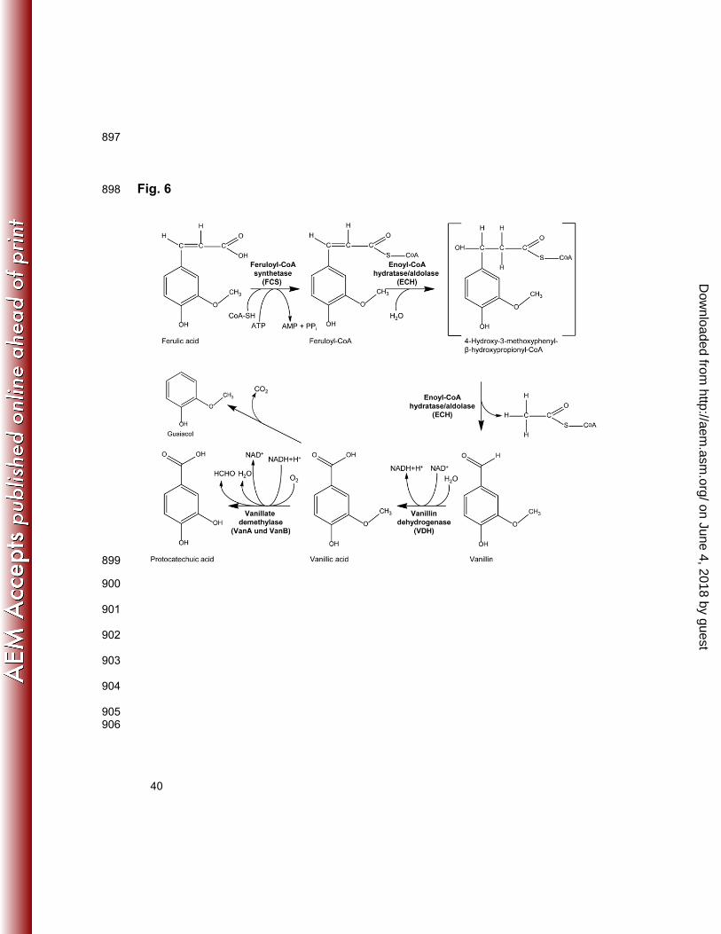
protocatechuic acid or guaiacol with VDHATCC 39116 as a key enzyme (Fig. 6). 461
Following the identification of the VDHATCC 39116 encoding gene, the physiological impact 462
of VDHATCC 39116 for the catabolism of vanillin and related compounds, such as ferulic acid, 463
was determined. Therefore, a precise vdh-deletion mutant was generated, and the 464
on June 4, 2018 by guesthttp://aem
.asm.org/
Dow
nloaded from

20
catabolism of ferulic acid of the mutant was investigated and compared to the wild type of 465
Amycolatopsis sp. ATCC 39116. A ‘resting cell’ approach was employed, and the occurring 466
catabolism intermediates, including vanillin and vanillic, were observed by HPLC. 467
Both, the wild type and Amycolatopsis sp. ATCC 39116 ∆vdh::KmR, degrade ferulic acid 468
in a comparable manner (Fig. 5A). In contrast to former studies no negative effect, 469
regarding the catabolism of ferulic acid was observed (21, 33, 34). The authors assumed 470
polar effects, as a consequence of the vdh deletion or disruption, respectively, to be 471
responsible for the resulting phenotype. Since the vdh gene of Amycolatopsis sp. 472
ATCC 39116, unlike in other studied strains, is not part of a cluster with other ferulic acid 473
catabolism genes, a similar effect was not to be expected for the present strain (9, 29, 34, 474
36, 46). A remarkable difference was, however, observed for the synthesis of vanillin and 475
vanillic acid (Fig. 5B, 5C). The vdh-deletion mutant showed a 2.3 times enhanced vanillin 476
accumulation, which started immediately after exposure of the cells to the substrate. A final 477
concentration of 6.8 mM was reached, and the excreted vanillin in the culture supernatants 478
was degraded only very slowly, even after ferulic acid was completely metabolized. The 479
wild type synthesized and excreted vanillin only up to 2.9 mM but only after an initial 480
accumulation of vanillic acid up to 1 mM had occurred. This observation has previously 481
also been made for the wild type Streptomyces sp. ATCC 39116 by Muheim and Lerch 482
(25). Since the accumulation of vanillin by the deletion mutant started independently from 483
vanillic acid, a regulatory effect, which influences the expression of vdh or the activity of 484
the enzyme in the wild type depending on the vanillic acid concentration, could be a 485
possible explanation. Moreover, as a result of the decreasing ferulic acid concentration, 486
vanillin was subsequently degraded by the wild type cells. In line with this, further 487
catabolism intermediates like guaiacol or protocatechuic acid were formed in detectable 488
amounts only in cultures of the wild type. 489
on June 4, 2018 by guesthttp://aem
.asm.org/
Dow
nloaded from

21
Growth analysis of the mutant strain revealed, as previously described for other 490
knockout mutants (21, 23), that the bacteria are no longer able to use vanillin as sole 491
source of carbon and energy. The subsequent enzyme assay showed in coincidence with 492
this that in contrast to the wild type, no oxidation of vanillin was detectable under the used 493
conditions. This observation is in accordance with the fact, that no other tested homologue 494
of VDH ATCC 39116 showed the desired enzyme activity. However, an alternative minor 495
pathway of vanillin oxidation might be present, since a slow degradation of vanillin was 496
observable in the biotransformation experiments, even despite of the knockout. 497
These experiments clearly showed that VDHATCC 39116 plays a significant role in the 498
course of ferulic acid degradation in Amycolatopsis sp. ATCC 39116. Therefore, the 499
deletion mutant represents a promising production strain to compete with the already 500
established vanillin production processes using wild type strains (26, 39). 501
Further metabolic engineering will improve the already obtained outcome since a slow 502
degradation of vanillin, probably due to unspecific aldehyde dehydrogenases or oxidases 503
activities (43), occurred even for Amycolatopsis sp. ATCC 39116 ∆vdh::KmR. Similar 504
observations were also made for different other vanillin production strains, in which VDH-505
encoding genes were deleted or disrupted (21, 31, 33, 34). In addition, ferulic acid could 506
be completely degraded to serve as source of energy through competing pathways, such 507
as decarboxylation to 4-vinylguaiacol (37). This could be the reason why vanillin or vanillic 508
acid, respectively, were not produced in equimolar amounts in the biotransformation 509
experiments (42% molar yield for the mutant, 19% for the wild type). Optimization of the 510
fermentation process is therefore aiming at an improved vanillin synthesis rate and at a 511
higher molar yield of vanillin with regard to ferulic acid. 512
Based on the present study, a highly efficient microbial production strain for natural 513
vanillin from ferulic acid can now be developed. Complete prevention of the catabolism of 514
on June 4, 2018 by guesthttp://aem
.asm.org/
Dow
nloaded from

22
vanillin during the biotransformation and optimizing the fermentation process will 515
significantly improve the production of natural vanillin using Amycolatopsis sp. 516
ATCC 39116 ∆vdh::KmR. 517
518
ACKNOWLEDGEMENTS 519
The project was financially supported by SYMRISE AG, Holzminden, Germany, which is 520
gratefully acknowledged. We thank Isabelle van Hamme for technical assistance. 521
522
REFERENCES 523
1. Achterholt S, Priefert H, Steinbüchel A. 2000. Identification of Amycolatopsis sp. 524
strain HR167 genes, involved in the bioconversion of ferulic acid to vanillin. Appl. Microbiol. 525
Biotechnol. 54:799-807. 526
2. Altschul SF, Madden TL, Schaffer AA, Zhang J, Zhang Z, Miller W, Lipman DJ. 527
1997. Gapped BLAST and PSI-BLAST: a new generation of protein database search 528
programs. Nucleic Acids Res. 25:3389-3402. 529
3. Altschul SF, Wootton JC, Gertz EM, Agarwala R, Morgulis A, Schaffer AA, Yu YK. 530
2005. Protein database searches using compositionally adjusted substitution matrices. 531
FEBS J. 272:5101-5109. 532
4. Bare G, Swiatkowski T, Moukil A, Gerday C, Thonart P. 2002. Purification and 533
characterization of a microbial dehydrogenase: a vanillin:NAD(P)+ oxidoreductase. Appl. 534
Biochem. Biotechnol. 98-100:415-428. 535
on June 4, 2018 by guesthttp://aem
.asm.org/
Dow
nloaded from

23
5. Barghini P, Di Gioia D, Fava F, Ruzzi M. 2007. Vanillin production using 536
metabolically engineered Escherichia coli under non-growing conditions. Microbial Cell 537
Factories. 6: Art. 13. 538
6. Becker J, Wittmann C. 2012. Systems and synthetic metabolic engineering for 539
amino acid production - the heartbeat of industrial strain development. Curr. Opin. 540
Biotechnol. doi: 10.1016/j.copbio.2011.12.025. 541
7. Bradford MM. 1976. A rapid and sensitive method for the quantitation of microgram 542
quantities of protein utilizing the principle of protein-dye binding. Anal. Biochem. 72:248-543
254. 544
8. Converti A, Aliakbarian B, Domínguez JM, Vázquez GB, Perego P. 2010. 545
Microbial production of biovanillin. Brazilian J. Microbiol. 41:519-530. 546
9. Davis JR, Goodwin LA, Woyke T, Teshima H, Bruce D, Detter C, Tapia R, Han S, 547
Han J, Pitluck S, Nolan M, Mikhailova N, Land ML, Sello JK. 2012. Genome sequence 548
of Amycolatopsis sp. strain ATCC 39116, a plant biomass-degrading actinomycete. J. 549
Bacteriol. 194:2396-2397. 550
10. Di Gioia D, Luziatelli F, Negroni A, Ficca AG, Fava F, Ruzzi M. 2011. Metabolic 551
engineering of Pseudomonas fluorescens for the production of vanillin from ferulic acid. J. 552
Biotechnol. 156:309-316. 553
11. European Commission Regulation No 1334/2008. of the European Parliament 554
and of the Council of 16 December 2008 on flavourings and certain food ingredients with 555
flavouring properties for use in and on foods and amending Regulation (EC) No 1601/91 of 556
the Council, Regulations (EC) No 2232/96 and (EC) No 110/2008 and Directive 557
2000/13/EC. 558
on June 4, 2018 by guesthttp://aem
.asm.org/
Dow
nloaded from

24
12. Fleige C, Hansen G, Kroll J, Hilmer JM, Lambrecht S, Pesaro M, Steinbüchel A. 559
2012. Microorganisms and methods for producing substituted phenols. Patent application 560
PCT/EP2012061600. 561
13. Gasson MJ, Kitamura Y, McLauchlan WR, Narbad A, Parr AJ, Parsons ELH, 562
Payne J, Rhodes MJC, Walton NJ. 1998. Metabolism of ferulic acid to vanillin: A bacterial 563
gene of the enoyl- SCoA hydratase/isomerase superfamily encodes an enzyme for the 564
hydration and cleavage of a hydroxycinnamic acid SCoA thioester. J. Biol. Chem. 565
273:4163-4170. 566
14. Gasteiger E, Gattiker A, Hoogland C, Ivanyi I, Appel RD, Bairoch A. 2003. 567
ExPASy: The proteomics server for in-depth protein knowledge and analysis. Nucleic Acids 568
Res. 31:3784-3788. 569
15. Greated A, Lambertsen L, Williams PA, Thomas CM. 2002. Complete sequence 570
of the IncP-9 TOL plasmid pWW0 from Pseudomonas putida. Environ. Microbiol. 4:856-571
871. 572
16. Hall TA. 1999. BioEdit: a user-friendly biological sequence alignment editor and 573
analysis program for Windows 95/98/NT. Nucleic Acids Symposium Series. 41:95-98. 574
17. Hansen EH, Møller BL, Kock GR, Bünner CM, Kristensen C, Jensen OR, 575
Okkels FT, Olsen CE, Motawia MS, Hansen J. 2009. De novo biosynthesis of vanillin in 576
fission yeast (Schizosaccharomyces pombe) and baker's yeast (Saccharomyces 577
cerevisiae). Appl. Environ. Microbiol. 75:2765-2774. 578
18. Hiessl S, Schuldes J, Thürmer A, Halbsguth T, Bröker D, Angelov A, Liebl W, 579
Daniel R, Steinbüchel A. 2012. Involvement of two latex-clearing proteins during rubber 580
degradation and insights into the subsequent degradation pathway revealed by the 581
genome sequence of Gordonia polyisoprenivorans strain VH2. Appl. Environ. Microbiol. 582
78:2874-2887. 583
on June 4, 2018 by guesthttp://aem
.asm.org/
Dow
nloaded from
25
19. Kroll J, Klinter S, Steinbüchel A. 2011. A novel plasmid addiction system for large-584
scale production of cyanophycin in Escherichia coli using mineral salts medium. Appl. 585
Microbiol. Biotechnol. 89:593-604. 586
20. Laemmli UK. 1970. Cleavage of structural proteins during the assembly of the head 587
of bacteriophage T4. Nature. 227:680-685. 588
21. Martínez-Cuesta MDC, Payne J, Hanniffy SB, Gasson MJ, Narbad A. 2005. 589
Functional analysis of the vanillin pathway in a vdh-negative mutant strain of 590
Pseudomonas fluorescens AN103. Enzyme Microb. Technol. 37:131-138. 591
22. Masai E, Harada K, Peng X, Kitayama H, Katayama Y, Fukuda M. 2002. Cloning 592
and characterization of the ferulic acid catabolic genes of Sphingomonas paucimobilis 593
SYK-6. Appl. Environ. Microbiol. 68:4416-4424. 594
23. Masai E, Yamamoto Y, Inoue T, Takamura K, Hara H, Kasai D, Katayama Y, 595
Fukuda M. 2007. Characterization of ligV essential for catabolism of vanillin by 596
Sphingomonas paucimobilis SYK-6. Biosci. Biotechnol. Biochem. 71:2487-2492. 597
24. McLeod MP, Warren RL, Hsiao WW, Araki N, Myhre M, Fernandes C, Miyazawa 598
D, Wong W, Lillquist AL, Wang D, Dosanjh M, Hara H, Petrescu A, Morin RD, Yang G, 599
Stott JM, Schein JE, Shin H, Smailus D, Siddiqui AS, Marra MA, Jones SJ, Holt R, 600
Brinkman FS, Miyauchi K, Fukuda M, Davies JE, Mohn WW, Eltis LD. 2006. The 601
complete genome of Rhodococcus sp. RHA1 provides insights into a catabolic 602
powerhouse. Proc. Natl. Acad. Sci. U. S. A. 103:15582-15587. 603
25. Muheim A, Lerch K. 1999. Towards a high-yield bioconversion of ferulic acid to 604
vanillin. Appl. Microbiol. Biotechnol. 51:456-461. 605
26. Muheim A, Müller B, Münch T. Wetli M.1998. Process for the production of vanillin. 606
Patent EP0885968 607
on June 4, 2018 by guesthttp://aem
.asm.org/
Dow
nloaded from

26
27. Narbad A, Gasson MJ. 1998. Metabolism of ferulic acid via vanillin using a novel 608
CoA-dependent pathway in a newly-isolated strain of Pseudomonas fluorescens. 609
Microbiology. 144:1397-1405. 610
28. Nelson KE, Weinel C, Paulsen IT, Dodson RJ, Hilbert H, Martins dos Santos 611
VAP, Fouts DE, Gill SR, Pop M, Holmes M, Brinkac L, Beanan M, DeBoy RT, 612
Daugherty S, Kolonay J, Madupu R, Nelson W, White O, Peterson J, Khouri H, Hance 613
I, Lee PC, Holtzapple E, Scanlan D, Tran K, Moazzez A, Utterback T, Rizzo M, Lee K, 614
Kosack D, Moestl D, Wedler H, Lauber J, Stjepandic D, Hoheisel J, Straetz M, Heim S, 615
Kiewitz C, Eisen J, Timmis KN, Düsterhöft A, Tümmler B, Fraser CM. 2002. Complete 616
genome sequence and comparative analysis of the metabolically versatile Pseudomonas 617
putida KT2440. Environ. Microbiol. 4:799-808. 618
29. Oliynyk M, Samborskyy M, Lester JB, Mironenko T, Scott N, Dickens S, 619
Haydock SF, Leadlay PF. 2007. Complete genome sequence of the erythromycin-620
producing bacterium Saccharopolyspora erythraea NRRL23338. Nat Biotech. 25:447-453. 621
30. Overhage J, Priefert H, Rabenhorst J, Steinbüchel A. 1999. Biotransformation of 622
eugenol to vanillin by a mutant of Pseudomonas sp. strain HR199 constructed by 623
disruption of the vanillin dehydrogenase (vdh) gene. Appl. Microbiol. Biotechnol. 52:820-624
828. 625
31. Overhage J, Priefert H, Steinbüchel A. 1999. Biochemical and genetic analyses of 626
ferulic acid catabolism in Pseudomonas sp. strain HR199. Appl. Environ. Microbiol. 627
65:4837-4847. 628
32. Overhage J, Steinbüchel A, Priefert H. 2003. Highly Efficient Biotransformation of 629
eugenol to ferulic Acid and further conversion to vanillin in recombinant strains of 630
Escherichia coli. Appl. Environ. Microbiol. 69:6569-6576. 631
on June 4, 2018 by guesthttp://aem
.asm.org/
Dow
nloaded from

27
33. Plaggenborg R, Overhage J, Loos A, Archer JAC, Lessard P, Sinskey AJ, 632
Steinbüchel A, Priefert H. 2006. Potential of Rhodococcus strains for biotechnological 633
vanillin production from ferulic acid and eugenol. Appl. Microbiol. Biotechnol. 72:745-755. 634
34. Plaggenborg R, Overhage J, Steinbüchel A, Priefert H. 2003. Functional 635
analyses of genes involved in the metabolism of ferulic acid in Pseudomonas putida 636
KT2440. Appl. Microbiol. Biotechnol. 61:528-535. 637
35. Priefert H, Achterholt S, Steinbüchel A. 2002. Transformation of the 638
Pseudonocardiaceae Amycolatopsis sp. strain HR167 is highly dependent on the 639
physiological state of the cells. Appl. Microbiol. Biotechnol. 58:454-460. 640
36. Priefert H, Rabenhorst J, Steinbüchel A. 1997. Molecular characterization of 641
genes of Pseudomonas sp. Strain HR199 involved in bioconversion of vanillin to 642
protocatechuate. J. Bacteriol. 179:2595-2607. 643
37. Priefert H, Rabenhorst J, Steinbüchel A. 2001. Biotechnological production of 644
vanillin. Appl. Microbiol. Biotechnol. 56:296-314. 645
38. Rabenhorst J, Hopp R. 1997. Process for the preparation of vanillin and suitable 646
microorganisms. Patent EP0761817. 647
39. Saitou N, Nei M. 1987. The neighbor-joining method: a new method for 648
reconstructing phylogenetic trees. Mol. Biol. Evol. 4:406-425. 649
40. Sambrook, J, Russell, D. 2001. Molecular Cloning: A Laboratory Manual. Cold 650
Spring Harbor Laboratory Press. 651
41. Sekine M, Tanikawa S, Omata S, Saito M, Fujisawa T, Tsukatani N, Tajima T, 652
Sekigawa T, Kosugi H, Matsuo Y, Nishiko R, Imamura K, Ito M, Narita H, Tago S, 653
Fujita N, Harayama S. 2006. Sequence analysis of three plasmids harboured in 654
Rhodococcus erythropolis strain PR4. Environ. Microbiol. 8:334-346. 655
on June 4, 2018 by guesthttp://aem
.asm.org/
Dow
nloaded from

28
42. Silby MW, Cerdeno-Tarraga AM, Vernikos GS, Giddens SR, Jackson RW, 656
Preston GM, Zhang XX, Moon CD, Gehrig SM, Godfrey SA, Knight CG, Malone JG, 657
Robinson Z, Spiers AJ, Harris S, Challis GL, Yaxley AM, Harris D, Seeger K, Murphy 658
L, Rutter S, Squares R, Quail MA, Saunders E, Mavromatis K, Brettin TS, Bentley SD, 659
Hothersall J, Stephens E, Thomas CM, Parkhill J, Levy SB, Rainey PB, Thomson NR. 660
2009. Genomic and genetic analyses of diversity and plant interactions of Pseudomonas 661
fluorescens. Genome Biol. 10:R51. 662
43. Sutherland JB, Crawford DL, Pometto AL. 1983. Metabolism of cinnamic, p-663
coumaric, and ferulic acids by Streptomyces setonii. Can. J. Microbiol. 29:1253-1257. 664
44. Tilay A, Bule M, Annapure U. 2010. Production of biovanillin by one-step 665
biotransformation using fungus Pycnoporous cinnabarinus. J. Agric. Food Chem. 58:4401-666
4405. 667
45. Towbin H, Staehelin T, Gordon J. 1992. Electrophoretic transfer of proteins from 668
polyacrylamide gels to nitrocellulose sheets: procedure and some applications. 1979. 669
Biotechnology. 24:145-149. 670
46. Venturi V, Zennaro F, Degrassi G, Okeke BC, Bruschi CV. 1998. Genetics of 671
ferulic acid bioconversion to protocatechuic acid in plant-growth-promoting Pseudomonas 672
putida WCS358. Microbiology. 144:965-973. 673
47. Weber K, Osborn M. 1969. The reliability of molecular weight determinations by 674
dodecyl sulfate-polyacrylamide gel electrophoresis. J. Biol. Chem. 244:4406-4412. 675
48. Wendisch VF, Bott M, Eikmanns BJ. 2006. Metabolic engineering of Escherichia 676
coli and Corynebacterium glutamicum for biotechnological production of organic acids and 677
amino acids. Curr Opin Microbiol. 9:268-274. 678
49. Zhao W, Zhong Y, Yuan H, Wang J, Zheng H, Wang Y, Cen X, Xu F, Bai J, Han X, 679
Lu G, Zhu Y, Shao Z, Yan H, Li C, Peng N, Zhang Z, Zhang Y, Lin W, Fan Y, Qin Z, Hu Y, 680
on June 4, 2018 by guesthttp://aem
.asm.org/
Dow
nloaded from

29
Zhu B, Wang S, Ding X, Zhao G-. 2010. Complete genome sequence of the rifamycin SV-681
producing Amycolatopsis mediterranei U32 revealed its genetic characteristics in 682
phylogeny and metabolism. Cell Res. 20:1096-1108. 683
684
685 686
on June 4, 2018 by guesthttp://aem
.asm.org/
Dow
nloaded from

30
LEGENDS TO FIGURES 687
FIG. 1. Phylogenetic tree of the putative VDH of Amycolatopsis sp. ATCC 39116 and 688
related enzymes with ALDH or VDH activity. Multiple sequence alignment using 689
“ClustalX 1.83“ based on the amino acid sequences of the following enzymes: putative 690
VDHATCC 39116 of Amycolatopsis sp. ATCC 39116; VDH of Pseudomonas sp. HR199 (36); 691
VDH of Pseudomonas fluorescens AN103 (12); VDH of P. putida WCS358 (46); VDH of 692
P. putida KT2440 (28); VDH of Sphingomonas paucimobilis SYK-6 (22); Benzaldehyde 693
dehydrogenase (NAD+) of Nocardioidaceae bacterium Broad-1 (ZP_08197264); 694
Benzaldehyde dehydrogenase of Rhodococcus erythropolis PR4 (41); Benzaldehyde 695
dehydrogenase (NAD+) of Saccharopolyspora erythraea NRRL2338 (29); Benzaldehyde 696
dehydrogenase (NAD+) of Rhodococcus jostii RHA1 (24); Aldehyde dehydrogenase of 697
Streptomyces lividans TK24 (ZP_06526522); Benzaldehyde dehydrogenase (NAD+) of 698
Burkholderia cenocepacia AU 1054 (YP_621190); Benzaldehyde dehydrogenase (NAD+) 699
of Pseudomonas fluorescens Pf0-1 (42); Benzaldehyde dehydrogenase of Pseudomonas 700
putida (15); Aldehyde dehydrogenase of Pseudonocardia sp. P1 (ZP_08121488); NAD+-701
dependent Aldehyde/ benzaldehyde dehydrogenase of Amycolatopsis mediterranei U32 702
(49). Calculations were performed using the neighbour joining method (39). Enzymes with 703
verified vanillin dehydrogenase activity are shown in bold. 704
705
FIG. 2. Documentation of the purification of His6-tagged VDHATCC 39116 by SDS-706
PAGE (A) and immuno detection of the His6-tag (B). The samples were separated in 707
two identically prepared 12.5 % (wt/vol) SDS-polyacrylamide gels. The first gel was stained 708
with Serva Blue G (A), while the proteins of the second gel were transferred onto a PVDF-709
membrane, and the His6-tag was immuno detected as described in materials and methods 710
on June 4, 2018 by guesthttp://aem
.asm.org/
Dow
nloaded from
31
(B). Lanes 1 and 8: 8 µl molecular weight marker; 2: 25 µg protein of crude extract of 711
E. coli BL21(DE3) pET23a; 3: 25 µg protein of crude extract of E. coli BL21(DE3) 712
pET23a::vdhHis; 4: 25 µg protein of soluble fraction of E. coli BL21(DE3) pET23a::vdhHis; 713
5: 25 µg protein of flowthrough of the purification; 6: 10 µg protein of the first washing step; 714
7: 10 µg protein of the eluate. 715
716
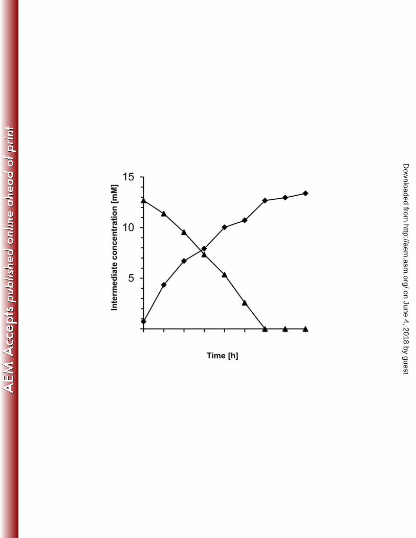
FIG. 3. Biotransformation of vanillin by E. coli BL21(DE3) pET23a::vdh. Cells were 717
grown in LB with 100 µg/ml ampicillin at 30 °C and harvested after 16 h in the stationary 718
growth phase. After two washing steps with 100 mM potassium phosphate buffer (pH 7.5), 719
cells were resuspended in 50 ml mineral salts medium containing 100 µg/ml ampicillin and 720
14 mM vanillin. The cultures were incubated at 30 °C and 100 rpm for 8 h and 721
biotransformation of vanillin to vanillic acid was observed at the given times using HPLC 722
analyses as described in materials and methods. () vanillin; () vanillic acid. 723
724
FIG. 4. Verification of the gene replacement in Amycolatopsis sp. ATCC 39116 725
∆vdh::KmR. In order to verify a specific gene replacement of the vdh gene by the 726
kanamycin resistance cassette different diagnostic PCRs were carried out using 727
chromosomal DNA as template. The resulting fragments were analyzed in a 1 % agarose 728
gel. Molecular weight marker: “GeneRuler™ 1 kb DNA Ladder” (Fermentas GmbH, St. 729
Leon-Rot, Germany). 730
Lanes 1 and 2 represent the amplificate using oligonucleotides vdhLF_for_diag and 731
vdhRF_rev2: Showing the difference between the wild type Amycolatopsis sp. 732
ATCC 39116 (1) and the mutant Amycolatopsis sp. ATCC 39116 ∆vdh::KmR (2). Lanes 3 733
and 4 present the results using primer vdhLF_for_diag and pRLE6for2 (3) as well as 734
KmR_rev_vdhLF and vdhRF_rev2 (4): Shows the proof of the specific integration of the 735
on June 4, 2018 by guesthttp://aem
.asm.org/
Dow
nloaded from

32
kanamycin resistance cassette into the vdh locus for the mutant Amycolatopsis sp. 736
ATCC 39116 ∆vdh::KmR 737
738
FIG. 5. Biotransformation of ferulic acid by Amycolatopsis sp. ATCC 39116 and 739
the mutant Amycolatopsis sp. ATCC 39116 ∆vdh::KmR. Cells were grown in 250 ml 740
Caso-Medium at 42 °C and harvested within the stationary growth phase. After two 741
washing steps with phosphate buffered saline (pH 7.5), cells were resuspended in 742
1,000 of the same buffer containing 2 mM ferulic acid. The cultures were incubated in a 743
2L-bioreactor at 42 °C for 30 h. Biotransformation of ferulic acid (A) to vanillin (B) and 744
vanillic acid (C) was observed at the given times using HPLC analyses as described in 745
materials and methods. Initially, ferulic acid was fed at a rate of 0.75 mM/h during the first 746
7 hours. Afterwards, substrate was fed for 3 hours at a rate of 3.1 mM/h, before feeding 747
was stopped. () Amycolatopsis sp. ATCC 39116; () Amycolatopsis sp. ATCC 39116 748
∆vdh::KmR 749
750
FIG. 6. Proposed pathway of the CoA-dependent catabolism of ferulic acid via 751
vanillin in Amycolatopsis sp. ATCC 39116 752
753
754
755
756
757
758
759
760
on June 4, 2018 by guesthttp://aem
.asm.org/
Dow
nloaded from

33
TABLES 761
TAB. 1: Bacterial strains and plasmids used in the present study 762
Bacterial strains
and plasmids
Description, sequence Reference
Strains
Amycolatopsis sp.
ATCC 39116
wild-type, kanamycin-sensitive American Type
Culture Collection,
No. 39116
Amycolatopsis sp.
ATCC 39116
∆vdh::KmR
vdh-deletion-mutant, kanamycin-resistant This study
E. coli Mach-1 T1 F - φ80(lacZ)ΔM15 ΔlacX74 hsdR (rK– mK
+)
ΔrecA1398 endA1 tonA
Invitrogen GmbH
(Karlsruhe,
Germany)
E. coli BL21(DE3) F – ompT hsdSB(rB– mB
–) gal dcm (DE3) Novagen Inc.
(Madison, WI,
USA)
Plasmids
pET23a T7-promotor, His6-tag, T7-terminator, ColE1 pBR322
ori, AmpR,f1 ori
Novagen Inc.
(Madison, WI,
USA)
pET23a::vdh pET23a harboring vdh (NdeI/XhoI) coding for
VDHATCC 39116 colinear to T7-promotor
This study
on June 4, 2018 by guesthttp://aem
.asm.org/
Dow
nloaded from

34
pET23a::vdhHis pET23a harboring vdh (NdeI/XhoI) without native
stop codon coding for VDHATCC 39116 with C-terminal
His6-tag, colinear to T7-Promotor
This study
763
764
765 766 767 768 769 770 771 772 773 774 775 776 777 778 779 780 781 782 783 784 785 786 787 788 789 790 791 792 793 794 795 796 797 798 799
on June 4, 2018 by guesthttp://aem
.asm.org/
Dow
nloaded from

35
FIGURES 800
Fig. 1 801
802 803 804 805 806 807 808
on June 4, 2018 by guesthttp://aem
.asm.org/
Dow
nloaded from

36
809
Fig. 2 810
811 812 813 814 815 816 817 818 819 820 821 822 823 824 825 826 827 828 829 830 831 832 833
on June 4, 2018 by guesthttp://aem
.asm.org/
Dow
nloaded from

37
834
Fig. 3 835
836 837 838 839 840 841 842 843 844 845 846 847 848 849 850 851 852 853
on June 4, 2018 by guesthttp://aem
.asm.org/
Dow
nloaded from

38
854
Fig. 4 855
856 857 858 859 860 861 862 863 864 865 866 867 868 869 870 871
on June 4, 2018 by guesthttp://aem
.asm.org/
Dow
nloaded from

39
872
Fig. 5 873
874
875 876 877 878 879 880 881 882 883 884 885 886 887 888 889 890 891 892 893 894
895
896
on June 4, 2018 by guesthttp://aem
.asm.org/
Dow
nloaded from
40
897
Fig. 6 898
899
900
901
902
903
904
905 906
on June 4, 2018 by guesthttp://aem
.asm.org/
Dow
nloaded from


C C
H
OH
O
CH3
H
C
O
OH
C C
H
OH
O
CH3
H
C
O
S CoAC C
OH
O
CH3
H
C
O
S CoA
H
OH
H
OHO
OH
O
CH3
OHO
OH
OH
VanillinVanillic acidProtocatechuic acid
Ferulic acid Feruloyl-CoA 4-Hydroxy-3-methoxyphenyl-
β-hydroxypropionyl-CoA
O2
NADH+H+NAD+
H2OHCHOH2O
NAD+NADH+H+
CoA-SH
ATP AMP + PPi
H2O
C
H
C
O
S CoA
H
H
HO
OH
O
CH3Vanillin
dehydrogenase
(VDH)
Vanillate
demethylase
(VanA und VanB)
Feruloyl-CoA
synthetase
(FCS)
Enoyl-CoA
hydratase/aldolase
(ECH)
Enoyl-CoA
hydratase/aldolase
(ECH)
Guaiacol
OH
O
CH3
CO2
on June 4, 2018 by guesthttp://aem
.asm.org/
Dow
nloaded from