investigation of renal function an overviewvictorjtemple.com/investigation of kidney function ppp...
TRANSCRIPT
INVESTIGATION OF RENAL FUNCTION – An Overview
UNIVERSITY OF PNGSCHOOL OF MEDICINE AND HEALTH SCIENCES
DISCIPLINE OF BIOCHEMISTRY & MOLECULAR BIOLOGYCLINICAL BIOCHEMISTRY LECTURE BMLS III & BDS IV
VJ. Temple
1

What are the major components of the kidneys?
• Kidneys are the major excretory system in humans and other Ureotelic Organisms;
• Nephron is the Functional unit of the Kidneys (Fig 1a and 1b)
• Major components of the Nephron:
• Glomerulus – where filtration occurs,
• Proximal tubule – where main reabsorption occurs,
• Loop of Henle – where concentration of filtrate occurs,
• Distal tubule – where secretion occurs,
• Collecting duct – where water reabsorption occurs,
2

3
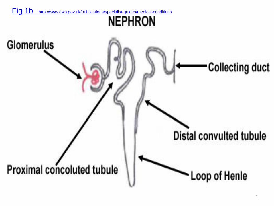
Fig 1b http://www.dwp.gov.uk/publications/specialist-guides/medical-conditions
4

5

What are some of the functions of the kidneys?
• Regulate Extracellular Fluid (ECF) Volume and Electrolyte composition to compensate for wide daily variations in Water and Electrolyte intake,
• Regulation of water,
• Regulation of Electrolyte,
• Regulation of Acid-Base balance, which involves maintaining the pH (acidity/alkalinity) in body fluids,
• Excretion of metabolic waste products (of Protein and Nucleic acid): Urea, Creatinine, Creatine, Uric acid, Sulphate and Phosphate;
6

What are the endocrine functions of the kidneys?
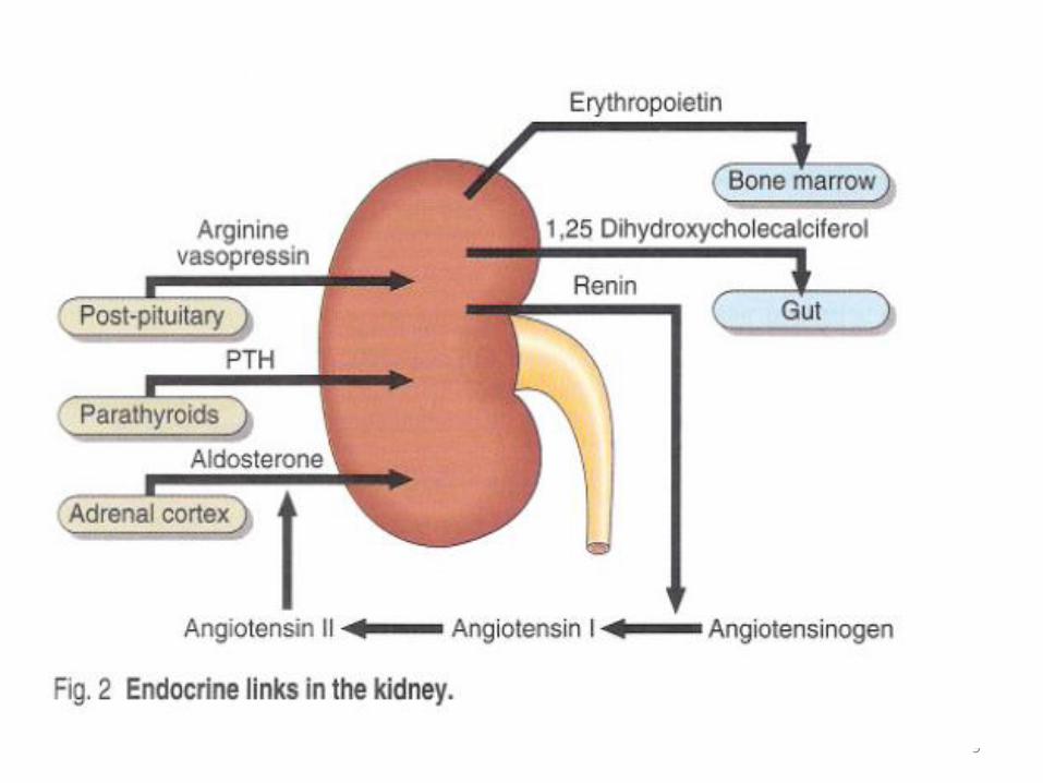
• Kidneys are under the control of some hormones and productions of some hormones are under the control of the kidneys (Fig. 2);
• Arginine Vasopressin (AVP), {Anti-diuretic Hormone ADH}: Acts on the kidneys to Regulate Water balance;
• Aldosterone: Acts on Kidney Tubules to Regulates Sodium balance;
• Parathyroid Hormone (PTH): Acts via the Kidneys:
• To promote Tubular Reabsorption of Calcium;
• To promote Phosphate excretion;
• For biosynthesis of 1,25-Dihydroxy-Cholecalciferol (Vitamin D3) that regulates Calcium absorption by Gastrointestinal Tract;
7
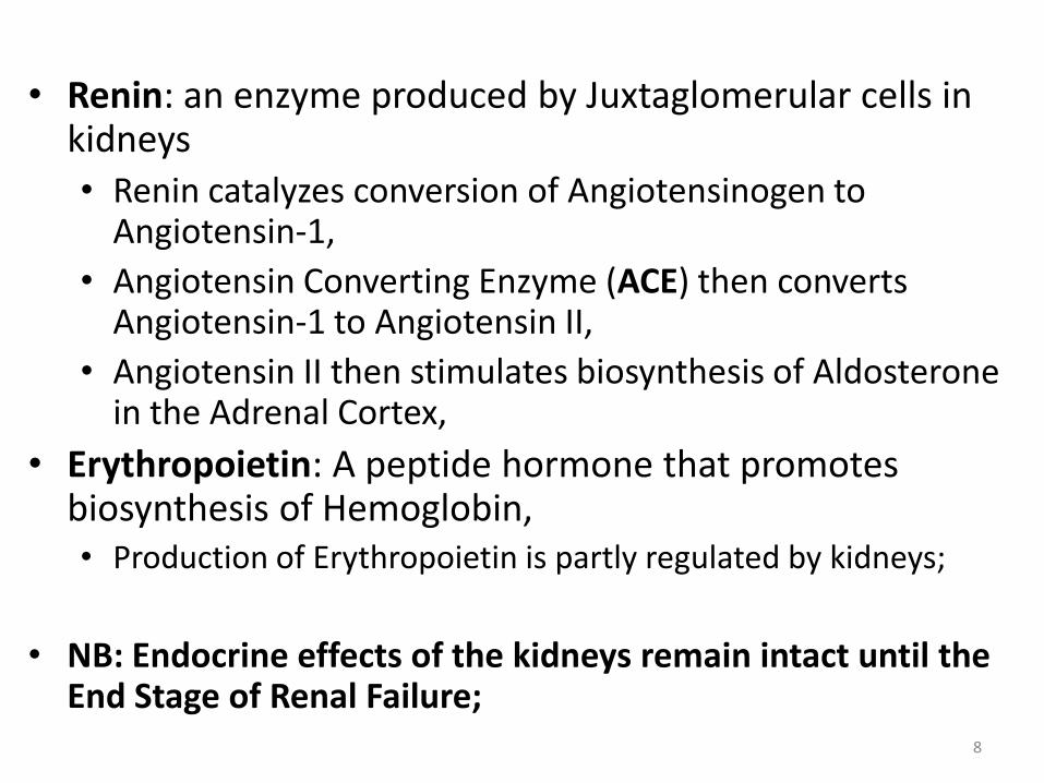
• Renin: an enzyme produced by Juxtaglomerular cells in kidneys• Renin catalyzes conversion of Angiotensinogen to
Angiotensin-1,
• Angiotensin Converting Enzyme (ACE) then converts Angiotensin-1 to Angiotensin II,
• Angiotensin II then stimulates biosynthesis of Aldosterone in the Adrenal Cortex,
• Erythropoietin: A peptide hormone that promotes biosynthesis of Hemoglobin, • Production of Erythropoietin is partly regulated by kidneys;
• NB: Endocrine effects of the kidneys remain intact until the End Stage of Renal Failure;
8
9
How are the functions of the kidneys assessed?
• Kidney functions are assessed by collection of tests called Renal Function Tests:
• Urinalysis is the first line Renal Function Tests;
• Tests for Glomerular Functions;
• Tests for Tubular Functions;
• Specimens for Renal Function Tests:
• Urine,
• Blood Plasma or Serum
10
What test are carried out during urinalysis?
• Randomly collected urine sample is examined:
• Physically,
• Chemically,
• Microscopically
• Physical Examination of Urine sample for:
• Color,
• Odor,
• Appearance,
• Concentration (specific gravity);
11

• Chemical Examination of urine sample for:
• Protein,
• Glucose,
• Urine pH (acidity/ alkalinity);
• Microscopic Examination of urine samples for:
• Cellular elements (red blood cells, white blood cells, and epithelial cells),
• Bacteria,
• Crystals,
• Casts (structures formed by the deposit of protein, cells, and other substances in the kidney tubules);
12

What is done if the Urinalysis tests are positive?
• If Urinalysis indicates disease or impaired kidney function, additional tests are performed to diagnose the problem; These include the following:
• Creatinine Clearance Test (CC): • CC evaluates how efficiently the kidneys clear Creatinine
from the blood;
• Creatinine is a waste product of muscle metabolism; it is produced at a constant rate that is proportional to the muscle mass of the individual;
• Creatinine is filtered and excreted mainly in the urine;
• CC is performed on urine samples collected over 24-hours;
• Determination of the concentration of Creatinine in blood is also required to calculate the Rate at which the Kidneys are clearing Creatinine from the blood;
13

• Urine Osmolality:
• Osmolality determines the number of particles dissolved in the urine;
• Osmolality is more precise than Specific Gravity for evaluating the ability of kidneys to Concentrate or Dilute the Urine;
• For a “healthy” person under normal Physiological conditions the Urine is more concentrated than Plasma
Urine Osmolality > Plasma Osmolality
14
• Measure the Osmolality on:
• Early morning urine samples,
• Multiple timed urine samples, or
• 24-hour urine sample;
• Inability of the kidneys to concentrate the urine in response to restricted fluid intake, or to dilute the urine in response to increased fluid intake during Osmolality testing may indicate decreased kidney function;
15
• Urine Protein (Proteinuria):
• Glomerular filtrate is an ultra-filtrate of plasma;
• Glomerular basement membrane does not usually allow passage of albumin and large proteins;
• Small amount of protein, usually less than 25mg/24h is in urine;
• A positive screening test for protein (included in a routine urinalysis) on a random urine sample is usually followed-up with a test on a 24-hour urine sample that more precisely measures the quantity of protein;
• Larger amount of protein, in excess of 250 mg/24h in urine, indicates significant damage to the Glomerular membrane;
• Persistent presence of significant amounts of protein in urine, is an important indicator of kidney disease;
16
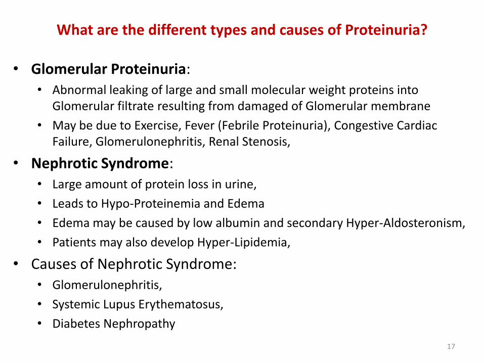
What are the different types and causes of Proteinuria?
• Glomerular Proteinuria: • Abnormal leaking of large and small molecular weight proteins into
Glomerular filtrate resulting from damaged of Glomerular membrane
• May be due to Exercise, Fever (Febrile Proteinuria), Congestive Cardiac Failure, Glomerulonephritis, Renal Stenosis,
• Nephrotic Syndrome: • Large amount of protein loss in urine,
• Leads to Hypo-Proteinemia and Edema
• Edema may be caused by low albumin and secondary Hyper-Aldosteronism,
• Patients may also develop Hyper-Lipidemia,
• Causes of Nephrotic Syndrome: • Glomerulonephritis,
• Systemic Lupus Erythematosus,
• Diabetes Nephropathy
17
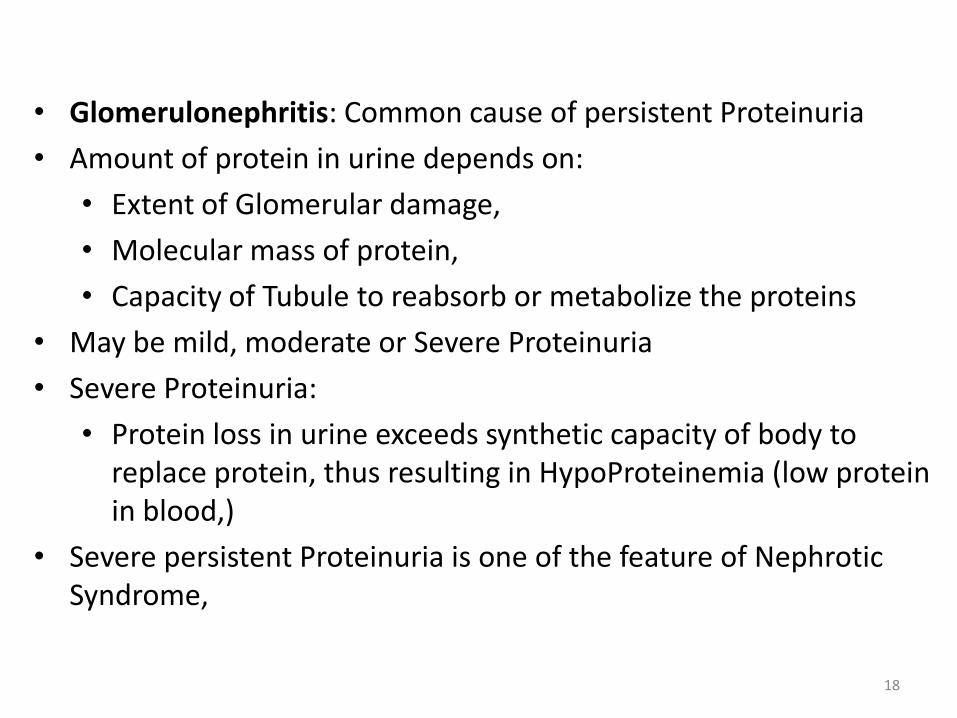
• Glomerulonephritis: Common cause of persistent Proteinuria
• Amount of protein in urine depends on:
• Extent of Glomerular damage,
• Molecular mass of protein,
• Capacity of Tubule to reabsorb or metabolize the proteins
• May be mild, moderate or Severe Proteinuria
• Severe Proteinuria:
• Protein loss in urine exceeds synthetic capacity of body to replace protein, thus resulting in HypoProteinemia (low protein in blood,)
• Severe persistent Proteinuria is one of the feature of Nephrotic Syndrome,
18

• Tubular Proteinuria:
• Failure of Tubules to reabsorb filtered plasma proteins,
• Abnormal secretion of protein into urinary tract,
• May be due to Tubular or Interstitial damage
• Proteins with low molecular mass excreted by Tubules,
• Loss of protein is mild about 2.0g/24hours,
• Sensitive test for assessment of Renal Tubular damage:
• Measure Urinary β2-Microglobulin (> 0.4mg/24hours indicates damage)
19

• Overflow Proteinuria:
• Large amount of low molecular weight proteins in urine,
• Proteins are filtered at Glomerulus, but are not reabsorbed or catabolized completely by Tubules
• Causes of Overflow Proteinuria:
• Acute Pancreatitis,
• Multiple Myeloma,
• Intravascular Hemolysis,
• Myelomonocytic Leukemia,
• Crush Injuries
• Orthostatic (Postural) Proteinuria:
• Proteinuria occurs after standing for a long time,
• Protein absent in early morning urine samples,20

BLOOD TEST FOR ASSESSING RENAL FUNCTION
• Plasma Creatinine: • Creatinine is filtered from the blood by the kidneys
and excreted in urine;
• Production of Creatinine depends on the muscle mass, which usually fluctuates very little over 24-hour period;
• For “Health” individuals with Normal cardiac and kidney functions amount of Creatinine in blood remains relatively constant and normal;
• Elevated Plasma or Serum Creatinine Concentration is a sensitive indicator of impaired kidney function, if the cardiac output and renal blood supply are normal;
21

• Blood Urea Nitrogen (BUN) {H2N-CO-NH2 }
• Urea is a by-product of protein metabolism,
• Urea is formed in the liver, released in the blood then filtered by the Glomerulus and excreted in the urine,
• BUN is the amount of Nitrogen contained in Urea,
• High concentration of BUN indicates kidney dysfunction, but because BUN is also affected by Protein intake and Liver Function, the test is usually done in conjunction with Plasma Creatinine, which is a more specific indicator of kidney function;
• An elevated BUN, by itself, is suggestive, but not diagnostic of kidney dysfunction, because BUN can be affected by other factors;
22
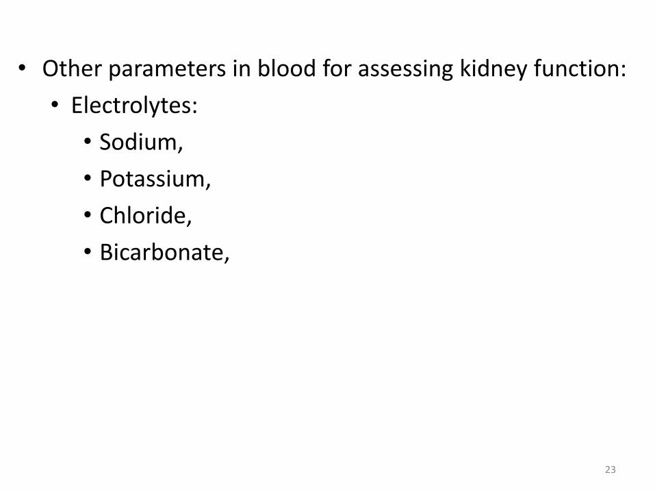
• Other parameters in blood for assessing kidney function:
• Electrolytes:
• Sodium,
• Potassium,
• Chloride,
• Bicarbonate,
23

What is Glomerular Filtration Rate (GFR)?
• Glomerular Filtrate is an Ultra filtrate of Plasma;• It has the same biochemical composition as plasma excluding
most of the Plasma Proteins;• Normally, Plasma is filtered by Glomeruli at a rate of 140ml/min,• Normal GFR depends on normal Renal Blood Flow and Pressure,• GFR is directly related to body size; higher in males than females;• GFR is affected by age, with a reduced rate in elderly;
• If GFR falls due to Restriction of Renal Blood Supply, or Low Cardiac Output, or Damage of Nephrons by Renal Disease, then waste products of metabolism will accumulate in blood;
• As the renal disease progresses, the amount of Creatinine and Urea in blood increase slowly over many months;
• In patients with Chronic Renal disease, a new “Steady State” is reached with a high plasma concentration of Creatinine and Urea;
24

How is Creatinine Clearance (CC) calculated?
• CC is a measure of GFR: amount (ml) of filtrate made by kidneys per minute;
• CC is the rate at which the kidneys clear a compound from blood
• GFR is the maximum rate that plasma can be ‘Cleared’ of any substance;
• GFR can be calculated from the Clearance of some plasma constituent, which is freely filtered at the Glomerulus, and is neither reabsorbed nor secreted by the kidney tubules;
• In clinical practice Creatinine fulfills the above requirements;
• GFR can be calculated from Creatinine content of a 24-hour urine collection, and the Plasma concentration of Creatinine within the period;
25
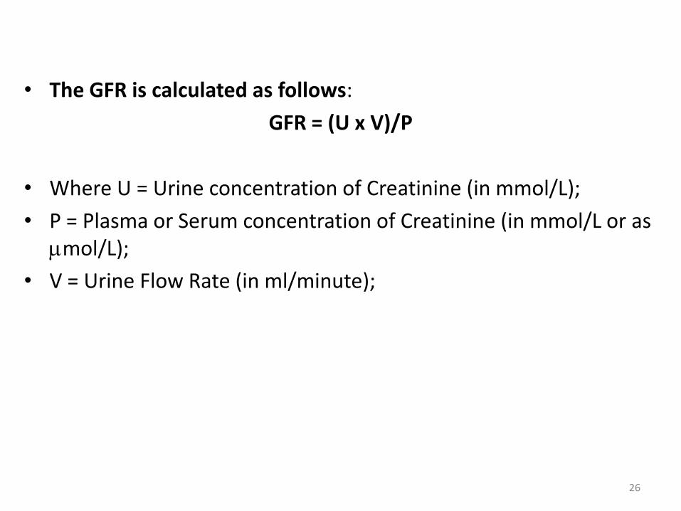
• The GFR is calculated as follows:
GFR = (U x V)/P
• Where U = Urine concentration of Creatinine (in mmol/L);
• P = Plasma or Serum concentration of Creatinine (in mmol/L or as mol/L);
• V = Urine Flow Rate (in ml/minute);
26
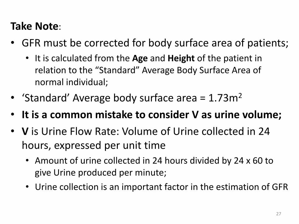
Take Note:
• GFR must be corrected for body surface area of patients;
• It is calculated from the Age and Height of the patient in relation to the “Standard” Average Body Surface Area of normal individual;
• ‘Standard’ Average body surface area = 1.73m2
• It is a common mistake to consider V as urine volume;
• V is Urine Flow Rate: Volume of Urine collected in 24 hours, expressed per unit time
• Amount of urine collected in 24 hours divided by 24 x 60 to give Urine produced per minute;
• Urine collection is an important factor in the estimation of GFR
27

Example for calculation of CC (GFR): QUESTION
• A male patient age 45 years was admitted with loin pain in the Clinical ward in PMG.
• Volume of urine collected in 24-hour was 2160ml;
• Concentration of Creatinine in the urine was 7.5mmol/L;
• Concentration of Creatinine in plasma was 150 mol/L;
• Calculate the Creatinine Clearance of the patient and comment on the results.
• Assume the correction factor for body surface area = 0.8;
• The nursing staff that collected the urine came to see you later and told you that the 2160ml of urine was collected in 17hours not 24-hours.
• How does this affect the result and its interpretation?
28
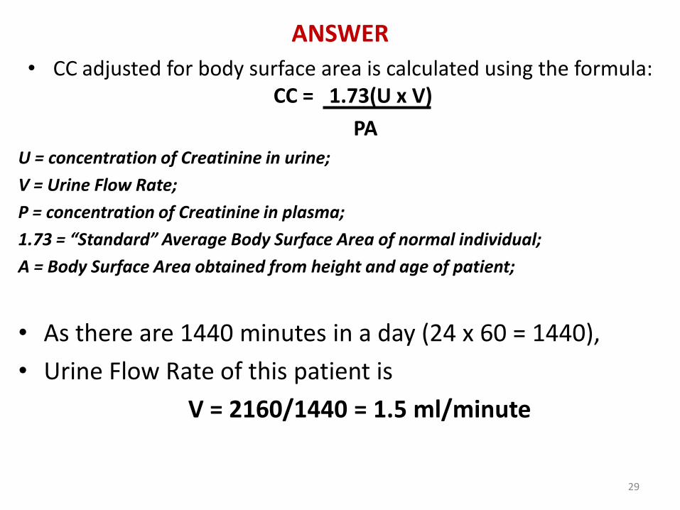
ANSWER
• CC adjusted for body surface area is calculated using the formula: CC = 1.73(U x V)
PAU = concentration of Creatinine in urine;
V = Urine Flow Rate;
P = concentration of Creatinine in plasma;
1.73 = “Standard” Average Body Surface Area of normal individual;
A = Body Surface Area obtained from height and age of patient;
• As there are 1440 minutes in a day (24 x 60 = 1440),
• Urine Flow Rate of this patient is
V = 2160/1440 = 1.5 ml/minute
29

• Urinary Creatinine concentration must be in the same units as the Plasma Creatinine concentration;
• U = 7.5 mmol/L;
• P = 150/1000 = 0.15 mmol/L;
• Correction factor for Body Surface Area 1.73/A is given as 0.8;
• Thus:
CC = 0.8 (7.5 x 1.5)
0.15
• CC = 60.0 ml/minute
• This value is low for a young male;
• The normal range for male is 80 – 130 ml/minute;
30

• When it was discovered that the urine collection was for 17 hours and not 24 hours, the Urine Flow Rate was recalculated.
V = 2160/1020 = 2.1 ml/minute
• Recalculating the CC value gives:
CC = 0.8 (7.5 x 2.1)
0.15
CC = 84.0ml/minute
• This is within the 80 – 130ml/min range for a young adult
31

COMMENTS
• Recalculated result is within the range for young male;
• Errors in timing and collection of urine can significantly influence calculation of Creatinine Clearance;
• Errors in collection are by far the most common and serious errors encountered when estimating Creatinine Clearance;
• Low Clearance values for Creatinine and Urea indicate diminished ability of the kidneys to filter these waste products from the blood and excrete them in the urine;
• As Clearance levels decrease, blood levels of Creatinine and Urea Nitrogen increase;
32

State some factors that can affect CC values?
CC value can be increased by:
• Exercise,
• Pregnancy (due in part to the increased load placed on the kidney by the growing fetus),
• High cardiac output syndromes (as blood flow increases to the kidney, GFR and CC increases);
CC value can be reduced by:
• Impaired kidney function,
• Congestive heart failure,
• Cirrhosis with Ascites,
• Shock,
• Dehydration,
• Conditions associated with decreased blood flow to kidneys33

How can the Creatinine Clearance be calculated using the Cockcroft and Gault equation?
140 – Age in yrs x Weight (Kg)
• CC (ml/min) =
0.814 x Serum Creatinine (umol/L)
• To correct for muscle mass:
• For Female multiply result by 0.85
• For Male multiply by 1.22
34
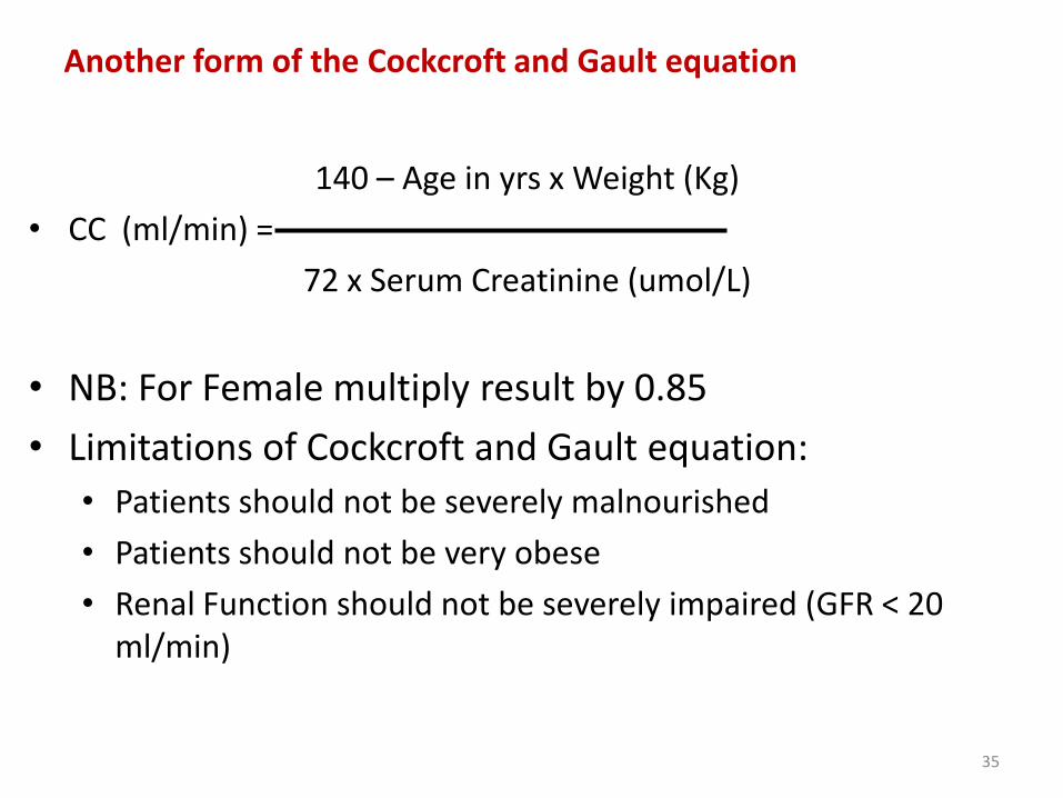
Another form of the Cockcroft and Gault equation
140 – Age in yrs x Weight (Kg)
• CC (ml/min) =
72 x Serum Creatinine (umol/L)
• NB: For Female multiply result by 0.85
• Limitations of Cockcroft and Gault equation:
• Patients should not be severely malnourished
• Patients should not be very obese
• Renal Function should not be severely impaired (GFR < 20 ml/min)
35
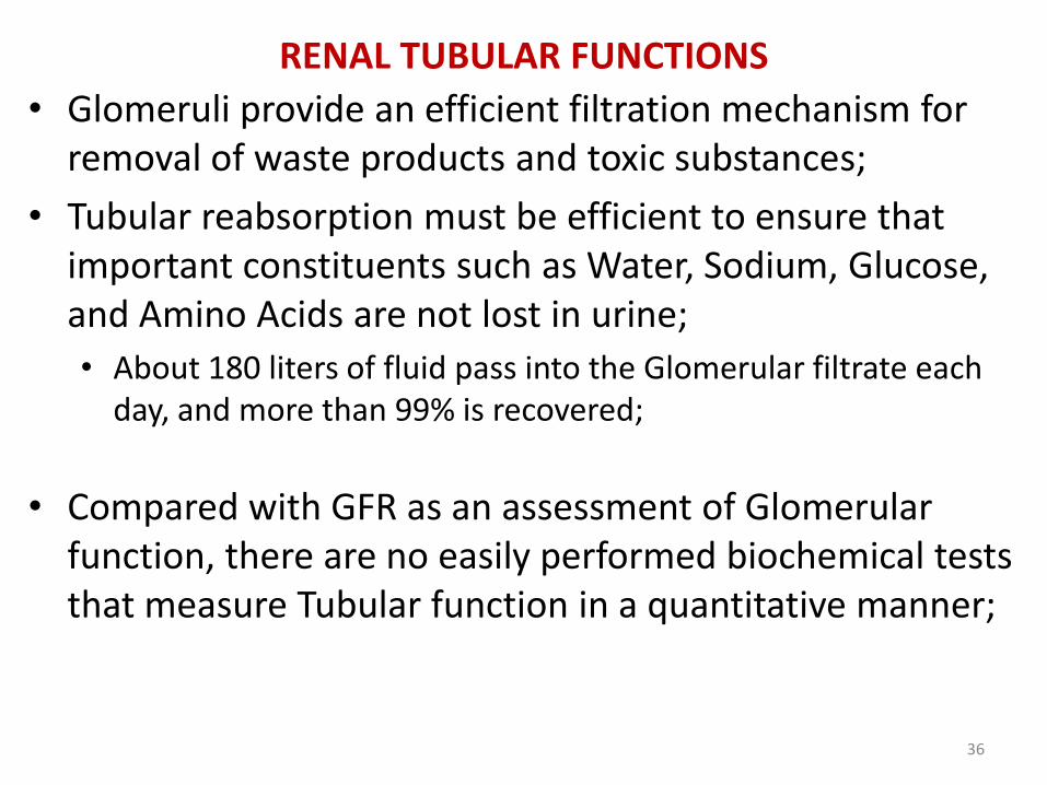
RENAL TUBULAR FUNCTIONS
• Glomeruli provide an efficient filtration mechanism for removal of waste products and toxic substances;
• Tubular reabsorption must be efficient to ensure that important constituents such as Water, Sodium, Glucose, and Amino Acids are not lost in urine;
• About 180 liters of fluid pass into the Glomerular filtrate each day, and more than 99% is recovered;
• Compared with GFR as an assessment of Glomerular function, there are no easily performed biochemical tests that measure Tubular function in a quantitative manner;
36

TUBULAR DYSFUNCTION
• Some disorders of tubular function are inherited;
• Some patients are unable to reduce their urine pH below 6.5, because of a specific failure of Hydrogen ion secretion;
• Of all the tubular functions, the most frequently affected by disease is the ability to concentrate the urine;
• Tubular function can be assessed by measuring the Concentration (Osmolality) of urine and compare it with plasma;
• Urine-Plasma Osmolality Ratio is between 1.0 and 3.0, because the urine is normally more concentrated than plasma;
37

• If the ratio is 1.0 or less, the renal tubules are not reabsorbing water;
• Urine / Plasma ratio < 1.0,
• Indicates poor reabsorption by Renal Tubules
• Some disorders of Tubular function are inherited:
• Example:
• Some patients are unable to reduce their urine pH below 6.5, because of a specific failure of Hydrogen ion secretion;
38
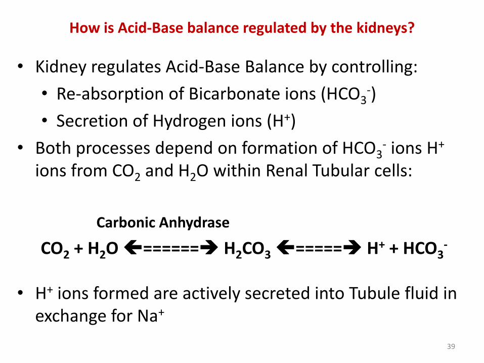
How is Acid-Base balance regulated by the kidneys?
• Kidney regulates Acid-Base Balance by controlling:
• Re-absorption of Bicarbonate ions (HCO3-)
• Secretion of Hydrogen ions (H+)
• Both processes depend on formation of HCO3- ions H+
ions from CO2 and H2O within Renal Tubular cells:
Carbonic Anhydrase
CO2 + H2O ====== H2CO3===== H+ + HCO3-
• H+ ions formed are actively secreted into Tubule fluid in exchange for Na+
39

What mechanisms are used in the kidney for elimination of Acids?
• Mechanisms for elimination of Acids:
• Re-absorption of Sodium Bicarbonate (NaHCO3) by Proximal Renal Tubules, (Fig. 3)
• Regeneration of HCO-3 by Distal Renal Tubules (Fig. 4)
• Formation of Phosphate buffer in Distal Tubules; (Fig. 5)
• Production of Ammonia (NH3) by Distal Renal Tubules for formation of Ammonium buffer; (Fig. 6)
• Secretion of H+ ions by Tubular cells serves initially to reabsorb HCO3
- ions from the Glomerular filtrate;
• After all the HCO3- ions have been reabsorbed, any
deficit that occurs is regenerated;
40
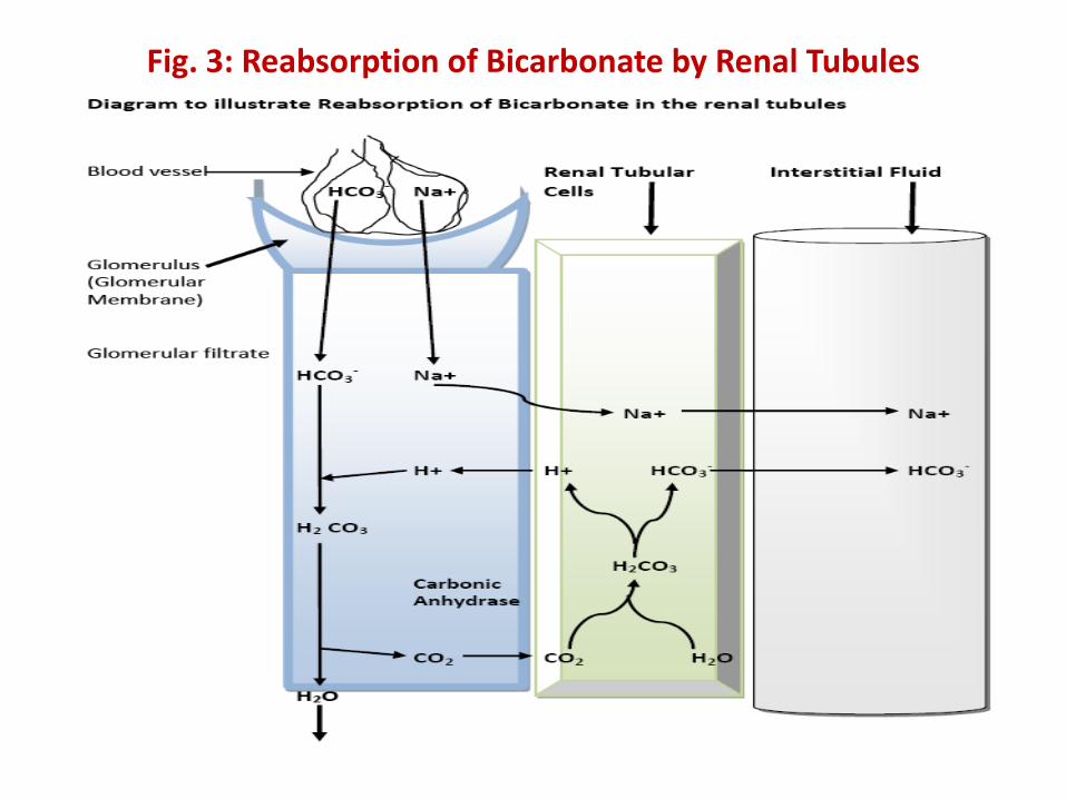
Fig. 3: Reabsorption of Bicarbonate by Renal Tubules
41

Fig. 4: Regeneration of Bicarbonate ions by Renal Tubules
42
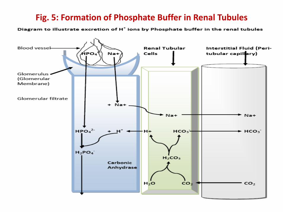
Fig. 5: Formation of Phosphate Buffer in Renal Tubules
43

Fig. 6: Formation of Ammonium Buffer in Renal Tubules
44

What is Anion Gap?
• Anion Gap (AG) calculation is the sum of routinely measured Cations minus routinely measured Anions:
Anion Gap = (Na+ + K+) – (Cl- + HCO3-)
• However, because K+ is a small value it is usually omitted from the AG equation; the most commonly use equation is:
Anion Gap = Na+ - (Cl- + HCO3-)
45
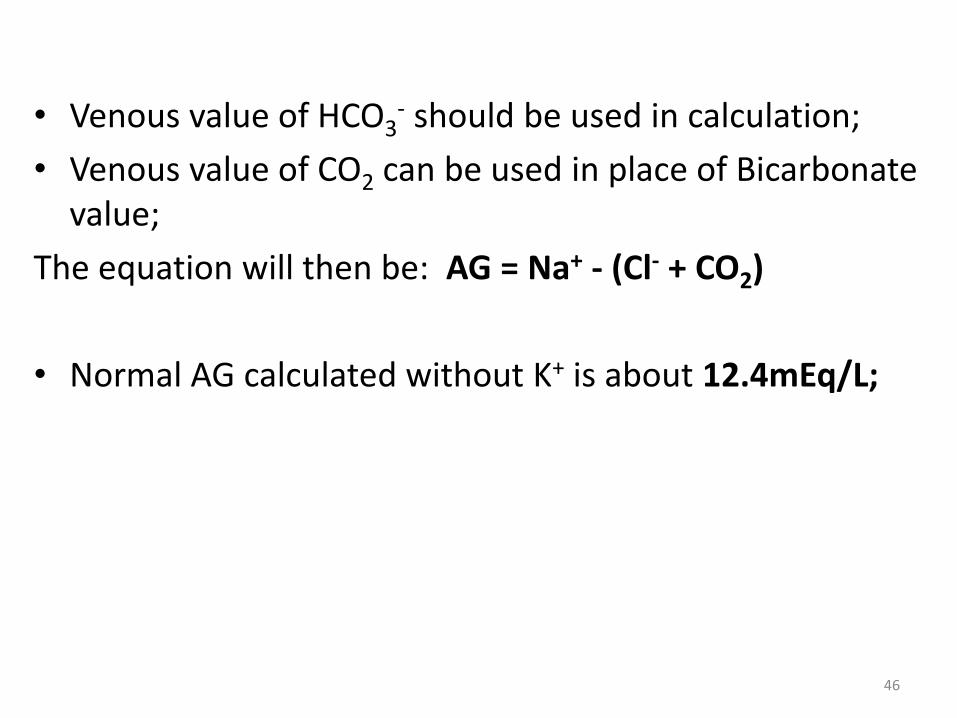
• Venous value of HCO3- should be used in calculation;
• Venous value of CO2 can be used in place of Bicarbonate value;
The equation will then be: AG = Na+ - (Cl- + CO2)
• Normal AG calculated without K+ is about 12.4mEq/L;
46

• Anion Gap exists because not all Electrolytes are routinely measured;
• Normally there is electrochemical balance in cells; thus the sum of all Anions equals the sum of all Cations;
• However, several Anions are not measured routinely, leading to the Anion Gap;
• Anion Gap is thus an artifact of measurement, and not a Physiologic reality;
47
Study questions
48