investigating lipid headgroup composition within
TRANSCRIPT

This journal is © The Royal Society of Chemistry 2021 Soft Matter, 2021, 17, 6773–6786 | 6773
Cite this: Soft Matter, 2021,
17, 6773
Investigating lipid headgroup composition withinepithelial membranes: a systematic review†
R. T. Coones, *a R. J. Green *a and R. A. Frazier *b
Membrane lipid composition is often quoted within the literature, but with very little insight into how or
why these compositions vary when compared to other biological membranes. One prominent area that
lacks understanding in terms of rationale for lipid variability is the human gastro-intestinal tract (GIT). We
have carried out a comprehensive systematic literature search to ascertain the key lipid components of
epithelial membranes, with a particular focus on addressing the human GIT and to use compositional
data to understand structural aspects of biological membranes. Both bacterial outer membranes and the
human erythrocyte membrane were used as a comparison for the mammalian [epithelial] membranes
and to understand variations in lipid presence. We show that phosphatidylcholine (PC) lipid types tend to
dominate (33%) with phosphatidylethanolamines (PE) and cholesterol having very similar abundances
(25 and 23% respectively). This systematic review presents a detailed insight into lipid headgroup
composition and roles in various membrane types, with a summary of the distinction between the major
lipid bilayer forming lipids and how peripheral lipids regulate charge and fluidity. The variety of lipids
present in biological membranes is discussed and rationalised in terms function as well as cellular position.
Introduction
Almost 50 years on from the proposal of the fluid mosaic modelof cell membranes by Singer and Nicholson in 1972, the modelis still relatively untouched and is accepted today as thefoundation of our understanding of cell surfaces.1 Since thenit has been amended and updated in order to agree withcurrent understanding, forming a cornerstone of a field thatis in constant change.2
The cell membrane is an extremely complex set of structureand functions. Because of this, being able to understand it at afundamental level is challenging and requires a number ofdifferent techniques to be able to provide information.Historically biological mimetic (biomimetic) membranes werestudied using monolayers at the air–water interface throughtechniques such as Langmuir–Blodgett deposition.3 Movingforwards, more complex analyses of increasingly advancedand accurate models have been developed, including usingsolid state NMR techniques to help understand bilayerdynamics, probing layers on a solid support through atomicforce microscopy, or investigations of bilayers using a quartz
crystal microbalance with dissipation monitoring, attenuatedtotal reflection FTIR, or much more powerful techniques suchas small angle X-ray scattering (SAXS), neutron reflectometry(NR), or small angle neutron scattering (SANS).4–10 For systemssuch as DSC, SANS, and NR bilayer deposition, micelle/vesiclesizes of 50–200 nm are common.11,12 In order to gain an indepth and accurate understanding of how the biologicalmembrane functions, a number of these techniques are neededin tandem. Fig. 1 shows some examples of systems used asfoundations for membrane modelling. It should be made clearthat even the most complex biomimetic models do not yetreflect the cell in its entirety. It is common for each model toexamine a small number of functions or components but giventhat biological membranes include all kinds of complex parts (ionchannels, receptors, surface proteins, surface carbohydrates, etc.)it is a hard task to investigate all of these at once. Whilstmodelling does not always completely imitate the real world stateof the system, these methods are also valuable tools in under-standing membrane interactions.13,14
At present, the mammalian membrane environment isconsidered to be dominated by glycerophospholipids andsupplemented by the presence of sphingolipids and sterols.15
This review sets out to bring together the understanding thatsurrounds membrane systems, in particular those that arerelevant to epithelial surfaces and identify potential gaps inthe literature as far as mammalian model systems areconcerned. Further, we show that bacterial modelling isextremely popular as a method for examining the effectiveness
a Department of Pharmacy, School of Chemistry, Food, and Pharmacy,
University of Reading, UK. E-mail: [email protected],
[email protected] Department of Food and Nutritional Sciences, School of Chemistry,
Food and Pharmacy, University of Reading, UK. E-mail: [email protected]
† Electronic supplementary information (ESI) available. See DOI: 10.1039/d1sm00703c
Received 12th May 2021,Accepted 23rd June 2021
DOI: 10.1039/d1sm00703c
rsc.li/soft-matter-journal
Soft Matter
PAPER
Ope
n A
cces
s A
rtic
le. P
ublis
hed
on 2
4 Ju
ne 2
021.
Dow
nloa
ded
on 1
2/1/
2021
10:
03:2
9 A
M.
Thi
s ar
ticle
is li
cens
ed u
nder
a C
reat
ive
Com
mon
s A
ttrib
utio
n 3.
0 U
npor
ted
Lic
ence
.
View Article OnlineView Journal | View Issue

6774 | Soft Matter, 2021, 17, 6773–6786 This journal is © The Royal Society of Chemistry 2021
of antimicrobial agents16 and compare the compositions of themammalian epithelium and the human erythrocyte. Within themembrane environment, epithelial cells are polarised and areable to maintain separation between the apical, lumen facinginterface, and the basolateral portion. Even within theseadjacent domains of a single cell, there is localised variationin the distribution of species across the surface and thatrelative concentrations of various components can have largeimplications in the function of a surface.17,18 Given that there issuch variation in lipid composition within the natural world,this invites questions concerning why there are so many kindsof lipids, and do differences in lipid species at a surface have an
effect on function?19 While lipids form such a ubiquitous partof the natural world, the reason for their variation and breadthremains relatively poorly understood.20 Cells expend a lot ofresources on the synthesis of the vast array of lipids within amembrane – but for what reason?21 It is prevalent throughoutthe biological world that structure is secondary to function; thatis that there is an implicit reason for such a diverse range oflipid species.22 Bacterial membranes can also be built from theneat lipid components, or alternatively by using bacterial lipidextracts, both of which are suitable for investigation using thetechniques outlined above.23 The large scope for variability inmembrane composition and structure gives rise to a range ofways to observe and model such membranes. As epithelia differin their specific composition from cell line models to bacterialto simple mammalian epithelial linings.20,24,25 With differingstructure and function comes differing methods for probingthe various aspects of a membrane.26 Quintessentially thesereviews highlight that as techniques and technology progresses,so too does our understanding.
When modelling lipid systems, as with any model, there area lot of components to consider. It is important to try and getthe model to describe its real-world counterpart, althoughnaturally as the accuracy becomes better the complexity oftenincreases as a result. In this article, the aim is to understandthe content of the scientific literature through a holisticapproach in order to gain insight into what is known aboutlipid composition in general, as well as in more detail at atissue/biological interface specific level in order to identifypossible gaps or areas of need in the scientific knowledge.We set out to answer the question ‘‘What is the composition ofepithelial membranes?’’ Throughout this article the kinds ofspecies shown in Fig. 2 are referred to. Fig. 2 gives a breakdownof how the species are related, the chemical class they belongto, as well as a brief schematic representation.
From Fig. 2, triacylglycerides are a large component inmammalian fats, as well as present in the blood. In all their
Fig. 1 Schematics of some typical methods for modelling lipid systemsand for observing their interactions; top left – micelles; typical ofsurfactants and other hydrophobic molecules when introduced to anaqueous environment. Top right – monolayers; very common set-up forLangmuir–Blodgett type experiments for measuring lipid surface pressureand tension. Bottom left – vesicles; these can adopt a large size regimeand are frequently used to mimic simple bilayer interactions when formedwith lipids. Bottom right – asymmetric bilayers; complex layer formation istaking centre stage in many surface sensitive techniques as they becomemore accurate and complex.
Fig. 2 Depiction of lipid classes, along with categorical examples of each class and a simplified schematic representations. Chain lengths and degree ofsaturation varies within natural membranes, typically ranging from around C-14 to C-24.
Paper Soft Matter
Ope
n A
cces
s A
rtic
le. P
ublis
hed
on 2
4 Ju
ne 2
021.
Dow
nloa
ded
on 1
2/1/
2021
10:
03:2
9 A
M.
Thi
s ar
ticle
is li
cens
ed u
nder
a C
reat
ive
Com
mon
s A
ttrib
utio
n 3.
0 U
npor
ted
Lic
ence
.View Article Online
This journal is © The Royal Society of Chemistry 2021 Soft Matter, 2021, 17, 6773–6786 | 6775
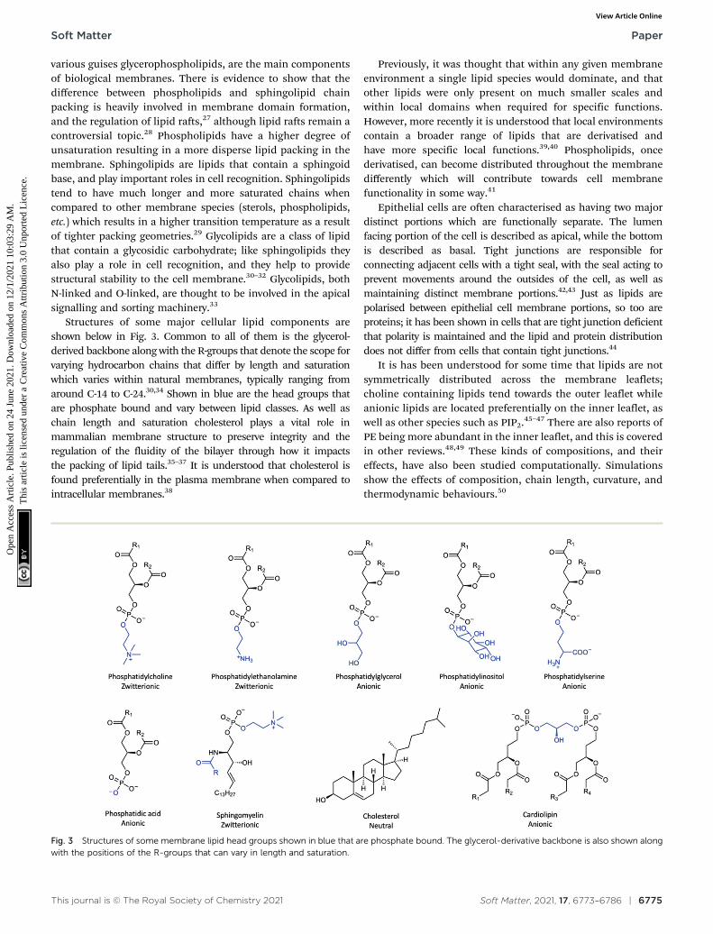
various guises glycerophospholipids, are the main componentsof biological membranes. There is evidence to show that thedifference between phospholipids and sphingolipid chainpacking is heavily involved in membrane domain formation,and the regulation of lipid rafts,27 although lipid rafts remain acontroversial topic.28 Phospholipids have a higher degree ofunsaturation resulting in a more disperse lipid packing in themembrane. Sphingolipids are lipids that contain a sphingoidbase, and play important roles in cell recognition. Sphingolipidstend to have much longer and more saturated chains whencompared to other membrane species (sterols, phospholipids,etc.) which results in a higher transition temperature as a resultof tighter packing geometries.29 Glycolipids are a class of lipidthat contain a glycosidic carbohydrate; like sphingolipids theyalso play a role in cell recognition, and they help to providestructural stability to the cell membrane.30–32 Glycolipids, bothN-linked and O-linked, are thought to be involved in the apicalsignalling and sorting machinery.33
Structures of some major cellular lipid components areshown below in Fig. 3. Common to all of them is the glycerol-derived backbone along with the R-groups that denote the scope forvarying hydrocarbon chains that differ by length and saturationwhich varies within natural membranes, typically ranging fromaround C-14 to C-24.30,34 Shown in blue are the head groups thatare phosphate bound and vary between lipid classes. As well aschain length and saturation cholesterol plays a vital role inmammalian membrane structure to preserve integrity and theregulation of the fluidity of the bilayer through how it impactsthe packing of lipid tails.35–37 It is understood that cholesterol isfound preferentially in the plasma membrane when compared tointracellular membranes.38
Previously, it was thought that within any given membraneenvironment a single lipid species would dominate, and thatother lipids were only present on much smaller scales andwithin local domains when required for specific functions.However, more recently it is understood that local environmentscontain a broader range of lipids that are derivatised andhave more specific local functions.39,40 Phospholipids, oncederivatised, can become distributed throughout the membranedifferently which will contribute towards cell membranefunctionality in some way.41
Epithelial cells are often characterised as having two majordistinct portions which are functionally separate. The lumenfacing portion of the cell is described as apical, while the bottomis described as basal. Tight junctions are responsible forconnecting adjacent cells with a tight seal, with the seal acting toprevent movements around the outsides of the cell, as well asmaintaining distinct membrane portions.42,43 Just as lipids arepolarised between epithelial cell membrane portions, so too areproteins; it has been shown in cells that are tight junction deficientthat polarity is maintained and the lipid and protein distributiondoes not differ from cells that contain tight junctions.44
It is has been understood for some time that lipids are notsymmetrically distributed across the membrane leaflets;choline containing lipids tend towards the outer leaflet whileanionic lipids are located preferentially on the inner leaflet, aswell as other species such as PIP2.45–47 There are also reports ofPE being more abundant in the inner leaflet, and this is coveredin other reviews.48,49 These kinds of compositions, and theireffects, have also been studied computationally. Simulationsshow the effects of composition, chain length, curvature, andthermodynamic behaviours.50
Fig. 3 Structures of some membrane lipid head groups shown in blue that are phosphate bound. The glycerol-derivative backbone is also shown alongwith the positions of the R-groups that can vary in length and saturation.
Soft Matter Paper
Ope
n A
cces
s A
rtic
le. P
ublis
hed
on 2
4 Ju
ne 2
021.
Dow
nloa
ded
on 1
2/1/
2021
10:
03:2
9 A
M.
Thi
s ar
ticle
is li
cens
ed u
nder
a C
reat
ive
Com
mon
s A
ttrib
utio
n 3.
0 U
npor
ted
Lic
ence
.View Article Online

6776 | Soft Matter, 2021, 17, 6773–6786 This journal is © The Royal Society of Chemistry 2021
In this study, we use a comprehensive literature search tosystematically investigate the lipid head group composition ofmammalian epithelial cells, with a particular focus on thosethat are intestinal and from the human GIT. Rationale forunderstanding the lipid composition of the GIT stems fromthe importance of helping to understand how these biologicalsurfaces are structured and how they behave. By using thecompositional data to inform models, there is scope for thiskind of information to develop how models behave duringillness or disease, and even to help study the effects ofpharmaceutical compounds on these kinds of biologicalsurfaces. Further, the principles used can be taken and appliedelsewhere, for example closely related surfaces such as otherepithelia can be similarly modelled. This article aims to identifygaps within the literature where data relating to GIT lipidcomposition is concerned, and identify some open questionsthat can help shape research in this area moving forwards.First, the contents of epithelial membranes are studied, andthen these are broken down into various subgroups dependingon the source of the epithelium. These head group compositionsare then compared to bacterial outer membranes and theepithelium of human erythrocytes. Similarities and differencesbetween the various membrane types are considered all with theintention of understanding reasons for lipid variability withinmembranes.
MethodologySystematic review of epithelial cell composition
The focus of this systematic review was to search terms for lipidmembranes that linked to the lipid head group composition ofthese lipids, as this will provide crucial information that willserve as a basis for (i) providing insight into how the membraneoperates as well as (ii) serving as a basis for model membranesynthesis. The composition of the epithelial membranes wasinvestigated, along with bacterial outer membrane compositionand that of the human erythrocyte for modes of comparison.Bacterial membranes were chosen because they are relativelywell characterised and understood, having long since beenused as membrane models. Human erythrocytes were chosento allow comparison to another human epithelial membrane,
especially for the case of comparing to human cell types. Thefirst step in this study was to screen the literature for articlescontaining relevant information on cell membrane lipidcomposition. This was done through a systematic search usingthe database Web of Sciences, searching all databases andscreening by document type for ‘Articles’ Search terms wereselected in order to remove unwanted hits and then secondarysearch terms were used in order to focus a broad result into alist of results that contained relevant information. The searchwas not filtered by year, and was from 1970–2020. Note – 1970was chosen as the start year as this is the earliest year listedwhen going through the filtering step.
Articles were collected through two independent searches,and the results were combined. Each search was comprised oftwo lists shown in Fig. 4b: lists A and B were used togetherwhile lists 1 and 2 were also paired. During searches, searchterms were combined using the Boolean operators OR betweengroups showing results that appeared through both. The twosets of lists were combined using the AND operator, whichresulted in only articles featuring in both searches beingdisplayed. Every possible combination of searches within therespective lists was used to generate results and the lists areshown below.
The literature search found studies that met the inclusioncriteria; (a) lipid composition, (b) epithelial membranes, and (c)membrane structure for healthy cells.51–105 Any studies fromthe search that did not contain quantitative information werediscarded from the results. Data from the remaining studieswere collated and analysed in order to assess the lipidcomposition of biological species and are presented below.The selection criteria were chosen to allow thousands ofpublications to be narrowed through the title or abstract ofthe publication. They were selected in a way that allowed amulti-layered analysis of the state of epithelial composition inmammalian cells as a whole, ending in a very specific view oflipid composition of the human GIT. The search was conductedindependently by 2 authors, and collation of the results wasfiltered and results collated by a single author examiningabstracts and applying inclusion/exclusion criteria. The resultsinclude a range of mammalian animal types. From the 54search results acquired there were 75 individual sets of data
Fig. 4 (a) Flow diagram that summarises the method for the selection of articles. (b) Schematic showing how search terms were combined between listsusing both AND and OR Boolean operators.
Paper Soft Matter
Ope
n A
cces
s A
rtic
le. P
ublis
hed
on 2
4 Ju
ne 2
021.
Dow
nloa
ded
on 1
2/1/
2021
10:
03:2
9 A
M.
Thi
s ar
ticle
is li
cens
ed u
nder
a C
reat
ive
Com
mon
s A
ttrib
utio
n 3.
0 U
npor
ted
Lic
ence
.View Article Online
This journal is © The Royal Society of Chemistry 2021 Soft Matter, 2021, 17, 6773–6786 | 6777
that related to lipid headgroup composition of one of therelevant tissues outlined above, that cover a very broad rangeof cell types. These can be divided down into subgroups fordata processing based on cell type, location, those that arespecific to humans, etc. These will be covered in more detailwithin the results section of the review and comparisonsbetween such subsets of data are shown in Fig. 8.
As a final note, while the head group composition was thefocus of this review, it is understood that tail length and level ofunsaturation all have important roles, these are commented onin later parts of this review, although not investigated andscrutinised as intensely as the head group composition.106
Further, composition across leaflets is not quantitativelyinvestigated, however it should be acknowledged that thisphenomenon occurs in most biomembranes.49
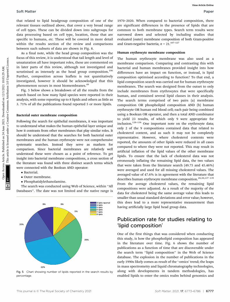
Fig. 5 below shows a breakdown of all the results from thesearch based on how many lipid species were reported in theiranalysis, with some reporting up to 8 lipids and others as little as3. 71% of all the publications found reported 5 or more lipids.
Bacterial outer membrane composition
Following the search for epithelial membranes, it was importantto understand what makes the human epithelial layer unique andhow it contrasts from other membranes that play similar roles. Itshould be understood that the searches for both bacterial outermembranes and the human erythrocyte were not comprehensivesystematic searches. Instead they serve as markers forcomparison. Since bacterial membranes are relatively wellunderstood these were chosen as a point of reference. To getinsight into bacterial membrane compositions, a cross section ofthe literature was found with three distinct search terms whichwere connected with the Boolean AND operator:� Bacterial.� Outer membrane.� Phosphatidylethanolamine.The search was conducted using Web of Science, within ‘‘All
Databases’’. The date was not limited and the native range is
1970–2020. When compared to bacterial composition, thereare significant differences in the presence of lipids that arecommon to both membrane types. Search term results werenarrowed down and selected by including studies thatevaluated (outer) membrane composition of both Gram-positiveand Gram-negative bacteria; n = 21.107–127
Human erythrocyte membrane composition
The human erythrocyte membrane was also used as amembrane comparison. Comparing and contrasting this withbacterial and human membranes provided insight into howdifferences have an impact on function, or instead, is lipidcomposition optimised according to function? To that end, alipid composition search was carried out for human erythrocytemembranes. The search was designed from the outset to onlyinclude membranes from erythrocytes that were specificallyhuman, and contained several lipid classes in their analysis.The search terms comprised of two pairs (a) membranecomposition OR phospholipid composition AND (b) humanerythrocyte OR human red blood cell; each pair being combinedusing a Boolean OR operator, and then a total AND combinatorto yield 23 results, of which only 9 were appropriate forinclusion.128–136 One important note on the erythrocyte data;only 2 of the 9 compositions contained data that related tocholesterol content, and as such it may not be completelyrepresentative. However, where cholesterol contents werereported, the amounts of other lipids were reduced in all casescompared to where they were not reported. This may result inartificial inflation of the lipid values of the other membranelipids. To ensure that the lack of cholesterol data was noterroneously inflating the remaining lipid data, the two valuesthat were taken from the literature search (49.75 and 45.46%)were averaged and used for all missing cholesterol values. Theaveraged value of 47.6% is in agreement with the literature thatdescribes human erythrocyte membrane composition.28,38,137–139
From the average cholesterol values, the remaining lipidcompositions were adjusted. As a result of the majority of thedata for cholesterol being the same average value this leads tosmaller than usual standard deviations and error value; however,this does lead to a more representative measurement thanhaving artificially large lipid head group data.
Publication rate for studies relating to‘lipid composition’
One of the first things that was considered when conductingthis study, is how the phospholipid composition has appearedin the literature over time. Fig. 6 shows the number ofpublications as a function of time that are discoverable underthe search term ‘‘lipid composition’’ in the Web of Sciencedatabase. The explosion in the number of publications in theearly 1990s likely comes as result of the ‘-omics’ trend; the leapsin mass spectrometry and liquid chromatography technologies,along with developments in tandem methodologies, hasenabled lipids to enter the omics realm behind genomics and
Fig. 5 Chart showing number of lipids reported in the search results bypercentage.
Soft Matter Paper
Ope
n A
cces
s A
rtic
le. P
ublis
hed
on 2
4 Ju
ne 2
021.
Dow
nloa
ded
on 1
2/1/
2021
10:
03:2
9 A
M.
Thi
s ar
ticle
is li
cens
ed u
nder
a C
reat
ive
Com
mon
s A
ttrib
utio
n 3.
0 U
npor
ted
Lic
ence
.View Article Online

6778 | Soft Matter, 2021, 17, 6773–6786 This journal is © The Royal Society of Chemistry 2021
proteomics.140 Within the figure, the blue bars are representativeof the total number of publications for a given year where a topicwas lipid composition. Within these, the other coloured barsrepresent categories by which the results can be sub-divided into‘‘human’’, ‘‘bacterial’’, ‘‘mammalian’’, or ‘‘intestine’’, etc. Thesmaller coloured bars are included within the total ‘‘Lipidcomposition’’ count and form part of the blue bars.
Lipid compositional studiesEpithelial membrane composition
It should be noted that when seeking out lipid compositionaland lipidomic studies there can be a large amount of variabilityin the lipids that are examined within these studies, which isdescribed and visualised in more detail in the ESI.† Throughsearching the literature our search provided 54 papers that havedefined lipid composition but from a variety of biologicalsources for epithelial cells. The data averaging lipid compositionfor all 54 is displayed in Fig. 7. While this hides any variabilitybetween different types of epithelial cells it does allow identificationof the major lipid components seen within these systems.We report lipids as a relative percentage of total lipid contentwithin each study. This serves to give a general overview of generallipid content within nature, and more specifically mammalianbiology, which lipids tend to dominate.
The 54 papers included in this study define lipid contentfrom a variety of epithelial membrane sources. It is clearthat within the majority of compositions, PC and PE lipidsdominate, along with cholesterol, with average compositions ofca. 37, 26 and 25% respectively It has been reported that PCaccounts for 450% of the lipids within eukaryotic membranes,although the results from this review show that this may be anover-estimation.22 No doubt, the variability within lipid
composition is dependent on the tissue type in terms of itsorigin, structure and function. These factors will have animpact on the phospholipid content and this is something thatthis text seeks to explore further.
When lipid membranes’ composition are taken as anaverage this does not by necessity take into account the functionof those membranes. In order to make judgements aboutdifferent membrane types, a more focussed analysis must be
Fig. 6 The number of publications for a given year as a result of thesearch term ‘lipid composition’. Colouring within the bars represents thetotal number of publications in a given filter term used to refine the overall‘lipid composition’ search. The smaller coloured bars are included withinthe total count and form part of the blue bars.
Fig. 7 Values of overall mean lipid content taken from the studies foundas a result of the initial literature search in Web of Science; n = 54. Thesepercentages represent the proportion of lipids present in all membranesirrespective of origin or type. Numerical values for the mean are presentedalong with their standard deviation. Tabular results can be found in theESI,† in Table S3.
Fig. 8 Mean compositional data for intestinal studies obtained from thesystematic review search for articles that pertained to intestinal tissues(n = 18), epithelial samples (n = 22), and samples that related to the humanGIT epithelium (n = 10). Values are plotted as relative means, and dataare given as relative means SEM in Table 1. Individual plots with theirassociated error bars are given in the ESI.†
Paper Soft Matter
Ope
n A
cces
s A
rtic
le. P
ublis
hed
on 2
4 Ju
ne 2
021.
Dow
nloa
ded
on 1
2/1/
2021
10:
03:2
9 A
M.
Thi
s ar
ticle
is li
cens
ed u
nder
a C
reat
ive
Com
mon
s A
ttrib
utio
n 3.
0 U
npor
ted
Lic
ence
.View Article Online
This journal is © The Royal Society of Chemistry 2021 Soft Matter, 2021, 17, 6773–6786 | 6779
conducted. Below comparisons are drawn from data obtained inthe review and will show specific data grouped together by celltype or by environment to investigate how lipid compositionaltrends differ as a function of biological geography. Onelimitation of the literature search was that not all papersreported exclusively cell membrane composition. Where themembrane was not isolated, these data may reflect more thewhole cell lipid isolate. As such, these data should be handledwith care and later are separated out into various surface types toreduce the effects of non-specific lipid extraction.
Studies that contained lipid compositional data for intestinalsamples (n = 18) showed that the three most dominant lipids arePE, PC, and Chol have relative percentages of 32.9 � 2.9, 29.5 �2.5, and 26.5 � 3.5% respectively (see Table 1). This is differentwhen compared with epithelial or human GIT epithelial mem-branes which show PC as the most abundant lipid. Interestingly,SM is around 3% lower from the mean data (Fig. 7); a decreasethat could be explained by the increased prevalence of otherbilayer lipids such as PS, PI, and PA, each with their ownstructural roles. Intestinal data, which is not strictly fromepithelial samples, shows elevated amounts of the anionic lipids.This may suggest that anionic lipids do not play as much ofa role in epithelial and surface cells as they might in basal ornon-lumen facing cell portions- shown in Fig. 8. Note that whilenot all these data are from human intestinal samples, they arestill deemed relevant in that they perform the same function:acting as a barrier for absorption of nutrients from the GIT.Conversely, cholesterol is the most abundant membrane speciesin human erythrocytes, going against the trend of being PC or PEdominant.
There is evidence that cholesterol and sphingomyelincomposition have an impact on each other. Cholesterol hasbeen shown to be associated with SM in preference of othermembrane species.141 The effect of SM presence within abilayer on cholesterol was demonstrated to be more importantthan cholesterol’s interactions with the layer lipids.142
Cholesterol and SM have both been linked to the productionof laterally organised microdomains (rafts) within membranes,and these topics have been reviewed elsewhere.29,143,144
Where epithelial surfaces are concerned (n = 22, althoughnot necessarily exclusive to the GIT) PC, PE, and Chol are stillthe dominant lipids with amounts of 33.6, 25.8, and 23.3%respectively. However, unlike for intestinal samples, PC is themost abundant lipid here. The relatively low proportion ofcholesterol in epithelial surfaces is likely related to theirfunction, as all interfaces that are lumen facing are requiredto be relatively fluid to allow transfer processes to occur. Thesedata are shown in Fig. 8 as the orange bars. Intestinal samples(n = 18) are of origin from the intestinal organs, but are notalways specified as either human or epithelial. Samples fromthe search that satisfied all the criteria of human, epithelial,and intestinal studies (n = 10) were collated to allow investiga-tion of specific types of epithelial surface. Again, in the humanintestinal epithelium it is shown that PC, PE, and cholesterolare clearly the most abundant. Peripheral lipids PS and PI eachmake up around 5–10% of the environment. These both helpregulate charge in the membrane, as in the other two categoriesthat were examined, and maintaining a balance of polarity/non-polarity across the membrane helps to only allowdesired species to cross the barrier. Human GIT epithelialcompositions are shown in Fig. 8 as yellow bars. The data forall three subsets can be found in Table 1.
Bacterial membrane composition
When compared to bacterial composition, there areobvious differences in the presence of lipids that are commonto both membrane types and can be seen in Fig. 9.Alongside this, comparisons are drawn to the main bacteriallipids (PE, PG, and CL) as they appear in mammalian cells inTable 2.
It is understood that CL plays a role in the membraneregions that surround proteins and their incorporation intothe bilayer, along with non-bilayer lipids such as PE.39,40,145
It has also been reported that PC can account for up to 50% oflipid species in eukaryotes.146 This can go some way towardsexplaining the lipid split as observed in Fig. 7 where PCdominates, with zwitterionic lipids being argued to playthe role of the dominant bilayer-forming lipid in bacterialmembranes, leaving PE and CL to provide structural supportand phase support around membrane proteins, which wouldaccount for the split of lipid species as shown in Fig. 9.
Bacterial membrane composition, has been well reviewedpreviously; with this in mind, the minor lipids of the bacterialcompositions were omitted from this review in order to betterhighlight the fundamental differences across membranetypes147 based on the lipids that tend to dominate. TheSohlenkamp and Geiger (2015) review outlines the presenceof how composition varies with species of bacteria, and howE. coli has long served as the model organism; hence, theauthors direct you to that work for a comprehensive breakdownof bacterial species. Crucially, the Sohlenkamp reviewsupports the findings gathered through the literature searchdescribed above as far as dominant lipid headgroup species areconcerned. The question of variation and diversity stillremains.
Table 1 Comparison of the lipid compositions of samples taken from theliterature study conducted as outlined above. Data below is from studiestaken from the total 54 articles found, and divided into subsets related tointestinal (n = 18), epithelial (n = 22), and human GIT epithelial samples (n =10). Values are given as rel% and quoted alongside are the SEM andstandard deviations of the mean
LipidEpithelia(n = 18) SEM
Stddev.
Intestinal(n = 22) SEM
Stddev.
hum. GITepi. (n = 10) SEM
Stddev.
PC 37.0 1.7 15.1 29.5 2.6 12.1 33.6 3.1 13.1PE 26.1 1.5 13.2 32.9 2.9 13.4 25.8 3.9 16.5PG 1.8 0.4 1.8 4.0 1.3 2.3 0.6 0.2 0.3PS 7.9 0.5 3.6 7.9 0.9 4.0 6.3 0.9 3.2PI 7.9 1.1 7.9 8.5 1.4 5.7 7.2 1.1 4.3PA 1.4 0.4 1.6 1.3 0.0 0.0 0.4 0.1 0.1SM 10.1 0.9 7.1 7.2 0.7 3.4 9.0 1.0 3.8Chol 24.5 1.7 9.1 26.4 3.5 10.4 23.3 2.1 6.7CL 4.9 0.8 3.0 5.4 1.1 3.1 3.3 1.4 3.4
Soft Matter Paper
Ope
n A
cces
s A
rtic
le. P
ublis
hed
on 2
4 Ju
ne 2
021.
Dow
nloa
ded
on 1
2/1/
2021
10:
03:2
9 A
M.
Thi
s ar
ticle
is li
cens
ed u
nder
a C
reat
ive
Com
mon
s A
ttrib
utio
n 3.
0 U
npor
ted
Lic
ence
.View Article Online

6780 | Soft Matter, 2021, 17, 6773–6786 This journal is © The Royal Society of Chemistry 2021
Human erythrocyte membrane lipid composition
In order to further develop understanding of these membranesurfaces, it is important to understand what makes themunique. How does their lipid composition have an impact onfunction, or instead, is lipid composition optimised accordingto function? To that end, a lipid composition search was carriedout for human erythrocyte membranes. The data for the humanerythrocyte compositions are graphically in Fig. 9 and valueswith their associated SEMs in Table 4. One important note onthe erythrocyte data; only 2 of the 9 compositions containeddata that related to cholesterol content, and as such it maynot be completely representative. The lack of cholesterol datawas corrected for as described earlier in the methodologysection. The average value taken was found to be in accordancewith the literature. While the standard errors and deviationsassociated with the cholesterol values are unusually small, thelipid values are more representative of the erythrocytemembrane.
While the human GIT epithelia remain higher with respectto the amount of PC and PE, the human erythrocyte membranecontains a far greater proportion of cholesterol. The largeproportion of cholesterol within erythrocyte membranes issomething that has been understood for some time, so whileonly 2 articles refer to cholesterol content, these data pointsshould not be excluded. Given that both of these membranetypes are human epithelia, the differences are markedly large interms of their lipid composition. It follows that there is a‘‘geographical’’ factor involved when membranes regulate lipidspecies.137,138
Membrane lipid composition comparison
Comparing across all three membranes types shows that lipidvariability is abundant. This has been covered in some detailin sections above that explore each individual membrane.The difference in both anatomical geography and function goessome way towards accounting for differences in the amounts oflipid present. Given that cholesterol plays such a role inmaintaining layer integrity, it follows that the erythrocytemembrane (in which the shape of the cell is critical for itsfunction needs to have a strictly regulated shape and containshigh amounts of cholesterol.148 Conversely, the humanGIT epithelium requires substances to pass across it for theabsorption of neutraceuticals and as such might be required tobe more fluid and permeable, so contains less cholesterol andmore PC and PE-like species.35–37
The bacterial OM is reported to be highly asymmetric withthe outer leaflet being enriched with lipopolysaccharide (LPS)and largely containing PE.149 The bacterial OM has much morePE as an external membrane component compared to either ofthe human-born membranes and in the bacterial membrane itis thought that LPS forms mixed bilayers with phospholipidsand proteins intercalated into the layer.150 The results ofIshinaga and colleagues are complementary in showing thatPE and PG are the dominant OM species, along withcardiolipin.151 Fig. 9 (below) shows the lipid compositions ofthe human GIT epithelium and the human erythrocytemembrane, as well as the bacterial outer membrane.
As summarised above the bacterial OM is PE and PGdominated, while it is clear that irrespective of the environmentfrom which a lipid sample is taken that PC, PE, and Chol aredominant as seen in Fig. 10. In samples of animal origin, a lackof PC lipids are replaced with increased amounts of cholesterolor peripheral lipids such as PS or PI. This is true for animalsamples that are epithelial not of the GIT (orange bars) as wellas from the animal GIT (green bars). In all cases SM appears toprovide a similar supplementary role, as does cardiolipin.The cardiolipin content is only around half of that seen inbacterial outer membranes. PG and PA in all cases are extremelyminor membrane components. Given that PG is predominantlya bacterial membrane lipid this is hardly surprising, and there isno need for PG on a charge basis as PS and PI both fulfil theseroles. An error bar is missing from the animal relative percentagefor PG in Fig. 10 as only a single study quoted PG levels. As astandard deviation and standard error cannot be calculated for a
Fig. 9 Figure showing the mean relative percentage lipid compositions ofthe human GIT epithelium (blue, n = 10), the human erythrocytemembrane (orange, n = 9), and a bacterial outer membrane (green, n =21). The GIT and erythrocyte membranes are compared according to thecommon lipid species that are contained within the membrane, while allthe lipid species taken from the bacterial membrane search are shown.Values are plotted along with their respective SEM values.
Table 2 Lipid composition of bacterial membranes as shown above in Fig.7 and 9 to show how individual lipid abundance varies between the twodifferent membrane types. Values are given as mean relative % SEM, andshown with the standard deviations as well as the range of the respectivedata sets
Lipid
Bacterial (n = 21) Mammalian (n = 54) Erythrocyte (n = 9)
Meanrel. (%) SEM
Stddev.
Meanrel. (%) SEM
Stddev.
Meanrel. (%) SEM
Stddev.
PE 62.6 3.5 23.0 26.1 1.5 13.2 10.3 1.3 3.7PG 25.6 2.8 19.9 4.9 1.8 0.4 — — —CL 13.0 2.1 13.9 4.9 0.8 3.0 — — —
Paper Soft Matter
Ope
n A
cces
s A
rtic
le. P
ublis
hed
on 2
4 Ju
ne 2
021.
Dow
nloa
ded
on 1
2/1/
2021
10:
03:2
9 A
M.
Thi
s ar
ticle
is li
cens
ed u
nder
a C
reat
ive
Com
mon
s A
ttrib
utio
n 3.
0 U
npor
ted
Lic
ence
.View Article Online

This journal is © The Royal Society of Chemistry 2021 Soft Matter, 2021, 17, 6773–6786 | 6781
single value, SEM of this value is omitted. The low abundance ofPA can be rationalised through its role as a mediator andintermediate in lipid biosynthesis.152 As the simplest membranelipid, it follows that it would be used as a precursor to otherderivatised lipids. It has been reported elsewhere to occur inextremely small amounts within a membrane.153 Other roles ofPA include regulation of membrane dynamics as well as lipidsignalling.
Open questions and further work
In this article, we have highlighted how the phospholipidcomposition of human epithelial membranes is reflected inthe scientific literature. These were compared to bacterialmembranes and the human erythrocyte membrane to displaythat different membranes vary in terms of their composition.When considering specifically the epithelial cell composition ofthe human GIT, we have shown that the amount of dataavailable when surveying the literature is small, with a gap inthe current understanding highlighted. In order to address thereason for such lipid variability as well as the role of lipids inthese environments, more information is required.
One question that remains largely unanswered surroundsthe need for lipid variability, especially on a small scale. Otherpublications have sought the answer to this question.154
However, no tangible and concise explanation has beenprovided, instead attention is only drawn to these problems.The framework that underlies these questions comes from aslightly updated version of the fluid mosaic model that describesseparation of the membrane into discrete regions called lipidrafts. This, however, is a topic which remains controversial andthe debate concerning lipid rafts is ongoing.28,155 While the
dynamics of lipid membranes have been under investigationfor years, the subtleties and nuances of the movement of lipidswithin bilayers remains somewhat elusive. Such aspects are stillunder contemporary investigation as efforts to understand theasymmetry and roles of lipids continue.156,157
This work shows that many membranes have a majority ofzwitterionic, bilayer forming lipids to drive layer self-assembly.Both the human erythrocyte and the different membranes fromFig. 8 are PC, PE and Chol rich. As mentioned previously, thePC and PE are bilayer forming and the cholesterol helps informlayer rigidity through lipid tail intercalation. As previouslymentioned, saturation of the hydrocarbon tails also plays arole in the fluidity of the system. Other lipids are responsible forallowing solubilisation of proteins into the membrane, helpingwith the regulation of charge in the layer and influencingcurvature.39,40,145
Given the need for more information on the species thatmake up biological membranes that this review has identified,some of this can be acquired through more lipidomic studies ofGIT epithelia, as well as epithelia at large. Some of the dataobtained experimentally and computationally is stored in theLipid MAPS database which collates structures, spectra, aswell as some software and nomenclature tools.158 Onerecommendation of this review is to establish a more in depthonline tool or platform where lipids can be explored throughvarious criteria: based on cell type, cellular region, or bio-geographical location. One such template for this could betaken from a data set that displayed the genetic epidemiologyof novel corona virus samples159 which allows variousconstraints or filters to be applied and is very interactive. Infor-mation on composition can also be gained from cell models, bothtop-down and bottom-up in their design. Having models that canbe verified both a posteriori and within the literature is essentialfor producing a fundamental understanding of the structureof biological interfaces. Through better modelling, moreexperimental evidence can be obtained, which in turn will providea more comprehensive method for learning about larger scalestructures as well as providing a platform for accurate drug testingmodels. Such cell mimetics could prove crucial in termsof targeting drug and pro-drug therapies if they can becompositionally accurate.
One weakness of our review is that minor lipid componentsthat may play a crucial role in terms of cell membrane functionmight have either been excluded or be too small to measure.Some papers only mentioned three or four lipid components,possibly due to only focusing on the major lipid components,or alternatively were either not able to measure or did not havesignificant resolving power in the techniques used to be able toidentify the more peripheral membrane components. Equally,they may have only been sensitive to certain kinds of lipids. As aresult, the errors may have been artificially inflated in caseswhere the values may be more spread out. Further, while theage of some of the publications involved may have had an effecton the experimental capabilities, these do not have any clearbearing on the introduction of errors. The shortcomings ofthese kinds of studies were consistently observed and the
Fig. 10 Grouped bar chart that shows lipid mean lipid composition thatvaries by sample environment as well as species. Lipid environments arecompared according to common lipid types. It should be noted that theanimal GIT value for PG has no error bar because there is onlyvalue reported. Values are given as mean relative percentage with theirassociated SEM values.
Soft Matter Paper
Ope
n A
cces
s A
rtic
le. P
ublis
hed
on 2
4 Ju
ne 2
021.
Dow
nloa
ded
on 1
2/1/
2021
10:
03:2
9 A
M.
Thi
s ar
ticle
is li
cens
ed u
nder
a C
reat
ive
Com
mon
s A
ttrib
utio
n 3.
0 U
npor
ted
Lic
ence
.View Article Online

6782 | Soft Matter, 2021, 17, 6773–6786 This journal is © The Royal Society of Chemistry 2021
number of lipids reported is assumed to be a product of thetime in which the work was conducted. Further, as all sampletypes taken here were similar, it is not surprising that the theyare all compositionally similar, although some differences areaccounted for.
There may be something that can be said about the lack inknowledge when compared to areas such as genomics andproteomics, given that lipidomics is still in its infancy; onlyreally beginning to be explored in the 1990s and some 20 or30 years behind its predecessors. Time could turn out to be akey factor in the volume of information available, as techniquesand methodologies are ever improving, so too will the amountof data available. However, when one considers the amount ofdata available for areas like the composition of bacterialmembranes, there is certainly no shortage. The turn of themillennium, and certainly the decade that followed, saw a risein the identification of bacterial and viral pathogens as a needfor concern, but that still doesn’t go all the way to explaining thegap between DNA/bacteria/viruses and phospholipids.160,161
Model biological interfaces are part of a rapidly growingfield, and taking advantage of surface sensitive techniques toexplore these interfaces are crucial in order to understand howthey behave.26 With membranes being so ubiquitous, it ishardly surprising that a huge number of drug targets are amembrane in some form or other.162 Having an understandingof the both the structure and composition of biologicalmembranes on a molecular scale is vital for being able to provideaccurate, well informed models. These models are then viable forelucidating complex mechanisms and awareness about howinteractions with biologically active compounds can be obtained.
Conclusions
To conclude, this paper has shown that developing a completeunderstanding of a complete, native biological membrane is acomplex task and requires a whole host of parallel streams ofwork. This comprehensive literature review has shown that PCand PE lipids tend to dominate within humans, as well as therelative proportions of some of the more abundant peripherallipid species such as PS, PI, SM, and PA. Further, we present forthe first time an in depth break down of lipid headgroupcomposition of membranes from various anatomicalenvironments including epithelia and intestinal samples of bothhuman and animal origin. Cholesterol is often present in highquantities as a structural component as an aid for lipid packingand organisation. The supporting role of PS and PI arerationalised through their role as membrane charge regulators,and PA in very small quantities as an intermediate in lipidbiosynthesis. SM is reported as having a critical role in theproduction of lateral organisation in the bilayer. A need for morework has been identified concerning the human GIT, and inparticular its epithelial layer. The use of lipidomic and cellcomposition studies in tandem with both surface sensitivetechniques and cell modelling and mimics could be the key togetting a wider and much more comprehensive understanding.
Author contributions
RTC – conceptualization, data curation, formal analysis,investigation, methodology, software, validation, visualization,writing (original draft), writing (review and editing). RJG –conceptualization, methodology, project administration,supervision, validation, writing (review and editing). RAF –conceptualization, project administration, supervision, valida-tion, writing (review and editing). Contributor role definitionstaken from https://casrai.org/credit/.
Abbreviations
Chol CholesterolCL CardiolipinFTIR Fourier transform infraredGIT Gastro-intestinal tractNR Neutron reflectometryPA Phosphatidic acidPC PhosphatidylcholinePE PhosphatidylethanolaminePG PhosphatidylglycerolPI PhosphatidylinositolPS PhosphatidylserineSANS Small angle neutron scatteringSAXS Small angle X-ray scatteringSEM Standard error of the meanSM Sphingomyelin
Conflicts of interest
The authors have no conflicts of interest to declare.
Acknowledgements
The authors would like to thank Dr Francesca Greco and Dr AlEdwards at the University of Reading for answering some of ourinitial questions, giving helping methodology advice, and takingthe time to provide feedback on early manuscripts. Medical artand biological drawings for lipids used in figures or the table ofcontents graphic were used with permission from ServierMedical Art (https://smart.servier.com/, June 2021).
References
1 S. Singer, The fluid mosaic model of membrane structure,Springer, 1977, pp. 443–461.
2 D. M. Engelman, Nature, 2005, 438, 578.3 F. Gambinossi, B. Mecheri, G. Caminati, M. Nocentini,
M. Puggelli and G. Gabrielli, Mater. Sci. Eng., C, 2002, 22,283–288.
4 D. Ding, L. Qin, J. Yang, G. Ren and Y. Wu, Thermochim.Acta, 2014, 576, 36–38.
5 X. Yu, S. Chu, A. E. Hagerman and G. A. Lorigan, J. Agric.Food Chem., 2011, 59, 6783–6789.
6 A. Drechsler and F. Separovic, IUBMB Life, 2003, 55, 515–523.
Paper Soft Matter
Ope
n A
cces
s A
rtic
le. P
ublis
hed
on 2
4 Ju
ne 2
021.
Dow
nloa
ded
on 1
2/1/
2021
10:
03:2
9 A
M.
Thi
s ar
ticle
is li
cens
ed u
nder
a C
reat
ive
Com
mon
s A
ttrib
utio
n 3.
0 U
npor
ted
Lic
ence
.View Article Online
This journal is © The Royal Society of Chemistry 2021 Soft Matter, 2021, 17, 6773–6786 | 6783
7 K. F. Wang, R. Nagarajan and T. A. Camesano, ColloidsSurf., B, 2014, 116, 472–481.
8 M. R. Sanders, L. A. Clifton, R. A. Frazier and R. J. Green,Langmuir, 2016, 32, 2050–2057.
9 J. E. Nielsen, V. A. Bjørnestad and R. Lund, Soft Matter,2018, 14, 8750–8763.
10 N. Paracini, L. A. Clifton, M. W. Skoda and J. H. Lakey,Proc. Natl. Acad. Sci. U. S. A., 2018, 115, E7587–E7594.
11 O. G. Mouritsen, Eur. J. Lipid Sci. Technol., 2011, 113,1174–1187.
12 A. Arouri, M. Dathe and A. Blume, Biochim. Biophys. Acta,Biomembr., 2009, 1788, 650–659.
13 S. A. Datta, F. Heinrich, S. Raghunandan, S. Krueger,J. E. Curtis, A. Rein and H. Nanda, J. Mol. Biol., 2011,406, 205–214.
14 Z. Jiang, F. Heinrich, R. P. McGlinchey, J. M. Gruschus andJ. C. Lee, J. Phys. Chem. Lett., 2017, 8, 29–34.
15 S. C. van IJzendoorn, J. Agnetti and A. Gassama-Diagne,Biochim. Biophys. Acta, Biomembr., 2019, 183145.
16 L. A. Clifton, F. Ciesielski, M. W. Skoda, N. Paracini,S. A. Holt and J. H. Lakey, Langmuir, 2016, 32, 3485–3494.
17 A. Shevchenko and K. Simons, Nat. Rev. Mol. Cell Biol.,2010, 11, 593–598.
18 J. Ikenouchi, M. Hirata, S. Yonemura and M. Umeda, J. CellSci., 2013, 126, 3585–3592.
19 J. Ikenouchi, Tissue Barriers, 2018, 6, 1–8.20 G. van Meer and K. Simons, J. Cell. Biochem., 1988, 36,
51–58.21 M. Sud, E. Fahy and D. Cotter, et al., J. Lipid Res., 2002,
(Database issue), D527–D532.22 D. Voelker and G. W. Feigenson, et al., Nat. Rev. Mol. Cell
Biol., 2008, 9, 112–124.23 T. K. Lind, H. Wacklin, J. Schiller, M. Moulin,
M. Haertlein, T. G. Pomorski and M. Cardenas, PLoSOne, 2015, 10, 1–16.
24 G. van Meer and K. Simons, EMBO J., 1986, 5, 1455–1464.25 F. Prossnigg, A. Hickel, G. Pabst and K. Lohner, Biophys.
Chem., 2010, 150, 129–135.26 L. A. Clifton, R. A. Campbell, F. Sebastiani, J. Campos-
Teran, J. F. Gonzalez-Martinez, S. Bjorklund, J. Sotres andM. Cardenas, Adv. Colloid Interface Sci., 2020, 277, 102118.
27 R. J. Schroeder, S. N. Ahmed, Y. Zhu, E. London andD. A. Brown, J. Biol. Chem., 1998, 273, 1150–1157.
28 I. Levental, K. R. Levental and F. A. Heberle, Trends CellBiol., 2020, 30, 341–353.
29 D. A. Brown and E. London, J. Biol. Chem., 2000, 275,17221–17224.
30 R. E. Brown, J. Cell Sci., 1998, 111, 1–9.31 R. Tauber, K. Reher, K. Helling and H. Scherer, Biol. Sci.
Space, 2002, 16, 22–26.32 A. Kusumi, I. Koyama-Honda and K. Suzuki, Traffic, 2004,
5, 213–230.33 L. Rajendran and K. Simons, J. Cell Sci., 2005, 118,
1099–1102.34 F. Foglia, M. J. Lawrence and D. J. Barlow, Curr. Opin.
Colloid Interface Sci., 2015, 20, 235–243.
35 F. De Meyer and B. Smit, Proc. Natl. Acad. Sci. U. S. A., 2009,106, 3654–3658.
36 E. J. Fernandez-Perez, F. J. Sepulveda, C. Peters,D. Bascunan, N. O. Riffo-Lepe, J. Gonzalez-Sanmiguel,S. A. Sanchez, R. W. Peoples, B. Vicente andL. G. Aguayo, Front. Aging Neurosci., 2018, 10, 1–14.
37 J. Lee, S.-t. Yang, V. Kiessling and L. K. Tamm, Biophys. J.,2017, 112, 9a.
38 P. L. Yeagle, Biochim. Biophys. Acta, Rev. Biomembr., 1985,822(3–4), 267–287.
39 D. Marsh, Biophys. J., 2007, 93, 3884–3899.40 W. Dowhan, M. Bogdanov and E. Mileykovskaya, Biochem-
istry of lipids, lipoproteins and membranes, Elsevier, 2008,pp. 1–37.
41 F. Martin-Belmonte, A. Gassama, A. Datta, W. Yu, U. Rescher,V. Gerke and K. Mostov, Cell, 2007, 128, 383–397.
42 D. G. Drubin and W. J. Nelson, Cell, 1996, 84, 335–344.43 K. Shin, V. C. Fogg and B. Margolis, Annu. Rev. Cell Dev.
Biol., 2006, 22, 207–235.44 K. Umeda, J. Ikenouchi, S. Katahira-Tayama, K. Furuse,
H. Sasaki, M. Nakayama, T. Matsui, S. Tsukita, M. Furuseand S. Tsukita, Cell, 2006, 126, 741–754.
45 J. Connor and A. J. Schroit, Biochemistry, 1987, 26,5099–5105.
46 P. Biitikofer, Z. W. Lin, D. T. Chiu, B. Lubin andF. A. Kuyperss, J. Biol. Chem., 1990, 265(27), 16035–16038.
47 S. Sun, C. Liu, D. R. Melendez, T. Yang and P. S. Cremer,J. Am. Chem. Soc., 2020, 142(30), 13003–13010.
48 W. van’t Hof and G. van Meer, Curr. Top. Membr., 1994, 40,539–563.
49 M. A. Kiselev and D. Lombardo, Biochim. Biophys. Acta,Gen. Subj., 2017, 1861, 3700–3717.
50 J. D. Perlmutter and J. N. Sachs, J. Am. Chem. Soc., 2011,133, 6563–6577.
51 D. Essaid, V. Rosilio, K. Daghildjian, A. Solgadi,J. Vergnaud, A. Kasselouri and P. Chaminade, Biochim.Biophys. Acta, Biomembr., 2016, 1858, 2725–2736.
52 G. Van Meer, D. R. Voelker and G. W. Feigenson, Nat. Rev.Mol. Cell Biol., 2008, 9, 112–124.
53 J. L. Sampaio, M. J. Gerl, C. Klose, C. S. Ejsing, H. Beug,K. Simons and A. Shevchenko, Proc. Natl. Acad. Sci. U. S. A.,2011, 108, 1903–1907.
54 M. Jakubec, E. Barias, F. Kryuchkov, L. V. Hjørnevik andØ. Halskau, ACS Omega, 2019, 4, 21596–21603.
55 G. W. Sewell, Y. A. Hannun, X. Han, G. Koster, J. Bielawski,V. Goss, P. J. Smith, F. Z. Rahman, R. Vega, S. L. Bloom,A. P. Walker, A. D. Postle and A. W. Segal, Int. J. Biochem.Cell Biol., 2012, 44, 1839–1846.
56 A. Stefanko, C. Thiede, G. Ehninger, K. Simons andM. Grzybek, PLoS One, 2017, 12, 1–12.
57 R. Busche, B. Schroder, K. Huber, H. P. Sallmann andG. Breves, J. Comp. Physiol., B, 2007, 177, 135–142.
58 M. G. Schmitz and W. Renooij, Gastroenterology, 1990, 99,1292–1296.
59 L. Mrnka, O. Novakova, F. Novak, E. Tvrzicka and J. Pacha,J. Steroid Biochem. Mol. Biol., 2000, 73, 11–17.
Soft Matter Paper
Ope
n A
cces
s A
rtic
le. P
ublis
hed
on 2
4 Ju
ne 2
021.
Dow
nloa
ded
on 1
2/1/
2021
10:
03:2
9 A
M.
Thi
s ar
ticle
is li
cens
ed u
nder
a C
reat
ive
Com
mon
s A
ttrib
utio
n 3.
0 U
npor
ted
Lic
ence
.View Article Online
6784 | Soft Matter, 2021, 17, 6773–6786 This journal is © The Royal Society of Chemistry 2021
60 K. Turnheim, J. Gruber, C. Wachter and V. Ruiz-Gutierrez,Am. J. Physiol.: Cell Physiol., 1999, 277, 83–90.
61 H. Weisser and M. Krieg, Prostate, 1997, 30, 41–46.62 S. M. Schwarz, H. E. Bostwick, M. D. Danziger,
L. J. Newman and M. S. Medow, Am. J. Physiol.: Gastro-intest. Liver Physiol., 1989, 257, 138–144.
63 T. Billington and P. R. Nayudu, Aust. J. Exp. Biol. Med. Sci.,1978, 56, 25–29.
64 S. E. Hancock, M. G. Friedrich, T. W. Mitchell,R. J. Truscott and P. L. Else, GeroScience, 2017, 39, 73–82.
65 C. Tessier, K. Sweers, A. Frajerman, H. Bergaoui, F. Ferreri,C. Delva, N. Lapidus, A. Lamaziere, J. P. Roiser, M. De Hertand P. Nuss, Transl. Psychiatry, 2016, 6(10), e906.
66 C. H. Cortie, A. J. Hulbert, S. E. Hancock, T. W. Mitchell,D. McAndrew and P. L. Else, Exp. Gerontol., 2015, 70, 135–143.
67 V. B. O’Donnell, R. C. Murphy and S. P. Watson, Circ. Res.,2014, 114, 1185–1203.
68 R. Bartz, W. H. Li, B. Venables, J. K. Zehmer, M. R. Roth,R. Welti, R. G. Anderson, P. Liu and K. D. Chapman,J. Lipid Res., 2007, 48, 837–847.
69 P. K. Dudeja, J. M. Harig, K. Ramaswamy and T. A. Brasitus, Am.J. Physiol. - Gastrointest. Liver Physiol., 1989, 257, G809–G817.
70 Y. Wang, S. Hinz, O. Uckermann, P. Honscheid, W. vonSchonfels, G. Burmeister, A. Hendricks, J. M. Ackerman,G. B. Baretton, J. Hampe, M. Brosch, C. Schafmayer,A. Shevchenko and S. Zeissig, Biochim. Biophys. Acta, Mol.Cell Biol. Lipids, 2020, 1865, 158579.
71 Y. P. Kang, J. H. Yoon, N. P. Long, G. B. Koo, H. J. Noh,S. J. Oh, S. B. Lee, H. M. Kim, J. Y. Hong, W. J. Lee, S. J. Lee,S. S. Hong, S. W. Kwon and Y. S. Kim, Front. Oncol., 2019,1865(3), 158579.
72 F. F. Eiriksson, O. Rolfsson, H. M. Ogmundsdottir,G. G. Haraldsson, M. Thorsteinsdottir and S. Halldorsson,Int. J. Biochem. Cell Biol., 2018, 103, 99–104.
73 L. Macchioni, M. Petricciuolo, M. Davidescu,K. Fettucciari, P. Scarpelli, R. Vitale, L. Gatticchi,P. L. Orvietani, A. Marchegiani, P. Marconi, G. Bassotti,A. Corcelli and L. Corazzi, Biochim. Biophys. Acta, Mol. CellBiol. Lipids, 2018, 1863, 895–908.
74 E. Morel, S. Ghezzal, G. Lucchi, C. Truntzer, J. P. Pais deBarros, F. Simon-Plas, S. Demignot, C. Mineo, P. W. Shaul,A. Leturque, M. Rousset and V. Carriere, Biochim. Biophys.Acta, Mol. Cell Biol. Lipids, 2018, 1863, 199–211.
75 K. Mida, A. Shamay and N. Argov-Argaman, J. Agric. FoodChem., 2012, 60, 10657–10665.
76 B. C. Cohen, A. Shamay and N. Argov-Argaman, PLoS One,2015, 10, 1–19.
77 R. Zarbock, M. Woischnik, C. Sparr, T. Thurm, S. Kern,E. Kaltenborn, A. Hector, D. Hartl, G. Liebisch, G. Schmitzand M. Griese, BMC Pulm. Med., 2012, 12(1), 1–13.
78 M. G. Marquez, F. Leocata Nieto, M. C. Fernandez-Tome,N. O. Favale and N. Sterin-Speziale, Lipids, 2008, 43,343–352.
79 M. G. Marquez, N. O. Favale, F. Leocata Nieto, L. G. Pescioand N. Sterin-Speziale, Biochim. Biophys. Acta, Biomembr.,2012, 1818, 491–501.
80 Q. Li, Q. Zhang, M. Wang, S. Zhao, J. Ma, N. Luo, N. Li,Y. Li, G. Xu and J. Li, Clin. Immunol., 2008, 126, 67–80.
81 L. Huang, R. Estrada, M. C. Yappert and D. Borchman, FreeRadical Biol. Med., 2006, 41, 1425–1432.
82 I. Diaz-Del Consuelo, Y. Jacques, G. P. Pizzolato, R. H. Guyand F. Falson, Arch. Oral Biol., 2005, 50, 981–987.
83 G. A. Bongiovanni, A. R. Eynard and R. O. Calderon, Mol.Cell. Biochem., 2005, 271, 69–75.
84 R. Prabhu and K. A. Balasubramanian, J. Gastroenterol.Hepatol., 2003, 18, 809–814.
85 R. E. Olsen, B. T. Dragnes, R. Myklebust and E. Ringø, FishPhysiol. Biochem., 2003, 29, 181–192.
86 M. Engelke, S. Tykhonova, M. Zorn-Kruppa and H. Diehl,Pharmacol. Toxicol., 2002, 91, 13–21.
87 I. O. Thompson, P. Van der Bijl, C. W. Van Wyk andA. D. Van Eyk, Arch. Oral Biol., 2001, 46, 1091–1098.
88 R. Prabhu and K. A. Balasubramanian, Anal. Biochem.,2001, 289, 157–161.
89 G. A. Fines, J. S. Ballantyne and P. A. Wright, Am. J. Physiol.- Regul. Integr. Comp. Physiol., 2001, 280, 16–24.
90 B. J. Tewes and H. J. Galla, Endothel. J. Endothel. Cell Res.,2001, 8, 207–220.
91 M. Madesh, R. Anup, O. Benard and K. A. Balasubramanian,Free Radical Biol. Med., 1999, 26, 836–843.
92 V. Calderon, A. Lazaro, R. G. Contreras, L. Shoshani,C. Flores-Maldonado, L. Gonzalez-Mariscal, G. Zampighiand M. Cereijido, J. Membr. Biol., 1998, 164, 59–69.
93 J. M. Lopez-Pedrosa, M. I. Torres, M. I. Fernandez, A. Riosand A. Gil, J. Nutr., 1998, 128, 224–233.
94 G. Griffiths, H. E. Jones, C. L. Eaton and A. K. Stobart,Prostate, 1997, 31, 29–36.
95 C. M. Ruiz-Guitierrez and V. Vasquez, Int. J. Gastroenterol.,1997, 58, 161–167.
96 G. S. Schuster, J. F. Erbland, C. A. Lefebvre,G. B. Caughman and K. L. Knoernschild, J. Biomater. Sci.,Polym. Ed., 1997, 8, 363–375.
97 H. Weisser and M. Krieg, Prostate, 1998, 36, 235–243.98 R. Acierno, M. Maffia, P. Sicuro, L. Fiammata, M. Rollo,
L. Ronzini and C. Storelli, Comp. Biochem. Physiol., Part A:Mol. Integr. Physiol., 1996, 115, 303–307.
99 H. G. Gulcan, R. A. Alvarez, M. B. Maude and R. E. Anderson,Investig. Ophthalmol. Vis. Sci., 1993, 34, 3187–3193.
100 E. Levy, L. Thibault and D. Menard, J. Lipid Res., 1992, 33,1607–1617.
101 V. Ruiz-Gutierrez, A. Cert and J. J. Rios, J. Chromatogr. B:Biomed. Sci. Appl., 1992, 575, 1–6.
102 M. A. Perez-Albarsanz, P. Lopez-Aparicio, S. Senar andM. N. Recio, Biochim. Biophys. Acta, Biomembr., 1991,1066, 124–130.
103 M. E. Turini, A. B. Thomson and M. T. Clandinin, Lipids,1991, 26, 431–440.
104 V. Duranthon, L. Fremont and C. L. Leger, Lipids, 1991, 26,175–181.
105 R. Christon, J. C. Meslin, J. Thevenoux, A. Linard,C. L. Leger and S. Delpal, Reprod. Nutr. Dev., 1991, 31,691–701.
Paper Soft Matter
Ope
n A
cces
s A
rtic
le. P
ublis
hed
on 2
4 Ju
ne 2
021.
Dow
nloa
ded
on 1
2/1/
2021
10:
03:2
9 A
M.
Thi
s ar
ticle
is li
cens
ed u
nder
a C
reat
ive
Com
mon
s A
ttrib
utio
n 3.
0 U
npor
ted
Lic
ence
.View Article Online

This journal is © The Royal Society of Chemistry 2021 Soft Matter, 2021, 17, 6773–6786 | 6785
106 K. Simons and W. L. Vaz, Annu. Rev. Biophys. Biomol.Struct., 2004, 33, 269–295.
107 R. Schmidt, D. Yonghong and R. Hoffmann, Diagn. Micro-biol. Infect. Dis., 2018, 90, 316–323.
108 L. Zheng, Y. Lin, S. Lu, J. Zhang and M. Bogdanov, Biochim.Biophys. Acta, Mol. Cell Biol. Lipids, 2017, 1862, 1404–1413.
109 V. W. Rowlett, V. K. P. S. Mallampalli, A. Karlstaedt,W. Dowhan, H. Taegtmeyer, W. Margolin and H. Vitrac,J. Bacteriol., 2017, 199, 1–22.
110 L. Davydova, S. Bakholdina, M. Barkina, P. Velansky,M. Bogdanov and N. Sanina, Biochimie, 2016, 123, 103–109.
111 H. Benamara, C. Rihouey, I. Abbes, M. A. Ben Mlouka,J. Hardouin, T. Jouenne and S. Alexandre, PLoS One, 2014,9(9), e108478.
112 N. N. Mishra, A. S. Bayer, T. T. Tran, Y. Shamoo,E. Mileykovskaya, W. Dowhan, Z. Guan and C. A. Arias,PLoS One, 2012, 7, 1–10.
113 D. K. Giles, J. V. Hankins, Z. Guan and M. S. Trent, Mol.Microbiol., 2011, 79, 716–728.
114 R. F. Epand, P. B. Savage and R. M. Epand, Biochim.Biophys. Acta, Biomembr., 2007, 1768, 2500–2509.
115 S. Laurinavicius, R. Kakela, P. Somerharju andD. H. Bamford, Virology, 2004, 322, 328–336.
116 F. R. Rana, C. M. Sultany and J. Blazyk, J. Microbiol.Methods, 1991, 14, 41–51.
117 G. V. Marinetti and K. Cattieu, Chem. Phys. Lipids, 1981,28(3), 241–251.
118 I. Contreras, L. Shapiro and S. Henry, J. Bacteriol., 1978,135, 1130–1136.
119 G. Barrett, M. Horiuchi, M. Paul, R. E. Pratt, N. Nakamuraand V. J. Dzau, Proc. Natl. Acad. Sci. U. S. A., 1992, 89,885–889.
120 W. Yuqian, W. Jianli, L. Ye, W. Biwen, G. Tao andW. Xiaoyuan, Eur. J. Mass Spectrom., 2015, 21, 739–746.
121 R. F. Epand, J. E. Pollard, J. O. Wright, P. B. Savage andR. M. Epand, Antimicrob. Agents Chemother., 2010, 54, 3708–3713.
122 S. Laurinavicius, D. H. Bamford and P. Somerharju, Bio-chim. Biophys. Acta, Biomembr., 2007, 1768, 2568–2577.
123 G. M. Loyd and N. J. Russell, Can. J. Microbiol., 1984, 30,1357–1366.
124 H. Anwar, M. R. Brown, R. M. Cozens and P. A. Lambert,J. Gen. Microbiol., 1983, 129, 499–507.
125 R. Masilamani, M. B. Cian and Z. D. Dalebroux, Infect.Immun., 2018, 86, 1–15.
126 A. Aloui, M. Mihoub, M. M. Sethom, A. Chatti, M. Feki,N. Kaabachi and A. Landoulsi, Foodborne Pathog. Dis.,2010, 7, 573–583.
127 R. M. Epand and R. F. Epand, Biochim. Biophys. Acta,Biomembr., 2009, 1788, 289–294.
128 H. G. Patel, R. V. Aras, S. S. Katyare and K. R. Dave, Z.Naturforsch., C: J. Biosci., 2000, 55, 770–777.
129 E. Weidekamm, B. Brdiczka and M. Wildermuth, Mol. Biol.Rep., 1978, 4(1), 25–28.
130 L. Li and W. Reserve, Biochem. J., 1991, 274(1), 121–132.131 Y. Kurumi, Y. Adachi, T. Itoh, H. Kobayashi, T. Nanno and
T. Yamamoto, Gastroenterol. Jpn., 1991, 26, 628–632.
132 J. Bramham and F. G. Riddell, J. Inorg. Biochem., 1995, 57,23–32.
133 J. A. Virtanen, K. H. Cheng and P. Somerharju, Proc. Natl.Acad. Sci. U. S. A., 1998, 95, 4964–4969.
134 E. Faloia, G. G. Garrapa, D. Martarelli, M. A. Camilloni,G. Lucarelli, R. Staffolani, F. Mantero, G. Curatola andL. Mazzanti, Eur. J. Clin. Invest., 1999, 29, 432–437.
135 K. G. Karageuzyan, Neurochem. Res., 1999, 24(9), 1161–1167.136 V. V. Revin, N. V. Gromova, E. S. Revina, I. P. Grunyushkin,
A. Y. Tychkov, A. Y. Samonova, A. N. Kukina,A. A. Moskovkin, J. C. Bourdon and N. Zhelev, Biotechnol.Biotechnol. Equip., 2018, 32, 1236–1250.
137 Y. Zhong, H. Tang, Q. Zeng, X. Wang, G. Yi, K. Meng,Y. Mao and X. Mao, Ups. J. Med. Sci., 2012, 117, 390–398.
138 F. D. Kolodgie, H. K. Gold, A. P. Burke, D. R. Fowler,H. S. Kruth, D. K. Weber, A. Farb, L. J. Guerrero, M. Hayase,R. Kutys, J. Narula, A. V. Finn and R. Virmani, N. Engl.J. Med., 2003, 349, 2316–2325.
139 A. Chabanel, M. Flamm, K. L. Sung, M. M. Lee,D. Schachter and S. Chien, Biophys. J., 1983, 44, 171–176.
140 A. Triebl, J. Hartler, M. Trotzmuller and H. C. Kofeler,Biochim. Biophys. Acta, Mol. Cell Biol. Lipids, 2017, 1862,740–746.
141 J. P. Slotte, Chem. Phys. Lipids, 1999, 102, 13–27.142 M. Porn, M. Ares and J. Slotte, J. Lipid Res., 1993, 34,
1385–1392.143 K. Simons and E. Ikonen, Nature, 1997, 387(6633), 569–572.144 D. A. Brown and E. London, Biochem. Biophys. Res. Com-
mun., 1997, 240, 1–7.145 W. Dowhan and M. Bogdanov, in Biochem. Lipids Lipopro-
teins Membr., ed. D. E. Vance and J. E. Vance, ElsevierScience, 4th edn, 2002, vol. 36, pp. 1–35.
146 G. Van Meer, D. R. Voelker and G. W. Feigenson, Nat. Rev.Mol. Cell Biol., 2008, 9, 112–124.
147 C. Sohlenkamp and O. Geiger, FEMS Microbiol. Rev., 2015,40, 133–159.
148 M. Girasole, G. Pompeo, A. Cricenti, A. Congiu-Castellano,F. Andreola, A. Serafino, B. Frazer, G. Boumis andG. Amiconi, Biochim. Biophys. Acta, Biomembr., 2007,1768, 1268–1276.
149 T. J. Silhavy, N. Ruiz and D. Kahne, Nat. Rev. Microbiol.,2006, 4, 57–66.
150 Y. Kamio and H. Nikaido, Biochemistry, 1976, 15,2561–2570.
151 M. Ishinaga, R. Kanamoto and M. Kito, J. Biochem., 1979,86(1), 161–165.
152 R. Thakur, A. Naik, A. Panda and P. Raghu, Front. Cell Dev.Biol., 2019, 7, 1–14.
153 E. E. Kooijman and K. N. Burger, Biochim. Biophys. Acta,Mol. Cell Biol. Lipids, 2009, 1791, 881–888.
154 L. A. Bagatolli, J. H. Ipsen, A. C. Simonsen andO. G. Mouritsen, Prog. Lipid Res., 2010, 49, 378–389.
155 K. Simons and G. Van Meer, Biochemistry, 1988, 27, 6197–6202.156 H. P. Wacklin, Langmuir, 2011, 27, 7698–7707.157 M. Chabanon, J. C. Stachowiak and P. Rangamani, Wiley
Interdiscip. Rev.: Syst. Biol. Med., 2017, 9, 20–22.
Soft Matter Paper
Ope
n A
cces
s A
rtic
le. P
ublis
hed
on 2
4 Ju
ne 2
021.
Dow
nloa
ded
on 1
2/1/
2021
10:
03:2
9 A
M.
Thi
s ar
ticle
is li
cens
ed u
nder
a C
reat
ive
Com
mon
s A
ttrib
utio
n 3.
0 U
npor
ted
Lic
ence
.View Article Online
6786 | Soft Matter, 2021, 17, 6773–6786 This journal is © The Royal Society of Chemistry 2021
158 E. Fahy, S. Subramaniam, R. C. Murphy, M. Nishijima,C. R. Raetz, T. Shimizu, F. Spener, G. van Meer,M. J. Wakelam and E. A. Dennis, J. Lipid Res., 2009, 50, S9–S14.
159 J. Hadfield, C. Megill, S. M. Bell, J. Huddleston, B. Potter,C. Callender, P. Sagulenko, T. Bedford and R. A. Neher,Bioinformatics, 2018, 34, 4121–4123.
160 R. M. Epand and R. F. Epand, J. Pept. Sci., 2011, 17,298–305.
161 V. C. Cheng, S. K. Lau, P. C. Woo and Y. Y. Kwok, Clin.Microbiol. Rev., 2007, 20, 660–694.
162 J. P. Overington, B. Al-Lazikani and A. L. Hopkins, Nat. Rev.Drug Discovery, 2006, 5, 993–996.
Paper Soft Matter
Ope
n A
cces
s A
rtic
le. P
ublis
hed
on 2
4 Ju
ne 2
021.
Dow
nloa
ded
on 1
2/1/
2021
10:
03:2
9 A
M.
Thi
s ar
ticle
is li
cens
ed u
nder
a C
reat
ive
Com
mon
s A
ttrib
utio
n 3.
0 U
npor
ted
Lic
ence
.View Article Online