investigating colonization of the healthy adult ... · studies (see, e.g., reference 6) and may...
TRANSCRIPT

Investigating Colonization of the Healthy AdultGastrointestinal Tract by Fungi
Thomas A. Auchtung,a Tatiana Y. Fofanova,a Christopher J. Stewart,a Andrea K. Nash,a Matthew C. Wong,a
Jonathan R. Gesell,a Jennifer M. Auchtung,a Nadim J. Ajami,a Joseph F. Petrosinoa
aAlkek Center for Metagenomics and Microbiome Research, Department of Molecular Virology andMicrobiology, Baylor College of Medicine, Houston, Texas, USA
ABSTRACT A wide diversity of fungi have been detected in the human gastrointes-tinal (GI) tract with the potential to provide or influence important functions. How-ever, many of the fungi most commonly detected in stool samples are also presentin food or the oral cavity. Therefore, to recognize which gut fungi are likely to havea sustained influence on human health, there is a need to separate transient mem-bers of the GI tract from true colonizers. To identify colonizing fungi, the eukaryoticrRNA operon’s second internal transcribed spacer (ITS2) was sequenced from thestool, saliva, and food of healthy adults following consumption of different con-trolled diets. Unlike most bacterial 16S rRNA genes, the only fungal ITS2 operationaltaxonomic units (OTUs) detected in stool DNA across multiple diets were also pres-ent in saliva and/or food. Additional analyses, including culture-based approachesand sequencing of the 18S rRNA gene, ITS2 cDNA, and DNA extracted using alterna-tive methods, failed to detect additional fungi. Two abundant fungi, Saccharomycescerevisiae and Candida albicans, were examined further in healthy volunteers. Sac-charomyces became undetectable in stool when a S. cerevisiae-free diet was con-sumed, and the levels of C. albicans in stool were dramatically reduced by more fre-quent cleaning of teeth. Extremely low fungal abundance, the inability of fungi togrow under conditions mimicking the distal gut, and evidence from analysis of otherpublic datasets further support the hypothesis that fungi do not routinely colonizethe GI tracts of healthy adults.
IMPORTANCE We sought to identify the fungi that colonize healthy GI tracts andthat have a sustained influence on the diverse functions of the gut microbiome. In-stead, we found that all fungi in the stool of healthy volunteers could be explainedby their presence in oral and dietary sources and that our results, together withthose from other analyses, support the model that there is little or no gastrointesti-nal colonization by fungi. This may be due to Westernization, primate evolution, fun-gal ecology, and/or the strong defenses of a healthy immune system. Importantly,fungal colonization of the GI tract may often be indicative of disease. As fungi cancause serious infections in immunocompromised individuals and are found at in-creased abundance in multiple disorders of the GI tract, understanding normal fun-gal colonization is essential for proper treatment and prevention of fungal patho-genesis.
KEYWORDS colonization, diet, fungi, gastrointestinal tract, mycobiome, saliva
There are fungi in the gastrointestinal (GI) tracts of all healthy people (1), where theycan impact the immune system (2) and produce secondary metabolites that
influence health (3, 4). One fungal strain, Saccharomyces cerevisiae var. boulardii, is alsocommonly used as a probiotic to treat gastrointestinal disorders (5). However, fungi inthe GI tract have been associated with numerous diseases based on case-control
Received 20 February 2018 Accepted 5March 2018 Published 28 March 2018
Citation Auchtung TA, Fofanova TY, StewartCJ, Nash AK, Wong MC, Gesell JR, Auchtung JM,Ajami NJ, Petrosino JF. 2018. Investigatingcolonization of the healthy adultgastrointestinal tract by fungi. mSphere3:e00092-18. https://doi.org/10.1128/mSphere.00092-18.
Editor Aaron P. Mitchell, Carnegie MellonUniversity
Copyright © 2018 Auchtung et al. This is anopen-access article distributed under the termsof the Creative Commons Attribution 4.0International license.
Address correspondence to Thomas A.Auchtung, [email protected].
RESEARCH ARTICLEHost-Microbe Biology
crossm
March/April 2018 Volume 3 Issue 2 e00092-18 msphere.asm.org 1
on August 23, 2019 by guest
http://msphere.asm
.org/D
ownloaded from

studies (see, e.g., reference 6) and may also contribute to the millions of invasive fungalinfections that occur in immunocompromised individuals every year (7). Consideringthat ~0.01% to 0.1% of metagenomic reads from adult stool samples have mapped tofungal species (8, 9) and that fungal genomes are ~3 to 10 times larger than bacterialgenomes, it is possible to estimate that of the �1013 microorganisms in the GI tract(10), about a billion compose what is often referred to as the gut mycobiota ormycobiome (11).
A recurring issue in studies of fungi in the GI tract is how many of the detected fungiare true colonizers in contrast to those transiently passing through the GI tract fromother sources such as food, the environment, or the nasal or oral cavities (12). Unlikebacteria, archaea, and some protists, the fungi detected in stool by ribosomal internaltranscribed spacer (ITS) amplification and sequencing typically have little longitudinalstability across samples from a single person (1). Most commonly detected fungalgenera have representatives present in food and/or the oral cavity (13–15), and someare known to increase in abundance in stool following consumption of certain foods(16). Therefore, any fungal commensals possibly colonizing the GI tract may be ob-scured in experiments that do not control for the presence of transient fungi.
We sought to differentiate transiently present fungal species from those thatcolonize the GI tract. However, our controlled-diet and dental hygiene experimentssuggested that all of the fungi detected were transiently associated and therefore thatpossibly no fungi had colonized the GI tracts of the individuals examined. Analyses oflarge public gut microbiome data sets and the failure of fungi to grow under gut-simulating conditions lend growing evidence to support the hypothesis that fungi areexcluded from colonizing the GI tracts of most healthy Western adults.
RESULTSExamining patterns in fungi detected in the stool of Human Microbiome
Project volunteers. Because the environment of the GI tract limits colonization tocertain organisms, with sufficient sampling, a large portion of the total diversity ofcolonizers can be identified and the detection of new organisms plateaus. In contrast,transient populations would not be required to survive in the gut and therefore wouldnot have the same restrictions on diversity. The fungi present in stool samples ofhealthy adults in the Human Microbiome Project (HMP) cohort were previously exam-ined through amplification and sequencing of the eukaryotic rRNA operon’s secondinternal transcribed spacer (ITS2) (1). We examined the rate at which new fungal ITS2operational taxonomic units (OTUs) could be detected in HMP stool samples andcompared this to the rate of detection of new 16S rRNA gene OTUs using all samplesthat had at least 1,000 reads from each analysis (148 shared samples from 100volunteers). Sequences from both regions were clustered into OTUs at 97% identity,which is roughly species level (17). For individual samples, the number of unique ITS2OTUs was typically lower than that of 16S rRNA gene OTUs. However, when at least 70samples were examined, ITS2 diversity surpassed that of 16S rRNA genes (Fig. 1). Whenall 148 samples were included, there were 501 ITS2 OTUs, compared to 385 16S rRNAgene OTUs. The addition of new OTUs most closely follows a power function for ITS2and a logarithmic curve for 16S rRNA genes. This difference in the rates of identifyingnew OTUs is consistent with the possibility that most of the detected fungi wereultimately derived from the consumption of different foods, which contain, as a whole,more unique microorganisms than those that may be colonizing the GI tract.
We further analyzed the data to identify the specific fungal taxa consistently presentover time. There were 42 volunteers with three longitudinal samples containing at least1,180 (mean, 54,880) nonplant ITS2 reads each. Although there was a mean of 10.3(range, 2 to 32) ITS2 OTUs present at �10 reads/sample, only OTUs representingSaccharomyces cerevisiae (30 volunteers) and Malassezia restricta (19 volunteers) werepresent at all sampling points for two or more volunteers (see Table S1 at https://github.com/auchtung/Auchtung2018). However, it is possible that additional, low-abundance fungi were present but obscured by the various fungal species found
Auchtung et al.
March/April 2018 Volume 3 Issue 2 e00092-18 msphere.asm.org 2
on August 23, 2019 by guest
http://msphere.asm
.org/D
ownloaded from

associated with foods and that they could be discovered by examining the stool ofvolunteers that consumed controlled diets of known fungal composition.
Examining fungi in the stool of volunteers across different controlled diets. Toidentify fungi colonizing the human GI tract, each of four healthy Western adultvolunteers (volunteer 1 [V1] to V4) of diverse geographical and dietary backgroundsconsumed the same sequence of four predefined diets of nuts and dried berries(A); beans, rice, and corn (B); pasta and marinara sauce (C); and an egg, sausage, andcheese sandwich and peas (D) (see Table S2A and B at https://github.com/auchtung/Auchtung2018). The diets were selected for their representation of majorfood groups, their relatively low fungal load, and their simplicity, which allowed allcomponents to be sequenced to identify the fungi present. Saliva samples werecollected throughout, and the second stool sample seen to contain food or dye fromthe particular diet was collected (the time of collection differed among the samples butwas typically 2 days after the volunteer commenced the diet). All volunteers alsoprovided stool and saliva samples on a day when they consumed their regular(uncontrolled) diet. Concurrently, a fifth healthy volunteer (V5) on an uncontrolled dietalso donated stool samples. Fungi present in stool, saliva, and controlled-diet compo-nents were examined by sequencing the ITS2 region from extracted DNA. Orthogonalapproaches were taken to ensure that no fungi in stool went undetected, includingamplifying and sequencing ITS2 cDNA from reverse-transcribed RNA and 18S rRNAgenes from DNA, examining fungi in DNA extracted by different methods, and culturingviable fungi. For comparison, the bacterial and archaeal community was examined by16S rRNA gene sequencing as necessary.
The abundance of ITS2 and 16S rRNA genes in DNA extracted from stool wasexamined by quantitative PCR. ITS2 copies were 680 to 7,600 (mean, 3,000) timesless abundant than 16S rRNA genes (see Fig. S1 at https://github.com/auchtung/Auchtung2018). The ITS2 region was also amplified from stool DNA for sequencing onan Illumina MiSeq platform (2 � 300 bp). A high fraction of sequence reads from allcontrolled-diet volunteers matched plant sequences (see Table S2C and Fig. S2 athttps://github.com/auchtung/Auchtung2018; V1 � 68%, V2 � 42%, V3 � 94%, andV4 � 91%). These matched either foods consumed in the controlled diets (see Ta-ble S2D at https://github.com/auchtung/Auchtung2018; Prunus almonds in diet A andPisum peas in diet D) or foods previously consumed by the volunteers, particularlythose associated with seeds (Rubus raspberries in V1, Linum flax in V2, Amaranthus ofunknown origin in V3, and Psidium guava and Solanum tomatoes in V4). Volunteers 3
FIG 1 Comparison of community fungal and bacterial/archaeal diversity levels. Numbers of unique ITS2(blue circles) or 16S rRNA gene (orange diamonds) OTUs in 148 Human Microbiome Project samples(from 100 volunteers) that had been rarefied to 1,000 reads for both regions were determined. Plotteddata represent the mean number of OTUs detected at the indicated number of samples when sampleorder was randomized over 1,000 iterations. The error bars represent the 95% confidence intervals. TheITS2 data most closely fit the power function y � 17.34x0.68 (R2 � 0.998), whereas the 16S rRNA gene datamost closely fit the logarithmic equation y � 79.03ln(x) �17.84 (R2 � 0.988).
Investigating Fungi in the Healthy Adult GI Tract
March/April 2018 Volume 3 Issue 2 e00092-18 msphere.asm.org 3
on August 23, 2019 by guest
http://msphere.asm
.org/D
ownloaded from

and 4 had no memory of having consumed amaranth or guava within 2 weeks of thestudy, suggesting that they represented a minor component of the fecal material andwere detected only because of the small amount of fungi present and the slow transittimes of seeds. The relative amount of plant reads was greater than that observed in V5and HMP volunteers (both 16%), possibly due to the controlled diets being relativelylow in fungi and high in plants that amplify well with the primers employed.
When plant reads were removed from the ITS2 DNA data, the volunteers had highlevels of fungi that corresponded to species abundant in their saliva, to speciesabundant in foods that the volunteers reported frequently consuming during theirregular diet, or to species abundant in foods that were components of the experimentaldiet: Candida albicans (saliva) or Penicillium sp. (blue cheese) in V1, C. albicans (saliva)and/or Saccharomyces (multiple foods) in V2, Phoma herbarum (saliva and beer) in V3,and Aspergillus niger (nut mix) (18) in V4 (Fig. 2; see also Table S2D and E at https://github.com/auchtung/Auchtung2018). In addition, all volunteers had detectable levelsof A. niger in diet A (multiple nuts and fruits) and Saccharomyces in diets B (corn) andD (bread).
Considering the unrarefied ITS2 DNA sequences from stool of the four controlleddiets, there were 60 fungal OTUs in total but just 10 OTUs that were found in two or
FIG 2 Relative abundances of fungal taxa detected by ITS2 DNA and cDNA analysis of stool samples from volunteers 1 to 4 following controlled diets A toD and the presence of those taxa in the saliva of the volunteers, components of the controlled diets, and known components of the volunteers’ regular diets.Stool DNA, stool cDNA, saliva, and controlled-diet samples had median read values of 24,655, 10,527, 18,540, and 18,500, respectively. For stool, the relativeabundances for triplicate samples were averaged and sequences mapping to plants were removed. Samples with �1,000 reads remaining were rarefied to 1,000reads. For saliva, there were 10 to 12 samples analyzed per volunteer that were collected across the diets. For controlled-diet components, two samples wereanalyzed (typically one whole sample and one sample rinse). A species was considered present if detected at �10 reads in any replicate of a sample. The speciesdetected from stool samples of �1 diet of a volunteer are indicated with coloring. Regular diet components were those foods volunteers reported typicallyeating.
Auchtung et al.
March/April 2018 Volume 3 Issue 2 e00092-18 msphere.asm.org 4
on August 23, 2019 by guest
http://msphere.asm
.org/D
ownloaded from
more stool samples from any volunteer (see Table S2C at https://github.com/auchtung/Auchtung2018; an OTU was considered present if there were at least 10 reads in anyreplicate of a sample). Of those 10 OTUs, 8 were also detected in saliva, and 8 were alsodetected in food. The 10 OTUs included those at the highest relative abundance(mentioned above): C. albicans, Penicillium sp., Saccharomyces, P. herbarum, and A. niger,plus Geotrichum candidum (other than in controlled-diet C, where it was detected in thepasta, it was detected only in V2 diet D stool samples, possibly due to the presence ofcheese in the regular diet consumed by V2), Cladosporium sp. (a common mold in theenvironment, detected at low levels from the volunteers and from seven foods acrossthree controlled diets), Penicillium citrinum (not detected in food but present in all fourvolunteers’ saliva samples), and Xanthophyllomyces dendrorhous (extraction or PCRcontaminant detected at very low levels in all sample types, including controls).Clavispora lusitaniae was the only OTU that was detected in multiple stool samples ofa volunteer but not in food or their saliva. However, its level narrowly surpassed thedefined limit of detection in stool (12 and 47 reads in one of three replicates of twodifferent V2 samples) and fell just short of the limit of detection in food (8 reads inblueberries). C. lusitaniae is commonly found in the environment and was also detectedin dust from a shelf in the laboratory where the samples were processed (see Table S2Fat https://github.com/auchtung/Auchtung2018). Although it could have been coloniz-ing V2, it was more likely detected by chance due to low-level contamination of foodthat the volunteer ate or during sample processing. Unlike ITS2, 138 of the 162 total 16SrRNA gene OTUs were detected in two or more stool samples from any volunteer, withonly 6/138 also present in saliva (see Fig. S3 and Table S2G and H at https://github.com/auchtung/Auchtung2018). The 16S rRNA genes in food were not analyzed.
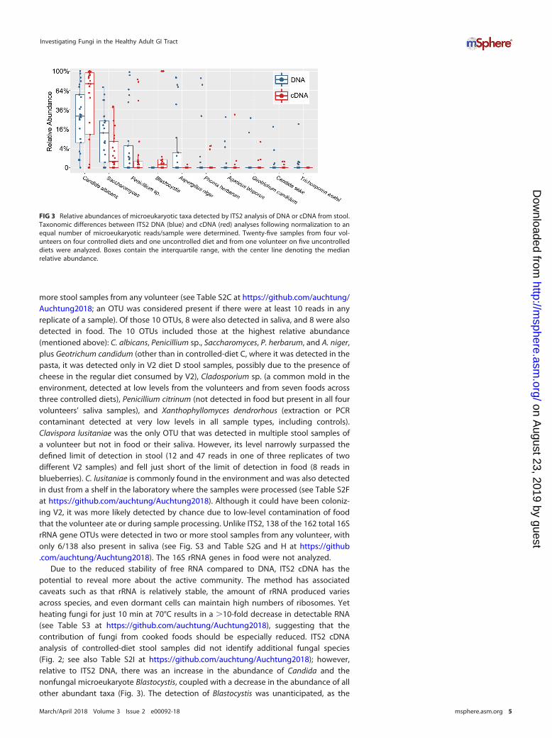
Due to the reduced stability of free RNA compared to DNA, ITS2 cDNA has thepotential to reveal more about the active community. The method has associatedcaveats such as that rRNA is relatively stable, the amount of rRNA produced variesacross species, and even dormant cells can maintain high numbers of ribosomes. Yetheating fungi for just 10 min at 70°C results in a �10-fold decrease in detectable RNA(see Table S3 at https://github.com/auchtung/Auchtung2018), suggesting that thecontribution of fungi from cooked foods should be especially reduced. ITS2 cDNAanalysis of controlled-diet stool samples did not identify additional fungal species(Fig. 2; see also Table S2I at https://github.com/auchtung/Auchtung2018); however,relative to ITS2 DNA, there was an increase in the abundance of Candida and thenonfungal microeukaryote Blastocystis, coupled with a decrease in the abundance of allother abundant taxa (Fig. 3). The detection of Blastocystis was unanticipated, as the
FIG 3 Relative abundances of microeukaryotic taxa detected by ITS2 analysis of DNA or cDNA from stool.Taxonomic differences between ITS2 DNA (blue) and cDNA (red) analyses following normalization to anequal number of microeukaryotic reads/sample were determined. Twenty-five samples from four vol-unteers on four controlled diets and one uncontrolled diet and from one volunteer on five uncontrolleddiets were analyzed. Boxes contain the interquartile range, with the center line denoting the medianrelative abundance.
Investigating Fungi in the Healthy Adult GI Tract
March/April 2018 Volume 3 Issue 2 e00092-18 msphere.asm.org 5
on August 23, 2019 by guest
http://msphere.asm
.org/D
ownloaded from

most closely related Blastocystis sequences in GenBank have five mismatches (includingthe two 3= nucleotides) with our reverse PCR primer, and suggests that Blastocystis RNAwas present in an especially high abundance relative to fungal RNA.
To determine whether any fungi were not detected due to primer bias, we alsoamplified and sequenced the V6/V7 region of 18S rRNA genes from all stool samples(see Fig. S4 and Table S2J at https://github.com/auchtung/Auchtung2018). There wasless diversity detected due to the greater sequence conservation of 18S rRNA genesthan of ITS regions, and there were no additional fungal lineages detected via 18S rRNAgenes that were not identified by ITS2 DNA analysis. However, three nonfungalmicroeukaryotes were identified which dominated stool samples from V1 (98.7%Dientamoeba) and V4 (91.1% Blastocystis and 8.9% Entamoeba).
To determine whether any fungi were undetected (despite being present in asample) due to uneven nucleic acid extraction by a Mo Bio PowerMag microbiome(PMM) kit, DNA was also extracted from replicates of a subset of stool samples (all fromV1) by two additional methods: a Mo Bio PowerSoil kit used by the HMP (19) and aprotocol used by MetaHIT (20). Whereas ITS2 analysis of PowerSoil-extracted samplesand 18S rRNA gene analysis of MetaHIT-extracted samples found no additional OTUs,ITS2 analysis of MetaHIT-extracted samples and 18S rRNA gene analysis of PowerSoil-extracted samples yielded four and two additional OTUs, respectively, that were notdetected in the corresponding PMM-extracted samples (see Fig. S5 and S6 and Ta-ble S2K to P at https://github.com/auchtung/Auchtung2018). However, the additionalOTUs were not abundant in V1 samples and were detected in PMM-extracted non-V1samples, suggesting that their lack of detection here was due to chance rather than toa bias in the protocol.
As an additional test to examine whether any fungi were not detected by molecularanalyses, and to gauge which fungi were able to survive gastrointestinal transit, stoolsamples were also plated on fungus-selective Sabouraud agar. Many of the mostabundant fungal taxa identified by ITS2 sequencing of V1, V2, and V4 samples were alsoisolated by culturing (see Table S2Q at https://github.com/auchtung/Auchtung2018).Little was isolated from V3, in agreement with molecular analyses. C. albicans andSaccharomyces were the only fungi identified from plates grown anaerobically, and nofungal growth was detected on anoxic YCFA media (77), which can support the growthof most gut bacteria (21). In summary, there is no evidence that any fungi in stool weremissed by biases in our approach to their detection; no fungi were consistently presentin the gut unless they also existed in the saliva or diet.
Examining fungal dynamics in gut-simulating bioreactors. The GI tract may notbe an accommodating environment for most fungi, as they must compete for nutrientswhile resisting inhibitory compounds produced by commensal bacteria such as short-chain fatty acids (22) and secondary bile acids (23). Fungal growth under the conditionsfound in the GI tract can be tested in vitro through the use of bioreactors, which areknown to support growth of complex fecal bacterial communities (24). We examinedthe identity and abundance of fungi present within the complex communities ofminibioreactor arrays after inoculation with stool from a healthy adult. Communitieswere cultivated at 37°C in media that varied in composition, under conditions of eitheran anoxic or hypoxic atmosphere (see Table S4 at https://github.com/auchtung/Auchtung2018). Samples were collected between days 2 and 7, when the communitiestypically had stabilized. Only one bioreactor, with high-sugar hypoxic media and a verylow diversity of bacteria, contained elevated levels of fungi (�104 ITS2 copies/ng totalDNA on days 4, 5, and 6). C. albicans was the only fungal species detected in thesesamples. Under conditions that resembled the healthy distal gut—low oxygen, simplesugars, and a diverse bacterial community—there was no evidence of fungal persis-tence.
Assessing Saccharomyces stability in the gut in the absence of dietary input.We examined whether Saccharomyces, a genus of yeast that can survive gastrointestinaltransit and one of the most commonly detected fungi in most examinations of humanstool, would continue to be detected in a healthy adult volunteer after consumption of
Auchtung et al.
March/April 2018 Volume 3 Issue 2 e00092-18 msphere.asm.org 6
on August 23, 2019 by guest
http://msphere.asm
.org/D
ownloaded from

a S. cerevisiae-free diet (see Table S5A at https://github.com/auchtung/Auchtung2018).Although it has been shown that the probiotic S. cerevisiae var. boulardii is clearedwithin a few days of consumption (25), the retention of other strains of Saccharomyceshas not been examined. Here, a volunteer collected all stool samples over the courseof a 15-day experiment as follows: 1 week on a regular diet, followed by 1 week on aS. cerevisiae-free diet, followed by a final day where some foods containing S. cerevisiaewere consumed. Samples were examined both by ITS2 and 18S rRNA gene analysis.Since there is only a single base pair in ITS2 distinguishing S. cerevisiae from relatedspecies (and since there are none in 18S rRNA genes), the OTU may contain multiplespecies of the genus and is referred to here as Saccharomyces. High levels of Saccha-romyces were detected over the course of a normal diet (days 1 to 7), with thefinal sample containing 86% Saccharomyces ITS2 reads (Fig. 4; see also Table S5Bat https://github.com/auchtung/Auchtung2018). After 2 days of consumption of aS. cerevisiae-free diet by the volunteer, Saccharomyces comprised just 0.1% of the reads(day 9). After unintentional consumption of a small amount of bread, there was a spikein the relative abundance of Saccharomyces on day 10 to 16% of the reads. However,by day 11 the abundance had again fallen to just 0.16% Saccharomyces. Reads ofSaccharomyces were below the level of detection in the remaining S. cerevisiae-free dietsamples (days 13 and 14). After the S. cerevisiae dietary exclusion had ended, Saccha-romyces again became one of the most abundant taxa detected (day 15). The 18S rRNAgene data were similar to the ITS2 data, with the relative abundances of Saccharomycesjust above the level of detection on days 9 and 11 and below the level of detectionon days 13 and 14 (see Fig. S7 and Table S5C at https://github.com/auchtung/Auchtung2018). These data suggest that the Saccharomyces in the volunteer’s stoolmay have entirely originated from dietary S. cerevisiae.
Tracing fluctuations in fecal Candida albicans to the oral cavity. Candida albicanshas long been studied for its role as an opportunistic pathogen (26). It is a leadingcause of nosocomial infection, particularly in immunosuppressed patients (27). Inour controlled-diet-experiment ITS2 analyses, it was the most abundant taxonoverall in both stool and saliva. C. albicans is frequently described as colonizing thehuman GI tract; however, to our knowledge, there has been no study that hasdemonstrated that this is true in healthy human individuals.
Since C. albicans is common in the oral cavity (15) and contributes to the formationof dental plaque and the development of caries (28), we wanted to assess thecontribution of C. albicans in swallowed saliva to what is detected in stool. For allexperiments, saliva or blended stool was plated on Sabouraud plates containingantibiotics. Possible C. albicans colonies were later spotted on chromogenic media todistinguish from closely related species. First, we confirmed that there was a differencein the levels of C. albicans in the saliva of a volunteer when the volunteer’s teeth were
FIG 4 Relative ITS2 abundance of Saccharomyces in stool over time as a Saccharomyces cerevisiae-freediet was consumed. A logarithmically scaled chart showing the unrarefied relative abundances ofSaccharomyces reads among ITS2 sequences amplified from stool DNA of a human volunteer is shown.Plant sequences were removed before analysis. The volunteer consumed a regular diet for 7 days and aSaccharomyces cerevisiae-free diet for 7 days and then resumed a regular diet. Day 8 data representaverages of results from two stool samples, and there was no sample on day 12.
Investigating Fungi in the Healthy Adult GI Tract
March/April 2018 Volume 3 Issue 2 e00092-18 msphere.asm.org 7
on August 23, 2019 by guest
http://msphere.asm
.org/D
ownloaded from

brushed with conventional fluoride toothpaste either once per day (at night) or afterevery meal (3 to 4 times/day). Toothpaste helps remove plaque but does not signifi-cantly impact the viability of C. albicans (29). The levels of C. albicans measured in theoral cavity were relatively variable but were higher on average in saliva and plaquewhen teeth were brushed just once per day (Fig. 5A). Next, we monitored the changein abundance of C. albicans in stool when teeth were cleaned after every meal insteadof once per day and saw that levels decreased from 106 cells to 104 to 105 cells (Fig. 5B).Finally, to confirm that the decrease was not due to unrelated artifacts, we monitoredC. albicans in stool while the protocols for cleaning of teeth were alternated: 2 dayscleaning after every meal, followed by 2 days cleaning once per day, each done fourtimes in total over 16 days. The total number of C. albicans colonies found in stool wassignificantly greater (two-sample equal variance t test P � 0.0007) after the volunteerbrushed just once per day (mean � 8.3 � 105) than after the volunteer brushed afterevery meal (mean � 2.5 � 105) (Fig. 5C). A second volunteer who repeated a shorterversion of the experiment on nonconsecutive days also had less C. albicans in stool afterthe volunteer brushed their teeth after every meal (Fig. 5D). Together, these experi-ments suggest that a large portion of C. albicans in the stool of the volunteers wasderived from the oral cavity.
FIG 5 Candida albicans levels were decreased in the mouth and stool of healthy human volunteer(s) when teeth werebrushed more often. (A) Concentrations of C. albicans in saliva throughout days when an adult volunteer either did or didnot perform tooth brushing after eating. The volunteer consumed the same diet on both tooth-brushing protocols, theexperiment was performed for two different diets, and levels of C. albicans were measured in a sample of plaque at theend of each day. (B) The total number of C. albicans cells in the stool of a volunteer over time (plotted on a log axis).Following a period of brushing teeth just once per day, the volunteer performed tooth brushing after every meal for 8consecutive days. The diet was not the same from day to day but contained similar levels of sugars throughout the timeperiod. (C and D) Lastly, the total number of C. albicans cells was measured in the stool of (C) a volunteer who alternatelyfollowed different tooth-brushing protocols for 2 days over the course of 16 days or of (D) a second volunteer who twiceconducted each tooth-brushing protocol on nonconsecutive days. For all experiments, saliva, plaque, or stool was platedon Sabouraud plates containing antibiotics. Possible C. albicans colonies were later spotted on chromogenic media todistinguish the C. albicans colonies from closely related species and to adjust C. albicans numbers.
Auchtung et al.
March/April 2018 Volume 3 Issue 2 e00092-18 msphere.asm.org 8
on August 23, 2019 by guest
http://msphere.asm
.org/D
ownloaded from

DISCUSSION
Here we investigated fungal colonization of the healthy adult GI tract from multipleperspectives. We conducted a controlled-diet experiment to examine the abundanceand identity of fungi present across multiple diets and then further investigated thedynamics of the two most abundant fungi, Saccharomyces and C. albicans, when dietaryor oral hygiene practices were altered. We also examined fungi in bioreactors and in alarge data set of healthy adults. From all avenues of investigation, the evidencesuggests that fungi may not readily colonize the GI tracts of healthy adults in Westernsociety.
Although some bacteria have been demonstrated to be consistently present at lowlevels in the GI tract (30), there was no evidence that that was the situation for any funginot derived from food or the oral cavity. Quantitative PCR analysis of controlled-diet-experiment stool samples demonstrated that ITS2 were on average 3,000� less abun-dant than 16S rRNA genes. Considering that many of the ITS2 amplicons were fromplants and that fungi often have higher ribosomal operon copy numbers than bacteria(31, 32), the fungi in our samples were at extremely low abundance (~0.001% to 0.01%of the population). This level of fungi is consistent with HMP metagenomic data (1);although early studies found fungal levels in adult stool that were ~10� higher (8, 9),the levels may have been overestimated due to extensive bacterial contamination offungal sequence databases.
Despite the volunteers having consumed the same controlled diets, the fungirepresented in volunteers’ stool DNA largely did not match by diet, due to the lowfungal content of the control foods, the variable composition of the volunteers’ oralfungi, and the contribution of residual fungi from the volunteers’ regular diets. Onlyfour taxa were present in at least one sample replicate of every diet of a volunteer(Candida albicans [V1, V2, and V3], Saccharomyces [V2], Aspergillus niger [V4], andPenicillium sp. [V1]). Both A. niger and Penicillium sp. were linked to the regular diets ofindividual volunteers and either were not detectable in the cDNA (A. niger) or were notdetectable in multiple other samples from the same volunteer in the S. cerevisiae-free diet experiment (Penicillium sp.). This suggests there is little or no A. niger andPenicillium colonization of the GI tracts of the volunteers. Saccharomyces and C. albicanscould have been from food and the mouth, respectively; however, to be certain of thesource, we examined these fungi further. When a healthy adult volunteer abstainedfrom consumption of food containing S. cerevisiae, the Saccharomyces OTU droppedbelow the limit of detection within a few days, suggesting that there had been little orno colonization. Malassezia, another fungal genus commonly detected in some studies(1, 13) and the only fungal genus other than Saccharomyces to be detected in all threelongitudinal samples of multiple HMP volunteers, was not consistently detected in anyvolunteer’s controlled-diet stool samples by any of our molecular approaches, despitebeing present in the saliva of all volunteers. Malassezia may contaminate oral and stoolsamples due to high abundance on skin and may not be as well adapted for survivingpassage through the GI tract.
C. albicans and related species have frequently been stated to colonize the GI tract(33, 34), though all evidence seems to be from their presence in human stool samplesand from studies of inbred, typically antibiotic-treated animals. However, since theaverage person is thought to swallow over a liter of saliva/day (35), the GI tract isconstantly dosed with oral microorganisms, including, for most people, species ofCandida (15). Here, we showed that when a healthy adult volunteer increased thefrequency of cleaning of teeth, the abundance of C. albicans in stool was lowered10-fold to 100-fold. This suggests that the oral cavity may be the primary source ofC. albicans detected in the stool of healthy people. Indeed, the fungal component ofdental plaque of at least one cohort has been shown to be dominated by C. albicans(36). Taking additional measures to further reduce oral C. albicans levels, such as extraflossing and attention to elimination of plaque and reduction of consumption of refinedsugars, may reduce levels of C. albicans in stool even further. This may explain why
Investigating Fungi in the Healthy Adult GI Tract
March/April 2018 Volume 3 Issue 2 e00092-18 msphere.asm.org 9
on August 23, 2019 by guest
http://msphere.asm
.org/D
ownloaded from

Hoffmann et al. (37) observed a positive correlation between fecal Candida levels andthe consumption of carbohydrates. C. albicans is also much more common in Westernpopulations, where refined sugar consumption is high (38, 39). Healthy mice are usuallyresistant to colonization by C. albicans (40); however, they are not typically maintainedon a diet containing the simple sugars that encourage the growth of C. albicans. Forone study where mice were fed a nutritionally adequate diet that included 10%sucrose, the mice had a sustained level of C. albicans in their feces (41). However,the oral cavities of the mice were not examined, so the role of that site incontributing to the C. albicans population is uncertain. Some work has been doneinvestigating the effects of certain dietary compounds on C. albicans (42, 43), butmore studies on the oral ecology of this opportunistic pathogen are needed. Inbioreactors designed to mimic in vivo conditions in parts of the GI tract, we saw thatC. albicans grew only when oxygen and a relatively high concentration of simplesugars were present. These conditions are relevant to the oral cavity and uppergastrointestinal tract, but not to the colon, where most bacteria are found. Thebacterial community in the C. albicans growth-supporting bioreactors was alsorelatively simple, unlike that of a healthy GI tract, and therefore might havepresented less resistance to fungal growth. It is possible that C. albicans colonizesboth the mouth and the upper GI tract; in this regard, however, C. albicans may beakin to the oral bacteria that exacerbate disease when they colonize the gut (44,45). Just as the Centers for Disease Control and Prevention recommends maintain-ing good oral health for preventing mouth, throat, or esophagus candidiasis(https://www.cdc.gov/fungal/diseases/candidiasis/thrush/), patients suffering fromor at risk for developing a gastrointestinal disease might also benefit from increasingthe attention that they pay to dental hygiene.
We conducted multiple analyses to ensure that fungi that might be colonizing thegastrointestinal tracts of volunteers were not missed by ITS2 sequencing of DNA. ITS2from cDNA, 18S rRNA genes from DNA, ITS2 and 18S rRNA genes from DNA extractedby alternate methods, and fungi that grew on agar plates were also analyzed. Noadditional fungal species were detected by ITS2 cDNA and 18S rRNA gene analysis,though both approaches did reveal the presence of nonfungal microeukaryotes. ITS2cDNA analysis specifically detected only high levels of C. albicans and the nonfungalmicroeukaryote Blastocystis, a known colonizer of the GI tract (46). The two alternativeDNA extraction methods tested did not reveal any problems with detecting fungiin DNA extracted by our standard protocol. Additionally, most fungi isolated inculture grew only on plates of rich media in the presence of oxygen. There was nofungal growth on an intestine-like medium which is able to support the growth ofnearly every bacterial species detected in stool by 16S rRNA gene analysis (21). Insummary, the additional analyses failed to find deficiencies in our approach tofungal detection.
Analysis of the accumulation of 16S rRNA gene and ITS2 OTUs detected in healthyadult HMP stool samples demonstrated that while bacterial populations are similarfrom one individual to another and community sampling approaches saturation, fungalpopulations are more variable, and the community continues a steady rate of revealingnew representatives as more samples are examined. In fact, in the entire cohort of 100individuals, there were more ITS2 OTUs than 16S rRNA gene OTUs. This may, in part,have been because unique food-derived bacteria are hard to detect among themembers of the enormous endemic bacterial population. An example is the genusLactobacillus; once perceived to be autochthonous members of the GI tract, mostspecies are actually derived from the mouth, from food, or from the upper GI tract (47).Whereas Lactobacillus sequences represent a minor fraction of the 16S rRNA gene datafrom stool, low-abundance fungal species from food and the oral cavity would be thepredominant ITS2 OTUs detected under conditions in which there had been little or nofungal colonization of the GI tract.
We have presented multiple lines of evidence supporting the hypothesis that thereare few or no fungal species indigenous to or colonizing the GI tracts of healthy adults
Auchtung et al.
March/April 2018 Volume 3 Issue 2 e00092-18 msphere.asm.org 10
on August 23, 2019 by guest
http://msphere.asm
.org/D
ownloaded from

in Western society. There are many possible reasons why gut-colonizing fungi mighthave been missed, but we consider them unlikely. They include the following possi-bilities. (i) Gut-specific strains might have been masked by other closely relatedfungi—at least for C. albicans, the same strain has been found in different parts of thebody (48). (ii) Disease-free gut fungus-colonized individuals might have been misseddue to our having examined only four volunteers. (iii) Fungi growing in the GI tractmight have been degraded to the extent that they became completely undetectable instool— even free DNA is known to survive transit (49). (iv) Fungi might colonize inextremely low numbers and bloom in the GI tract under healthy conditions notexamined. (v) Fungi might exist in close association with host cells such that very fewfungi are shed with fecal material. However, there are many reasons why fungi mightnot colonize, including inadequate physiological machinery for success in the environ-ment, defenses of the mature human immune system, and a loss of gastrointestinalmicrobial diversity through recent or long-term evolution or exposures.
Many of the fungi detected by molecular analyses are not optimized for growth at37°C (50, 51) and lack the genes and physiological traits reflective of an adaptation tothe environment of the GI tract. Although some fungi such as C. albicans are able tosurvive the healthy adult GI tract, in order to colonize it, they face intense bacterialcompetition for nutrients and the threat of challenge by antimicrobials not present inthe oral cavity, such as reactive nitrogen intermediates (52), peptides (53), and second-ary bile acids (23). Physiologically relevant concentrations of short-chain fatty acids canalso be fungistatic (22). Notably, the gut environment is not fully established in youngchildren, where the immune system and competing bacterial communities are stilldeveloping. Though more data are needed, some studies have shown increased fungaldiversity in children (54, 55).
Fungi may not persist in the GI tracts of those with a healthy immune system, andconversely, fungal colonization may be a symptom of illness. Those with a suppressedimmune system are particularly susceptible to fungal invasion (56); the immune re-sponse mediated via multiple pattern recognition receptor families is important in thedefense against fungi (57, 58). Furthermore, there are a multitude of studies showingfungal differences between healthy individuals and sick patients. However, there is adifference between detection of a microbe and proving that it colonizes that location—since the sick may spend more time indoors and/or eat differently, they could beexposed to different fungi. Therefore, it is necessary to consider confounding lifestyleand dietary differences to know whether any fungal gut colonizers are associated withthe disease. If fungal colonization of the gut is associated only with disease, however,it might be beneficial to treat more patients with antifungals, especially if side effectscan be avoided. In addition, the detection of increased levels of gastrointestinal fungicould alert clinicians to undiagnosed disorders.
Any indigenous or colonizing fungi might have disappeared as human diet andsociety has evolved. With modernization have come activities that limit microbialsurvival, transmission, and exposure, resulting in a loss of bacterial diversity andhelminths from the GI tracts of humans (59, 60). There have been few microbiomestudies conducted on non-Western communities, and most did not examine fungi. Inthe stool sample data set generated by Yatsunenko et al. (8), we found that subjectsfrom Malawi and Venezuela had an increased proportion of fungal reads and anincreased incidence of having any fungi compared to subjects from the United States(see Text S1 at https://github.com/auchtung/Auchtung2018). Yet, with the low overallabundance of fungi in human guts, deeper sequencing is needed to confidentlydetermine whether there are increased levels of fungi in non-Western populations. Inaddition, measurements of oral and dietary fungi are needed to assess whetherdifferences in stool fungi are due to increased levels of transient fungi or to genuine GItract colonization.
It is also possible that humans lost gut fungi when they diverged from chimpanzees,primates, or more distant relatives, corresponding to changes in our diet and GI tractmorphology (61). It has been shown that humans have had a loss of bacterial and
Investigating Fungi in the Healthy Adult GI Tract
March/April 2018 Volume 3 Issue 2 e00092-18 msphere.asm.org 11
on August 23, 2019 by guest
http://msphere.asm
.org/D
ownloaded from

archaeal diversity relative to other apes (62, 63). However, there have been few studieson fungi in the GI tracts of animals. Examination of stool from domesticated dogs anda wild gorilla found high fungal diversity (64, 65). Fungal populations in the guts oflaboratory rodents show large variation from study to study, and many of the detectedspecies can be explained by the presence of fungi in their chow and bedding (2, 66–69)(see Text S2 at https://github.com/auchtung/Auchtung2018). Clearly, much more re-search on fungal communities in the GI tracts of wild animals is needed.
In summary, we have shown that fungi detected from the GI tracts of volunteersacross multiple diets were at very low abundance, were only transiently present fromthe diet or oral cavity, and did not grow when placed in gut-like conditions. In addition,the levels of two common fungal species in stool were shown to be directly affected byaltering the oral hygiene or the dietary consumption of the volunteers, and analyses ofdata from larger cohorts were consistent with the possibility that fungi may notcolonize the guts of healthy adults in Western society. More studies are needed that aremindful of dietary and oral fungi and that examine the relative contributions ofimmunological defenses, fungal ecology, and human evolution to limiting fungalpersistence in the healthy modern adult GI tract.
MATERIALS AND METHODSComparison of fungi with bacteria/archaea in HMP stool samples. ITS2 and 16S rRNA gene
sequences from the Human Microbiome Project are available at NCBI (BioProject PRJNA356769) and theHuman Microbiome Project website (http://www.hmpdacc.org), respectively. There were 148 samplesthat had at least 1,000 plant-filtered ITS2 and 16S rRNA gene reads. The data sets were rarefied to 1,000reads by the use of single_rarefaction.py (70). Rarefaction curves were calculated with mothur v1.36.1,which uses a resampling-without-replacement approach (71).
Controlled-diet experiment. Four healthy people (ages between 25 and 37), all born and primarilyraised on different continents (but who were residing in Houston, TX, for the duration of the study), withdiverse dietary habits, consumed four identical diets (see Table S2A and B at https://github.com/auchtung/Auchtung2018). The diets were composed of a variety of food groups, largely low in fungi(except diet D), and were kept simple to facilitate analysis of all components. Volunteers did not consumeprobiotics or vitamins and did not chew gum but were allowed coffee, tea, and salt, as long as thosefoods were submitted for fungal sequencing analysis. There was no limit to the amount of foodconsumed. The diets were consumed nonconsecutively and in no set order over the course of 1 month,April 2016. Volunteers had the option to consume a 100-mg Brilliant Blue Lake (FD&C Blue 1 dye) pill atthe start of each diet to clarify via staining that stool samples were primarily resulting from the particulardiet and were ready to be collected. Volunteers collected a stool sample (12 to 222 g; mean, 77 g)following the appearance of the particular diet (or dye) in their stool. Saliva samples (typically three/diet)were collected at time points that were at least 2 h apart and at least 1 h after eating to reduce thenumber of plant particles that might obscure the detection of fungi. All samples were frozen at �80°Cuntil nucleic acid extraction.
Using a Mo Bio PowerMag microbiome kit (including the phenol step), nucleic acids were extractedfrom dietary components, saliva (200 �l), and total stool which had been blended in 2 vol of prereducedphosphate-buffered saline (PBS)– 0.5 g/liter cysteine (50 �l). For V1, replicate stool samples wereextracted using the MetaHIT (20) and Mo Bio, Inc., PowerSoil protocols (19). All stool samples used forPCR were sampled in triplicate. Nucleic acids were treated with either RNase A (Qiagen) or DNase I (NewEngland Biolabs). Representative RNA samples were analyzed by spectrophotometry and visualized on anagarose gel to confirm purity and integrity. rRNA was reverse transcribed using a Maxima H Minus kit(Thermo Fisher) with primer-specific conversion using ITS4R. As a control, RNA sample replicates with noreverse transcriptase were processed concurrently; DNA was rarely amplified, and taxa abundant in testsamples were not detected (see Table S2F at https://github.com/auchtung/Auchtung2018). The ITS2region, 18S rRNA gene V6/V7, and 16S rRNA gene V4 were amplified with primers ITS3F-ITS4R, 1152F-1428R, and 515F-806R, respectively, and sequenced on an Illumina MiSeq platform; reads were merged,clustered, subjected to chimera filtration, and mapped against databases as described previously (1).Nucleic acid from each of the three stool samples was amplified for ITS2 DNA, ITS2 cDNA, and 18S rRNAgenes, but only a single randomly chosen replicate was amplified for 16S rRNA gene analysis. Everysequenced region averaged �10,000 reads/sample. Because the maximum amount of sequencingbarcode bleed-over seen in 42 control samples was 7 reads, an OTU was considered present if there wereat least 10 reads detected in a sample. Three PBS/collection dish/blender controls yielded no isolates and�10 ITS2 DNA and cDNA reads (see Table S2F at https://github.com/auchtung/Auchtung2018). Forblending, nucleic acid extraction, and PCR amplification, the order of sample processing was randomized.All primer sequences are listed in Table S8 at https://github.com/auchtung/Auchtung2018. Taxon boxplots and stacked bar plots were generated in R (72), utilizing the phyloseq package (73).
Replicates of all blended stool samples (50 �l) were plated on Sabouraud agar (4% dextrose, 1%peptone, 100 �g/ml ampicillin, 50 �g/ml chloramphenicol) and grown at both room temperature and37°C under conditions of ambient and anoxic (90% N2, 5% CO2, 5% H2) atmospheres. Colonies wereidentified by Sanger sequencing following PCR amplification with ITS3F-ITS4R and, where necessary, 18S
Auchtung et al.
March/April 2018 Volume 3 Issue 2 e00092-18 msphere.asm.org 12
on August 23, 2019 by guest
http://msphere.asm
.org/D
ownloaded from

rRNA gene-targeting 1152F-1428R or 16S rRNA gene-targeting 27F-907R (74). To look for fungi that growunder conditions more physiologically similar to those of the gut, frozen replicates of the uncontrolled-diet samples were later plated on YCFA media (77) containing 50 �g/ml chloramphenicol, 100 �g/mlampicillin, 10 �g/ml tetracycline, and 50 �g/ml kanamycin with or without 250 �g/ml bovine bile andplaced in an anoxic atmosphere for at least 2 weeks.
ITS2-specific primers ITS3F and ITS4R and 16S rRNA gene-specific primers Bact1369F and Prok1492Rwere used in quantitative PCR analysis of controlled-diet fecal DNA samples using Applied BiosystemsPower SYBR green PCR master mix following the manufacturer’s instructions, with 60 s (ITS2) or 30 s (16SrRNA gene) of annealing/extension at 60°C. To create nonsupercoiled standard curve templates (75), DNAfrom Malassezia cuniculi (450-bp ITS2) or Escherichia coli rrsA was amplified using the primers specifiedabove, except that the procedure also used an additional 64 (ITS3F and ITS4R), 18 (Bact1369F), or 19(Prok1492R) 5= nucleotides (nt) so that quantitative PCR primers would not have to anneal to the veryend of the template.
Bioreactors. Experiments were conducted in minibioreactor arrays as previously described (24) usingfive different media under conditions of either an anoxic (5% CO2, 95% N2) or hypoxic (2% O2, 5% CO2,93% N2) atmosphere (see Table S4 at https://github.com/auchtung/Auchtung2018). For each medium/atmosphere condition, triplicate 15-ml reactors were inoculated with a 25% fecal slurry (containing 75%reduced PBS) and allowed to sit for 16 h (anoxic reactors) or 4 h (hypoxic reactors) before flow wasinitiated at 1.825 ml/h (anoxic) or 3.75 ml/h (hypoxic). The hypoxic bioreactors had a shorter initial waitand higher flow rate to more closely resemble the upper GI tract. Samples for DNA extraction (asdescribed in reference 76) and sequencing were collected daily from each reactor over the course of7 days. Quantitative PCR was performed as described above.
Saccharomyces experiment. A single healthy adult volunteer ate regularly for 1 week and thenavoided eating foods containing S. cerevisiae for 1 week, followed by 1 day of eating foods with a largeamount of S. cerevisiae (see Table S5A at https://github.com/auchtung/Auchtung2018). Over the courseof the 15 days, the stool samples were collected and frozen, typically once daily. Two months later,samples were collected during 3 days of a regular diet. DNA from approximately 250 mg frozen stool wasextracted using the Mo Bio PowerSoil protocol and analyzed as described above, with an average of�55,000 ITS2 and �25,000 18S rRNA gene reads/sample. We defined the limit of detection as 10 readsof Saccharomyces, since 27 pure culture controls had �10 reads from barcode bleed-over.
Candida albicans experiments. To test the effect of increased tooth brushing (after every meal,within 30 min) on C. albicans levels in saliva of a healthy adult volunteer, for every hour from 7 a.m. to9 p.m. over 2 consecutive days during which the same diet was consumed, 200 �l of saliva was spreadon Sabouraud media containing ampicillin and chloramphenicol. The numbers of colonies were countedafter 24 h at 37°C and were adjusted after 48 h following verification of the presence of previously smallcolonies. Up to 10 colonies/plate were spotted onto chromogenic BBL CHROMagar Candida (BD) andgrown for at least 48 h to distinguish C. albicans from related species. Since the cells do not grow as wellon the chromogenic agar, resulting in fewer colonies, the medium was used only to assess the identityof Sabouraud-grown colonies. The reported number of C. albicans cells in each sample was correctedusing these data.
To test how the levels of C. albicans in stool of healthy volunteers were affected by variations intooth-brushing protocols, the following experiments were conducted. First, over the course of 8 days,during which time the volunteer brushed their teeth after every meal instead of once per day, entire stoolsamples were collected and blended with PBS, and 150 �l was plated in triplicate on Sabouraud mediacontaining antibiotics. In a second experiment, the following protocol was repeated by the samevolunteer four times: brushing of teeth once per day (at night) for 2 days and then after each meal for2 days. For the course of the experiment, the volunteer defecated once per day at the same time(�30 min). On the day after each 2-day protocol, a full stool sample was collected and blended with PBS,and 50 �l was plated in triplicate. A variation of this experiment was conducted by a second volunteerbut on nonconsecutive days and following each protocol twice. Volunteers were instructed to eat similaramounts of sugary foods between the protocols, avoid Penicillium-rich cheeses, and avoid heavyconsumption of alcohol. At the end of the stool experiments, all presumed C. albicans colonies from atleast one random replicate plate/day were spotted onto chromogenic agar as described above. The t testwas performed in Excel.
Ethics statement. The Institutional Review Board of Baylor College of Medicine approved all studyprotocols. All participants provided written informed consent (protocols H-32521 and H-29645).
Data availability. Sequence data were deposited in NCBI’s Sequence Read Archive (BioProjectPRJNA379437) and GenBank (accession no. KY931930 to KY932673 and KY932725 to KY933059).
ACKNOWLEDGMENTSWe thank Auriole Tamegnon, Nguyen Truong, Tulin Ayvaz, Ryan Pepper, Daniel
Smith, and Shelly Buffington for technical assistance.Work on Human Microbiome Project samples was funded by grant 2 U54 HG003273
from NIH/NHGRI. Portions of this research were made possible by funding from theAlbert and Margaret Alkek Foundation. The funders had no role in study design, datacollection and interpretation, or the decision to submit the work for publication.
Investigating Fungi in the Healthy Adult GI Tract
March/April 2018 Volume 3 Issue 2 e00092-18 msphere.asm.org 13
on August 23, 2019 by guest
http://msphere.asm
.org/D
ownloaded from

REFERENCES1. Nash AK, Auchtung TA, Wong MC, Smith DP, Gesell JR, Ross MC, Stewart
CJ, Metcalf GA, Muzny DM, Gibbs RA, Ajami NJ, Petrosino JF. 2017. Thegut mycobiome of the Human Microbiome Project healthy cohort.Microbiome 5:153. https://doi.org/10.1186/s40168-017-0373-4.
2. Iliev ID, Funari VA, Taylor KD, Nguyen Q, Reyes CN, Strom SP, Brown J,Becker CA, Fleshner PR, Dubinsky M, Rotter JI, Wang HL, McGovern DP,Brown GD, Underhill DM. 2012. Interactions between commensal fungiand the C-type lectin receptor Dectin-1 influence colitis. Science 336:1314 –1317. https://doi.org/10.1126/science.1221789.
3. Guillamón E, García-Lafuente A, Lozano M, D’Arrigo M, Rostagno MA,Villares A, Martínez JA. 2010. Edible mushrooms: role in the preventionof cardiovascular diseases. Fitoterapia 81:715–723. https://doi.org/10.1016/j.fitote.2010.06.005.
4. Ianiro G, Tilg H, Gasbarrini A. 2016. Antibiotics as deep modulators of gutmicrobiota: between good and evil. Gut 65:1906 –1915. https://doi.org/10.1136/gutjnl-2016-312297.
5. Palma ML, Zamith-Miranda D, Martins FS, Bozza FA, Nimrichter L,Montero-Lomeli M, Marques ET, Jr, Douradinha B. 2015. Probiotic Sac-charomyces cerevisiae strains as biotherapeutic tools: is there room forimprovement? Appl Microbiol Biotechnol 99:6563– 6570. https://doi.org/10.1007/s00253-015-6776-x.
6. Hoarau G, Mukherjee PK, Gower-Rousseau C, Hager C, Chandra J, Re-tuerto MA, Neut C, Vermeire S, Clemente J, Colombel JF, Fujioka H,Poulain D, Sendid B, Ghannoum MA. 2016. Bacteriome and mycobiomeinteractions underscore microbial dysbiosis in familial Crohn’s disease.MBio 7:e01250-16. https://doi.org/10.1128/mBio.01250-16.
7. Brown GD, Denning DW, Gow NA, Levitz SM, Netea MG, White TC. 2012.Hidden killers: human fungal infections. Sci Transl Med 4:165rv13.https://doi.org/10.1126/scitranslmed.3004404.
8. Yatsunenko T, Rey FE, Manary MJ, Trehan I, Dominguez-Bello MG, Con-treras M, Magris M, Hidalgo G, Baldassano RN, Anokhin AP, Heath AC,Warner B, Reeder J, Kuczynski J, Caporaso JG, Lozupone CA, Lauber C,Clemente JC, Knights D, Knight R, Gordon JI. 2012. Human gut micro-biome viewed across age and geography. Nature 486:222–227. https://doi.org/10.1038/nature11053.
9. Arumugam M, Raes J, Pelletier E, Le Paslier D, Yamada T, Mende DR,Fernandes GR, Tap J, Bruls T, Batto JM, Bertalan M, Borruel N, Casellas F,Fernandez L, Gautier L, Hansen T, Hattori M, Hayashi T, Kleerebezem M,Kurokawa K, Leclerc M, Levenez F, Manichanh C, Nielsen HB, Nielsen T,Pons N, Poulain J, Qin J, Sicheritz-Ponten T, Tims S, Torrents D, Ugarte E,Zoetendal EG, Wang J, Guarner F, Pedersen O, de Vos WM, Brunak S,Doré J; MetaHIT Consortium, Antolín M, Artiguenave F, Blottiere HM,Almeida M, Brechot C, Cara C, Chervaux C, Cultrone A, Delorme C, et al.2011. Enterotypes of the human gut microbiome. Nature 473:174 –180.https://doi.org/10.1038/nature09944.
10. Sender R, Fuchs S, Milo R. 2016. Revised estimates for the number ofhuman and bacteria cells in the body. PLoS Biol 14:e1002533. https://doi.org/10.1371/journal.pbio.1002533.
11. Huseyin CE, O’Toole PW, Cotter PD, Scanlan PD. 2017. Forgotten fungi-the gut mycobiome in human health and disease. FEMS Microbiol Rev41:479 –511. https://doi.org/10.1093/femsre/fuw047.
12. Hallen-Adams HE, Suhr MJ. 2017. Fungi in the healthy human gastroin-testinal tract. Virulence 8:352–358. https://doi.org/10.1080/21505594.2016.1247140.
13. Hallen-Adams HE, Kachman SD, Kim J, Legge RM, Martínez I. 2015. Fungiinhabiting the healthy human gastrointestinal tract: a diverse and dy-namic community. Fungal Ecol 15:9 –17. https://doi.org/10.1016/j.funeco.2015.01.006.
14. Oh J, Byrd AL, Deming C, Conlan S, NISC Comparative SequencingProgram, Kong HH, Segre JA. 2014. Biogeography and individualityshape function in the human skin metagenome. Nature 514:59 – 64.https://doi.org/10.1038/nature13786.
15. Ghannoum MA, Jurevic RJ, Mukherjee PK, Cui F, Sikaroodi M, Naqvi A,Gillevet PM. 2010. Characterization of the oral fungal microbiome (my-cobiome) in healthy individuals. PLoS Pathog 6:e1000713. https://doi.org/10.1371/journal.ppat.1000713.
16. David LA, Maurice CF, Carmody RN, Gootenberg DB, Button JE, Wolfe BE,Ling AV, Devlin AS, Varma Y, Fischbach MA, Biddinger SB, Dutton RJ,Turnbaugh PJ. 2014. Diet rapidly and reproducibly alters the human gutmicrobiome. Nature 505:559 –563. https://doi.org/10.1038/nature12820.
17. Blaalid R, Kumar S, Nilsson RH, Abarenkov K, Kirk PM, Kauserud H. 2013.
ITS1 versus ITS2 as DNA metabarcodes for fungi. Mol Ecol Resour13:218 –224. https://doi.org/10.1111/1755-0998.12065.
18. Tournas VH, Niazi NS, Kohn JS. 2015. Fungal presence in selected treenuts and dried fruits. Microbiol Insights 8:1– 6. https://doi.org/10.4137/MBI.S24308.
19. Aagaard K, Petrosino J, Keitel W, Watson M, Katancik J, Garcia N, Patel S,Cutting M, Madden T, Hamilton H, Harris E, Gevers D, Simone G, McInnesP, Versalovic J. 2013. The Human Microbiome Project strategy for com-prehensive sampling of the human microbiome and why it matters.FASEB J 27:1012–1022. https://doi.org/10.1096/fj.12-220806.
20. Wesolowska-Andersen A, Bahl MI, Carvalho V, Kristiansen K, Sicheritz-Pontén T, Gupta R, Licht TR. 2014. Choice of bacterial DNA extractionmethod from fecal material influences community structure as evalu-ated by metagenomic analysis. Microbiome 2:19. https://doi.org/10.1186/2049-2618-2-19.
21. Browne HP, Forster SC, Anonye BO, Kumar N, Neville BA, Stares MD,Goulding D, Lawley TD. 2016. Culturing of “unculturable” human micro-biota reveals novel taxa and extensive sporulation. Nature 533:543–546.https://doi.org/10.1038/nature17645.
22. Cottier F, Tan AS, Xu X, Wang Y, Pavelka N. 2015. MIG1 regulates resistanceof Candida albicans against the fungistatic effect of weak organic acids.Eukaryot Cell 14:1054–1061. https://doi.org/10.1128/EC.00129-15.
23. Marshall SE, Marples BA, Salt WG, Stretton RJ. 1987. Aspects of the effectof bile salts on Candida albicans. J Med Vet Mycol 25:307–318. https://doi.org/10.1080/02681218780000351.
24. Auchtung JM, Robinson CD, Britton RA. 2015. Cultivation of stable,reproducible microbial communities from different fecal donors usingminibioreactor arrays (MBRAs). Microbiome 3:42. https://doi.org/10.1186/s40168-015-0106-5.
25. Moré MI, Swidsinski A. 2015. Saccharomyces boulardii CNCM I-745 sup-ports regeneration of the intestinal microbiota after diarrheic dysbio-sis—a review. Clin Exp Gastroenterol 8:237–255. https://doi.org/10.2147/CEG.S85574.
26. Barnett JA. 2008. A history of research on yeasts 12: medical yeasts part1, Candida albicans. Yeast 25:385– 417. https://doi.org/10.1002/yea.1595.
27. Yapar N. 2014. Epidemiology and risk factors for invasive candidiasis.Ther Clin Risk Manag 10:95–105. https://doi.org/10.2147/TCRM.S40160.
28. Pereira D, Seneviratne CJ, Koga-Ito CY, Samaranayake LP. 2017. Is theoral fungal pathogen Candida albicans a cariogen? Oral Dis https://doi.org/10.1111/odi.12691.
29. de Rossi A, Ferreira DC, da Silva RA, de Queiroz AM, da Silva LA,Nelson-Filho P. 2014. Antimicrobial activity of toothpastes containingnatural extracts, chlorhexidine or triclosan. Braz Dent J 25:186 –190.https://doi.org/10.1590/0103-6440201300027.
30. David LA, Materna AC, Friedman J, Campos-Baptista MI, Blackburn MC,Perrotta A, Erdman SE, Alm EJ. 2014. Host lifestyle affects human micro-biota on daily timescales. Genome Biol 15:R89. https://doi.org/10.1186/gb-2014-15-7-r89.
31. Torres-Machorro AL, Hernández R, Cevallos AM, López-Villaseñor I. 2010.Ribosomal RNA genes in eukaryotic microorganisms: witnesses of phy-logeny? FEMS Microbiol Rev 34:59 – 86. https://doi.org/10.1111/j.1574-6976.2009.00196.x.
32. Stoddard SF, Smith BJ, Hein R, Roller BR, Schmidt TM. 2015. rrnDB:improved tools for interpreting rRNA gene abundance in bacteria andarchaea and a new foundation for future development. Nucleic AcidsRes 43:D593–D598. https://doi.org/10.1093/nar/gku1201.
33. Nobile CJ, Johnson AD. 2015. Candida albicans biofilms and humandisease. Annu Rev Microbiol 69:71–92. https://doi.org/10.1146/annurev-micro-091014-104330.
34. Kumamoto CA. 2011. Inflammation and gastrointestinal Candida colo-nization. Curr Opin Microbiol 14:386 –391. https://doi.org/10.1016/j.mib.2011.07.015.
35. Humphrey SP, Williamson RT. 2001. A review of saliva: normal compo-sition, flow, and function. J Prosthet Dent 85:162–169. https://doi.org/10.1067/mpr.2001.113778.
36. Vesty A, Biswas K, Taylor MW, Gear K, Douglas RG. 2017. Evaluating theimpact of DNA extraction method on the representation of human oralbacterial and fungal communities. PLoS One 12:e0169877. https://doi.org/10.1371/journal.pone.0169877.
37. Hoffmann C, Dollive S, Grunberg S, Chen J, Li H, Wu GD, Lewis JD,Bushman FD. 2013. Archaea and fungi of the human gut microbiome:
Auchtung et al.
March/April 2018 Volume 3 Issue 2 e00092-18 msphere.asm.org 14
on August 23, 2019 by guest
http://msphere.asm
.org/D
ownloaded from
correlations with diet and bacterial residents. PLoS One 8:e66019.https://doi.org/10.1371/journal.pone.0066019.
38. Angebault C, Djossou F, Abélanet S, Permal E, Ben Soltana M, DiancourtL, Bouchier C, Woerther PL, Catzeflis F, Andremont A, d’Enfert C,Bougnoux ME. 2013. Candida albicans is not always the preferentialyeast colonizing humans: a study in Wayampi Amerindians. J Infect Dis208:1705–1716. https://doi.org/10.1093/infdis/jit389.
39. Xu J, Mitchell TG. 2003. Geographical differences in human oral yeastflora. Clin Infect Dis 36:221–224. https://doi.org/10.1086/345672.
40. Kennedy MJ, Volz PA. 1985. Effect of various antibiotics on gastrointes-tinal colonization and dissemination by Candida albicans. Sabouraudia23:265–273. https://doi.org/10.1080/00362178585380391.
41. Yamaguchi N, Sonoyama K, Kikuchi H, Nagura T, Aritsuka T, Kawabata J.2005. Gastric colonization of Candida albicans differs in mice fed com-mercial and purified diets. J Nutr 135:109 –115. https://doi.org/10.1093/jn/135.1.109.
42. Weig M, Werner E, Frosch M, Kasper H. 1999. Limited effect of refinedcarbohydrate dietary supplementation on colonization of the gastroin-testinal tract of healthy subjects by Candida albicans. Am J Clin Nutr69:1170 –1173. https://doi.org/10.1093/ajcn/69.6.1170.
43. Gunsalus KT, Tornberg-Belanger SN, Matthan NR, Lichtenstein AH, Ku-mamoto CA. 2016. Manipulation of host diet to reduce gastrointestinalcolonization by the opportunistic pathogen Candida albicans. mSphere1:e00020-15. https://doi.org/10.1128/mSphere.00020-15.
44. Sokol H, Leducq V, Aschard H, Pham HP, Jegou S, Landman C, Cohen D,Liguori G, Bourrier A, Nion-Larmurier I, Cosnes J, Seksik P, Langella P,Skurnik D, Richard ML, Beaugerie L. 2017. Fungal microbiota dysbiosis inIBD. Gut 66:1039 –1048. https://doi.org/10.1136/gutjnl-2015-310746.
45. Atarashi K, Suda W, Luo C, Kawaguchi T, Motoo I, Narushima S, KiguchiY, Yasuma K, Watanabe E, Tanoue T, Thaiss CA, Sato M, Toyooka K, Said HS,Yamagami H, Rice SA, Gevers D, Johnson RC, Segre JA, Chen K, Kolls JK,Elinav E, Morita H, Xavier RJ, Hattori M, Honda K. 2017. Ectopic colonizationof oral bacteria in the intestine drives TH1 cell induction and inflammation.Science 358:359–365. https://doi.org/10.1126/science.aan4526.
46. Stensvold CR, Clark CG. 2016. Current status of blastocystis: a personalview. Parasitol Int 65:763–771. https://doi.org/10.1016/j.parint.2016.05.015.
47. Walter J. 2008. Ecological role of lactobacilli in the gastrointestinal tract:implications for fundamental and biomedical research. Appl EnvironMicrobiol 74:4985– 4996. https://doi.org/10.1128/AEM.00753-08.
48. Moorhouse AJ, Rennison C, Raza M, Lilic D, Gow NA. 2016. Clonal strainpersistence of Candida albicans isolates from chronic mucocutaneouscandidiasis patients. PLoS One 11:e0145888. https://doi.org/10.1371/journal.pone.0145888.
49. Palka-Santini M, Schwarz-Herzke B, Hösel M, Renz D, Auerochs S,Brondke H, Doerfler W. 2003. The gastrointestinal tract as the portal ofentry for foreign macromolecules: fate of DNA and proteins. Mol GenetGenomics 270:201–215. https://doi.org/10.1007/s00438-003-0907-2.
50. Bergman A, Casadevall A. 2010. Mammalian endothermy optimally re-stricts fungi and metabolic costs. MBio 1:e00212-10. https://doi.org/10.1128/mBio.00212-10.
51. Robert V, Cardinali G, Casadevall A. 2015. Distribution and impact ofyeast thermal tolerance permissive for mammalian infection. BMC Biol13:18. https://doi.org/10.1186/s12915-015-0127-3.
52. Björne H, Weitzberg E, Lundberg JO. 2006. Intragastric generation ofantimicrobial nitrogen oxides from saliva—physiological and therapeu-tic considerations. Free Radic Biol Med 41:1404 –1412. https://doi.org/10.1016/j.freeradbiomed.2006.07.020.
53. Fan D, Coughlin LA, Neubauer MM, Kim J, Kim MS, Zhan X, Simms-Waldrip TR, Xie Y, Hooper LV, Koh AY. 2015. Activation of HIF-1alpha andLL-37 by commensal bacteria inhibits Candida albicans colonization. NatMed 21:808 – 814. https://doi.org/10.1038/nm.3871.
54. Chehoud C, Albenberg LG, Judge C, Hoffmann C, Grunberg S, BittingerK, Baldassano RN, Lewis JD, Bushman FD, Wu GD. 2015. Fungal signaturein the gut microbiota of pediatric patients with inflammatory boweldisease. Inflamm Bowel Dis 21:1948 –1956. https://doi.org/10.1097/MIB.0000000000000454.
55. Strati F, Di Paola M, Stefanini I, Albanese D, Rizzetto L, Lionetti P, CalabròA, Jousson O, Donati C, Cavalieri D, De Filippo C. 2016. Age and genderaffect the composition of fungal population of the human gastrointes-tinal tract. Front Microbiol 7:1227. https://doi.org/10.3389/fmicb.2016.01227.
56. Low CY, Rotstein C. 2011. Emerging fungal infections in immunocom-promised patients. F1000 Med Rep 3:14. https://doi.org/10.3410/M3-14.
57. Netea MG, Joosten LA, van der Meer JW, Kullberg BJ, van de VeerdonkFL. 2015. Immune defence against Candida fungal infections. Nat RevImmunol 15:630 – 642. https://doi.org/10.1038/nri3897.
58. Gow NAR, Netea MG. 24 October 2016. Medical mycology and fungalimmunology: new research perspectives addressing a major worldhealth challenge. Phil Trans R Soc B https://doi.org/10.1098/rstb.2015.0462.
59. Clemente JC, Pehrsson EC, Blaser MJ, Sandhu K, Gao Z, Wang B, MagrisM, Hidalgo G, Contreras M, Noya-Alarcón Ó, Lander O, McDonald J, CoxM, Walter J, Oh PL, Ruiz JF, Rodriguez S, Shen N, Song SJ, Metcalf J,Knight R, Dantas G, Dominguez-Bello MG. 17 April 2015. The micro-biome of uncontacted Amerindians. Sci Adv https://doi.org/10.1126/sciadv.1500183.
60. Ramanan D, Bowcutt R, Lee SC, Tang MS, Kurtz ZD, Ding Y, Honda K,Gause WC, Blaser MJ, Bonneau RA, Lim YA, Loke P, Cadwell K. 2016.Helminth infection promotes colonization resistance via type 2 immu-nity. Science 352:608 – 612. https://doi.org/10.1126/science.aaf3229.
61. Milton K. 2003. The critical role played by animal source foods in human(Homo) evolution. J Nutr 133:3886S–3892S. https://doi.org/10.1093/jn/133.11.3886S.
62. Raymann K, Moeller AH, Goodman AL, Ochman H. 2017. Unexploredarchaeal diversity in the great ape gut microbiome. mSphere 2:e00026-17. https://doi.org/10.1128/mSphere.00026-17.
63. Moeller AH, Li Y, Mpoudi Ngole E, Ahuka-Mundeke S, Lonsdorf EV, PuseyAE, Peeters M, Hahn BH, Ochman H. 2014. Rapid changes in the gutmicrobiome during human evolution. Proc Natl Acad Sci U S A 111:16431–16435. https://doi.org/10.1073/pnas.1419136111.
64. Hamad I, Keita MB, Peeters M, Delaporte E, Raoult D, Bittar F. 2014.Pathogenic eukaryotes in gut microbiota of western lowland gorillas asrevealed by molecular survey. Sci Rep 4:6417. https://doi.org/10.1038/srep06417.
65. Foster ML, Dowd SE, Stephenson C, Steiner JM, Suchodolski JS. 2013.Characterization of the fungal microbiome (mycobiome) in fecal sam-ples from dogs. Vet Med Int 2013:658373. https://doi.org/10.1155/2013/658373.
66. Dollive S, Chen YY, Grunberg S, Bittinger K, Hoffmann C, Vandivier L, CuffC, Lewis JD, Wu GD, Bushman FD. 2013. Fungi of the murine gut:episodic variation and proliferation during antibiotic treatment. PLoSOne 8:e71806. https://doi.org/10.1371/journal.pone.0071806.
67. Heisel T, Montassier E, Johnson A, Al-Ghalith G, Lin YW, Wei LN, KnightsD, Gale CA. 2017. High-fat diet changes fungal microbiomes and interk-ingdom relationships in the murine gut. mSphere 2:e00351-17. https://doi.org/10.1128/mSphere.00351-17.
68. Botschuijver S, Roeselers G, Levin E, Jonkers DM, Welting O, HeinsbroekSEM, de Weerd HH, Boekhout T, Fornai M, Masclee AA, Schuren FHJ, deJonge WJ, Seppen J, van den Wijngaard RM. 2017. Intestinal fungaldysbiosis is associated with visceral hypersensitivity in patients withirritable bowel syndrome and rats. Gastroenterology 153:1026 –1039.https://doi.org/10.1053/j.gastro.2017.06.004.
69. Wheeler ML, Limon JJ, Bar AS, Leal CA, Gargus M, Tang J, Brown J, FunariVA, Wang HL, Crother TR, Arditi M, Underhill DM, Iliev ID. 2016. Immu-nological consequences of intestinal fungal dysbiosis. Cell Host Microbe19:865– 873. https://doi.org/10.1016/j.chom.2016.05.003.
70. Caporaso JG, Kuczynski J, Stombaugh J, Bittinger K, Bushman FD,Costello EK, Fierer N, Peña AG, Goodrich JK, Gordon JI, Huttley GA, KelleyST, Knights D, Koenig JE, Ley RE, Lozupone CA, McDonald D, Muegge BD,Pirrung M, Reeder J, Sevinsky JR, Turnbaugh PJ, Walters WA, Widmann J,Yatsunenko T, Zaneveld J, Knight R. 2010. QIIME allows analysis ofhigh-throughput community sequencing data. Nat Methods 7:335–336.https://doi.org/10.1038/nmeth.f.303.
71. Schloss PD, Westcott SL, Ryabin T, Hall JR, Hartmann M, Hollister EB,Lesniewski RA, Oakley BB, Parks DH, Robinson CJ, Sahl JW, Stres B,Thallinger GG, Van Horn DJ, Weber CF. 2009. Introducing Mothur: open-source, platform-independent, community-supported software for de-scribing and comparing microbial communities. Appl Environ Microbiol75:7537–7541. https://doi.org/10.1128/AEM.01541-09.
72. R Core Team. 2017. R: a language and environment for statistical com-puting. R Foundation for Statistical Computing, Vienna, Austria. https://www.R-project.org.
73. McMurdie PJ, Holmes S. 2013. Phyloseq: an R package for reproducibleinteractive analysis and graphics of microbiome census data. PLoS One8:e61217. https://doi.org/10.1371/journal.pone.0061217.
74. Muyzer G, Teske A, Wirsen CO, Jannasch HW. 1995. Phylogenetic rela-tionships of Thiomicrospira species and their identification in deep-sea
Investigating Fungi in the Healthy Adult GI Tract
March/April 2018 Volume 3 Issue 2 e00092-18 msphere.asm.org 15
on August 23, 2019 by guest
http://msphere.asm
.org/D
ownloaded from

hydrothermal vent samples by denaturing gradient gel electrophoresisof 16S rDNA fragments. Arch Microbiol 164:165–172. https://doi.org/10.1007/BF02529967.
75. Hou Y, Zhang H, Miranda L, Lin S. 2010. Serious overestimation inquantitative PCR by circular (supercoiled) plasmid standard: microalgalpcna as the model gene. PLoS One 5:e9545. https://doi.org/10.1371/journal.pone.0009545.
76. Antonopoulos DA, Huse SM, Morrison HG, Schmidt TM, Sogin ML, Young
VB. 2009. Reproducible community dynamics of the gastrointestinal micro-biota following antibiotic perturbation. Infect Immun 77:2367–2375. https://doi.org/10.1128/IAI.01520-08.
77. Duncan SH, Hold GL, Harmsen HJM, Stewart CS, Flint HJ. 2002. Growthrequirements and fermentation products of Fusobacterium prausnitzii,and a proposal to reclassify it as Faecalibacterium prausnitzii gen. nov.,comb. nov. Int J Syst Evol Microbiol 52:2141–2146. https://doi.org/10.1099/00207713-52-6-2141.
Auchtung et al.
March/April 2018 Volume 3 Issue 2 e00092-18 msphere.asm.org 16
on August 23, 2019 by guest
http://msphere.asm
.org/D
ownloaded from