introduction to molecular contrast agents: atherosclerosis
TRANSCRIPT

Introduction to molecular contrast agents: atherosclerosis
Klaas Nicolay Eindhoven University of Technology
WMIC 2012
Educational:Cardiovascular
Potential molecular imaging targets in atherosclerosis

Outline of the talk
• Choice of contrast agent and imaging modality
• Examples of molecular and cellular imaging in atherosclerosis
• Use in therapy guidance and evaluation
• Concluding remarks
Design considerations of contrast materials
• The imaging target: • Type• Location• Abundance
• Characteristics of the imaging modality of choice

Some examples
• Receptors on vascular endothelium:• Present in low concentrations in early phase of atherosclerosis • Expressed in thin endothelial cell layer
• Targeted nanoparticles are attractive in this setting: • High contrast agent payload • Mainly restricted to vascular compartment, slowly permeating into plaque • Employed by all major imaging modalities (often in multimodality fashion)
Some examples
• Structural components of the extracellular matrix (e.g., elastin,collagen):• Abundant target • Often densely packed
• Low-molecular weight agents are typically preferred: • Effective interaction with target
• Unless one aims at probing ECM disorganization during remodeling • In that case, nanoparticles may be preferred

Some examples
• Metabolic status of the plaque: • Inflammation is associated with elevated glucose use • Probed with closely related glucose analogue (18F-deoxyglucose for PET)
Molecular structure of typical targeted contrast agents
• Ligand for target recognition:• Antibody• Peptide, peptidomimetic• Aptamer, etc
• Imaging signal generating moiety: • Direct detection:
− Positron emitter (PET) − Gamma emitter (SPECT) − Stable cavitation (ultrasound) − Fluorescent emission (optical imaging)
• Indirect detection: − Gd-chelate (MRI) − FeO nanoparticle (MRI)

Molecular imaging needs very effective agents small ligands active-site binders protein tags sensing probes
supramolecularstructures
engineeredproteins
inorganicnanoparticles bio-nanoparticles
Choice of imaging modality
• Sensitivity for contrast agent detection • Spatial resolution • Scan time
• Versatility (e.g., can it also provide anatomical, structural and/or functional information)
• Translatability (from mouse to man?)
• Practicalities (e.g., cost, availability, radiation dose)

Targets for molecular imaging of atherosclerosis
• Endothelial cell activation • Macrophage activity • Oxidative stress • Proteinases• Extracellular matrix • Thrombus
• Therapeutic interventions
BoundMPIO
RBC�s
Nanoparticles for molecular and cellular MRI
Gd-micellesGd-lipopoteins
Gd-perfluorocarbons
USPIO CLIO
MPIO

Adhesion molecule-targeted MPIO in apo-E-/- mouse
McAteer et al., Atheroslerosis 209: 18-27, 2010
Contrast-enhanced ultrasound of endothelial markers
Inaba et al., Transl Res 159: 140-148, 2012 Nico de Jong et al., EMC, Rotterdam
5 µm

Contrast-enhanced ultrasound of endothelial markers
Inaba et al., Transl Res 159: 140-148, 2012 Nico de Jong et al., EMC, Rotterdam
Contrast-enhanced ultrasound of VCAM-1 expression
Inaba et al., Transl Res 159: 140-148, 2012

USPIO-enhanced MRI of drug therapy in apo-E -/- mice
Sigovan et al., Invest Radiol 47: 546-552, 2012
USPIO-enhanced MRI of drug therapy in apo-E -/- mice
Sigovan et al., Invest Radiol 47: 546-552, 2012

Lipid-based nano-structures for molecular MRI
micelles liposomeslipid-coated
quantum dots
15 - 30 nm
Gd-DOTA-DSPE
Mulder et al., Acc Chem Res 42: 904-914, 2009 Agrawal et al., Adv Drug Deliv Rev 62: 42-58, 2010
20 - 60 nm 100 – 500 nm
Macrophage targeting with CD204-micelles inapo-E -/- mice
abdominalaorta
Size of bare micelles: 15 nm T1-weighted MRI
pre post analysis
Mulder et al., Magn Reson Med 58: 1164-1170, 2007

Myeloperoxidase-targeted imaging of inflammation
Chen et al., Brain 131: 1123-33, 2008
• The CA radicalizes in the presence of myeloperoxidase and forms oligomers, which can also bind to proteins
• This leads to improved detection sensitivity and prolonged retention • Single enzyme can “activate” many CA molecules
MPO in atherosclerotic plaques in rabbit model
Ronald et al., Circulation 120: 592-599, 2009
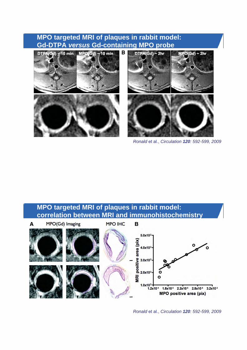
MPO targeted MRI of plaques in rabbit model: Gd-DTPA versus Gd-containing MPO probe
Ronald et al., Circulation 120: 592-599, 2009
MPO targeted MRI of plaques in rabbit model: correlation between MRI and immunohistochemistry
Ronald et al., Circulation 120: 592-599, 2009

18FDG-PET/CT in apo-E -/- mouse
ApoE-/- mouse
carotid cast
[18F]FDG-PET/CT
Courtesy of Michael Schäfers et al., Münster
PET imaging of MMP activity in apo-E -/- mouse
Hermann et al., J Nucl Cardiol 19: 609-617, 2012

Fluorescence Molecular Tomography (FMT)Visible light
Trans-illumination
Reconstructed3-D FMT data
Nahrendorf et al., Circ Cardiovasc Imaging 2: 56-70, 2009
FMT/CT of protease activity
Nahrendorf et al., ATVB 29: 1444-1451, 2009

Protease sensing: FMT/CT of atorvastatin treatment
apoE-/- apoE-/- on statin
60
40
FMT
sign
al [p
mol
]
no Tx Statin
20*
0
Nahrendorf et al., ATVB 29: 1444-1451, 2009
High-resolution MRI of mouse vascular anatomy
1 mm 1 mm
Right commoncarotid artery
Right sub- clavian artery Brachiocephalic
artery
Ascending aorta
Rik Moonen et al., TU/e
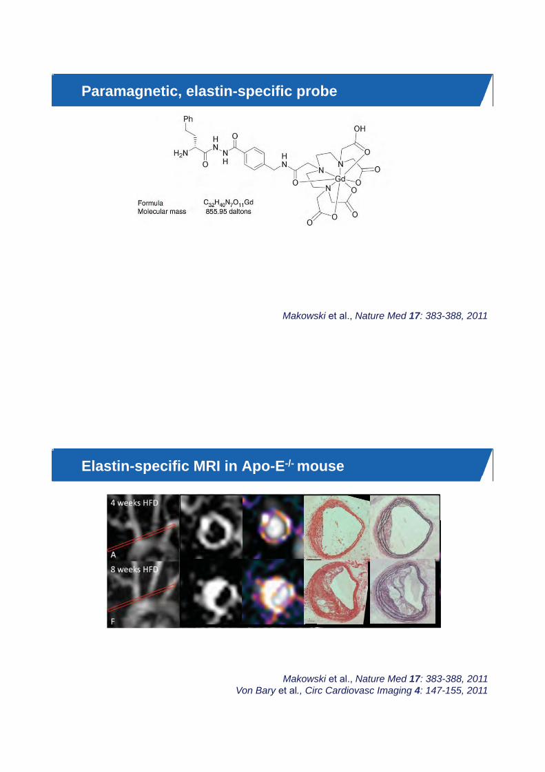
Paramagnetic, elastin-specific probe
Makowski et al., Nature Med 17: 383-388, 2011
Elastin-specific MRI in Apo-E-/- mouse
Makowski et al., Nature Med 17: 383-388, 2011 Von Bary et al., Circ Cardiovasc Imaging 4: 147-155, 2011

Elastin-specific MRI in Apo-E-/- mouse
Makowski et al., Nature Med 17: 383-388, 2011 Von Bary et al., Circ Cardiovasc Imaging 4: 147-155, 2011
Con
trast
-to-N
oise
Elastin-enhanced aortic MRI in pig model
Makowski et al., Invest Radiol 47: 438-444, 2012

Elastin-enhanced aortic MRI in pig model
Makowski et al., Invest Radiol 47: 438-444, 2012
Apo-E knock-out mouse with carotid artery cast
Van Bochove et al., MAGMA 23: 77-84, 2010Van Bochove et al., CMMI 6: 35-45, 2011
Kuhlmann et al., JoVE, 2012
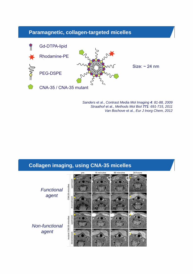
Paramagnetic, collagen-targeted micelles
PEG-DSPE
Rhodamine-PE
Gd-DTPA-lipid
CNA-35 / CNA-35 mutant
Size: ~ 24 nm
Sanders et al., Contrast Media Mol Imaging 4: 81-88, 2009 Straathof et al., Methods Mol Biol 771: 691-715, 2011
Van Bochove et al., Eur J Inorg Chem, 2012
Collagen imaging, using CNA-35 micelles
Non-functionalagent
Functionalagent
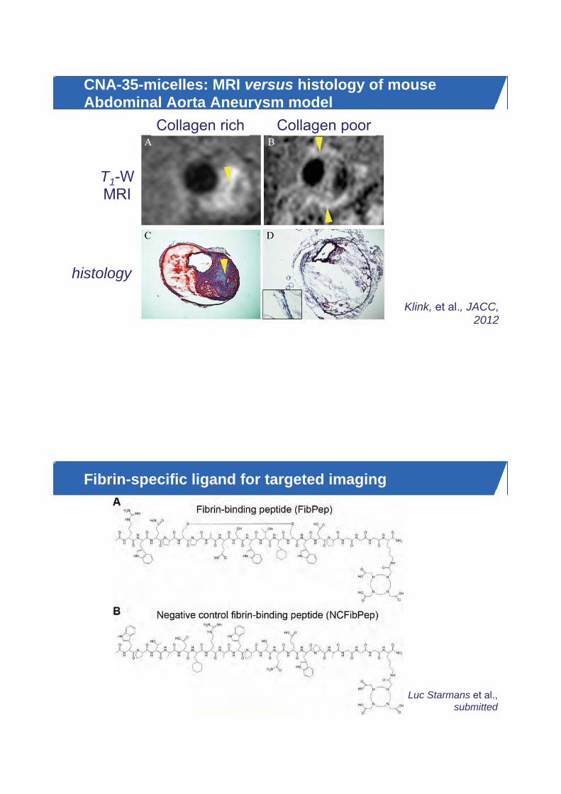
CNA-35-micelles: MRI versus histology of mouse Abdominal Aorta Aneurysm model
Klink, et al., JACC, 2012
Collagen rich Collagen poor
T1-WMRI
histology
Fibrin-specific ligand for targeted imaging
Luc Starmans et al.,submitted
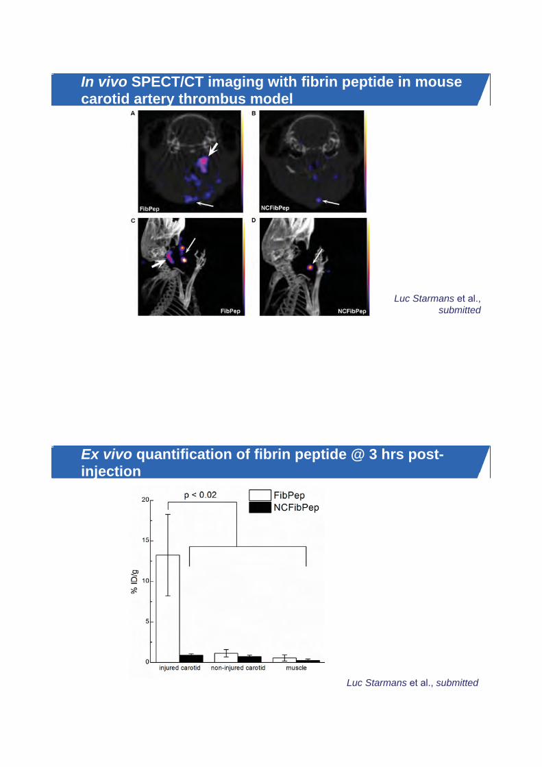
In vivo SPECT/CT imaging with fibrin peptide in mouse carotid artery thrombus model
Luc Starmans et al.,submitted
Ex vivo quantification of fibrin peptide @ 3 hrs post-injection
Luc Starmans et al., submitted

Clinical translation: Gd-containing fibrin agent
Spuentrup et al., Eur Radiol 18: 1995-2005, 2008
post-contrast T1-weighted MRI CT First-in-man MRI-based fibrin imaging
EP-2104R, a fibrin-specific agent
Clinical translation: Gd-containing fibrin agent
First-in-man MRI-based fibrin imaging
Spuentrup et al., Eur Radiol 18: 1995-2005, 2008

Plaque progression and therapeutic options
Quillard and Libby, Circulation Res 111: 231-244, 2012
Liposome-based anti-inflammatory therapy of atherosclerosis
Mark Lobatto et al., Mol Pharmaceutics 7: 2020-2029, 2010

MRI of steroid-loaded paramagnetic liposomes
24 hrs after liposomeinjection
beforeinjection
Mark Lobatto et al., Mol Pharmaceutics 7: 2020-2029, 2010
Monitoring anti-inflammatory therapy with 18FDG-PET
Mark Lobatto et al., Mol Pharmaceutics 7: 2020-2029, 2010
FreeLiposome
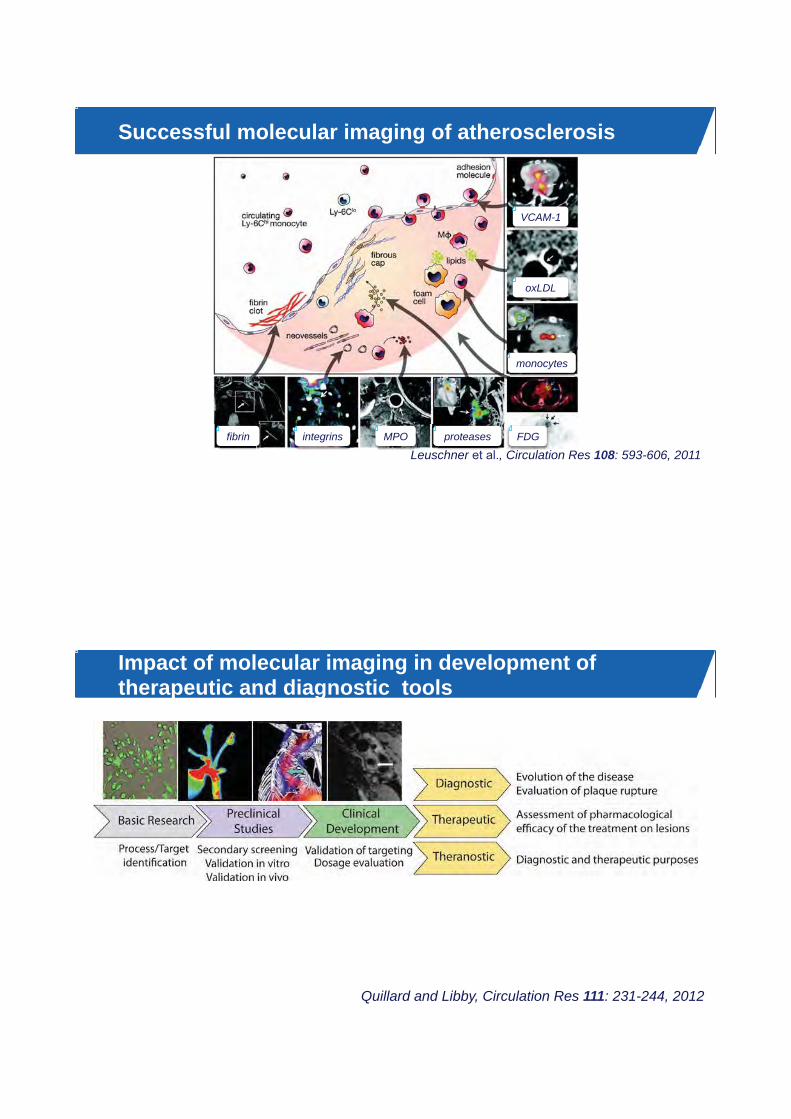
Successful molecular imaging of atherosclerosis
Leuschner et al., Circulation Res 108: 593-606, 2011
VCAM-1
oxLDL
monocytes
Ci l tiFDG
h t lproteases
LMPOintegrinsfibrin
Impact of molecular imaging in development of therapeutic and diagnostic tools
Quillard and Libby, Circulation Res 111: 231-244, 2012

Acknowledgements of MI collaboratorsMount Sinai, New YorkDavid CormodeZahi FayadAhmed Klink Willem Mulder Esad Vucic
AMC, Amsterdam Mat DaemenYigal Pinto
Maastricht University Chris Reutelingsperger
Leiden UMC Brigit den Adel Erik KaijzelClemens LöwikRob PoelmannLouise van der Weerd
SyMO-ChemHenk Janssen Henk Keizer
TU/eLuc BurnsveldMaarten MerkxBert Meijer
Erasmus University Rotterdam Dirk DunckerNico de Jong
University of Torino Silvio AimeEnzo Terreno
University of TwenteGert Storm Michel Versluis
Utrecht University Twan LammersRaymond Schiffelers
Philips ResearchHolger GrüllMarc Robillard
Bruker BioSpinWulf-Ingo Jung Arno Nauerth
University of MünsterMichael Kuhlman Michael SchäfersLars Stegger
University of Bonn Bernd Fleischmann Willy Roell
Imperial College LondonRob Krams
UMC Utrecht Wilbert Bartels Willem Mali Chrit MoonenGerard Pasterkamp
Vienna University Franz Gabor
Acknowledgements: Biomedical NMR @ TU/eGroup members
Desirée AbdurrachimOt BakermansBernard te BoekhorstSander van DuijnhovenMartijn FroelingTessa GeelenLarry de GraafWolter de GraafFloortje de Groot Holger GrüllJo HabetsStefanie Hectors Nicole HijnenIgor Jacobs Sharon JanssensRichard JonkersEsther Kneepkens
Master students Wouter DijkRobbert van GorkumNicole HaazenArjan HendriksJean-Paul KleijnenMariët KoopmanMarloes MarteijnJules NelissenTom PeetersTim SchakelTom SchreursJolanda SpijkermanBjorn StemkensSophie PeereboomPieternel van der TolSiem Wouters
Abdallah Mohamed Rik MoonenTiemen van MourikMiranda NabbenBastiaan van NieropLéonie NiesenLéonie PaulisJeanine PrompersPedro SanchesTom SchreursMariska de SmetLuc StarmansGustav StrijkersDavid VeraartBart WesselsChu Wong Sin Yuin Yeo

Funding @ Biomedical NMR � Center for Translational Molecular Medicine
� Equipment grants
� VIDI-grants Jeanine Prompers and Gustav Strijkers
� EMBO
� Netherlands Consortium for Systems Biology (NCSB)
� EU
� Program grant Netherlands Heart Foundation
� COST Action TD1004 “Theranostics Imaging and Therapy”
� High-Tech Systems and Materials (HTS&M) project NanoNextNL
s

Publications Klaas Nicolay “Introduction to Molecular Contrast Agents and New Devices ‐
Atherosclerosis”
1. Quillard T, Libby P. Molecular imaging of atherosclerosis for improving diagnostic and
therapeutic development. Circ Res 111: 231‐244, 2012
2. Leuschner F, Nahrendorf M. Molecular imaging of coronary atherosclerosis and myocardial
infarction: considerations for the bench and perspectives for the clinic. Circ Res 108: 593‐606, 2011
3. Mulder WJ, Strijkers GJ, van Tilborg GA, Cormode DP, Fayad ZA, Nicolay K. Nanoparticulate
assemblies of amphiphiles and diagnostically active materials for multimodality imaging. Acc Chem
Res 42: 904‐914, 2009