intravitreal injection
TRANSCRIPT

INTRAVITREAL INJECTION
HOW TO GIVE , AGENTS , INDICATIONS , COMPLICATIONS
INDOREDRISHTI.WORDPRESS.COM

DR DINESH MITTAL DR SONALEE MITTAL
DRISHTI EYE HOSP VIJAYNAGAR INDORE

INTRAVITREAL INJECTION
Intravitreal Injections Introduction• The delivery of medication directly into the vitreous cavity via injection or implant has become commonplace in ophthalmology. This method allows for higher concentrations in the eye with less systemic absorption compared to other methods of medication administration.
• In 1911, the injection of air into the vitreous cavity for retinal detachment (RD) repair was initially reported.
INTRAVITREAL INJECTION• The use of anti-vascular endothelial growth factor (anti-
VEGF) agents has resulted in a dramatic increase in intravitreal injections in recent years. An analysis of the Medicare claims database revealed fewer than 5,000 intravitreal injections in 2001 and 812,413 in 2007. Today, intravitreal injection is one of the most commonly performed medical procedures in the United States. More than 2.3 million intravitreal injections were performed in the United States in 2012, and projections call for more than 6 million annually by 2016
What is intravitreal injection• Intravitreal inj are an effective mode of treatment of many
retinal vascular diseases like diabetic maculopathy , age related macular degeneration and retinal vascular occlusions .
• The development of vascular endothelial growth factor VEGF inhibitors for the treatment of ocular neovascularization and macular edema can be regarded as the beginning of a new era in ophthalmological therapy. Before the year 2000, the treatment of any vascular abnormality in the macular region was merely restricted to laser photocoagulation
Quantifying Risk of Endophthalmitis
• With the widespread use of the intravitreal injection technique has come an increased concern regarding the risk of post-injection endophthalmitis. Because most patients treated with anti-VEGF agents receive a series of injections over months or years, it is important to distinguish between per-injection rates of endophthalmitis versus per-patient (or cumulative) rates of endophthalmitis over the course of treatment. While reported per-injection rates of endophthalmitis are generally very low in series of patients treated with anti-VEGF agents, per-patient rates may be close to 1 percent over a two-year course of therapy .
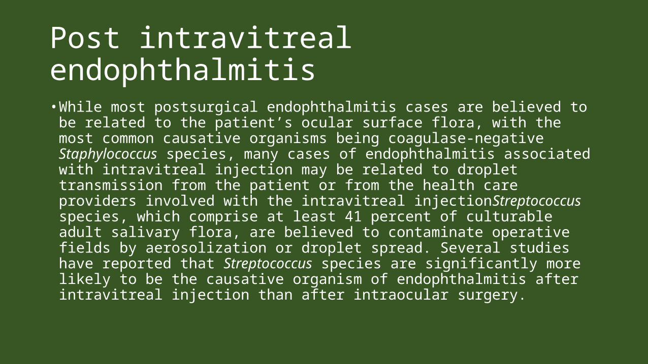
Post intravitreal endophthalmitis• While most postsurgical endophthalmitis cases are believed to be
related to the patient’s ocular surface flora, with the most common causative organisms being coagulase-negative Staphylococcus species, many cases of endophthalmitis associated with intravitreal injection may be related to droplet transmission from the patient or from the health care providers involved with the intravitreal injectionStreptococcus species, which comprise at least 41 percent of culturable adult salivary flora, are believed to contaminate operative fields by aerosolization or droplet spread. Several studies have reported that Streptococcus species are significantly more likely to be the causative organism of endophthalmitis after intravitreal injection than after intraocular surgery.
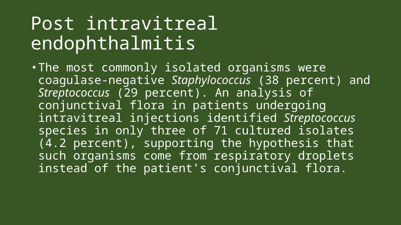
Post intravitreal endophthalmitis• The most commonly isolated organisms were
coagulase-negative Staphylococcus (38 percent) and Streptococcus (29 percent). An analysis of conjunctival flora in patients undergoing intravitreal injections identified Streptococcus species in only three of 71 cultured isolates (4.2 percent), supporting the hypothesis that such organisms come from respiratory droplets instead of the patient’s conjunctival flora.
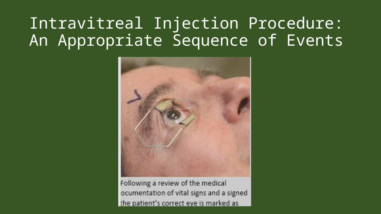
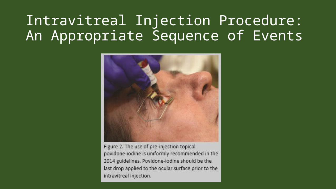
Intravitreal Injection Procedure: An Appropriate Sequence of Events
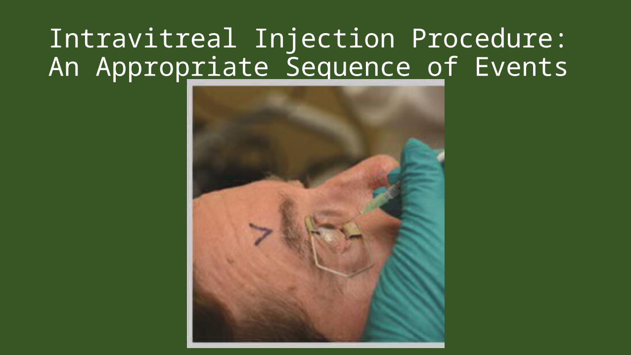
Intravitreal Injection Procedure: An Appropriate Sequence of Events
Intravitreal Injection Procedure: An Appropriate Sequence of Events
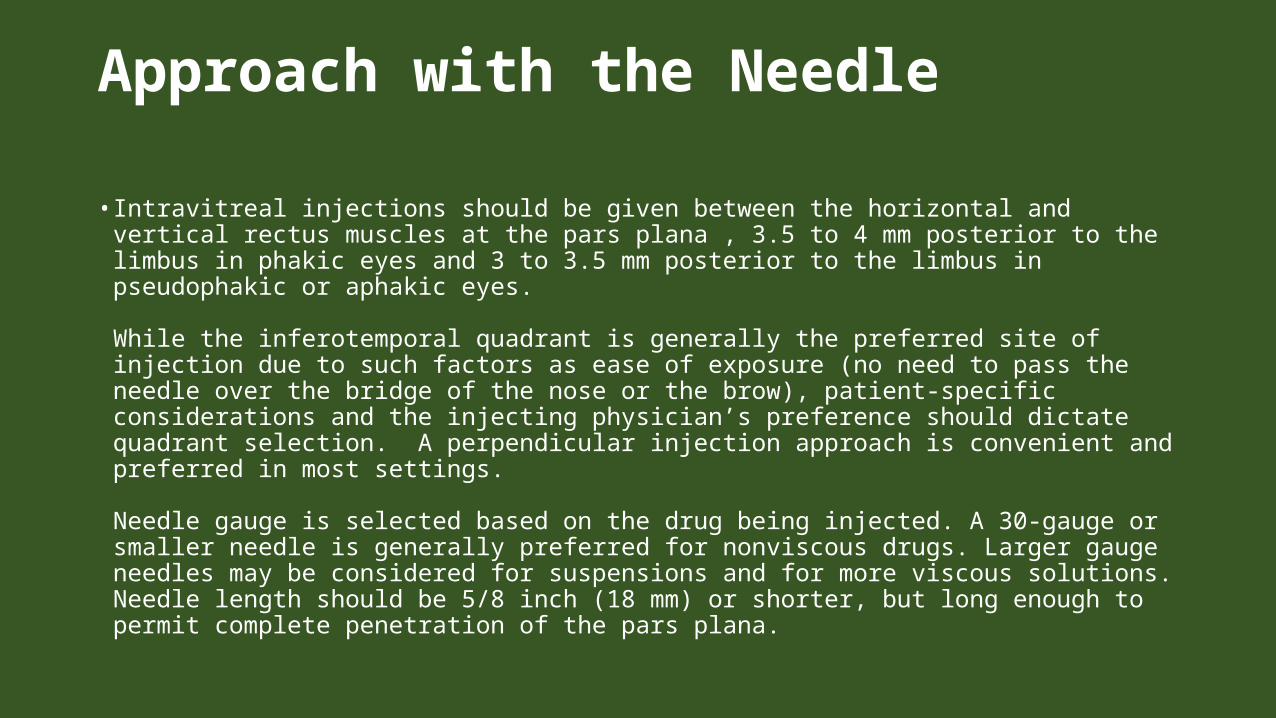
Approach with the Needle
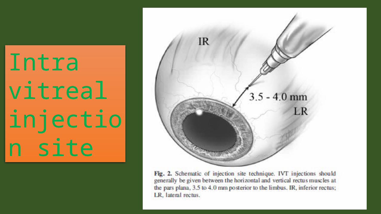
• Intravitreal injections should be given between the horizontal and vertical rectus muscles at the pars plana , 3.5 to 4 mm posterior to the limbus in phakic eyes and 3 to 3.5 mm posterior to the limbus in pseudophakic or aphakic eyes.
While the inferotemporal quadrant is generally the preferred site of injection due to such factors as ease of exposure (no need to pass the needle over the bridge of the nose or the brow), patient-specific considerations and the injecting physician’s preference should dictate quadrant selection. A perpendicular injection approach is convenient and preferred in most settings.
Needle gauge is selected based on the drug being injected. A 30-gauge or smaller needle is generally preferred for nonviscous drugs. Larger gauge needles may be considered for suspensions and for more viscous solutions. Needle length should be 5/8 inch (18 mm) or shorter, but long enough to permit complete penetration of the pars plana.
INTRAVITREAL INJECTIONS• Intravitreal injections and implants are a safe, effective and common method of delivering medication to the eye.
• Topical anesthesia usually sufficient• Sterile technique with eyelid speculum used• Small gauge needles 30-gauge can be used for intravitreal injections
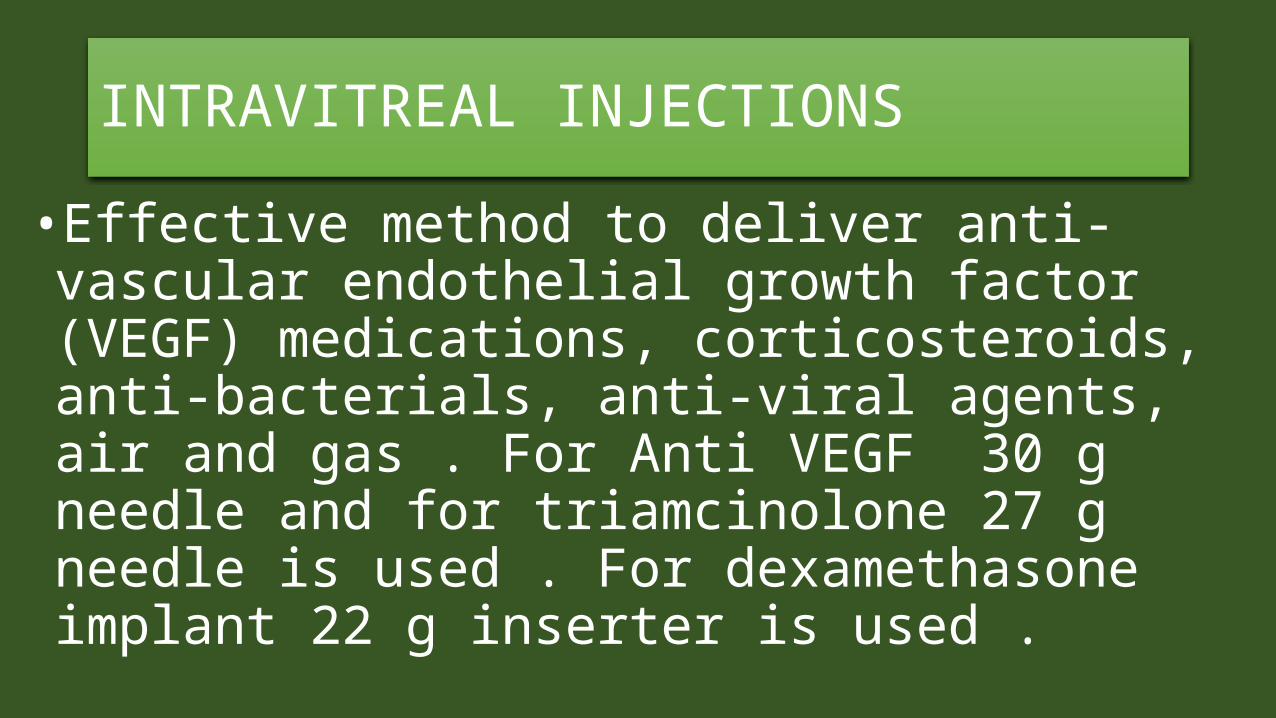
INTRAVITREAL INJECTIONS•Effective method to deliver anti-vascular endothelial growth factor (VEGF) medications, corticosteroids, anti-bacterials, anti-viral agents, air and gas . For Anti VEGF 30 g needle and for triamcinolone 27 g needle is used . For dexamethasone implant 22 g inserter is used .
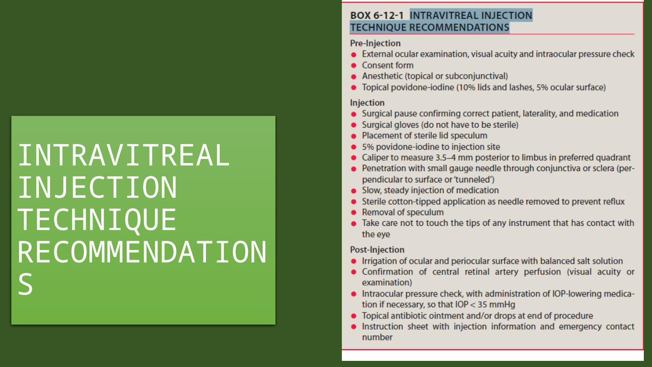
INTRAVITREAL INJECTIONTECHNIQUE RECOMMENDATIONS
Intravitreal injection site
PRE-INJECTION PREPARATION• Treatment of overt, active blepharitis should be done prior to injection to attempt to decrease bacterial load, which may increase risk of infection. Patients with bacterial or viral conjunctivitis should be treated to manage infection and have their injection rescheduled. As with any surgical procedure all patients should sign an informed consent after being explained the risks, benefits, and alternatives to injection
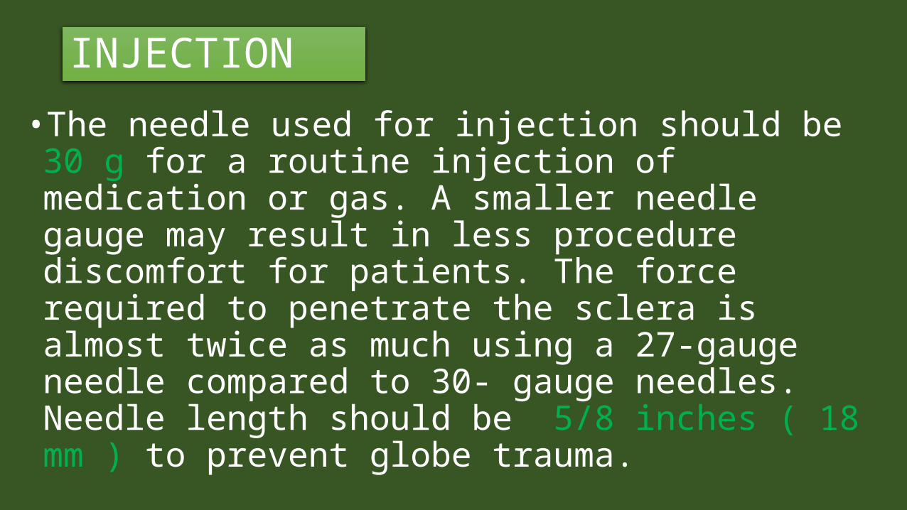
INJECTION• The needle used for injection should be 30 g for a routine injection of medication or gas. A smaller needle gauge may result in less procedure discomfort for patients. The force required to penetrate the sclera is almost twice as much using a 27-gauge needle compared to 30- gauge needles. Needle length should be 5/8 inches ( 18 mm ) to prevent globe trauma.
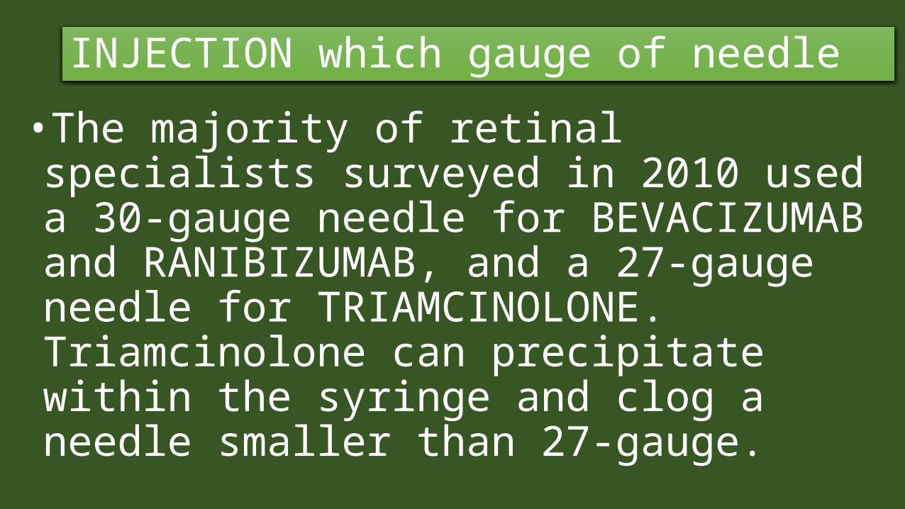
INJECTION which gauge of needle•The majority of retinal specialists surveyed in 2010 used a 30-gauge needle for BEVACIZUMAB and RANIBIZUMAB, and a 27-gauge needle for TRIAMCINOLONE. Triamcinolone can precipitate within the syringe and clog a needle smaller than 27-gauge.
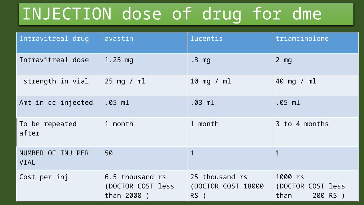
INJECTION dose of drug for dmeIntravitreal drug avastin lucentis triamcinolone
Intravitreal dose 1.25 mg .3 mg 2 mg
strength in vial 25 mg / ml 10 mg / ml 40 mg / ml
Amt in cc injected .05 ml .03 ml .05 ml
To be repeated after 1 month 1 month 3 to 4 months
NUMBER OF INJ PER VIAL
50 1 1
Cost per inj 6.5 thousand rs(DOCTOR COST less than 2000 )
25 thousand rs (DOCTOR COST 18000 RS )
1000 rs (DOCTOR COST less than 200 RS )
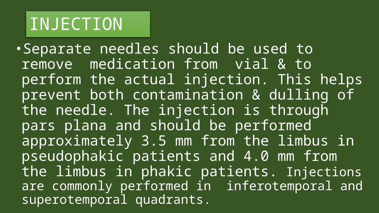
INJECTION• Separate needles should be used to remove medication from vial & to perform the actual injection. This helps prevent both contamination & dulling of the needle. The injection is through pars plana and should be performed approximately 3.5 mm from the limbus in pseudophakic patients and 4.0 mm from the limbus in phakic patients. Injections are commonly performed in inferotemporal and superotemporal quadrants.
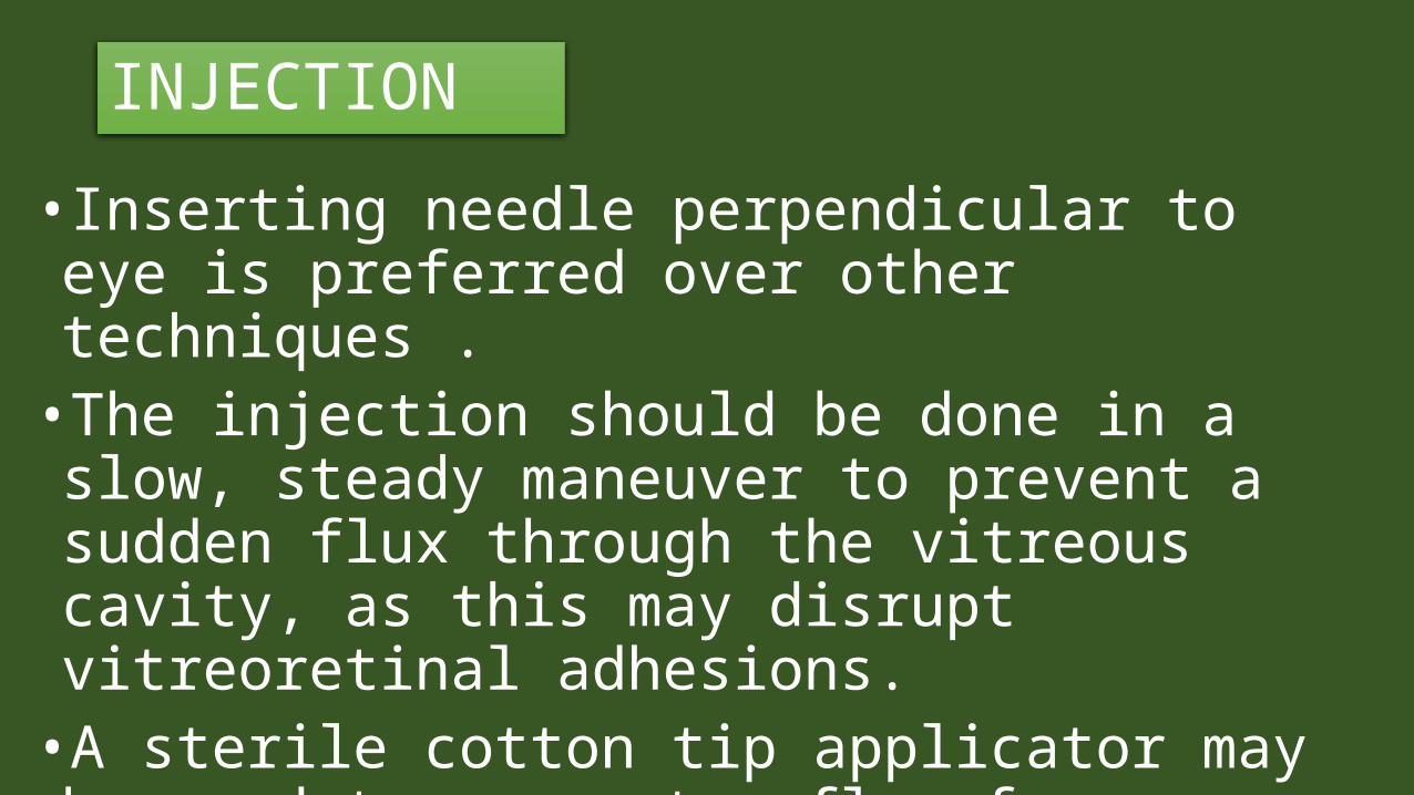
INJECTION• Inserting needle perpendicular to eye is preferred over other techniques .
•The injection should be done in a slow, steady maneuver to prevent a sudden flux through the vitreous cavity, as this may disrupt vitreoretinal adhesions.
•A sterile cotton tip applicator may be used to prevent reflux for injections
Injection Technique
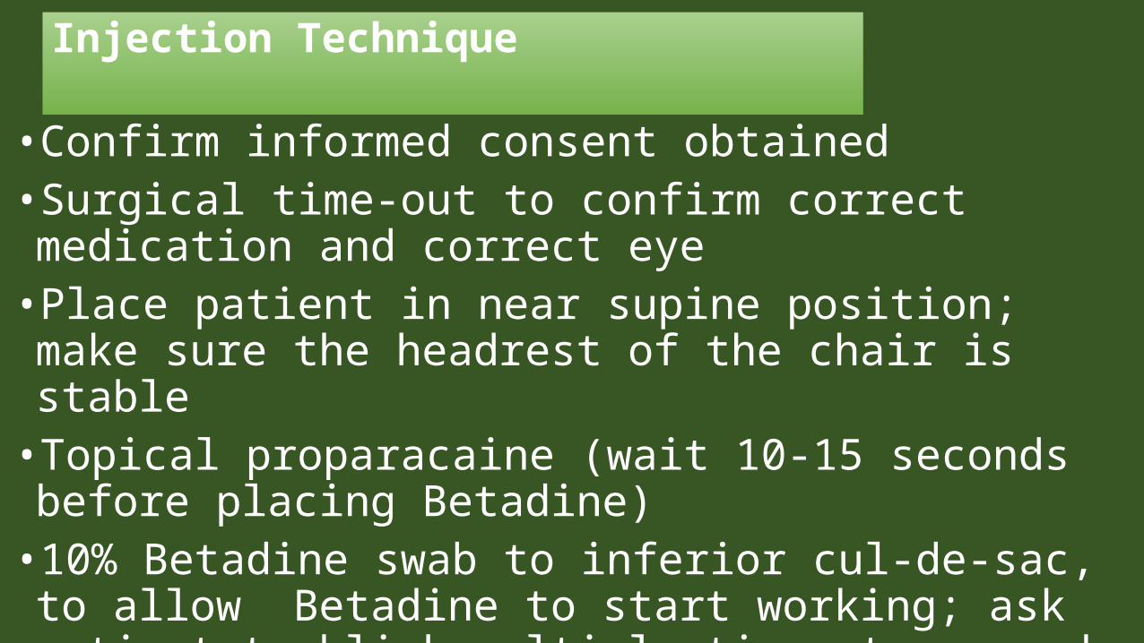
• Confirm informed consent obtained • Surgical time-out to confirm correct medication and correct eye
• Place patient in near supine position; make sure the headrest of the chair is stable
• Topical proparacaine (wait 10-15 seconds before placing Betadine)
• 10% Betadine swab to inferior cul-de-sac, to allow Betadine to start working; ask patient to blink multiple times to spread Betadine .
Injection Technique
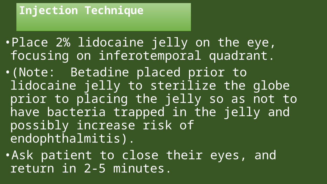
• Place 2% lidocaine jelly on the eye, focusing on inferotemporal quadrant.
• (Note: Betadine placed prior to lidocaine jelly to sterilize the globe prior to placing the jelly so as not to have bacteria trapped in the jelly and possibly increase risk of endophthalmitis).
• Ask patient to close their eyes, and return in 2-5 minutes.
Injection Technique
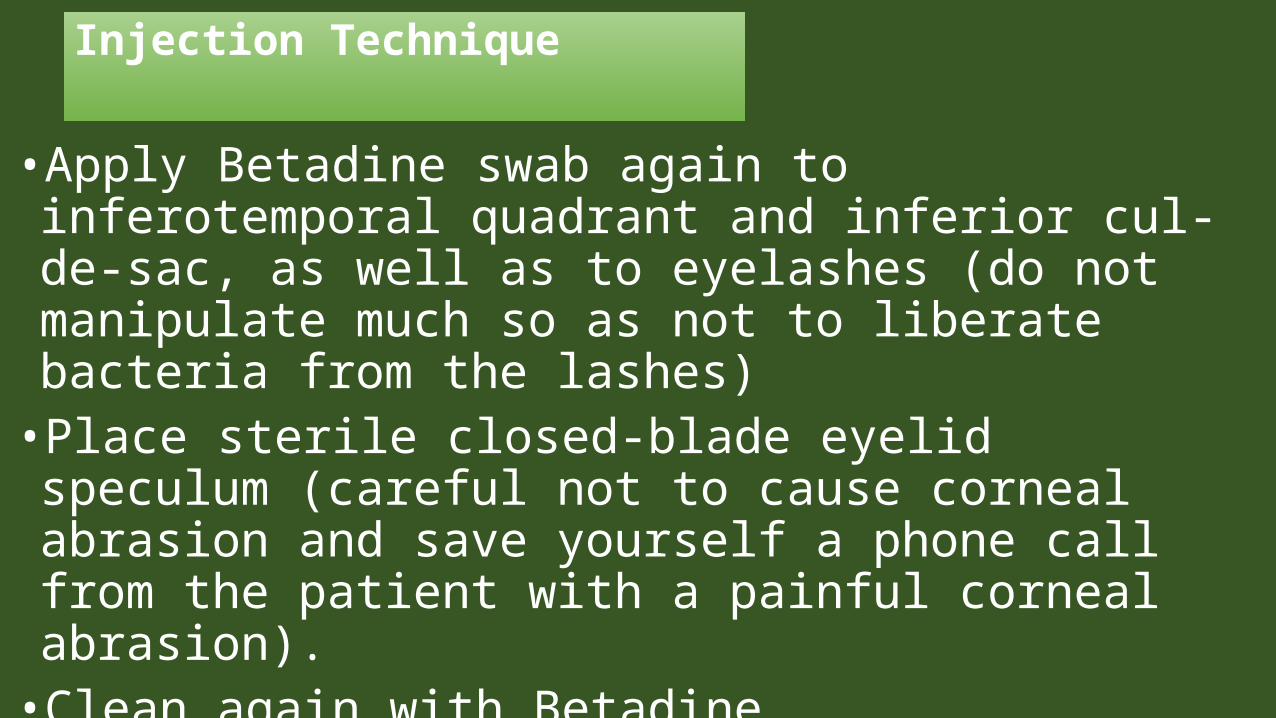
• Apply Betadine swab again to inferotemporal quadrant and inferior cul-de-sac, as well as to eyelashes (do not manipulate much so as not to liberate bacteria from the lashes)
• Place sterile closed-blade eyelid speculum (careful not to cause corneal abrasion and save yourself a phone call from the patient with a painful corneal abrasion).
• Clean again with Betadine
Injection Technique
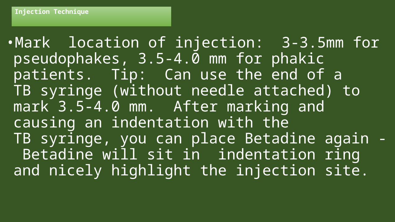
• Mark location of injection: 3-3.5mm for pseudophakes, 3.5-4.0 mm for phakic patients. Tip: Can use the end of a TB syringe (without needle attached) to mark 3.5-4.0 mm. After marking and causing an indentation with the TB syringe, you can place Betadine again - Betadine will sit in indentation ring and nicely highlight the injection site.
Injection Technique
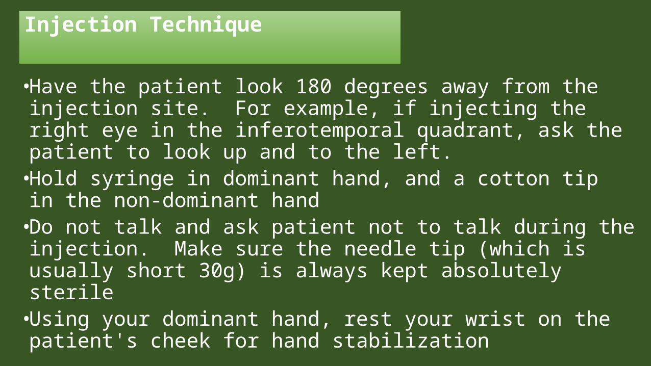
• Have the patient look 180 degrees away from the injection site. For example, if injecting the right eye in the inferotemporal quadrant, ask the patient to look up and to the left.
• Hold syringe in dominant hand, and a cotton tip in the non-dominant hand
• Do not talk and ask patient not to talk during the injection. Make sure the needle tip (which is usually short 30g) is always kept absolutely sterile
• Using your dominant hand, rest your wrist on the patient's cheek for hand stabilization
Injection Technique
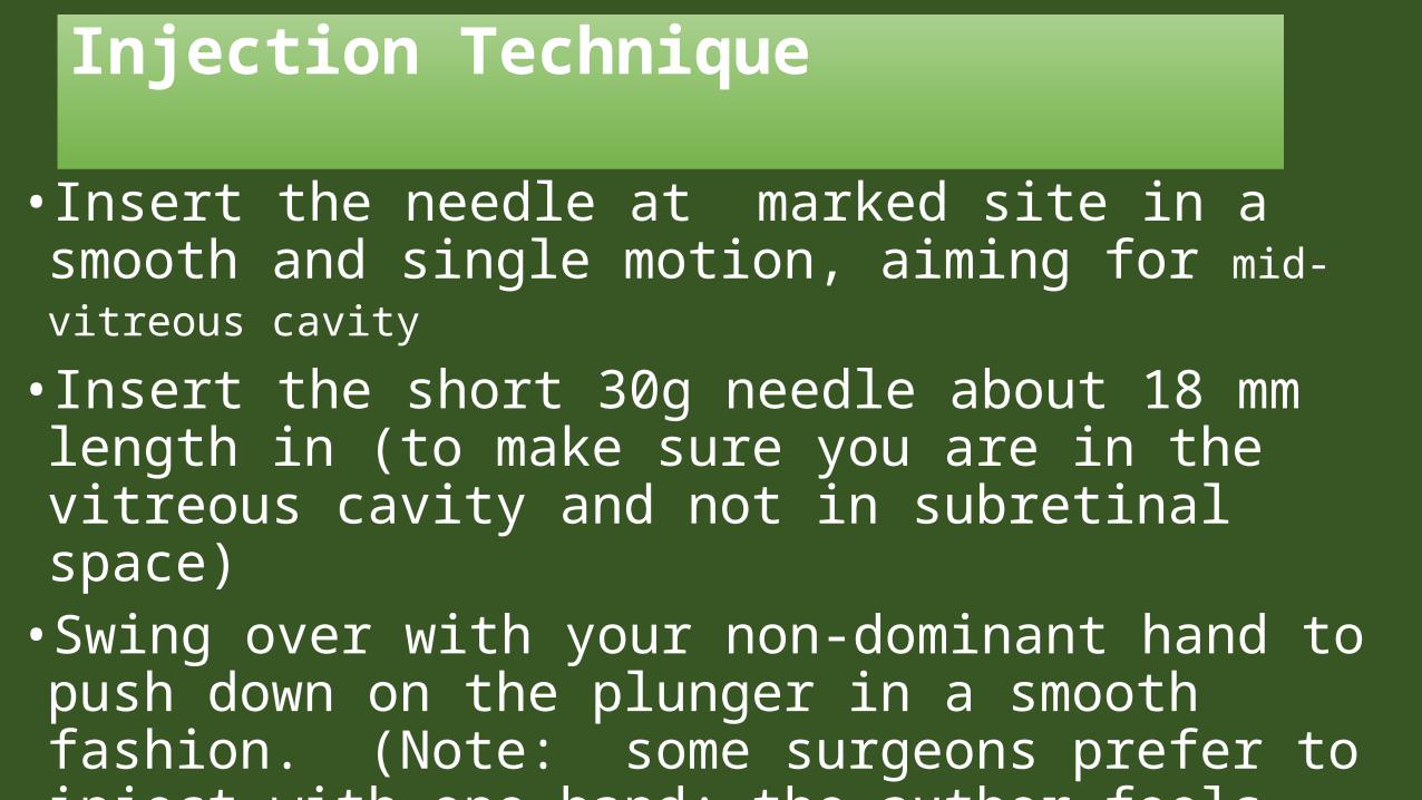
• Insert the needle at marked site in a smooth and single motion, aiming for mid-vitreous cavity
• Insert the short 30g needle about 18 mm length in (to make sure you are in the vitreous cavity and not in subretinal space)
• Swing over with your non-dominant hand to push down on the plunger in a smooth fashion. (Note: some surgeons prefer to inject with one-hand; the author feels that using two hands is more stable).
Injection Technique
• Do not move the needle while inside the eye so as to not cause traction on the vitreous and potentially cause a retinal tear / detachment.
•As you remove the needle out, cover the injection site with a cotton tip that is in your non-dominant hand
•Rinse the Betadine off of the patients eye
Injection Technique
• Ensure optic nerve perfusion (patient should be at least light perception). Paracentesis is usually not required, unless a large volume of medication is injected. Some Retina Specialists prefer to check and document the IOP and not let the patient leave until the IOP has reduced to an acceptable level.

ANTI-VEGF THERAPY
ANTI-VEGF THERAPY• In 2006, intraocular anti-VEGF therapy for exudative
age-related macular degeneration (AMD) was ranked among the top 10 breakthroughs of the year by Science Magazine. Since then, antiangiogenic therapy has broadened its impact from AMD treatment to various other diseases of the eye like macular oedema in diabetic retinopathy or retinal vein occlusion. In other areas, for example, ROP, antiangiogenic therapy is just beginning to find its place and is currently being evaluated in clinical studies .
WHICH ANTI VEGF INHIBITOR•BEVACIZUMAB ( AVASTIN ) & RANIBIZUMAB ( LUCENTIS ) are two of anti VEGF agents and both are owned by GENTECH USA .
•Both are equally effective but AVASTIN is not FDA approved .
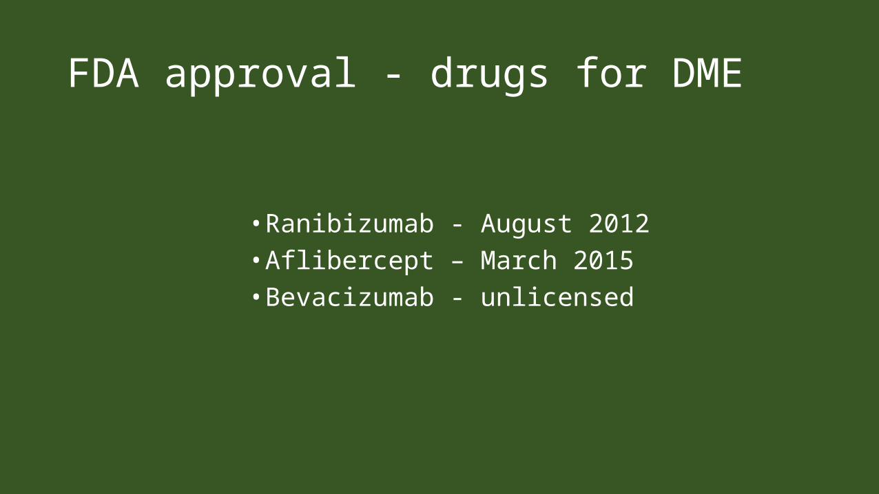
FDA approval - drugs for DME
• Ranibizumab - August 2012• Aflibercept – March 2015• Bevacizumab - unlicensed
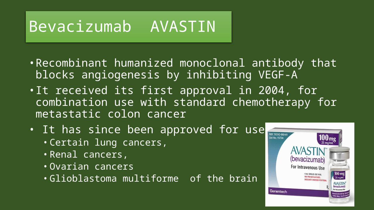
Bevacizumab AVASTIN
• Recombinant humanized monoclonal antibody that blocks angiogenesis by inhibiting VEGF-A
• It received its first approval in 2004, for combination use with standard chemotherapy for metastatic colon cancer
• It has since been approved for use in • Certain lung cancers, • Renal cancers, • Ovarian cancers• Glioblastoma multiforme of the brain

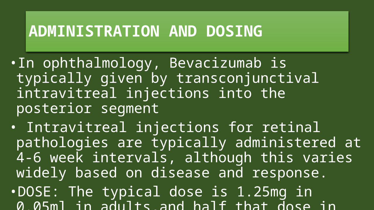
ADMINISTRATION AND DOSING• In ophthalmology, Bevacizumab is typically given by transconjunctival intravitreal injections into the posterior segment
• Intravitreal injections for retinal pathologies are typically administered at 4-6 week intervals, although this varies widely based on disease and response.
• DOSE: The typical dose is 1.25mg in 0.05ml in adults,and half that dose in babies.
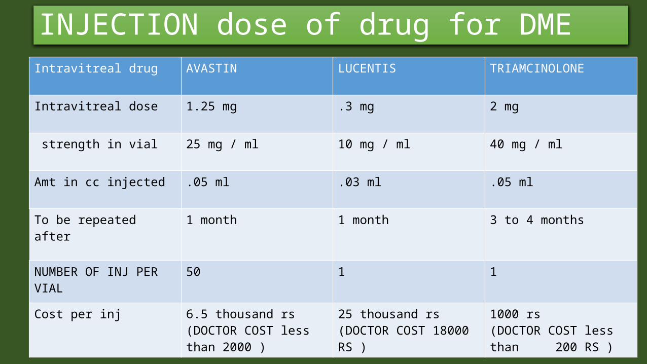
INJECTION dose of drug for DMEIntravitreal drug AVASTIN LUCENTIS TRIAMCINOLONE
Intravitreal dose 1.25 mg .3 mg 2 mg
strength in vial 25 mg / ml 10 mg / ml 40 mg / ml
Amt in cc injected .05 ml .03 ml .05 ml
To be repeated after 1 month 1 month 3 to 4 months
NUMBER OF INJ PER VIAL
50 1 1
Cost per inj 6.5 thousand rs(DOCTOR COST less than 2000 )
25 thousand rs (DOCTOR COST 18000 RS )
1000 rs (DOCTOR COST less than 200 RS )
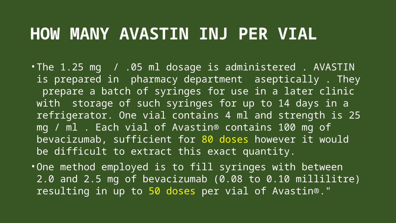
HOW MANY AVASTIN INJ PER VIAL• The 1.25 mg / .05 ml dosage is administered . AVASTIN is
prepared in pharmacy department aseptically . They prepare a batch of syringes for use in a later clinic with storage of such syringes for up to 14 days in a refrigerator. One vial contains 4 ml and strength is 25 mg / ml . Each vial of Avastin® contains 100 mg of bevacizumab, sufficient for 80 doses however it would be difficult to extract this exact quantity.
• One method employed is to fill syringes with between 2.0 and 2.5 mg of bevacizumab (0.08 to 0.10 millilitre) resulting in up to 50 doses per vial of Avastin®."
RANIBIZUMAB
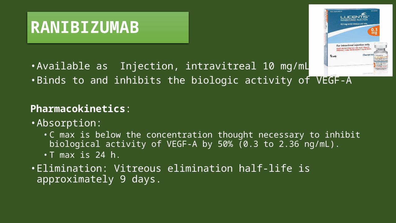
• Available as Injection, intravitreal 10 mg/mL• Binds to and inhibits the biologic activity of VEGF-A
Pharmacokinetics:• Absorption:
• C max is below the concentration thought necessary to inhibit biological activity of VEGF-A by 50% (0.3 to 2.36 ng/mL).
• T max is 24 h.• Elimination: Vitreous elimination half-life is approximately
9 days.

Mediators in DME (VEGF)• Vascular Endothelial Growth Factor (VEGF) is increased in human
ocular fluids and plays a crucial role in ischemic retinal diseases, such as DR and retinal vein occlusions . The VEGF family includes different isoforms: VEGF-A has been shown to be upregulated in hypoxic tissues. In DR, the loss of retinal capillaries is believed to lead to hypoxia, which is the main stimulus for increased retinal expression of VEGF-A, mediated through hypoxia-inducible factors. VEGF-A has an angiogenic role that is responsible for the progression of DR to the proliferative stage. Apart from its angiogenic role, VEGF-A increases vascular permeability . Thus, the role of VEGF-A may be central to DME pathogenesis. Moreover, several studies have demonstrated the efficacy of anti-VEGF treatment of DME, thus supporting the role of VEGF.
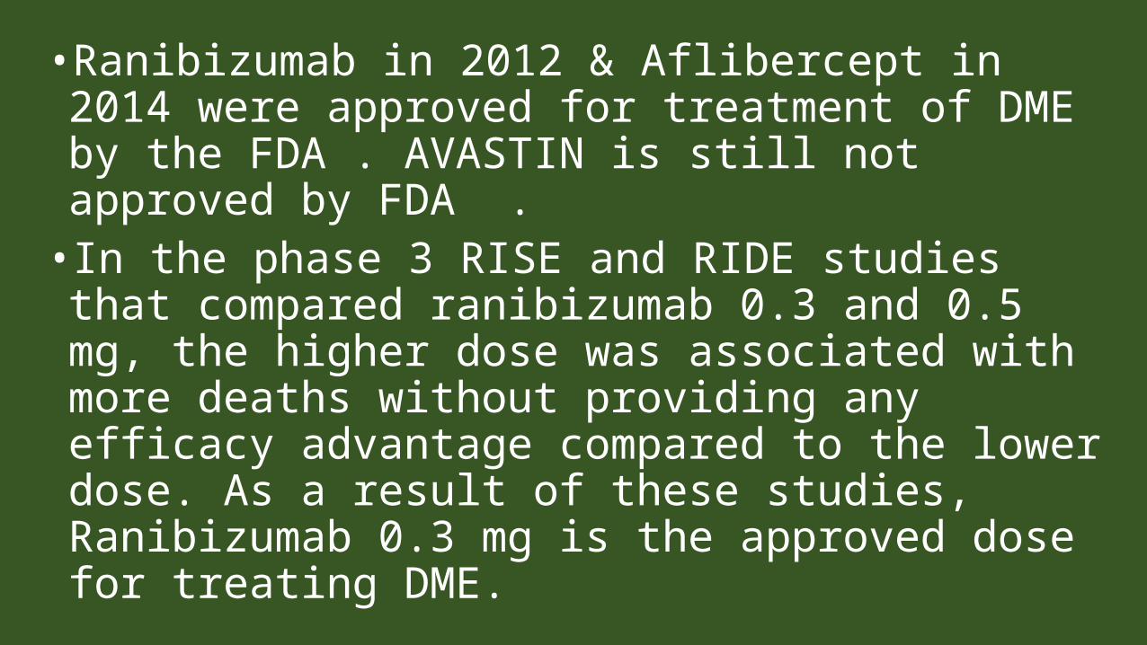
• Ranibizumab in 2012 & Aflibercept in 2014 were approved for treatment of DME by the FDA . AVASTIN is still not approved by FDA .
• In the phase 3 RISE and RIDE studies that compared ranibizumab 0.3 and 0.5 mg, the higher dose was associated with more deaths without providing any efficacy advantage compared to the lower dose. As a result of these studies, Ranibizumab 0.3 mg is the approved dose for treating DME.

•As we know, corticosteroid agents are not without attendant risks. IOP elevation and cataract formation are most notable side effects, for which we regularly monitor our patients started on steroids . In our experience, these side effects, although common, are effectively managed with topical glaucoma medications and cataract surgery, respectively .
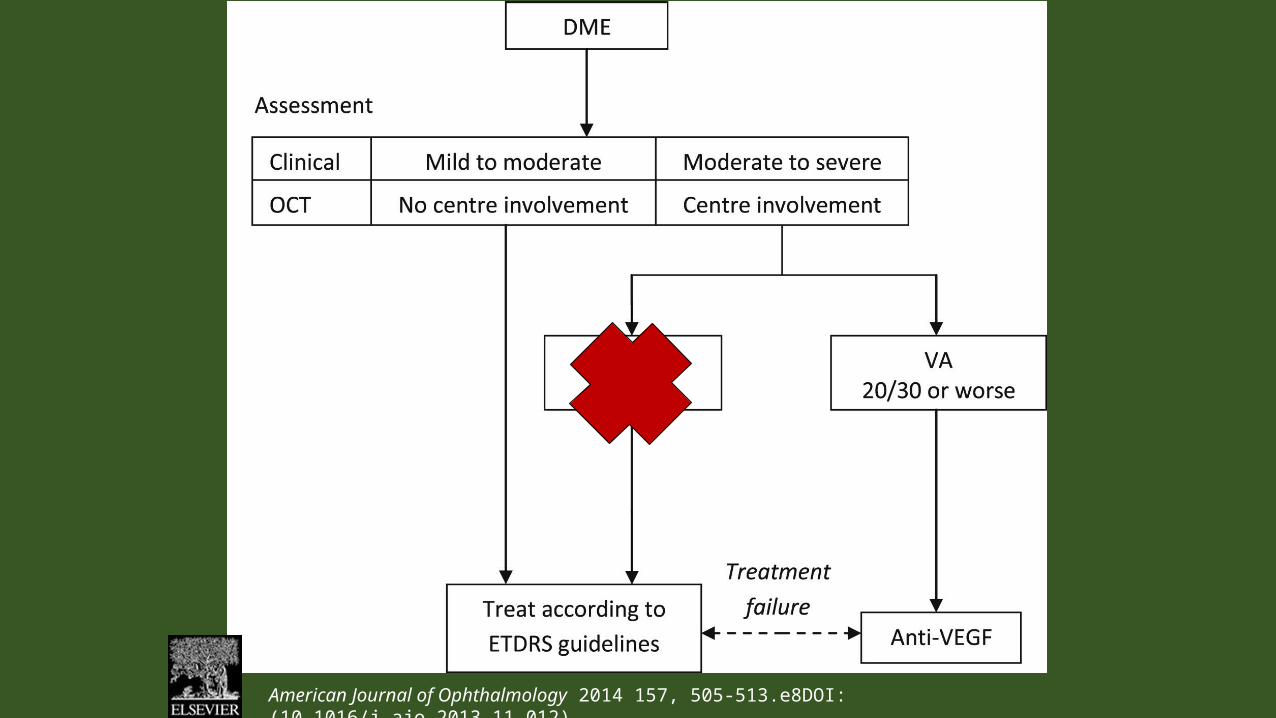
American Journal of Ophthalmology 2014 157, 505-513.e8DOI: (10.1016/j.ajo.2013.11.012)
threshold for starting anti- VEGF injections in a patient?
•If patient is symptomatic, begin treatment. But DME differs from AMD in several ways. For one, you do not have to treat a patient with DME immediately. If a patient is 20/20 or 20/25 and asymptomatic, even with 350 to 400 μm of edema, wait and watch .
threshold for starting anti- VEGF injections in a patient?
•Improving this patient’s condition may require seven to eight ocular injections during the first year, and outcomes might not be discernable to the patient.
If you choose to include steroids in your treatment regimens, which one do you use first • I use triamcinolone suspension, 1 mg to 2 mg as recommended by the Protocol B and the SCORE studies. I used to use a 4-mg dose, but now we know that the same biologic effect occurs with 1 mg, and that greatly reduces the complication rate.
IST YEAR ANTI DME TREATMENT• The great outcomes in RISE/RIDE, VIVID/VISTA and Protocol I require a very heavy burden, especially in that first year. That is one of the real key points. In AMD, the initiation phase/loading dose has usually involved three injections . However, in DME, there is a much heavier 9 OR 10 injection burden in the first year. It is a very steady, gradual improvement in both visual acuity and OCT outcomes .
DRCR.net Protocol I• DRCR.net Protocol I, patients were started off with a loading dose of four injections, but if patients did not have 20/20 vision and no edema, they were given an additional two injections. So in reality, it was a six-dose loading phase. Comparatively speaking, in AMD it is traditionally a three-dose loading phase.
DRCR.net Protocol I• Most of the AMD studies that utilized a p.r.n. dosing strategy required six to seven injections in the first year in comparison to Protocol I and T, where the average was higher with nine to 10 injections . Protocol I was instrumental in showing us that monthly treatment is not necessary to achieve vision stabilization and letter gain comparable with the more continuous treatment regimens in RISE/RIDE .

TREATMENT OF DME VS ARMD• It is important to distinguish wet AMD from DME—these are, indeed, two very different diseases. For patients with wet AMD, we are treating choroidal neovascularization with exudation. For patients with DME, we are only treating edema, the source of which is retinovascular incompetence. While we are trying to get that macula to a satisfactory dry status, the burden over time tends to fall off in DME.
TREATMENT OF DME VS ARMD• In wet AMD, there is more of a continuous burden year after year for an indefinite time frame in most patients. In DME, studies with discontinuous or P.R.N. style therapy such as Protocol I and Protocol T, many patients were eventually able to cease therapy .
TREATMENT OF DME VS ARMD• With wet AMD, whether it is P.R.N. or TREAT-AND-EXTEND (TAE), the mean number of treatments will typically reach a plateau after the first year. It does not tend to decrease much thereafter. But with DME, P.R.N. studies that we have seen, Protocol I in particular, started with a mean of eight to nine injections in year 1, then three to four in year 2, two to three in year 3 and by years 4 and 5, many had no further treatment .
How to reduce numer of injections• Steroids can be used for reduced burden of treatment as a combination therapy, but probably not as a low-burden monotherapy for most diabetic patients. There may be special cases where steroids are best choice as a monotherapy, such as in a newly pseudophakic patient who develops macular edema post-operatively, but results are simply not as robust as with anti VEGF therapy.
Type of response to anti VEGF• My patients typically fall into one of three categories. The first is what we all want—a patient who is exquisitely sensitive to anti-VEGF therapy and needs only one injection to have a very dramatic improvement. The second group is what we dread—no measurable response with anti-VEGFs after three or four injections. The third group, however, is what most of us see—people who have some response to the anti-VEGF therapy.
final• After all the studies, I think it is safe to say anti-VEGF is our
first-line defense. We have long-term outcomes from the studies, and those results are extremely efficacious. But it is a complex disease. Even when you look at the results from Protocol T, the agent that worked the best still left one-third of the patients with more than 250 microns of edema at 1 year. Most of us are using bevacizumab, and many of our patients are going to have persistent fluid even after 10 treatments in the first year. . We don’t have large studies to dictate whether or not the intravitreal triamcinolone should be our preferred second-line agent.


Pre-packaged syringes• "Pre-packaged syringes of bevacizumab for intravitreal
use are available to purchase from special manufacturing units. Moorfields Pharmaceuticals, of Moorfields NHS hospital, can supply syringes of 1.25 mg in 0.05 ml at £85 per syringe, excluding VAT and delivery charges. The syringes must be stored in a refrigerator and have an expiry date of six weeks from the date of manufacture, which would mean an effective expiry of about two to four weeks from receipt of delivery."
COMPOUNDING AVASTIN• "Any compounding of a single vial of drug into multiple
dose units will carry some risk of microbial and particulate cross contamination beyond that associated with preparation of a single dose. This risk can be minimised by performing the procedure in an aseptic clean room using trained staff and storing the finished product in a refrigerator."
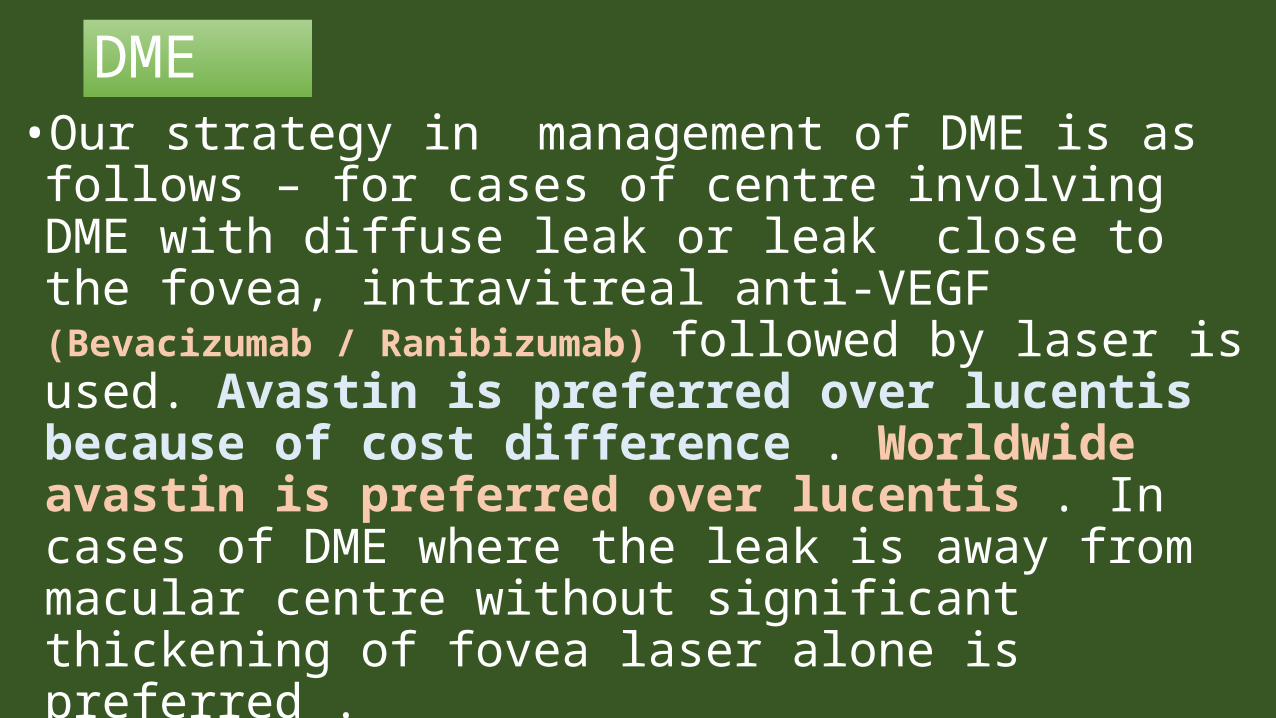
DME• Our strategy in management of DME is as follows – for cases of centre involving DME with diffuse leak or leak close to the fovea, intravitreal anti-VEGF (Bevacizumab / Ranibizumab) followed by laser is used. Avastin is preferred over lucentis because of cost difference . Worldwide avastin is preferred over lucentis . In cases of DME where the leak is away from macular centre without significant thickening of fovea laser alone is preferred .
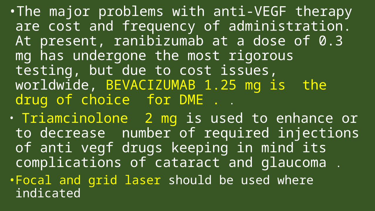
• The major problems with anti-VEGF therapy are cost and frequency of administration. At present, ranibizumab at a dose of 0.3 mg has undergone the most rigorous testing, but due to cost issues, worldwide, BEVACIZUMAB 1.25 mg is the drug of choice for DME . .
• Triamcinolone 2 mg is used to enhance or to decrease number of required injections of anti vegf drugs keeping in mind its complications of cataract and glaucoma .
• Focal and grid laser should be used where indicated

COMPLICATIONS
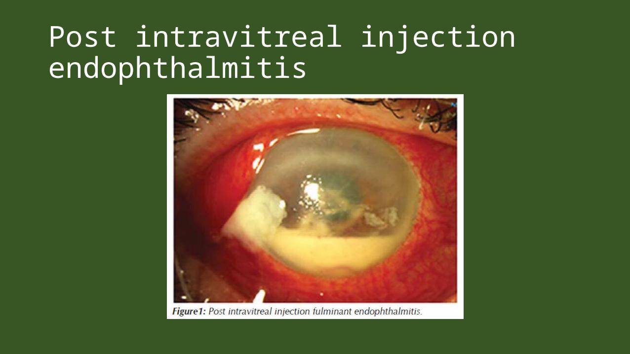
Post intravitreal injection endophthalmitis• The time period for occurrence of PIE from injection to
presentation is early and ranges from within 24 hrs. to even upto 26 days as reported, with average of 4 days.
• There has been some reports of culture-negative sterile endophthalmitis after intravitreal injection for different retinal pathologies, resembling toxic anterior segment syndrome (TASS-like) seen after intraocular surgery .
Post intravitreal injection endophthalmitis

Post intravitreal injection endophthalmitis•post-injection endophthalmitis (PIE) has early presentation and worser prognosis , especially with
streptococcus viridans. The incidence is especially more in office-based setting as compared to the operating-room setting.
Indication based risk factor
• The risk of endophthalmitis seems to be lower in eyes with macular edema secondary to retinal vein occlusion as the indication for injection. The risk is more in patients with diabetic eye disease and neovascular age-related macular degeneration (AMD), with impaired or waning immunity as the hypothesized mechanism in both.
TREATMENT OF POST-INJECTIONendophthalmitis• Patients receiving intravitreal injection of bevacizumab
may present with sterile endophthalmitis. In cases of doubt, it is important to consider all unexpected inflammatory response following injection or surgery to be endophthalmitis unless proven otherwise. The diagnosis can be confirmed by culture of causative organism in-vitro from intraocular samples. Samples that can be collected are aqueous tap or vitreous sample (higher yield) or both (preferred).
TREATMENT OF POST-INJECTIONEndophthalmitis• Vitreous sample can be obtained by vitreous tap using 23-
G needle through pars plana route before intravitreal antibiotic injections. However, due to inadequacy of sample for analysis and theoretical risk of producing vitreous traction during aspiration, vitreous biopsy is preferred by many surgeons especially without infusion line to safely obtain adequate volume of sample that provides higher yield of organisms. This may be performed as a sole procedure or just before pars plana vitrectomy for endophthalmitis. The sample is then sent for staining for microscopic evaluation and culture & sensitivity
TREATMENT OF POST-INJECTIONendophthalmitis• Apart from confirmation of diagnosis in patients
presenting with intraocular inflammation, it is also important to maintain and check records of the batch number of the drug used, and patients receiving injection on same day or injection from the same batch number. Thus helping to trace the source of infection and early detection of other cases of endophthalmitis in the cluster if any.
TREATMENT OF POST-INJECTIONendophthalmitisThis includes anti-bacterial therapy in form of intravitreal antibiotics, anti- inflammatory therapy, supportive therapy or surgery. Concentrated topical antibiotics should be considered empirically till the culture results are awaited, especially if route of infection spread seems to be from anterior segment. Concentrated topical antibiotics may include cefazolin 5% and tobramycin 1.3%. Commonly used empiric intravitreal antibiotics include vancomycin 1mg/0.1ml and ceftazidime 2.25 mg/0.1 ml. Though, EVS concluded no additional benefits of parentral antibiotics in post-cataract surgery endophthalmitis, parentral antibiotics help in augmenting and sustaining an adequate concentration of antibiotics in the vitreous cavity for a more prolonged period .
TREATMENT OF POST-INJECTIONendophthalmitis• Treatment of post-intravitreal endophthalmitis needs be tailored
depending upon individual cases. The treatment in post-intravitreal endophthalmitis should be more aggressive as the infection tends to have a worser prognosis. While, intravitreal antibiotics seem to be the most common first treatment in post cataract surgery endophthalmitis, Early surgical intervention should be preferred in postintravitreal endophthalmitis. With advancement and advent in surgical techniques and equipments, the aim of surgery is to achieve complete vitrectomy with PVD induction, thereby removing the infectious nidus and substantially decreasing toxic and inflammatory load.
TREATMENT OF POST-INJECTIONendophthalmitis• Undiluted specimen should be sent for culture studies
and antibiotics should be instilled in the vitreous cavity at the end of the surgery thereby achieving increased intraocular antibiotic concentration.
AVASTIN INDUCED ENDOPHTHALMITIS• Vial itself may be adulterated and hence containing toxins .
So even if the vial is opened on the table and pt given intravit injection pt. will develop symptoms like TASS . In case of avastin symptoms will be localized in posterior segment so this should be called TOXIN POSTERIOR SEGMENT SYNDROME and not TOXIN ANTERIOR SEGMENT SYNDROME ( TASS ) . These symptoms will develop in few hours and are a type of sterile reaction to toxins . And if the avastin gets infected during compounding then infective endoph will develop after some days .
AVASTIN INDUCED ENDOPHTHALMITIS
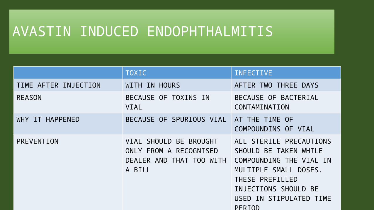
TOXIC INFECTIVETIME AFTER INJECTION WITH IN HOURS AFTER TWO THREE DAYSREASON BECAUSE OF TOXINS IN VIAL BECAUSE OF BACTERIAL
CONTAMINATIONWHY IT HAPPENED BECAUSE OF SPURIOUS VIAL AT THE TIME OF
COMPOUNDINS OF VIAL PREVENTION VIAL SHOULD BE BROUGHT
ONLY FROM A RECOGNISED DEALER AND THAT TOO WITH A BILL
ALL STERILE PRECAUTIONS SHOULD BE TAKEN WHILE COMPOUNDING THE VIAL IN MULTIPLE SMALL DOSES. THESE PREFILLED INJECTIONS SHOULD BE USED IN STIPULATED TIME PERIOD
HOW TO PREVENT AVASTIN INDUCED REACTIONS IN EYES• 1 BEST IS FDA SHOULD APPROVE AVASTIN and
GENETECH should be persuaded to make available avastin in small aliquots for intravitreal use .this will help in three ways
• It will reduce price further .• Spurious drugs will be knocked out and hence toxin
induced reactions will be avoided .• Contamination of bacteria at the time of dispensing
AVASTIN will be avoided .
HOW TO PREVENT AVASTIN INDUCED REACTIONS IN EYES• 2. governments should appoint nodal agency in country
which will procure avastin directly from genetech and it will dispense in small aliquots to whole of country .
• It will again prevent us from spurious drugs and also minimize the chances of bacterial contamination .

HOW TO PREVENT AVASTIN INDUCED REACTIONS IN EYES• 3 . If above two steps are not feasible then is the 3rd
step at the injecting surgeon level .• MUST DO’S FOR INJECTING SURGEON• 1 buy avastin vial from a well reputed and authrised
dealer and always insist for bill .it will save us from spurios avastin vials .
• 2 dispense the vial in multiple preloaded injections under asceptic conditions preferably in pharmacy deptt.it will avoid bacterial contamination .
HOW TO PREVENT AVASTIN INDUCED REACTIONS IN EYES• 3. do not inject in all the pts together . Ist day inject in
two three eyes . Donot inject in both eyes simultaneously .so that if disaster happens it will happen only in one eye . See the response next day and only then inject in other pts .
• 4 use all the prefilled intravitreal injections in stipulated time period say two weeks .
• AVASTIN IS THERE TO HELP MAN TO REDUCE BLINDNESS & MAN SHOULD ALSO USE IT ACCORDINGLY TO DO THE SAME .
Conclusion
• To optimize the outcomes associated with intravitreal injection, the retina specialist and staff should pay careful attention to reducing the risk of complications. Treatment outcomes depend not only on the safety and efficacy of the pharmacotherapy being delivered, but also on the safety and potential adverse events associated with the procedure itself .


THANK YOU
DR DINESHDR SONALEE