intraportal insulin protects from the liver injury of portacaval shunt in dogs
TRANSCRIPT
1241
Preliminary Communications
INTRAPORTAL INSULIN PROTECTS FROMTHE LIVER INJURY OF PORTACAVAL SHUNT
IN DOGS
THOMAS E. STARZL KENDRICK A. PORTERCHARLES W. PUTNAM
Department of Surgery, University of Colorado MedicalCenter, Denver, Colorado, 80220; and Department of
Pathology, St. Mary’s Hospital Medical School, London W2
Summary 4 days after portacaval shunt, the liversof normal dogs had pronounced atrophy
and other structural abnormalities. These changes weregreatly reduced in the left liver lobes, but not in theright, by a constant infusion to the left portal vein of in-sulin in non-hypoglycæmic doses. These experimentalfindings should have implications in clinical medicine.
INTRODUCTION
DURING the past ten years we have developed evidencethat substances, termed hepatotrophic factors, in the
portal venous blood of dogs can profoundly influenceliver function as well as the size, internal structure,chemical composition, and dividing capability of thehepatocytes.1-5 In most of these experiments, techniqueswere exploited that permitted comparative study of twoportions of the same liver which were given differentkinds of portal venous inflow under diabetic or non-dia-betic conditions. The concept emerged that manifoldhepatic processes are controlled or influenced by hor-mones that are generated by splanchnic organs and deli-vered straight to the liver, with a presumably augmentedsignificance because of the episodically high concentra-tions of nutrient substrate in the same blood.2-5 Insulinhas been identified as the most important of these un-doubtedly multiple portal hepatotrophic constituents.2-5
If the foregoing conclusions were correct, the actiology
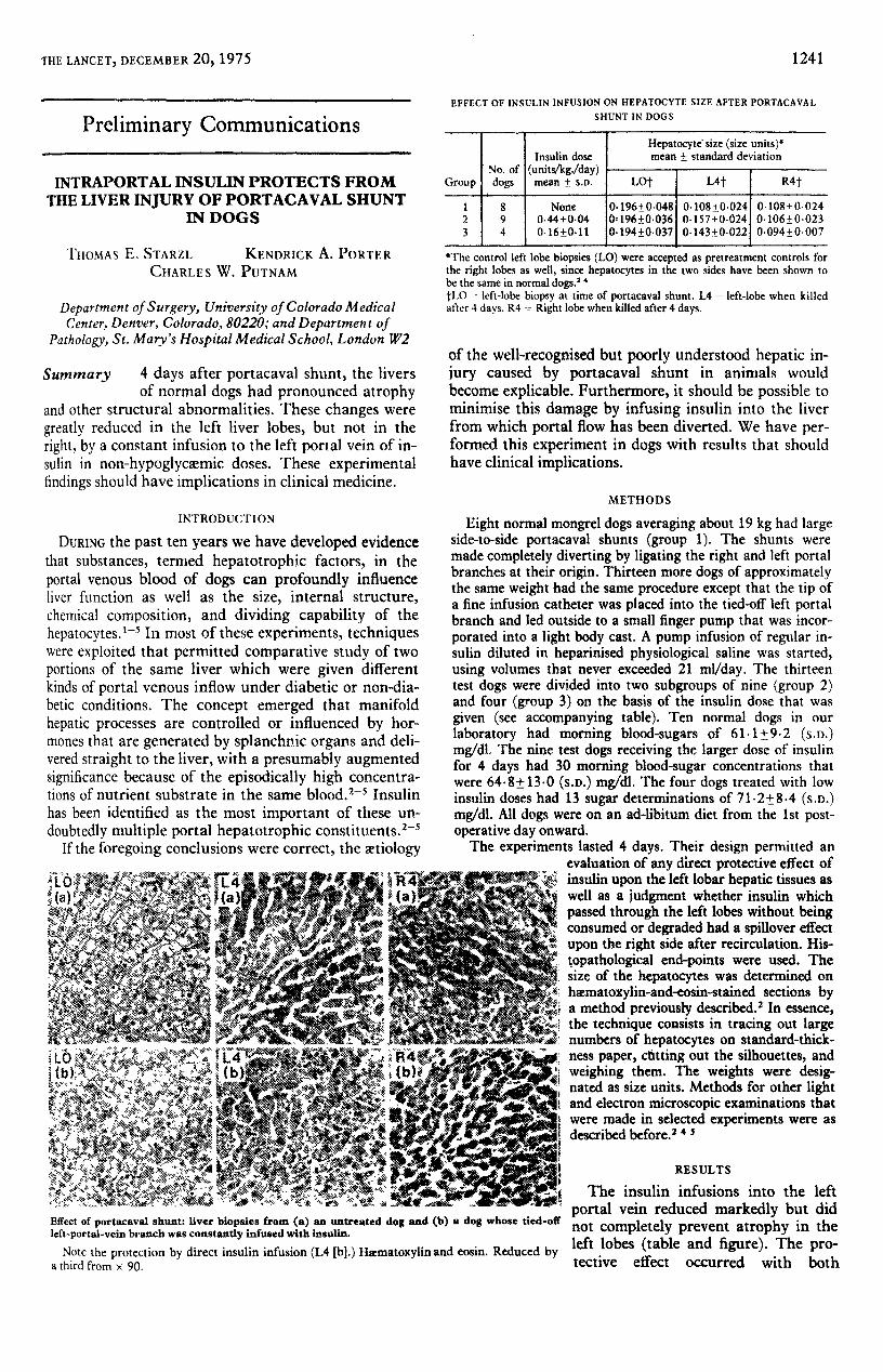
Effect of portacaval shunt: liver biopsies from (a) an untreated dog and (b) a dog whose tied-otleft-portal-vein branch was constantly infused with insulin.
Note the protection by direct insulin infusion (L4 [b].) Haematoxytinand eosin. Reduced bya third from x 90.
EFFECT OF INSULIN INFUSION ON HEPATOCYTE SIZE AFTER PORTACAVAL
SHUNT IN DOGS
"The control left lobe biopsies (LO) were accepted as pretreatment controls forthe right lobes as well, since hepatocytes in the two sides have been shown tobe the same in normal dogs.1 . 4f!,0 ’ -- left-lobe biopsy at time of portacaval shunt. L4 = left-lobe when killedafter 4 days. R4 right lobe when killed after 4 days.
of the well-recognised but poorly understood hepatic in-jury caused by portacaval shunt in animals wouldbecome explicable. Furthermore, it should be possible tominimise this damage by infusing insulin into the liverfrom which portal flow has been diverted. We have per-formed this experiment in dogs with results that shouldhave clinical implications.
METHODS
Eight normal mongrel dogs averaging about 19 kg had largeside-to-side portacaval shunts (group 1). The shunts weremade completely diverting by ligating the right and left portalbranches at their origin. Thirteen more dogs of approximatelythe same weight had the same procedure except that the tip ofa fine infusion catheter was placed into the tied-off left portalbranch and led outside to a small finger pump that was incor-porated into a light body cast. A pump infusion of regular in-sulin diluted in heparinised physiological saline was started,using volumes that never exceeded 21 ml/day. The thirteentest dogs were divided into two subgroups of nine (group 2;and four (group 3) on the basis of the insulin dose that wasgiven (see accompanying table). Ten normal dogs in out
laboratory had morning blood-sugars of 61.l:!:9.2 (S.D.]mgldl. The nine test dogs receiving the larger dose of insulirfor 4 days had 30 morning blood-sugar concentrations thaiwere 64.8±13.0 (s.D.) mg/dl. The four dogs treated with lovinsulin doses had 13 sugar determinations of 71.2±8.4 (s.D.mg/dl. All dogs were on an ad-libitum diet from the 1st postoperative day onward.The experiments lasted 4 days. Their design permitted at
evaluation of any direct protective effect ofinsulin upon the left lobar hepatic tissues aswell as a judgment whether insulin whichpassed through the left lobes without beingconsumed or degraded had a spillover effectupon the right side after recirculation. His-topathological end-points were used. Thesize of the hepatocytes was determined onhaematoxylin-and-eosin-stained sections bya method previously described In essence,the technique consists in tracing out largenumbers of hepatocytes on standard-thick-ness paper, catting out the silhouettes, andweighing them. The weights were desig-nated as size units. Methods for other lightand electron microscopic examinations thatwere made in selected experiments were as
described before .24 s
RESULTS
I The insulin infusions into the left
portal vein reduced markedly but didnot completely prevent atrophy in theleft lobes (table and figure). The pro-tective effect occurred with both

1242
higher and lower insulin dose ranges, but somewhat lesswith the low doses. There was no spillover protectionwhatever for the right lobes (table). The differences inthe day-4 left lobar biopsies between all thirteen treateddogs versus the eight controls were significant (r< 0-001)as were the differences between the day-4 left-versus-right lobar biopsies in the pooled thirteen treated ani-mals (r<0001).The maintenance of the size of the hepatocytes in the
left lobes of the animals treated with insulin was not dueto an accumulation of fat or glycogen in the cytoplasm(figure). This was confirmed by electron microscopywhich showed that these hepatocytes had an essentiallynormal ultrastructure. In contrast, the atrophic right-lobe hepatocytes showed an increased number of smalllipid vacuoles and other inclusion bodies, reduction inamount and dilatation of the rough endoplasmic reticu-lum, and swelling of the mitochondria.
DISCUSSION
These experiments were planned after it was realisedin earlier work with what remarkable rapidity liver tis-sue in dogs was altered if it was denied exposure toeither total splanchnic venous blood or that portion ofit which returns from the pancreas.5 Thus, it was not
surprising that in untreated control animals hepatocyteatrophy and other morphological changes within 4 daysafter portacaval shunt were almost as pronounced asthose we have seen 2 months after this operation.3 Norwas it unexpected that intraportal insulin in non-hypo-glycaemic doses sharply reduced these structural abnor-malities in the liver lobes directly exposed to the infu-sion, but not in the contralateral lobes-presumablybecause the liver avidly removes insulin on first pass.6The failure of insulin to completely protect even the di-rectly infused liver probably means that other indivi-dually less important substances in splanchnic bloodhave a significant composite, hepatotrophic effect as wehave suggested in the past from other lines of evi-dence.2-sThe concept of an interplay between the liver and the
other splanchnic organs in which the liver is the targetand not just the monitor and integrator of hormonalmessages has many specific implications apart from abroader understanding of hepatic physiology. It has
already permitted an approach to such diverse clinicalissues as the correct revascularisation of liver homo-
grafts; the means by which portal diversion can benefitpatients with hyperlipidxmia 1 and glycogen-storagedisease;8 the mechanism by which portal blood affectsliver regeneration;5 9-11 and the proper selection ofshunt procedures to decompress oesophageal varices.The observations of this report hold open two addi-
tional possibilities of investigation. First the therapeuticeffect of intraportal insulin alone and in combinationwith other possible collaborating hepatotrophic sub-stances (such as glucagon) should be investigated in
appropriate animal models of acute liver injury otherthan the Eck fistula preparation which we used. Itwould be surprising if the course of recovery from otherkinds of injury could not be influenced. Second, thesame approach of hormone therapy should be evaluatedin an attack on the root cause of the hepatic encephalo-pathy that can occur in animals and man with liver fail-ure or after completely diverting portacaval shunt. Past
efforts have tended to focus upon the toxic productscausing the encephalopathic syndromes rather than
upon the organ whose malfunction is responsible.This work was supported by research grants from the Veterans
Administration; by grants AI-AM-08898 and AM-07772 from theNational Institutes of Health; by grants RR-00051 and RR-00069from the general clinical research centers program of the Divtsion ofResearch Resources, National Institutes of Health; and by a grantfrom the Medical Research Council of Great Britain.
Requests for reprints should be addressed to T. E. S., Departmentof Surgery, University of Colorado Medical Center, Denver, Colorado80220, U.S.A.
REFERENCES
1. Marchioro, T. L., Porter, K. A., Dickinson, T. C., Fans, T D., Starzl, TE. Surgery Gynec. Obstet. 1965, 121, 17.
2. Starzl, T. E., Francavilla, A., Halgrimson, C. G., Francavilla, F. R , Porter,K. A., Brown, T. H., Putnam, C. W. ibid. 1973, 137, 179.
3. Starzl, T. E., Lee, I-Y., Porter, K. A., Putnam, C. W. ibid 1975, 140, 3814. Starzl, T. E., Porter, K. A., Kashiwagi, N., Lee, I-Y., Russell, W J I., Put-
nam, C. W. ibid. p. 549.5. Starzl, T. E., Porter, K. A., Kashiwagi, N., Putnam, C. W ibid. 1975,141,
843.6. Field, J. B. A. Rev. Med. 1973, 24, 309.7. Starzl, T. E., Chase, H. P., Putnam, C. W., Porter, K. A Lancet, 1973, ii,
940.8. Starzl, T. E., Putnam, C. W., Porter, K. A., Halgrimson, C. G., Corman,
J., Brown, B. J., Gotlin, R. W., Rodgerson, D. O., Greene, H. L. AnnSurg. 1973, 178, 525.
9. Bucher, N. L. R., Swaffield, M. N. Proc. natn. Acad Sci., USA 1975, 72,1157.
10. Sgro, J-C., Charters, A. C., Chandler, J. G., Grambort, D. E , Orloff, MJ. Surg. Forum, 1973, 24, 377.
11. Broelsch, C. E., Lee, S., Charters, A. C. III, Chandler, J. G., Grambort, DE., Orloff, M. J. ibid. 1974, 25, 394.
IMMUNOTHERAPY OF NON-CLINICALVAGINAL CANCER
D. GUTHRIE S. WAY
Department of Gynæcological Oncology, Queen ElizabethHospital, Gateshead, and Department of Obstetrics and
Gynæcology, University of Newcastle upon Tyne
Summary In six women who had had a hysterec-tomy and three or more vaginal smears
positive for malignant cells but no symptoms, a courseof immunotherapy, based on delayed hypersensitivityreaction to dinitrochlorobenzene, was tried. Repeatsmears done every three months have all been normal,and the patients were well when last seen 2-35 monthsafter treatment.
INTRODUCTION
IN patients who have had a hysterectomy, the
appearance of malignant cells in a vaginal smear with-out clinical evidence of cancer presents a problem.Skilled colposcopic examination of the vagina maydemonstrate the site of the microscopic lesion, allowinglocal surgery to be done, but usually, in the absence ofa colposcope, the whole vagina is removed with or with-out vaginal replacement, depending upon the patient’ssexual needs.
In 1968, Klein described a method of immunotherapyof cutaneous and mucosal neoplasmsl based on the
delayed-hypersensitivity response to triaziquone (’Treni-mon’). Following correspondence with Dr Klein, wehave used the delayed-hypersensitivity response to
D.N.C.B. (dinitrochlorobenzene) in an attempt to treatnon-clinical vaginal cancer.