intraoperative cell salvage - transfusion guidelines
TRANSCRIPT
1
UK CELL SALVAGE ACTION GROUP
EDUCATION WORKBOOK
Intraoperative Cell Salvage (Cell washing devices)
Education Workbook
Edition 2 2018
2
Trainee Details
Name:
Department/Hospital:

3
Acknowledgements
This education workbook has been produced by the UK Cell Salvage Action Group. The images and diagrams in this workbook have been reproduced with the kind permission of:
• Medical Illustration Department – Manchester University NHS Foundation Trust
• Serious Hazards of Transfusion (SHOT)
• Members of the UK Cell Salvage Action Group
• Fresenius HemoCare (UK) Limited
• Haemonetics Limited
• Sorin Group (UK) Limited
• Medtronic Limited
The information contained within this workbook has been sourced from members of the UK Cell Salvage Action Group and is generally agreed as ‘good practice’. However, we do not accept any legal responsibility for errors or omissions.

4
Contents
Section Contents Page
1 Using the Education Workbook 7
1.1 How to use the Education Workbook 8 1.2 Associated Competency Assessment Skills 9
2 Training Pathway 11
3 Basic Blood Facts 13 3.1 Functions of Blood 13 3.2 Composition of Blood 14 3.3 Coagulation 16 3.4 Allogeneic (Donor) Blood Components 18 3.5 Risks of Allogeneic (Donor) Blood Transfusion 19 3.6 Allogeneic (Donor) Blood Products 19 3.7 Recombinant Therapies 20
4 Blood Conservation 24
4.1 Patient Blood Management (PBM) 25
4.2 Reasons for Blood Conservation 26
4.3 Autologous Transfusion Techniques 26
4.4 Strategies for Blood Conservation 27
4.4.1 Preoperative Strategies 28
4.4.2 Intraoperative Strategies 29
4.4.3 Postoperative Strategies 30
5 Haemovigilance 33
5.1 Serious Hazards of Transfusion (SHOT) 33
5.2 Serious Adverse Blood Reactions and Events (SABRE) 37
6 Principles of Intraoperative Cell Salvage 39
6.1 Fixed Volume Bowl System 40
6.2 Variable Volume Disk System 41
6.3 Continuous Rotary System 42
6.4 Stages of the Process 43
7 Indications and Contraindications 47
7.1 Indications and Patient Selection 48
7.2 Patient Consent 49
7.3 Relative Contraindications, Warnings and Cautions 49
7.4 Areas for Further Consideration 51

5
Section Contents Page
8 Practicalities – Blood Collection 60 8.1 Decision to Collect Blood 60 8.2 Equipment 61 8.3 Anticoagulant 62 8.4 Preparation of Equipment for Blood Collection 63 8.5 Blood Collection 67 8.6 Maximising Blood Collection 68 8.7 Swab Washing 69 8.8 Troubleshooting 70 8.9 Documentation 70
9 Practicalities – Blood Processing 73 9.1 Decision to Process Blood 74 9.2 Equipment 76 9.3 Choice of Bowl Size 76 9.4 Preparation of Equipment for Blood Processing 77 9.5 Blood Processing 80 9.6 Incomplete Bowls 80 9.7 Completing the Process 81 9.8 Troubleshooting 82 9.9 Blood Loss Calculations 82 9.10 Documentation 83
10 Practicalities – Reinfusion 85 10.1 ICS (Intraoperative Cell Salvage) End Product 85 10.2 Authorising ICS Blood 87 10.3 Equipment 87 10.4 Filters 87 10.5 Reinfusion 89 10.6 Administration of ICS Blood 91 10.7 Transfusion Reactions 93 10.8 Documentation 93
11 Unloading and Discarding 96
Appendices 1 Link to Intraoperative Cell Salvage Competency Assessments 100 2 Cell Salvage in Jehovah’s Witness patients 101 3 Autologous Transfusion Label 104 4 Intraoperative Cell Salvage Data Collection Form 105

6
Workbook Figures, Tables and Graphs Figure Title Page 1 Red Blood Cells, White Blood Cells and Platelets 15
2 Blood Separated into its Constituent Parts 15
3 The Coagulation Cascade 17
4 Strategies for Blood Conservation 27
5 Cumulative SHOT Data from 1996 – 2016; n=18,258 34
6 SHOT reporting of cell salvage incidents, 2008-2014 36
7 Separation of Red Blood Cells in a Fixed Volume Bowl 40
8 Variable Volume Disk System 41
9 Continuous Rotary System 42
10 Stages of the Process 43
11 Heparin Mechanism of Action 62
12 “Collect Only” Set Up 63
13 Swab Washing 69
14 Estimating Salvaged Blood Volume in the Collection Reservoir 75
15 Movement of Fluid Through the Cell Salvage Machine 77
16 Estimated Blood Loss Calculation 83
17 Haematocrit 86
18 Representation of a Continuous Circuit 102
19 Replacing a Filter Without Breaking Continuity 103
20 Autologous Transfusion Label 104
21 Example of an Intraoperative Cell Salvage Data Collection Form 105
Graph Title Page 1 Changes in Plasma Haemoglobin from Baseline Measurements 68
Table Title Page 1 Properties of the Main Components of Blood 14
2 Allogeneic (Donor) Blood Components 18
3 Blood Collection Equipment 61
4 Blood Processing Equipment 76
5 Blood Reinfusion Equipment 87
6 Filters for ICS Blood Reinfusion 88

7
Section 1 Using the Education Workbook
Aim • To introduce the learner to the education workbook Learning Outcomes • Identify how to use the workbook
• Identify who the workbook is produced for
• Identify different learning styles Introduction The aim of this education workbook is to provide learners with the necessary knowledge to assist them in the safe use of Intraoperative Cell Salvage (ICS) machines and disposables that wash red cells in the operating theatre and other areas. It should be used in conjunction with practical training covering the following skills:
• Set up of machines/disposables
• Processing red cells for reinfusion
• Reinfusion
• Disposal of cell salvage waste This education workbook has been developed by the UK Cell Salvage Action Group. The group was established to help support the wider implementation of cell salvage as an alternative to allogeneic (donor) blood, and to facilitate a UK-wide approach to its use. This workbook is intended for Doctors, Operating Department Practitioners, Perfusionists, Nurses, Midwives, Health Care Support Workers and all other staff responsible for the setup and/or running of ICS machines. This workbook does not currently cover any specialised requirements for paediatric practice.
It is essential that all staff involved in the operation of Intraoperative Cell Salvage machines are trained to the level at which they are expected to operate. Training should include both theory and practice.

8
1.1 How to use the Education Workbook This publication is designed to be used by the learner as a workbook and once completed can be kept and used for reference. Each section follows the same format and contains the following:
• Aims
• Learning Outcomes
• Subheadings for each “topic”
– These will include the body of the text, pictures, and boxes containing information and best practice guidance/cautions
• Documentation (if applicable)
• Key Points
• Further Reading
• Self-Directed Learning section
The theory component of each section can be accessed as:
• Face to face training, either classroom style and/or delivered by “key trainers”
• An e-learning package “Learn Cell Salvage” (available at www.transfusionguidelines.org.uk)
• Part of a manufacturer’s training programme
• A combination of these
9
Self-directed learning allows the learner to identify practice within their own organisation and reinforces the theory component of each section. In some of the sections the learner will find the following symbols:
1.2 Associated Competency Assessment Skills This workbook is designed to be used in conjunction with the UK Intraoperative Cell Salvage Competency Assessments. The related competency assessments for each section are listed in Appendix 1. The ICS competency assessment workbook can be downloaded through the UK Cell Salvage Action Group section of the Better Blood Transfusion Toolkit. www.transfusionguidelines.org.uk.
Relates to information and best practice
Cautions: critical points to be aware of
Self-directed learning questions

10
Self-Directed Learning
Who will deliver training and how will it be delivered in your organisation?
Have you accessed the e-learning package Learn Cell Salvage at www.transfusionguidelines.org.uk? If yes, identify three key learning points.

11
Section 2 Training Pathway
Introduction This education workbook can be used as part of a training pathway. A suggested pathway designed to offer comprehensive and flexible learning in the use of Intraoperative Cell Salvage (ICS) is outlined below, followed by an explanation of each of the stages at the end of this section.
Date achieved
/ /
/ /
/ /
/ /
Ongoing
Ref
lect
ive
pra
ctic
e sh
ou
ld b
e under
take
n
at e
ach
sta
ge
of
the
trai
nin
g p
athw
ay.
Safe Transfusion Practice
Learn Cell Salvage
ICS Education Workbook
Practical Session
Competency Assessment
Maintenance of Case Logs
/ /

12
The basics to safe practice in allogeneic blood transfusion are covered in the Safe Transfusion Practice course of Learn Blood Transfusion, an online package aimed at all staff groups involved in the transfusion process. There is an online assessment at the end of each of the seven modules in this course. A more advanced course, Blood components and Indications for Use, is aimed at members of staff who authorize blood and blood components. Learn Blood Transfusion is part of the Transfusion Practice Toolkit (www.transfusionguidelines.org.uk).
Learn Cell Salvage is another e-learning course within Learn Blood Transfusion. This course provides you with an introduction to blood conservation in general and ICS in particular. An online assessment can be taken at the end of this course.
A device-specific practical session on setting up, operating and unloading of the ICS equipment should be undertaken. Training is usually offered by manufacturers but may also be delivered by “in-house” trainers.
An ICS Competency Assessment Workbook is available to download from www.transfusionguidelines.org.uk and has been created based on standards developed by Skills for Health (PCS 19-22 www.skillsforhealth.org.uk). In-house trainers may offer competency assessment.
The ICS Competency Assessment Workbook also provides the learner with a case log for recording ICS cases that they have been involved in.
Recognition of the need for safe practice in transfusion is essential. Safe transfusion training should highlight the risks associated with allogeneic (donor) blood transfusion and the importance of the appropriate use of blood.

13
Section 3 Basic Blood Facts
Aim • To introduce the basic concepts of haematology, blood components and blood
products and how they interlink with Intraoperative Cell Salvage (ICS) and blood conservation
Learning Outcomes • Describe the main functions of blood
• Identify the main components of blood and describe their individual functions
• Describe basic coagulation
• List the allogeneic (donor) blood components available for clinical use
• Identify the allogeneic (donor) blood products available for clinical use
• Identify the recombinant therapies available for clinical use
Introduction Before considering ICS it is important to understand the composition and function of whole blood as well as the functions of the main components of blood and how these components can be separated. 3.1 Functions of Blood Human blood is a collection of cells suspended in liquid and has the following definable functions:
• Transport:
– Dissolved gases (e.g. oxygen, carbon dioxide)
– Waste products of metabolism (e.g. water, urea)
– Hormones, enzymes and nutrients
– Plasma proteins (associated with defence, such as blood clotting and antibodies)
– Blood cells (including white blood cells and red blood cells)
• Maintenance of body temperature
• Control of pH:
– The pH of blood must remain in the range 6.8 to 7.4 otherwise cells become damaged
• Removal of toxins from the body:
– The kidneys filter all of the blood in the body (approximately 8 pints), 36 times every 24 hours. Toxins removed from the blood by the kidneys leave the body in the urine. Toxins also leave the body in the form of sweat.
• Regulation of body fluid electrolytes:
– Excess salt is removed from the body

14
3.2 Composition of Blood Blood has both cellular and non-cellular components, each accounting for approximately half of the total volume. The cellular components, which are produced in the bone marrow, include red blood cells (RBCs), white blood cells (WBCs) and platelets. The non-cellular component of blood is plasma which is primarily water. Plasma contains proteins such as albumin, clotting factors, immunoglobulin and electrolytes. Blood constitutes about 7% of body weight, which is 70ml/kg. Haemoglobin (Hb) is a complex protein-iron compound in the blood that carries oxygen to the cells from the lungs and carbon dioxide away from the cells to the lungs. Each red blood cell contains 200 to 300 million molecules of haemoglobin. Each molecule of haemoglobin contains several molecules of haem, each of which can carry one molecule of oxygen. The normal concentration of haemoglobin is between 125 and 160g/l. Haematocrit (Hct) is a measure of the number of red cells found in the blood, stated as a percentage of the total blood volume. The normal range is between 43 and 49% in men and between 37 and 43% in women. Table 1. Properties of the Main Components of Blood
*Normal ranges will vary according to age and gender and also depending on the technology used to measure the cells.
Properties Red Blood Cells White Blood Cells Platelets
Size 7 microns 7 – 20 microns 2 – 5 microns
Survival 120 days Hours – few days 5 – 9 days
Normal ranges* 4.5 – 5.8 million 5,000 – 10,000 150,000 – 400,000
Function Transport of O2 Immune response, fight infection
Clotting

15
Figure 1. Red Blood Cells, White Blood Cells and Platelets
Because the components of blood have different densities, if they are allowed to settle in a test tube or spun in a centrifuge, they will separate according to their densities (Figure 2). Figure 2. Blood Separated into its Constituent Parts

16
3.3 Coagulation The clotting cascade is initiated by either the intrinsic or extrinsic pathway both leading to a series of coagulation events. The intrinsic pathway is initiated when blood comes into contact with a foreign (non-endothelial) surface such as tissue grafts or artificial heart valves, or when blood is removed from the body. The extrinsic pathway is normally activated by an external tissue injury such as a cut or ruptured vessel. Regardless of the origin, an amplification of the coagulation process leads to a common pathway where fibrinogen is converted to fibrin. During surgical procedures both the intrinsic and extrinsic pathway are stimulated.
Coagulation tests
• The APTT is a test of the intrinsic pathway of coagulation. (Activated Partial Thromboplastin Time (APTT, KCCT, PTTK, KPTT, PTT)). All the above abbreviations refer to the same test and terminology varies between laboratories.
• The PT tests the extrinsic pathway of coagulation (One Stage Prothrombin Time (OSPT, PT)).
• TEG®/ROTEM® are tests of whole blood coagulation measuring the viscoelastic properties of the developing blood clot. These tests can be performed near to the patient i.e. they are Point of Care Tests (POCT).
• Platelet function tests (e.g. Platelet mapping™, Multiplate®, Verifynow®) measure the effect of platelet inhibitory drugs on platelet function.

17
Figure 3. The Coagulation Cascade
(Adapted from the American Association for Clinical Chemistry1)

18
3.4 Allogeneic (Donor) Blood Components All blood components in the UK are collected from blood donors who are unpaid volunteers. They are very carefully selected and tested to make sure that the blood they donate is as safe as possible. Compared to other everyday risks, the likelihood of getting an infection from a blood transfusion is very low. All units supplied in the UK are leucodepleted (white blood cells removed) and have been since 1999 as a precaution against variant Creutzfeldt-Jakob Disease (vCJD), with the exception of Granulocytes, which are the white blood cells. Table 2 lists the blood components available for clinical use.
Table 2. Allogeneic (Donor) Blood Components
Component Volume Storage Clinical indications in the surgical setting
Red cells 220-340ml Designated temperature controlled fridge 2-6ºC.
Shelf life: 35 days.
To raise the oxygen-carrying capacity of the blood when it is symptomatically reduced due to red cell loss or reduced red cell production.
Platelets Apheresis ~199ml
Pooled ~300ml
Temperature controlled ‘room temperature’ (20-24ºC) - gentle agitation to promote gaseous exchange.
Shelf Life: 5-7 days.
The prevention and treatment of bleeding due to:
• Thrombocytopenia associated with large volume blood transfusions.
• Consumption due to disseminated intravascular coagulation (DIC), major surgery.
Fresh frozen plasma*
*Patients born on or after 1 January 1996 should only receive plasma sourced from countries with a low risk of vCJD.
~274ml Designated temperature controlled freezer <-25ºC.
Shelf life: 36 months (24 hours at 4ºC after thawing).
• Clinically abnormal haemostasis following massive blood transfusion or major surgery.
• Multiple coagulation factor deficiencies and disseminated intravascular coagulation (DIC).
• Haemostatic defects associated with liver disease if bleeding/invasive procedure.
Cryo-precipitate Single ~43ml
Pooled ~189ml
Designated temperature controlled freezer <-25ºC.
Shelf life: 36 months (use within 4 hours of thawing, do not refrigerate).
Bleeding associated with hypofibrinogenaemia. This most commonly occurs in:
• DIC
• massive transfusion
• advanced liver disease.
Granulocytes Single ~40-70ml
Pooled ~207ml
Temperature controlled ‘room temperature’ (20-24ºC).
Shelf-life: 24 hours.
Patients with/at high risk of developing life-threatening bacterial or fungal infection secondary to neutropenia caused by bone marrow failure or neutrophil dysfunction.

19
3.5 Risks of Allogeneic (Donor) Transfusion It is rare for someone to develop a viral infection from a blood transfusion, as the blood services use strict testing processes, however there will always be a small risk of this.
. The risk of getting vCJD from a blood transfusion is extremely low with a single blood transfusion, but the risk of any infection will increase with additional blood transfusions. . One of the biggest risks is from getting the “wrong blood” as evidenced by the Serious Hazards of Transfusion (SHOT) annual reports2.
3.6 Allogeneic (Donor) Blood Products Human Albumin 4.5%
4.5 % human albumin is iso-oncotic with human plasma. It is usually supplied in a 400ml bottle which is stored at room temperature. The dosage should reflect circulating blood volume, rather than measures of albumin levels, and will vary according to patient size and the severity of the illness or fluid/protein losses. It is usually administered through a standard infusion set at rates of 5-15ml per minute, although this varies according to clinical need. There is no firm evidence that the use of colloids, including albumin, is advantageous over the use of balanced crystalloid solutions for fluid resuscitation in patients with trauma, burns or following surgery3.
Blood and blood components must always be stored under controlled storage conditions in designated fridges, freezers, and agitators.
Simply raising a patient’s albumin level does not improve outcome and other fluids may be effective for raising blood pressure: e.g. balanced crystalloid solutions

20
Human Albumin 20%
20% albumin has an oncotic pressure approximately 3-4 times higher than that of normal human plasma and infusion will therefore expand plasma volume by drawing in extravascular fluid. It is supplied in 100ml bottles and again is infused through a standard infusion set at rates of 1-2ml per minute. 20% albumin solutions are used in the management of:
• Hypoproteinaemic oedema associated with nephrotic syndrome (diuretic resistant oedema)
• Ascites in liver disease Immunoglobulin Products
Immunoglobulins are the antibodies produced by B-lymphocytes in response to infection. Immunoglobulins are important for the correct functioning of the immune system, fighting bacterial infections, neutralising viruses and activating the complement systems. Fractionated Plasma Derivatives
Fractionated plasma derivatives such as prothrombin complex concentrate (combined Factor II, VII, IX, X concentrates), fibrinogen concentrate and other single Factor concentrates (e.g. Factor VIII or IX) are used in the management of both hereditary and acquired clotting disorders. 3.7 Recombinant Therapies Recombinant Clotting Factors
Recombinant clotting Factors VIII and IX are used as a treatment for people with Haemophilia A and B, respectively.
Allogeneic blood products fall under the Human Medicines Regulations (2012), are classed as medicines and must be prescribed.
Allogeneic blood components do not fall under these regulations, are not classed as medicines and do not need to be prescribed, but must be authorised by an appropriately qualified healthcare professional.

21
Key Points • Red cells are the heaviest component of blood and it is this property that allows
the separation of washed red cells from the waste products in ICS.
• Heparin and citrate both inhibit coagulation and this allows for blood to be collected without clotting.
• Allogeneic blood and blood components are extremely safe and the greatest risk is in giving the wrong blood.
References 1. AnaesthesiaUK. Coagulation – classical model (2005).
http://www.frca.co.uk/article.aspx?articleid=100096
2. Serious Hazards of Transfusion (1996 – 2016) Annual Reports www.shotuk.org
3. Perel P, Roberts I, Ker K. Colloids versus crystalloids for fluid resuscitation in critically ill patients. Cochrane Database Syst Rev. 2013 Feb; 28;2:CD000567
Further Reading • Hoffbrand’s Essential Haematology (Essentials), V. Hoffbrand and P Moss
(2015) (ISBN-‐13:978-‐1118408674)
• ABC of Transfusion (ABC Series), Marcela Contreras (2009) (ISBN 10-‐1405156465)
• Handbook of Transfusion Medicine, 5th Edition ed D Norfolk (2013) (ISBN 9780117068469)

22
Self-Directed Learning
What are the normal ranges for Haemoglobin (Hb), Haematocrit (Hct), and platelets in your hospital?
What are the normal ranges for Prothrombin Time (PT), Activated Partial Thromboplastin Time (APTT) and Fibrinogen in your hospital?

23
What allogeneic blood components are available from your blood transfusion laboratory?
What allogeneic blood products are available in your hospital?

24
Section 4
Blood Conservation
Aim
• To introduce the learner to the basic concepts of blood conservation Learning Outcomes • Identify the principles of blood conservation
• Identify the areas where blood conservation can be undertaken in surgical patients
• Describe the main strategies of blood conservation Introduction Allogeneic (donor) blood is a valuable but limited resource and although potentially life-saving, is not without risks e.g. wrong blood incidents, transmission of infection and immunosuppression.
Concerns over a future blood shortage, have resulted in increased efforts to manage the blood supply more effectively.
Reducing the demand (blood conservation) takes many forms and can occur in both medical and/or surgical patients. Blood conservation strategies are a major component of Patient Blood Management, particularly in the surgical setting.

25
4.1 Patient Blood Management (PBM) Patient Blood Management is a multidisciplinary, evidence-based approach to optimising the care of patients who might need blood transfusion. Patient Blood Management puts the patient at the heart of decisions made about blood transfusion to ensure they receive the best treatment and avoidable, inappropriate use of blood and blood components is reduced.
In June 2012 a panel of experts and influencers in the field were invited to a one-day conference in order to consider international best practice and what can be done to ensure a Patient Blood Management approach is adopted across England and North Wales.
The aim of the multi-disciplinary conference was to share views on how blood transfusion practice could be improved to:
• Build on the success of previous Better Blood Transfusion initiatives and to further
• promote appropriate use of blood components.
• Improve the use of routinely collected data to influence transfusion practice.
• Provide practical examples of high quality transfusion practice and measures for the avoidance of transfusion, wherever appropriate.
• Consider the resources needed to deliver better transfusion practice including support from NHSBT.
• Understand the patient perspective on transfusion practice.
Following this conference an initial series of recommendations1 were published by the National Blood Transfusion Committee supported by NHS England and NHS Blood and Transplant in June 2014.
A copy of these recommendations can be found at: http://www.transfusionguidelines.org.uk/uk-transfusion-committees/national-blood-transfusion-committee/patient-blood-management
A toolkit to assist NHS Trusts is being developed and posted on the NBTC website: http://www.transfusionguidelines.org.uk/transfusion-practice

26
4.2 Reasons for Blood Conservation Concerns over possible future blood shortages have resulted in increased efforts to manage the blood supply more effectively. This includes efforts to increase the supply and to reduce the demand for blood. Reducing the demand (blood conservation) takes many forms and can occur in both medical and/or surgical patients. This section focuses on surgical patients.
Emergency Plans for Blood Shortages Blood services and the hospitals across the UK have made plans to manage the supply of blood in the event of a prolonged shortage. The UK blood services and hospitals have a responsibility to develop an integrated Emergency Blood Management Plan to ensure shortages are handled in a fair way and, once implemented, will invoke a controlled response to a shortage situation. For this reason, efforts at better and more appropriate management of the blood supply are being advocated.
4.3 Autologous Transfusion Techniques The following techniques involve the collection and reinfusion of the patient’s own blood or blood components. Preoperative Autologous Donation (PAD) PAD is a form of autologous transfusion where blood is collected from the patient, stored and reinfused at surgery, if appropriate.
Preoperative Autologous Donation prior to planned surgery has been used extensively in the USA. In practice the patient goes to theatre with a lower than normal Hb and there is no evidence that these patients receive any less allogeneic (donor) blood, so this technique is no longer recommended as routine. In rare cases of unusual antibody formation or in a situation of blood shortage, it may be considered but it can only be carried out in premises licensed by the Medicines and Healthcare products Regulatory Agency (MHRA) as a blood establishment.
Autologous blood transfusion is one of many blood conservation strategies which should be considered when developing a blood conservation programme.
Preoperative Autologous Donation (PAD) is currently only recommended in exceptional circumstances.

27
Acute Normovolaemic Haemodilution (ANH) This is a procedure where the patient donates their own blood in the anaesthetic room with full monitoring in place. At the same time, a plasma expanding fluid is infused to maintain the circulating volume. The patient’s whole blood is collected, labelled and kept by the patient’s side, then reinfused when surgical bleeding has ceased.
Adverse events include myocardial ischaemia, pulmonary oedema and mis-identification of blood. A meta-analysis2 suggested only modest benefits and therefore this technique has limited benefit.
Intraoperative Cell Salvage (ICS)
ICS is used during surgery to collect whole blood which would otherwise be lost. Most systems then concentrate and wash the red cell component before reinfusion back to the patient.
Post-operative Cell Salvage (PCS)
Generally used in orthopaedic surgery, blood that is lost from the wound post-operatively is collected into special autologous wound drains where it is filtered before being reinfused to the patient. There are also machines available that extend the intraoperative cell salvage process into the post-operative period providing washed red blood cells for reinfusion.
4.4 Strategies for Blood Conservation in surgical patients
Figure 4. Strategies for Blood Conservation
Acute Normovolaemic Haemodilution (ANH) is not currently recommended
Strategies for Blood Conservation
Pre-operative Intraoperative Post-operative
Maximum Surgical Blood Order schedule
Assessment clinics
Iron
Erythropoetin
Cell salvage
Anaesthestic technique
Normothermia
Tranexamic acid
Surgical technique
Haemostats and sealants
Point of care tests
Minimize blood loss
Cell salvage
Transfusion thresholds
Review requirement for transfusion
Iron
Erythropoetin

28
4.4.1 Preoperative Strategies
Maximum Surgical Blood Order Schedule:
A maximum surgical blood order schedule (MSBOS) is an agreed number of units of blood (red cells) that will be cross-matched for a patient undergoing a specific surgical procedure. MSBOS reduces excessive requesting of blood. These days many surgical procedures have no units listed in an MSBOS and rely simply on a valid group and save sample which may be used to issue group-specific or cross-matched units on request.
Assessement Clinics (Preoperative Planning)
- Manage Hb (correct anaemia)
- Manage haemostasis (detect and manage coagulation disorders, stop anti-coagulants and anti-platelet drugs if safe to do so)
- Cell salvage (arrange for blood salvage to be available if it is appropriate for the planned surgery, ensuring availability of kit and operator)
- Discuss potential need for transfusion and possible alternatives with the patient.
Iron:
Iron supplements are usually used to treat iron-deficiency anaemia. It is also common to administer supplements to individuals who are not anaemic but who have evidence of absent body iron stores. Iron can be an oral or intravenous preparation.
Oral Iron
Oral iron is available in a variety of preparations and is the recommended treatment for mild to moderate iron deficiency anaemia. The recommended dose is 80-100mg elemental iron per day but compliance is often poor because of gastrointestinal side effects. Oral iron therapy may fail in the presence of chronic diseases, e.g. Crohn’s, ulcerative colitis, coeliac disease, renal failure, parasitic disease, and drugs that inhibit erythropoiesis (red blood cell production). NICE guidance on blood transfusion (2015) recommended that oral iron be offered before and after surgery to patients with iron deficiency anaemia.
Intravenous (IV) Iron
This is an alternative to oral iron and may be required if there is insufficient or no response to oral iron, intolerance of oral iron, severe anaemia or a need for a rapid response. IV iron should only be administered if the patient’s iron status is known, to prevent iron overload.

29
NICE guidance recommends:
Consider intravenous iron before or after surgery for patients who: - Have iron deficiency anaemia and cannot tolerate or absorb oral iron or are
unable to adhere to oral iron treatment - Are diagnosed as having functional iron deficiency, or, are diagnosed as having
iron deficiency anaemia and the interval between the diagnosis of anaemia and surgery is predicted to be too short for oral iron to be effective.
Erythropoietin (EPO):
Erythropoietin (EPO) is a glycoprotein hormone produced primarily by cells of the endothelium of the kidney and is responsible for regulating red cell production and stimulating red cell production where necessary. A recombinant form of this hormone, given as an injection, is available to boost manufacture of red cells and is acceptable to Jehovah’s Witness patients. However, NICE recommendations state that recombinant erythropoietin should not be offered to reduce the need for blood transfusion in patients having surgery, unless: the patient has anaemia and meets the criteria for blood transfusion, but declines it because of religious beliefs or other reasons; or, the appropriate blood type is not available because of the patient's red cell antibodies.
4.4.2 Intraoperative strategies Intraoperative cell salvage (ICS) - blood lost during the operation can be collected, washed and given back to the patient.
Anaesthetic techniques - this could include deliberate hypotension e.g. with regional anaesthesia, commonly spinal or epidural anaesthesia, which lowers the patient’s blood pressure during surgery and therefore reducing blood loss from the surgical site.
Normothermia - if patients get cold their clotting system is impaired
Tranexamic acid acts against breakdown of clots (by inhibiting or stopping plasminogen activation and fibrinolysis), and so it is useful in stopping severe blood loss as it increases clot formation. NICE has recommended that it be offered to adults undergoing surgery who are expected to have at least moderate blood loss. It can also be used in children undergoing surgery who are expected to have a blood loss greater than 10% of their blood volume.
Surgical techniques - this could include meticulous surgical techniques during dissection, use of minimally invasive technique, use of harmonic scalpel.
Haemostats and sealants– e.g. Haemostatic products containing gelatin, collagen, cellulose or polysaccharide spheres that form a barrier to stop the flow of blood and create a surface on which blood may rapidly clot. Also products containing thrombin +/- exogenous plasma derived fibrin which may be applied to tissues to activate clot formation.

30
POCT (Point of Care Testing) - Blood samples are drawn from the patient and tested for Hb concentration and coagulation abnormalities. The tests are performed close to the patient, often in the operating theatre. During large blood loss and transfusion the patient’s haemoglobin and coagulation status can change considerably. POCT rapidly provides the clinician with information that permits targeted, appropriate treatment of low Hb and rapid correction of a coagulopathy.
4.4.3 Postoperative strategies Minimise blood loss - this could include adequate oxygenation, management of drugs that impair clotting and postoperative cell salvage. Minimising blood sampling is also important.
Cell salvage - PCS blood is collected from wound drains, filtered and/or washed and given back to the patient.
Transfusion threshold - A lower Hb level is accepted before an allogeneic (donor) red cell transfusion is considered. The acceptable Hb level varies between patient groups and often between individual patients. NICE recommends the use of restrictive transfusion thresholds for patients who do not have major haemorrhage, acute coronary syndrome, or need regular blood transfusions for chronic anaemia. The recommended threshold is 70g/l and an haemogblobin (Hb) target of 70-90g/l after transfusion. In patients with acute coronary syndrome, this is increased to 80g/l and an Hb concentration target of 80-100g/l after transfusion.
Review requirement for transfusion – i.e. check Hb / other signs and symptoms of anaemia. Unless the patient is actively bleeding: after each single‑unit red blood cell transfusion (or equivalent volumes calculated based on body weight for children or adults with low body weight), the patient should be clinically reassessed and their haemoglobin levels checked, and further transfusions given if needed.

31
Key Points • Blood conservation requires a team approach if it is to be successful.
• Safe and appropriate use of allogeneic (donor) blood should be a priority for all staff.
• Developing a blood conservation policy for each organisation is essential.
References
1. JE Martin (2014) on behalf of the National Blood Transfusion Committee. Patient Blood Management: An evidence-based approach to patient care. http://www.transfusionguidelines.org.uk/uk-transfusion-committees/national-blood-transfusion-committee/patient-blood-management
2. Segal, J. B., Blasco-Colmenares, E., Norris, E.J., Guallar, E. (2004) Preoperative acute normovolaemic haemodilution: A meta-analysis. Transfusion, 44(5) 632-644
Further Reading
• All Blood Counts: A manual for blood conservation and patient blood management (2016) ed Thomas, Thompson and Ridler (ISBN: 978-‐1-‐903378-‐95-‐3)
• Gombotz H. Patient Blood Management: A Patient-Orientated Approach to Blood Replacement with the Goal of Reducing Anemia, Blood Loss and the Need for Blood Transfusion in Elective Surgery. Transfus Med Hemother. 2012 Apr;39(2):67-72
• Blood transfusion; NICE guideline, Published: 18 November 2015 nice.org.uk/guidance/ng24

32
Self-Directed Learning
What pre-operative assessment clinics are run in your organisation?
What methods of blood conservation are you aware or in your theatre / department

33
Section 5 Haemovigilance
Aim
• To introduce the learner to the basic concepts of haemovigilance Learning Outcomes • Demonstrate an understanding of the principles of haemovigilance
• Identify the risks associated with administration of allogeneic (donor) blood Introduction Haemovigilance comprises organised surveillance procedures relating to serious adverse or unexpected events or reactions in blood donors and recipients. 5.1 Serious Hazards of Transfusion (SHOT) SHOT is the United Kingdom’s independent, professionally-led haemovigilance scheme. Since 1996 SHOT has been collecting and analysing anonymised information on adverse events and reactions in blood transfusion from all healthcare organisations that are involved in the transfusion of blood and blood components in the United Kingdom. Where risks and problems are identified, SHOT produces recommendations to improve patient safety. The recommendations are put into its annual report which is then circulated to all the relevant organisations including the four UK Blood Services, the Departments of Health in England, Wales, Scotland and Northern Ireland and all the relevant professional bodies as well as circulating it to all of the reporting hospitals. As haemovigilance is an ongoing exercise, SHOT can also monitor the effect of the implementation of its recommendations. Over an twenty year period, from 1996 to 2016, 18,258 reported incidents have been analysed. The cumulative incidents, reported to SHOT, within each category from 1996 to 2016 are shown in Figure 5. Annual reports can be downloaded from: http://www.shotuk.org/shot-reports/

34
Figure 5. Cumulative SHOT Data from 1996 – 2016; n=18,258

35
Reporting adverse events and reactions relating to Cell Salvage to SHOT In addition to reporting to the hospital transfusion team, any adverse events or reactions associated with intraoperative (ICS) and postoperative (PCS) cell salvage (washed or unwashed) should be reported to SHOT. A list of trigger events to report and the categories they fall into is given below:
A data collection form to collate the data required to make a report on an adverse event or reaction associated with cell salvage can be downloaded from the SHOT website: http://www.shotuk.org/wp-content/uploads/2010/03/Cell-salvage-dataset-Jan2010.pdf
Category What to report Operator error Patient Identification error -‐ Incorrect blood component transfused (IBCT)
Equipment not assembled correctly to include both collection and processing equipment Incorrect dilution of heparinised saline Inadequate anticoagulation -‐ clotting reservoir Non IV saline used for wash Contraindicated substances aspirated into the collection reservoir
Reinfusion bag not labelled for the patient -‐ either ICS or post-‐operative cell salvage (PCS) Time exceeded for collection and/or reinfusion for wither ICS or PCS PCS system not assembled correctly Incorrect swab washing Contraindicated procedure e.g. infected hip
Machine/System failure
Any stoppage of the machine where the operator has not made the decision to halt the procedure Reinfusion bag falls off (PCS)
Clinical events Air embolism Fat embolism Signs of acute haemolytic transfusion reaction -‐ pyrexia, rigors etc. Hypotensive episode on reinfusion of processed red cells -‐ not related to hypovolaemia Bacterial contamination Anaphylaxis or other allergic reaction Other -‐ please state

36
Reporting of cell salvage incidents has been ongoing since a pilot scheme was launched in 2008. Reports are generally categorized as adverse events (operator error, machine or equipment failure) or adverse reactions (clinical events). The total number of reports received for cell salvage (both ICS and PCS) between 2008 and 2014 are shown in figure 6. The most common adverse reaction reported was hypotension following reinfusion of intraoperatively salvaged blood with 27 reports between 2008-2014.
Figure 6. SHOT reporting of cell salvage incidents, 2008-2014 (*2008 pilot data gathered over a 6 month period)

37
5.2 Serious Adverse Blood Reactions and Events (SABRE) The European Union (EU) Blood Safety Directive2 introduced a legal requirement for the reporting of serious adverse reactions and serious adverse events occurring within EU Member States to the relevant Competent Authority. The Department of Health has designated the Medicine and Healthcare products Regulatory Agency (MHRA) as the UK Competent Authority. To facilitate reporting, in 2005 the MHRA developed an online reporting system: SABRE (Serious Adverse Blood Reactions and Events). Serious Adverse Events
Definition “any untoward occurrence associated with the collection, testing, processing, storage and distribution of blood or blood components that might lead to death or life-‐threatening, disabling or incapacitating conditions for patients or which results in, or prolongs, hospitalisation or morbidity.” Serious Adverse Reactions
Definition “an unintended response in a donor or in a patient that is associated with the collection or transfusion of blood or blood components that is fatal, life-‐threatening, disabling or incapacitating, or which results in or prolongs hospitalisation or morbidity.” Key points • All staff involved in the transfusion process are responsible for haemovigilance
and the reporting of adverse events and reactions.
References 1. SHOT Annual reports/Annual summaries available at
www.shotuk.org/home.htm
2. Directive 2002/98/EC of the European Parliament and of the Council of 27 January 2003 (2003) Setting Standards of Quality and Safety for the Collection, Testing, Processing, Storage and Distribution of Human Blood and Blood Components and Amending Directive 2001/83/EC. Official Journal of the European Union, V46: L33/30-40 http://eur-lex.europa.eu/JOIndex.do

38
Self Directed Learning
Can you identify any events which may occur in your area of practice which would be reportable to SHOT/SABRE?
What is the most frequently reported risk from having a blood transfusion?
a) Transfusion transmitted infection?
b) Administration of blood intended for another patient?

39
Section 6 Principles of Intraoperative Cell Salvage
Aim • To enable the learner to develop an understanding of the various stages of
Intraoperative Cell Salvage (ICS) Learning Outcomes • Identify the three main stages of ICS
• Identify the different ICS systems that exist
• Describe the end product of ICS Introduction As highlighted in Figure 2 (Section 3), if whole blood is allowed to settle, it will separate into its constituent components. Red blood cells (RBC) are the most dense component of blood and consequently will settle at the bottom. A centrifuge can significantly increase this rate of separation. It is through this process of centrifugation that many cell salvage machines separate red blood cells from the mixture of whole blood and anticoagulant that is salvaged from the surgical field. ICS begins with the collection of shed blood from the surgical field. The blood is anticoagulated as it is aspirated with low suction into a collection reservoir where it passes through a filter. Separation of RBCs from whole anticoagulated blood occurs through centrifugation. The RBCs are washed using IV normal saline (0.9% NaCl) solution and then pumped into a bag for reinfusion to the patient. There are a variety of ICS systems available. All of the systems produce a comparable end product, i.e. the patient’s own RBCs suspended in IV normal saline (0.9% NaCl). This section looks at the various stages of processing and the different systems that exist.

40
6.1 Fixed Volume Bowl System The fixed volume bowl rotates at speeds of up to 6,000rpm, and processes the salvaged blood in fixed volume batches. As anticoagulated whole blood is pumped into the spinning bowl, the centrifugal force separates the blood into its components as the bowl fills. As more blood is pumped into the bowl the RBCs are retained in the bowl while the supernatant, which is made up of the remaining components plus the anticoagulant, is expressed through the outlet port and into the waste bag. When the machine detects an adequate amount of RBCs within the bowl, a wash solution of IV normal saline (0.9% NaCl) is pumped into the bowl passing through the red cell layer and displacing most of the remaining non-red cell component into the waste bag. Excess IV normal saline (0.9% NaCl) is also expressed through the outlet port and into the waste bag. The fixed volume bowl may be available in a range of sizes (depending on the manufacturer) to suit the anticipated blood loss. In order to provide a consistent and high quality end product, fixed volume bowls require a predetermined volume of RBCs to be reached within the bowl before the machine will trip automatically into the wash stage.
Figure 7. Separation of Red Blood Cells in a Fixed Volume Bowl

41
6.2 Variable Volume Disk System
Figure 8. Variable Volume Disk System The variable volume disk (dynamic disk) system is similar in principle to the fixed volume bowl in the separation of RBCs through centrifugation and washing with IV normal saline (0.9% NaCl).
However, this system has an elastic silicone diaphragm which permits a variable volume of RBCs to be processed, i.e. it does not require a set volume of RBCs for processing to take place. The elastic silicone diaphragm changes shape and size during processing so that the machine
delivers an end product of variable volume with a fixed haematocrit (Hct). The variable volume disk system will process 100ml of reservoir contents at a time. If the volume of RBCs being drawn into the disk from the reservoir is under 15mls, the system will concentrate several batches of blood before washing. This system is therefore more advantageous for procedures where lower volume blood losses occur or during long procedures where the blood loss is constant and slow.
Choosing to operate an ICS machine in manual mode will remove the safety benefits and will affect the consistent, high quality end product offered by the automatic mode.
(Haemonetics)

42
6.3 Continuous Rotary System
Figure 9. Continuous Rotary System
The continuous rotary system works by continuously removing the supernatant and concentrating and washing the RBCs. It requires only a very small volume of blood loss to process, however, this does not automatically mean processing should progress. The decision to process should always be made on an individual patient basis.

43
6.4 Stages of the Process Opposite (Figure 9) is a description of each of the three main processing stages of the ICS process. The fixed and variable volume systems follow a pattern similar to that described below. In the continuous rotary system, washing, separation and reinfusion take place concurrently.
Figure 10. Stages of the Process
1 - Collection Blood is aspirated and mixed with anticoagulant through an aspiration and anticoagulation (A&A) line, into a collection reservoir. The collection reservoir contains a filter that removes clots and other gross particulate matter. 2 - Processing a. Separation
The reservoir contents are pumped/drawn into a spinning centrifuge system. The RBC component is retained within the bowl while the lighter components are forced out into a waste line. As the reservoir contents continue to enter the system and separate, the Hct within the system increases.
b. Washing
In some systems, optical sensors are positioned to detect a precise Hct during automatic mode. When this Hct is reached, the machine trips into the wash stage. IV normal saline (0.9% NaCl) is pumped into the spinning centrifuge system, passing through the heavier RBC component and out into the waste line displacing the remaining waste products (anticoagulant, cell debris, free haemoglobin, plasma etc.).
3 - Reinfusion The end product of washed, packed, RBCs, suspended in IV normal saline (0.9% NaCl) is pumped into a bag ready for reinfusion.

44
Key Points • The key stages of ICS are:
– Collection
– Processing (cell separation and washing)
– Reinfusion
• ICS produces an end product of packed RBCs suspended in IV normal saline (0.9% NaCl) solution.
• Where large blood loss occurs, transfusion of allogeneic (donor) blood products may be required.
Most systems have a minimum wash volume recommended by the manufacturer. It is not advisable to decrease the wash volume below this level.
It is advisable to increase the wash volume for procedures where there is a high risk of contamination of salvaged blood, e.g. obstetrics and orthopaedics. See Section 9 for further details.
ICS can reduce and sometimes eliminate the need to transfuse allogeneic (donor) RBCs. In cases where large blood loss occurs, patients receiving ICS may still become depleted of clotting factors and platelets. In such cases transfusion of allogeneic (donor) components such as fresh frozen plasma (FFP), platelets or cryoprecipitate may be required.

45
Further Reading
• UK Cell Salvage Action Group (UKCSAG) – Policy for the provision of Intraoperative Cell Salvage (available to download at www.transfusionguidelines.org.uk)
• American Association of Blood Banks – Standards for Perioperative Autologous Blood Collection and Administration 6th Edition (ISBN: 9781563958953)
• Manufacturers’ ICS Machine Specific Guidance

46
Self-Directed Learning
What system(s) of ICS are in use in your hospital?
How many ICS machines do you have in your hospital?

47
Section 7 Indications and Contraindications
Aim • To highlight the surgical areas where Intraoperative Cell Salvage (ICS) is
indicated or may be contraindicated
Learning Outcomes • To identify the indications for ICS
• To identify the relative contraindications for ICS
• To outline when the risks/benefits of using/not using ICS change
Introduction The routine use of ICS is recommended in many surgical procedures providing there are no local factors which may make its use inappropriate e.g. lack of competent staff. There is evidence from randomised controlled trials (RCT)1 and observational reports2 of decreases in allogeneic (donor) blood transfusion when ICS has been used.
The decision to collect blood is often based on a number of factors including:
• The anticipated blood loss for a particular surgical procedure
• Patient factors including:
– Risk factors for bleeding
– A low preoperative haemoglobin
– Religious or other objections to receiving allogeneic (donor) blood
These factors are discussed in more detail in this section.
Each organisation should have a policy in place for ICS which includes the indications and contraindications for use. A generic policy is available within the UKCSAG section of the JPAC website at: http://www.transfusionguidelines.org.uk/transfusion-practice/uk-cell-salvage-action-group

48
7.1 Indications and Patient Selection • ICS systems may be used in elective and/or emergency surgical procedures
where the surgical field is not contaminated by faecal or infective matter and where no other contraindications exist (see 7.2).
• Patient selection for ICS is at the discretion of the surgeon and anaesthetist caring for the patient.
• Providing that none of the contraindications listed in Section 7.3 exist, patients to be considered for ICS include:
– Adult and paediatric patients undergoing elective or emergency surgical procedures, where the anticipated blood loss is greater than 20% of the patient’s estimated blood volume.
– Adult and paediatric patients undergoing elective or emergency surgical procedures who have risk factors for bleeding or low preoperative haemoglobin levels.
– Patients who have rare blood groups or multiple antibodies for whom it may be difficult to obtain allogeneic (donor) blood.
– Patients who, for moral, religious or other reasons, are unwilling to receive allogeneic (donor) blood and have given their consent to receiving autologous blood collected using ICS (all such decisions should be documented). Reference should be made to the patient’s Advance Medical Directive where one exists.
– ICS is used in laparoscopic surgery as well as open surgery.
– Areas where there seems little debate that ICS can be employed are listed below (this is not an exhaustive list).
• Total knee replacement (if no tourniquet is used)
• Revision total hip replacement
• Total hip replacement
• Spinal surgery
• Abdominal aortic aneurysm surgery
• Traumatic liver or spleen injury not associated with perforated bowel
• Thoracic aneurysm surgery
• Cardiac surgery
• Benign urological surgery

49
7.2 Patient Consent If a patient is likely to have cell salvage as part of their operation the process should be discussed with them pre-operatively whenever possible and documented accordingly. A patient information leaflet from the UK Cell Salvage Action Group to support this is available to downloaded from the transfusionguidelines.org.uk website.
If it is not possible to discuss the process with the patient pre-operatively (e.g. in an emergency procedure), it is good practice to inform the patient retrospectively.
Autologous transfusion may be accepted for use by Jehovah Witnesses, but must be discussed pre-operatively with the individual and their decision documented accordingly. If the Jehovah’s Witness patient does not already have an advance decision document or another document indicating treatments that are acceptable, this should also be discussed. Cell salvage itself will not prevent patients from donating blood once they have fully recovered from their operation, but associated perioperative treatments that necessitate deferral as a blood donor should be discussed with the patient. This includes transfusion of allogeneic blood.
7.3 Relative Contraindications, Warnings and Cautions The risk/benefit ratio of ICS should be assessed for each individual patient by the surgeon and anaesthetist responsible for the patient’s care.
Relative Contraindications
ICS should not be used in the following situations:
• Bowel contents in the surgical field (this is discussed in more detail later – see 7.3)
• Infected surgical fields - the use of ICS in the presence of infection may result in bacterial contamination of the salvaged blood. The aspiration of blood from an infected site should be avoided and antibiotics should be given as appropriate.
• Sickle cell disease - there are concerns relating to the use of ICS in patients with sickle cell disease (SCD). Several reports have been published describing successful cell salvage use in patients with sickle cell trait. However, in SCD, limited case reports describe no useable red blood cells recovered with a high percentage of cells showing characteristic sickle shape under light microscopy after processing. The use of ICS in patients with abnormal red cell disorders should be made on a clinical, individual patient basis.
• Heparin induced thrombocytopenia if heparin is the only available anticoagulant for ICS (a citrate anticoagulant solution may be used instead
Warnings
• ICS should be temporarily discontinued when substances not licensed for intravenous (IV) use are used within the surgical field and could potentially be aspirated into the collection reservoir. The standard theatre suction should be used to aspirate the surgical field and the wound should be irrigated with copious IV normal saline (0.9% NaCl) before resuming ICS.

50
Examples of non-IV materials that should not be aspirated into the ICS system include:
– Antibiotics not licensed for IV use
– Iodine
– Topical clotting agents
– Orthopaedic cement
A list of potential contaminants and their associated problems can be found in the UK Cell Salvage Action Group document “Technical Factsheet 9 – Contraindications to ICS”1.
• Gastric/pancreatic secretions should not be aspirated into the system as they may cause enzymatic haemolysis and are not reliably removed by the washing procedure.
• Pleural effusions should not be aspirated and should be drained prior to cell salvage. However, blood which subsequently accumulates in the pleural space may be aspirated.
• Metal fragments from implants can be present in the surgical field in some orthopaedic procedures, e.g. Although metallosis is rare, with an incidence of around 5% for metal on metal joint implants, it is unclear as to how successful ICS devices are in removing metal fragments. If there is evidence of metallosis, in most situations cell salvage should be avoided or the risk benefit carefully assessed in cases of high blood loss
Cautions
• The use of Hartmann’s Solution will inhibit the action of citrate based anticoagulants (e.g. ACD) if used as an irrigant or wash solution.
• Air will be present in the primary reinfusion bag when it is still connected to the cell saver or when it has been disconnected but air has not been evacuated. Where possible, all air should be evacuated from the primary reinfusion bag prior to reinfusion. Manufacturers advise NOT to use a pressure cuff as there is a risk of air embolus and some devices may also detect a back pressure if the reinfusion line is open.
• Manual mode – It is recommended that ICS devices are not run in manual mode as this may lead to reduced quality, insufficient washing of the final red blood cell product and the possible reinfusion of potentially harmful contaminants e.g. heparin. Machines should be run in automatic mode and manual mode should only be used when the benefits of doing so outweigh the risks, e.g. emergency situations where the need to reinfuse the red cells quickly outweighs the risks associated with running the machine in manual mode.

51
7.4 Areas for Further Consideration The remainder of this section examines the use of ICS in procedures where there is the potential for contamination from within the surgical field.
Bowel Contamination
As outlined earlier, the use of ICS in the presence of bowel contents is contraindicated. However, in cases of catastrophic haemorrhage, a clinical decision to use ICS may be made as the benefit may outweigh the risk of bacterial contamination. If deemed clinically necessary the following practical tips may help:
• Initial evacuation of the soiled abdominal contents
• Additional washing (increasing the volume of IV normal saline (0.9% NaCl) the machine uses to wash the salvaged blood)
• Reinfuse using a leucodepletion filter4 (Waters et al).
• Ensure use of broad spectrum antibiotics
It is unlikely that bowel contamination in such traumatised individuals will lead to problems in decision making about the use of ICS, but hopefully the points raised can enable all concerned to make informed management choices. Malignancy
The use of ICS in patients undergoing surgery for malignant disease is not recommended by the manufacturers of ICS devices. This is due to concern about the possibility of malignant cells being reinfused and giving rise to metastases. However, there are now a number of published reports outlining the use of ICS in cancer surgery without obviously leading to early metastasis, some hospitals now use ICS routinely during surgery for malignant disease. Aspiration of blood from around the tumour site should be avoided to minimise contamination of salvaged blood with malignant cells. The salvaged blood should be reinfused through a leucodepletion filter to minimise the reinfusion of any malignant cells which may have been aspirated into the collection reservoir.
The decision to use blood that is potentially contaminated with bacteria, amniotic fluid or malignant cells should be made by the clinicians caring for the patient, taking into account the latest evidence and considering the risk and benefits for the individual patient. Where possible, the options should be discussed with the patient prior to surgery.

52
Theoretical context
If there is concern that circulating malignant cells may lead to systemic spread then it is inadvisable to reinfuse any malignant cells. If the cancer cells are present in the final ICS blood for reinfusion, they must have been contaminating the collected blood prior to processing. These cells can only be present in the blood if:
• The tumour margins had been compromised at the time of resection making the whole operation palliative (as the likelihood of local recurrence would be high).
• The cancer cells were already blood borne at the time of surgery as resection of blood vessels distant from the tumour margins led to spillage of cancer cells directly from the circulating systemic blood.
• Cancer cells had already spread to the lymphatic system.
Practical Issues
• The use of a Leukoguard® RS filter (Haemonetics Ltd), a leucodepletion filter, is likely to lead to a 99.99% reduction in the number of nucleated (including malignant) cells present in the ICS blood for reinfusion.
• In large cancer centres it may be possible to safely organise irradiation of the collected blood. This would destroy all viable cancer cells within the ICS blood for reinfusion (see the ‘Caution Box’ on the next page). It has been recommended that a dose of 50Gy be used.5
Obstetrics
The main concern surrounding the use of ICS during obstetric haemorrhage is the risk of reinfusing fetal contaminants with the theoretical risk of causing amniotic fluid embolus.
ICS is being increasingly used in the UK in obstetrics for women at risk from massive obstetric haemorrhage during caesarean section. In the year 2005-06, 38% of UK maternity units used ICS, and 28% included the use of ICS in their Massive Obstetric Haemorrhage (MOH) protocol. Early theoretical concerns over amniotic fluid embolism have not been borne out in clinical practice, and 80% of maternity units identified lack of training, rather than safety concerns as the barrier to more frequent use of ICS.
Under European legislation6, the irradiation of red cells requires hospitals to register as a Blood Establishment and the irradiated ICS blood product would be subject to the requirements of the Medicines and Healthcare products Regulatory Agency (MHRA).
In addition, if the red cells are removed from the patients side (i.e. to another area of the hospital) to be irradiated, the risk of administration errors (the most frequently reported allogeneic (donor) blood incident) increases.

53
The use of ICS in obstetrics has been endorsed by:
• Centre for Maternal and Child Enquiries (CMACE) (formerly CEMACH)Joint Association of Anaesthetists of Great Britain and Ireland/Obstetric Anaesthetists Association (AAGBI/OAA) Guidelines
• National Institute for Health and Clinical Excellence (NICE) It is strongly recommended that any health care professional involved with obstetric ICS is familiar with all these guidelines. Patient Selection and Preparation
Wherever possible, the advantages and risks of ICS and allogeneic (donor) blood transfusion should be discussed with the woman prior to undergoing an obstetric surgical procedure (see section 7.2 Patient Consent).
Indications for ICS in Obstetrics
Case selection for ICS is at the discretion of the obstetrician and anaesthetist caring for the woman. The type of obstetric cases that should be considered for selection include:
• Emergency situations:
- Ruptured ectopic pregnancy
- Placental abruption
- Any emergency caesarean section where there is:
- An anticipated blood loss of >1000mls
Or where any of the following are present:
- Risk factors for bleeding
- Low pre-operative haemoglobin
- Rare blood group / multiple antibodies
- The woman has objections to receiving allogeneic blood but has consented to receiving cell salvage blood
• Surgical management of postpartum haemorrhage Elective situations:
- Patients with an anticipated blood loss of >1,000mls e.g. placenta praevia with placenta accrete/increta or percreta, large uterine fibroids, and other predictable causes of MOH.
- Women who, for religious or other reasons refuse allogeneic blood and have consented to the use of ICS in elective or emergency bleeding situations or in the presence of significant anaemia.

54
Practical Measures in Obstetric ICS • Amniotic fluid and use of leucodepletion filter – Amniotic fluid should ideally not be
aspirated into the ICS collection reservoir. A separate suction can be used to aspirate amniotic fluid prior to starting cell salvage. This recommendation will reduce the initial contamination, but it should be noted that the in vitro evidence suggests that the ICS process can effectively remove plasma phase elements of amniotic fluid (i.e. those less dense than red blood cells) whatever the initial load. Therefore, in life- threatening haemorrhage, a clinical decision to use ICS from the start of the procedure could be carefully considered and is supported by current in vitro evidence. The UK Cell Salvage Action Group is aware that since 2008, when the paper by Sullivan et al7 provided evidence that the one suction approach could be safely considered, a number of hospitals in the UK have adopted this approach irrespective of estimated blood loss.
• To ensure efficient washing, use a quality wash programme and consider increasing the standard saline wash volume. Do not process incomplete bowls as this will compromise the washing efficiency (use “concentrate” where appropriate).
• After processing, a Leukoguard® RS filter (Haemonetics Ltd) should be used to reinfuse the cell salvaged blood*. This is the only filter proved to effectively eliminate residual particulate elements of amniotic fluid8. It should be remembered that prior to the year 2000, this filter was not available, but over 250 cases worldwide had safely received cell-salvaged blood without a problem. This filter slows infusion rates considerably. When blood loss is rapid, the flow rate through the filter may not be sufficient to give back large volumes of blood quickly. Using a filter in each port will double the flow rate. The use of a pressure cuff is not advised due to the risk of air embolus and the unknown impact of pressure on the retention of amniotic contaminants within the filter. In life-threatening haemorrhage, however, where allogeneic blood may not be readily available or is refused, a clinical decision to remove the filter completely should be carefully considered.
• Rh Immunisation and Kleihauer testing – In any pregnancy, if the mother is RhD negative and the fetus is RhD positive there is a danger of RhD immunisation if the maternal circulation is exposed to fetal red cells. Antibodies against the fetal red cells can cause haemolytic disease of the newborn in subsequent pregnancies if untreated, consequently all RhD negative women who deliver an RhD positive baby will have a FMH test performed post delivery. FMH testing is required to establish the amount of fetal red cell exposure and ensure the recipient receives an appropriate dose of anti-D immunoglobulin (usually 125 iu/ml of fetal blood). Depending on the results of this (and if the baby is RhD positive) a minimum of 500iu anti-D will be given. The same protocol should apply for RhD negative women who have received salvaged red cells. If cell salvage is used in such women, exposure to fetal red cells is very likely because the cell saver centrifuge cannot distinguish fetal from maternal red cells. Where cell savage is used and where cord blood group is confirmed as RhD positive (or unknown) an initial dose of 1500iu anti-D is recommended following reinfusion of the ICS blood. The sample for Kleihauer testing should be taken 30 – 40 minutes after the reinfusion of the ICS blood and depending on the results it may be that further doses of anti-D will need to be administered9. Administration of anti-D should occur within 72 hours of delivery.

55
It should be remembered that the risk of sensitisation to other antigens may also be higher as a result of cell salvage being used. It has therefore been suggested that all women receiving cell salvaged blood should be followed up between 4 – 6 months post-delivery to check for antibody formation, however this is not currently practicable in most centres.
Key Points • ICS is of proven benefit in certain elective and emergency surgical procedures where
the predicted blood loss is in excess of 20% of the patient’s estimated blood volume.
• ICS should only be used in malignancy when the benefits outweigh the risks.
• ICS should be available for obstetric cases where there is the potential for massive haemorrhage.
The sample for Kleihauer testing should be taken after the reinfusion of ICS blood and administration of Anti-‐D should occur within 48-72 hours of delivery.

56
References 1. Carless PA, Henry DA, Moxey AJ, O'Connell D, Brown T, Fergusson DA. Cell
salvage for minimising perioperative allogeneic blood transfusion. Cochrane Database Syst Rev. 2010 Mar 17;(3):CD001888. doi: 10.1002/14651858.CD001888.pub3. Review. Update in: Cochrane Database Syst Rev. 2010;(4):CD001888
2. Ashworth A, Klein AA. Cell salvage as part of a blood conservation strategy in anaesthesia. Br J Anaesth. 2010 Oct;105(4):401-16
3. UK Cell Salvage Action GroupTechnical Factsheet 9 “Contraindications to ICS”. Better Blood Transfusion Toolkit www.transfusionguidelines.org.uk
4. Waters JH, Tuohy MJ, Hobson DF, Procop G (2003). Bacterial reduction by cell salvage washing and leukocyte depletion filtration. Anesthesiology; 99:652-5
5. Hansen E., Bechmann V. and Altmeppen J. (2002) Intraoperative blood salvage in oncologic surgery. Answers to current questions. Infus Therap Transfus Med; 29: 138-‐41
6. Directive 2002/98/EC of the European Parliament and of the Council of 27 January 2003 (2003) Setting Standards of Quality and Safety for the Collection, Testing, Processing, Storage and Distribution of Human Blood and Blood Components and Amending Directive 2001/83/EC. Official Journal of the European Union, V46: L33/30-40 http://eur-lex.europa.eu/JOIndex.do
7. Sullivan I, Faulds J, Ralph C. Contamination of salvaged maternal blood by amniotic fluid and fetal red cells during elective Caesarian section. Br J Anaesth 2008 ;101:225-229
8. Catling SJ Williams S. Fielding AM. (1999) Cell salvage in obstetrics: an evaluation of the ability of cell salvage combined with leucocyte depletion filtration to remove amniotic fluid from operative blood loss at caesarean section. Int J Obs Anesth; 8; 79 84.
9. Qureshi H et al. (2014) BCSH guideline for the use of anti-D immunoglobulin for the prevention of haemolytic disease of the fetus and newborn. Transfusion Medicine; 24; 8-20. www.bcshguidelines.com

57
Further reading UK Cell Action Group Publications.
The following publications are available to download at: www.transfusionguidelines.org.uk
• Policy for the provision of Intraoperative Cell Salvage
• Technical Factsheets:
8 – Intraoperative Cell Salvage in Obstetrics
9 – Contraindications to ICS
Meta-analyses and Reviews
• Waters JH. Intraoperative blood recovery. ASAIO J. 2013 Jan-Feb;59(1):11-7
• Li J, Sun SL, Tian JH, Yang K, Liu R, Li J. Cell salvage in emergency trauma surgery. Cochrane Database Syst Rev. 2015 Jan 23;1:CD007379.
• Theusinger OM, Spahn DR. Perioperative blood conservation strategies for major spine surgery. Best Pract Res Clin Anaesthesiol. 2016 Mar;30(1):41-52
• Wang G, Bainbridge D, Martin J, Cheng D. The efficacy of an intraoperative cell saver during cardiac surgery: a meta-analysis of randomized trials. Anesth Analg. 2009 Aug;109(2):320-30
Malignancy
• Trudeau JD, Waters T, Chipperfield K. Should intraoperative cell-salvaged blood be used in patients with suspected or known malignancy? Can J Anaesth. 2012 Nov;59(11):1058-70
• Waters JH, Yazer M, Chen YF, Kloke J. Blood salvage and cancer surgery: a meta-analysis of available studies. Transfusion. 2012 Oct;52(10):2167-73.
• Zhai B, Sun XY. Controversy over the use of intraoperative blood salvage autotransfusion during liver transplantation for hepatocellular carcinoma patients. World J Gastroenterol. 2013 Jun 14;19(22):3371-4.
• National Institute For Health & Clinical Excellence (NICE) (2008) Intraoperative red blood cell salvage during radical prostatectomy or radical cystectomy – Guidance http://www.nice.org.uk/nicemedia/pdf/IPG258Guidance.pdf

58
Obstetrics • Goucher H, Wong CA, Patel SK, Toledo P. Cell Salvage in Obstetrics. Anesth
Analg. 2015 Aug;121(2):
• Hall M. (2000 – 2002) Why Mothers Die – Report on confidential enquiries into maternal deaths in the United Kingdom. Chapter 4 (Haemorrhage). Confidential Enquiry into Maternal and Child Health (CEMACH); p91-92
• Obstetric Anaesthetists Association (OAA)/Joint Association of Anaesthetists of Great Britain and Ireland (AAGBI) (2005) Guidelines for Obstetric Anaesthetic Services revised Edition; p25
• Obstetric Anaesthetists Association (OAA) (2007) Survey of UK Maternity Units • National Institute For Health & Clinical Excellence (NICE) (2005) Intraoperative
Blood Cell Salvage in Obstetrics – Guidance http://www.nice.org.uk/nicemedia/pdf/ip/IPG144guidance.pdf
• Waters J.H. Biscotti C. Potter P.S. Phillipson E. (2000) Amniotic fluid removal during cell salvage in the cesarean section patient. Anesthesiology; 92; 1531-1536
• Teig M, Harkness M, Catling S, Clark V. Survey of cell salvage use in obstetrics in the UK. International Journal of Obstetric Anesthesia (2007) 16, S30
• Why Mothers Die 2000-2002 (2004) Confidential Enquiry into Maternal and Child Health (CEMACH) http://www.hqip.org.uk/assets/NCAPOPLibrary/ CMACE-Reports/33.-2004-Why-Mothers-Die-2000-2002-The- Sixth-Report-of-the-Confidential-Enquiries-into-Maternal-Deaths-inthe- UK.pdf
• Saving Mothers’ Lives: Reviewing maternal deaths to make motherhood safer 2003-2005 (2007) Confidential Enquiry into Maternal and Child Health (CEMACH) http://www.oaaanaes.ac.uk/assets/_managed/editor/File/Reports/2003- 2005_saving_mothers_full_report.pdf
• Saving Mothers’ Lives: Reviewing maternal deaths to make motherhood safer 2006-2008 (2011) Centre for Maternal and Child Enquiries (CMACE) http://www.oaaanaes. ac.uk/assets/_managed/editor/File/Reports/2006- 2008%20CEMD.pdf
• The Association of Anaesthetists of Great Britain and Ireland Obstetric Anaesthetists’ Association. Guidelines for Obstetric Anaesthetic Services Revised Edition 2005, p25 http://www.aagbi.org/sites/default/files/obstetric05.pdf
• National Institute for Health & Care Excellence. 2005. Guideline IPG144: Intraoperative blood cell salvage in obstetrics (issued November 2005). Available from: http://www.nice.org.uk/nicemedia/live/11038/30690/30690.pdf
Professional Standards and Guidelines • Blood transfusion; NICE guideline, Published: 18 November 2015
nice.org.uk/guidance/ng24
• American Association of Blood Banks – Standards for Perioperative Autologous Blood Collection and Administration 3rd Edition (ISBN-978-1-56395-248-7)

59
Self-Directed Learning
Do you Use ICS in obstetrics in your organisation? If so, for what procedures is ICS routinely set up for?
Do you keep leucodepletion filters in your department? If yes, describe how to prime the filter.
Is ICS in your organisation for cases of malignancy? If yes, what procedures are performed using ICS?

60
Section 8 Practicalities – Blood Collection Aim
• To introduce the basic theory and principles of collecting blood for Intraoperative Cell Salvage (ICS)
Learning Outcomes • To identify the equipment used for blood collection and describe the function of
each component
• To describe the steps required in preparing for and commencing blood collection
• To name the two main types of anticoagulant used in ICS, describe their function and mechanism of action
• To describe methods of maximising blood salvage
• To identify areas for potential problems during blood collection
Introduction Whilst the practical set up of the equipment for the blood collection phase of ICS is specific to the machine in use, the basic theory and principles are the same. During the blood collection phase of ICS, blood lost during surgery is aspirated from the surgical field, mixed with anticoagulant to prevent clotting, filtered to remove large particulate debris and stored in a collection reservoir ready for processing. 8.1 Decision to Collect Blood The decision to collect blood is often based on a number of factors including:
• The anticipated blood loss for a particular surgical procedure
• Patient has risk factors for bleeding
• The presence of low preoperative haemoglobin
• Patient’s religious or other objection to receiving allogeneic (donor) blood
• The decision to use ICS should be discussed with the patient prior to surgery and their acceptance recorded in the clinical record
Collect Only – In situations where it is difficult to predict if the blood loss will be large enough to be processed, it’s good practice to set the ICS system for “collect only” whereby only the equipment required for blood collection is prepared. The processing set can be loaded later if a sufficient volume of blood has been collected for processing.
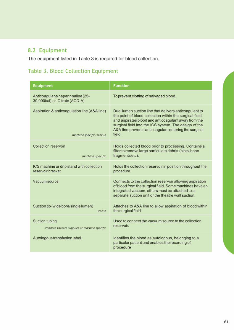
61
8.2 Equipment The equipment listed in Table 3 is required for blood collection. Table 3. Blood Collection Equipment
Equipment Function
Anticoagulant (heparin saline (25-30,000iu/l) or Citrate (ACD-A)
To prevent clotting of salvaged blood.
Aspiration & anticoagulation line (A&A line)
machine specific/sterile
Dual lumen suction line that delivers anticoagulant to the point of blood collection within the surgical field, and aspirates blood and anticoagulant away from the surgical field into the ICS system. The design of the A&A line prevents anticoagulant entering the surgical field.
Collection reservoir
machine specific
Holds collected blood prior to processing. Contains a filter to remove large particulate debris (clots, bone fragments etc).
ICS machine or drip stand with collection reservoir bracket
Holds the collection reservoir in position throughout the procedure.
Vacuum source Connects to the collection reservoir allowing aspiration of blood from the surgical field. Some machines have an integrated vacuum, others must be attached to a separate suction unit or the theatre wall suction.
Suction tip (wide bore/single lumen) sterile
Attaches to A&A line to allow aspiration of blood within the surgical field.
Suction tubing
standard theatre supplies or machine specific
Used to connect the vacuum source to the collection reservoir.
Autologous transfusion label Identifies the blood as autologous, belonging to a particular patient and enables the recording of procedure

62
8.3 Anticoagulant To prevent clotting, the aspiration and anticoagulation line (A&A line) delivers anticoagulant to the point of collection within the surgical field. Blood aspirated from the surgical field mixes with the anticoagulant as it enters the A&A line and is therefore anticoagulated before it enters the collection reservoir. If the rate of flow of the anticoagulant is insufficient, the salvaged blood will clot. This may result in contamination of the processed blood and/or may prevent processing. Types of anticoagulant used are:
• Heparin saline:
– 25-30,000iu heparin/1,000ml intravenous (IV) normal saline (0.9% NaCl)
– Heparin works by activating Antithrombin III which in turn inactivates both Factor Xa and Factor IIa (Thrombin) in the coagulation cascade (Figure 11). This prevents the conversion of Fibrinogen to Fibrin and the formation of clots.
– The recommended ratio is approximately 1:5 e.g. 20ml of anticoagulant to 100ml of blood (check your machine manufacturer recommendations)
Figure 11. Heparin Mechanism of Action
Heparin anticoagulant – will be ineffective if the patient suffers from Antithrombin III deficiency. It is recommended that a citrate anticoagulant is used for these patients.
Factor X Factor Xa
X Factor II Factor IIa
(Prothrombin) (Thrombin)
Inactive Antithrombin III
Active Antithrombin III Heparin
X Fibrinogen Fibrin

63
• Acid citrate dextrose anticoagulant (ACD-A):
– Citrate based anticoagulant
– Pre-prepared
– Citrate based anticoagulants work by binding to free calcium in the blood. Calcium is a required cofactor in the activation of clotting factors; the action of the citrate removes calcium from the coagulation cascade, therefore preventing clot formation by inhibiting the coagulation cascade.
– The recommended ratio is approximately 1:7 e.g. approximately 15ml of anticoagulant to 100ml of blood (check your machine manufacturer recommendations)
The typical (minimum) flow rate for anticoagulant is around 45-60 drops per minute for ACD-A and around 60-80 drops per minute for heparin saline. The anticoagulant flow rate may need to be increased during the procedure to accommodate increased levels of bleeding, this can then be returned to the minimum flow rate once bleeding is under control. Minor adjustments to the flow rate of the anticoagulant may be necessary throughout the duration of the procedure to accommodate varying levels of bleeding. 8.4 Preparation of Equipment for Blood Collection
Figure 12. “Collect Only” Set Up
Citrate anticoagulants – fluids containing calcium e.g. Hartmann’s, (if used for irrigation) may inhibit citrate based anticoagulants and should be avoided.

64
The set up of the blood collection equipment for ICS is represented in Figure 12. As discussed earlier in this section, the practical set up of the equipment for ICS is specific to the machine in use. However, the basic principles and theory are the same. The main steps in the preparation of the blood collection equipment are outlined below. Clean/aseptic technique should be used as appropriate and protective clothing should be worn in accordance with local policy.
Suction Tip– to minimise damage to the red blood cells (RBCs) being aspirated, a wide bore (minimum 4mm), single lumen suction tip e.g. Yankauer sucker, should be used.
A&A Line/Suction Tip • Pass aseptically to the scrubbed person within the sterile field.
• Ask them to attach a large bore/single port suction tip to the A&A line.
Anticoagulant • Aseptically add 25-30,000iu of heparin to 1,000ml of IV normal saline (0.9% NaCl) and label clearly with an appropriate “drugs added label” or select a bag of pre-prepared citrate anticoagulant. In both cases check the expiry date of the products before use.
• Hang the anticoagulant on the drip stand on the machine or the drip stand with the collection reservoir bracket on if the machine is not available.
Collection Reservoir/Autologous Transfusion Label
• Load the collection reservoir into the bracket on the machine or drip stand.
• If appropriate (see manufacturer’s instructions) clamp off the port that leads to the processing line.
• Enter the patient’s details (from the patient’s identification band) onto the autologous transfusion label (Appendix 3) and attach it to the collection reservoir.

65
Time limits for consumable set up – unprimed disposables can be used for up to 24 hours providing they have not been primed. Once primed, they should be used within 8 hours or discarded.
Labelling the collection reservoir – to avoid errors in patient identification, an autologous transfusion label should be completed at the patient’s side, at the start of blood collection. The patient’s details should be taken from the identification band attached to the patient. The label should be securely attached to the collection reservoir. If a processing set is subsequently loaded into the machine (see section 9), the autologous transfusion label should be transferred to the reinfusion bag immediately, or a new label completed and attached to the bag.

66
Vacuum Source/ Suction Tubing
• Attach the suction tubing to the vacuum source either on the machine or the theatre wall suction, making sure there is a secure connection.
• Attach the other end of the suction tubing to the appropriate port on the collection reservoir (see manufacturer’s guidelines).
Connect A&A Line • Prior to the start of the operation, ask the scrub practitioner to pass the spiked end of the A&A line out of the sterile field.
• Attach the wide bore line to the appropriate port on the collection reservoir (see manufacturer’s guidelines).
• Close the roller clamp on the small bore line and spike the line into the port on the anticoagulant bag.
Turn on Vacuum
• Turn on either the machine vacuum, (if available see manufacturer’s guidelines), or the wall vacuum source.
• For non-self regulating vacuums, set the vacuum to approximately –100mmHg to –150mmHg (follow your manufacturer’s guidance).
Vacuum Levels – High vacuum levels cause haemolysis (destruction of the RBCs). Maintaining low vacuum levels minimises haemolysis and maximises the red blood cells available for reinfusion.

67
8.5 Blood Collection During the blood collection phase, it may be necessary for the operator to make minor adjustments to the system:
• Regulating the vacuum – it may become necessary during periods of high blood loss to increase the level of the vacuum at the request of the surgical team. The vacuum should be returned to a standard level, (approximately –100mmHg to –150mmHg) as soon as the bleeding is under control. This will minimise damage to the RBCs.
• Regulating the anticoagulant flow – the flow rate of the anticoagulant must be regulated depending on the level of bleeding. Insufficient anticoagulant will result in the system clotting off.
• Monitoring the volume of blood loss – when using a “collect only” system, the cell salvage operator must decide if it is appropriate to process the blood based on the volume of blood collected (see Section 9).
Prime A&A Line/ Collection Reservoir
• Fully open the roller clamp on the A&A line.
• Allow approximately 100ml – 150ml of anticoagulant to run through the line to prime the collection reservoir.
Regulate Anticoagulant Flow
• Regulate the flow of anticoagulant to approximately one drop per second.
Begin Blood Collection • Inform the surgical team that blood collection can begin.
• Record the time blood first enters the collection reservoir.
Patients with religious requirements – the set up of ICS equipment for patients with religious requirement may differ. The requirements should be discussed with the patient prior to use, and all relevant staff should be made aware of these requirements. Further information can be found in Appendix 2.
IV Grade fluids – Remember, anything that is aspirated from the surgical field could potentially go back into the patient’s circulation. Only IV grade fluids should be aspirated into the ICS system. To avoid contaminating the ICS blood, the standard theatre suction should be used for aspirating when non-IV substances are being used within the surgical field. e.g. orthopaedic cement, betadine, antibiotics not licensed for IV use etc.

68
8.6 Maximising Blood Collection
There are several techniques that can be used to maximise the volume of RBCs available for reinfusion. These include:
• Low vacuum level – Maintaining a low vacuum level minimises haemolysis, and therefore maximises the RBCs available for reinfusion. High vacuum levels cause RBC haemolysis, which can be measured by the concentration of plasma (free) haemoglobin (haemoglobin that has been released from haemolysed RBCs).
• Suction technique – where possible, the suction tip should be immersed in the blood and not skimmed across the surface of tissues or pools of blood. Skimming results in a large quantity of air mixing with the aspirated blood, this air interface causes haemolysis and therefore reduces the number of viable RBCs available for reinfusion.
Graph 1 (below) shows plasma haemoglobin at different vacuum levels and using two types of suction technique1. The graph demonstrates that when blood only is aspirated (i.e. when the suction tip is immersed in a pool of blood), even high vacuum levels do not result in excessive RBC haemolysis. This supports increasing vacuum levels during excessive bleeding.
However, when blood and air are aspirated, as occurs naturally during most of the ICS process, even low vacuum levels result in excessive haemolysis and therefore reduces the available RBCs for reinfusion. Graph 1. Changes in Plasma Haemoglobin from Baseline Measurements
• Suction tip – as already mentioned, a wide bore, single lumen suction tip minimises damage to the RBCs during collection
• Swab washing – see next page

69
8.7 Swab Washing
Swab washing (Figure 13) allows blood that would normally be lost in swabs to be salvaged during ICS and can significantly increase the volume of RBCs for reinfusion.
• Equipment:
– Sterile bowl
– 1,000mls IV normal saline (0.9% NaCl)
Figure 13. Swab Washing
Swabs are placed in a bowl, within the sterile field, containing 1,000mls IV normal saline (0.9% NaCl). The swabs are left for approximately five minutes and are then gently (to avoid damaging the RBCs) squeezed out. The swabs are then disposed of as per department protocol. At the end of the procedure (or sooner if necessary) the swab wash is suctioned into the ICS reservoir and processing is
undertaken as normal (see section 9).
In high blood loss procedures, it may be appropriate to suction the swab wash into the ICS reservoir before the end of the procedure, to allow the blood to be processed and returned to the patient. Once the contents of the bowl have been aspirated into the collection reservoir, a further 1,000mls of IV normal saline (0.9% NaCl) should be added to the sterile bowl to allow swab washing to continue.
Ensure the swab wash bowl is maintained within the sterile field. Ensure no substances not intended for IV use enter the swab wash bowl e.g. Betadine soaked swabs.

70
8.8 Troubleshooting
As with any technical procedure, there is a potential for problems to arise during the process, e.g.
• Loss of suction:
– Switch off flow of anticoagulant until vacuum is restored
– Check the vacuum source
– Check the suction tubing is securely connected to the vacuum source and the collection reservoir
– Check the A&A line is securely connected to the collection reservoir
– Check the A&A line has not been clamped or otherwise obstructed
• Clotting in the collection reservoir:
– Check the anticoagulant is still flowing
– Increase the anticoagulant flow rate
– If excessive clotting has occurred it may be necessary to change the collection reservoir.
• Contamination with non-IV substances:
– Contamination of the salvaged blood with substances not intended for IV use should be discussed with the lead clinician taking responsibility for ICS in the procedure, (normally the lead anaesthetist, however, in some cases it may be the lead surgeon). A clinical decision on how to proceed should be made by this lead clinician. A list of potential contaminants and their associated problems can be found in the UK Cell Salvage Action Group document “Technical Factsheet 9 – Contraindications to ICS”2.
– The decision to use blood that is potentially contaminated with bacteria, amniotic fluid or malignant cells should be made by the clinicians caring for the patient, taking into account the latest evidence and consideration of the risks and benefits of proceeding for the individual patient.
8.9 Documentation The documentation required during the blood collection includes:
• Autologous transfusion label (Appendix 3)
• ICS data form (Appendix 4)

71
Key Points • The main equipment for blood collection includes an A&A line, a collection
reservoir and anticoagulant.
• The operator must maintain awareness throughout the procedure in order to prevent errors occurring.
• In order to maximise blood collection, a number of techniques can be used in conjunction with one another e.g. low vacuum levels, swab washing and suction technique.
References 1. Gregoretti, S. (1996) Suction-Induced Haemolysis at Various Vacuum Pressures:
Implications for Intraoperative Blood Salvage. Transfusion; 36:57-60
2. UK Cell Salvage Action GroupTechnical Factsheet 9 “Contraindications to ICS”. Better Blood Transfusion Toolkit www.transfusionguidelines.org.uk
Further Reading UK Cell Salvage Action Group Publications.
The following publications are available to download at: www.transfusionguidelines.org.uk • Policy for the provision of Intraoperative Cell Salvage
• Technical Factsheets
• Swab Washing
1 Anticoagulation
2 Blood Collection
3 Use of ICS in Jehovah’s Witness Patients Other
• American Association of Blood Banks – Standards for Perioperative Autologous Blood Collection and Administration 3rd Edition
• Manufacturer’s ICS Machine Specific Guidance

72
Self-Directed Learning
List the procedures in your department where ICS blood collection is routinely set up.
What type of anticoagulant is used in your department?
List the types of machine used in your department.

73
Section 9 Practicalities – Blood Processing
Aim • To introduce the basic theory and principles of processing blood during
Intraoperative Cell Salvage (ICS)
Learning Outcomes • To identify the steps taken in making the decision to process
• To list the equipment used for blood processing and describe the function of each component
• To describe the steps required in preparing for and commencing blood processing
• To describe the risks of overriding the automatic functions of the machine
• To identify the steps necessary to complete the blood processing phase Introduction While the practical set up of the equipment for the blood processing phase of ICS is specific to the machine in use, the basic theory and principles are the same for all machines. During the blood processing phase of ICS, blood that has been collected in the collection reservoir is processed by the ICS machine to separate the red blood cells (RBCs) from the waste products (plasma, clotting factors, platelets, anticoagulant etc). The RBCs are concentrated to produce a high haematocrit and washed with intravenous (IV) normal saline (0.9% NaCl). At the end of the processing phase the RBCs, now suspended in IV normal saline (0.9% NaCl), are pumped to a re-infusion bag. The types of processing systems (Fixed Volume Bowl, Continuous Rotary and Variable Volume Disk systems) were discussed in Section 6. All of the systems work on the principle of separating the dense RBCs from the less dense waste products using centrifugal forces.
Collection – Collection of blood can continue as outlined in Section 8 throughout the processing phase. The anticoagulant and vacuum should remain on at all times until the end of the procedure.

74
9.1 Decision to Process Blood If a “collect only” system has been used for blood collection, the ICS operator must make an informed decision to proceed to set up for processing the salvaged blood. The processing set usually comes packaged separately from the blood collection equipment (A&A line/collection reservoir). This reduces unnecessary waste and is more cost effective if there is insufficient blood loss to warrant processing. The decision to load the processing set can be based on a number of factors including:
• Adequate blood loss in the collection reservoir
• Anticipated adequate blood loss due to rapid bleeding during the procedure
• Patient factors e.g.
– Low haemoglobin (Hb)
– Anticipated post-operative benefit
Adequate Blood Loss
Adequate blood loss relates to two issues:
• The benefit of returning the blood to the patient e.g. will reinfusing 60mls of RBCs be of any benefit to a patient with a pre-operative Hb of 140g/l and low intraoperative blood loss?
• The minimum volume of blood necessary for processing in volume dependent systems (fixed volume bowl systems)
While there is no absolute way to determine if sufficient blood has been collected to warrant loading the processing kit, an experienced operator can make a judgement based on an estimate of the volume of blood in the collection reservoir. The collection reservoir will contain blood and anticoagulant as well as any irrigation fluids from the sterile field. This is illustrated in Figure 14 (opposite).
Emergency Procedures – for emergency procedures when blood loss is likely to be rapid, it may be considered appropriate to set up the collection and processing equipment before the procedure begins to ensure blood can be processed and returned to the patient without delay.
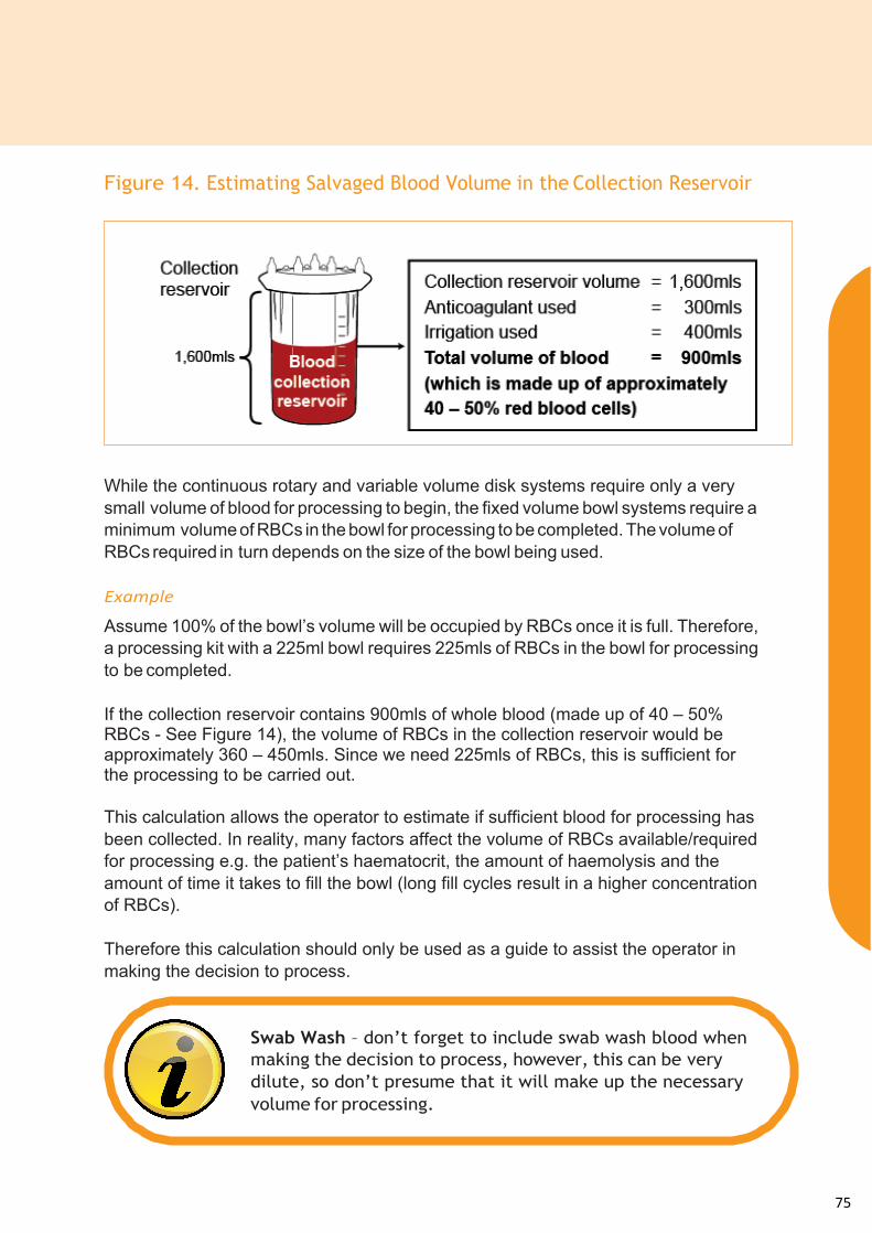
75
Figure 14. Estimating Salvaged Blood Volume in the Collection Reservoir
While the continuous rotary and variable volume disk systems require only a very small volume of blood for processing to begin, the fixed volume bowl systems require a minimum volume of RBCs in the bowl for processing to be completed. The volume of RBCs required in turn depends on the size of the bowl being used. Example
Assume 100% of the bowl’s volume will be occupied by RBCs once it is full. Therefore, a processing kit with a 225ml bowl requires 225mls of RBCs in the bowl for processing to be completed. If the collection reservoir contains 900mls of whole blood (made up of 40 – 50% RBCs - See Figure 14), the volume of RBCs in the collection reservoir would be approximately 360 – 450mls. Since we need 225mls of RBCs, this is sufficient for the processing to be carried out. This calculation allows the operator to estimate if sufficient blood for processing has been collected. In reality, many factors affect the volume of RBCs available/required for processing e.g. the patient’s haematocrit, the amount of haemolysis and the amount of time it takes to fill the bowl (long fill cycles result in a higher concentration of RBCs). Therefore this calculation should only be used as a guide to assist the operator in making the decision to process.
Swab Wash – don’t forget to include swab wash blood when making the decision to process, however, this can be very dilute, so don’t presume that it will make up the necessary volume for processing.

76
9.2 Equipment The equipment listed in Table 4 (below) is required for blood processing. Table 4. Blood Processing Equipment
9.3 Choice of Bowl Size The fixed volume bowl systems are generally available in a range of sizes that differ depending on the ICS machine being used. As outlined above, bowl systems require a minimum volume of RBCs in the bowl for processing to be completed, whereas the continuous rotary and the variable volume disk systems are one size and not dependent on volume. For fixed volume bowl systems, where there is a choice of bowl size, the appropriate size will depend on the anticipated blood loss. A small bowl requires a lower volume of RBCs for processing than a larger bowl.
Processing rates will also differ between manufacturers/ICS systems.
Equipment Function
ICS device Contains pump, sensors, centrifuge mechanism and control panel.
Processing set machine specific
Contains tubing, centrifuge system (bowl, disk etc), re-infusion bag and waste bag.
IV normal saline (0.9% NaCl) Used to wash the RBCs during processing.
Autologous transfusion label Identifies the blood as autologous, belonging to a particular patient and enables the recording of procedure specific details.
Where there is a choice of bowl size for a particular machine:
Smaller bowls – Will take longer to process a large volume of blood, as the blood is processed in small batches.
Larger bowls – Require a large volume of blood to “fill” them, if the bowl is not filled (i.e. if there is only a small volume of blood available), processing cannot be completed.

77
9.4 Preparation of Equipment for Blood Processing
Figure 15. Movement of Fluid Through the Cell Salvage Machine
The processing set includes the tubing that will carry the fluids through the machine. This tubing consists of:
• Collection reservoir line
• Wash solution line – Y-connector allowing two bags of IV normal saline (0.9% NaCl) to be connected
• Reinfusion bag line
The three lines join to a single line that carries fluid from the collection reservoir, and the wash solution (IV normal saline (0.9% NaCl)) into the centrifugal system, and the final processed RBCs away from it and into the reinfusion bag. The flow of fluids through these lines is controlled by a pump and a series of valves that open and close particular lines depending on the stage of the process.
In addition to these lines, there is a separate line that carries waste products from the centrifugal system into the waste bag.
Figure 15 (above) outlines the flow of fluids through the cell salvage machine.
The set up of the blood processing set for ICS is specific to the machine in use. However the basic principles and theory are the same. The key steps in the preparation of the blood processing equipment are outlined opposite. The order in which these are carried out should be completed according to the manufacturer’s training/guidance.
Clean/aseptic technique should be used as appropriate and protective clothing should be worn in accordance with local policy.

78
Centrifugal System • The centrifugal system should be loaded into the machine carefully according to the manufacturer’s guidelines. Damage to the centrifugal device could result in spillage.
Tubing Network • Ensure there are no kinks or twists in the tubing.
• Ensure the tubing is correctly placed through the pump, valves and machine sensors.
• Securely close any tubing retainers.
Waste Bag • Connect the waste bag to the waste line, (for some machines this is already done by the manufacturer) and hang on the machine according to the manufacturer’s guidelines.
• Ensure the waste line is not clamped.
• Check the outlet port on the bag is closed.
Reinfusion Bag • Hang the reinfusion bag on the machine dripstand.
• Connect the reinfusion line (for some machines this is already done by the manufacturer).
• Close the clamps on the reinfusion bag outlet/giving ports.
• Ensure the reinfusion bag line is not clamped.
• Transfer the autologous transfusion label (Appendix 3) from the collection reservoir (see Section 8) and securely attach to the reinfusion bag, or complete a new label and securely attach to the reinfusion bag.
Labelling – Section 8 outlines the importance of how and when the autologous label is completed. It is recommended that if a “collect only” system has been set up, the label is attached to the collection reservoir, and subsequently transferred to the reinfusion bag when the processing set is loaded.
If the entire system is set up from the start of the procedure, the autologous label should be completed at the patient’s side at the start of blood collection, and attached immediately to the reinfusion bag. To avoid errors in patient identification, the patient’s details should be taken from the identification band on the patient’s wrist.

79
IV Saline Wash Solution
• Hang one or two bags of IV normal saline (0.9% NaCl) onto the machine dripstand.
• Clamp both of the wash solution lines.
• Connect (spike) the wash solution line(s) to the IV normal saline (0.9% NaCl) bag(s) and open the clamp on one line.
Collection Reservoir Line
• Connect the collection reservoir line to the collection reservoir.
• If there is a clamp on the collection reservoir/collection reservoir line, ensure this is open.
Machine • Securely close all covers on the machine, ensuring none of the tubing is trapped.
• Follow the manufacturer’s guidance with regard to checking machine parameter (if applicable).
• Follow the manufacturer’s guidance with regard to initiating blood processing.
Patients with religious requirements – the set up of ICS equipment for patients with religious requirement may differ. The requirements should be discussed with the patient prior to use, and all relevant staff should be made aware of these requirements. Further information can be found in Appendix 2.
Automatic mode – Most machines are fully automatic. The responsibility of the operator is to start the processing cycle and then allow the machine to complete the cycle automatically. It is highly recommended that ICS machines are run in automatic mode. Running the machine in manual mode could result in residual contaminants in the RBCs, which could be potentially harmful to the patient.

80
9.5 Blood Processing During the blood processing phase, it may be necessary for the operator to make minor adjustments to the system (but not the processing functions of the machine):
• Replacing the wash solution – it is likely that the IV normal saline (0.9% NaCl) will need to be replaced regularly. The operator can anticipate this and change the bag, (while the machine is in standby) or wait for the machine alarm during processing (following the onscreen instructions to restart the process).
• Changing the reinfusion bag – most manufacturers can supply replacement reinfusion bags. The operator can replace the bag as necessary between processing cycles. The disconnected bag should be kept with the patient until it is reinfused, and the new bag should be labelled with an autologous label, which has been completed as outlined in Section 8.
• Emptying the waste bag – the waste bag can be emptied through a port on the bottom of the bag into a bucket. This can be disposed of as per local policy. The waste bag should never be fully emptied during the procedure, as the loss of air from the bag will prevent fluid movement through the machine.
Ensure protective clothing is worn in accordance with local policy. 9.6 Incomplete Bowls Machines that use the fixed volume bowl system require a minimum volume of RBCs to be reached in the bowl before the machine will wash them automatically and send them to the reinfusion bag.
Wash volumes – The minimum wash volume outlined by the manufacturers should be used. This will be set in the machine parameters and should not be changed. Reducing the wash volume may lead to residual contaminants in the RBCs, which could be potentially harmful to the patient.
In some circumstances it may be acceptable to increase the wash volume e.g. in orthopaedics or neurological procedures, where there are contaminants such as fat within the surgical field that could be potentially harmful. However, many machines have a high quality wash programme for this purpose, in which case the manufacturer’s guidance should be followed for selecting this programme.

81
Having processed all the blood from the collection reservoir and this minimum bowl volume is not reached, there are several things the operator can do: Wait for more blood loss – the machine will return to standby mode after a few minutes. The operator can then wait for more fluid in the collection reservoir, before starting processing in automatic mode again.
• Concentrate Function – This function should only be used at the end of the procedure when no more intraoperative blood loss is expected, and the swab wash has also been processed. The concentrate function uses RBCs that have already been processed and sent to the reinfusion bag to fill the bowl and complete the process as normal. This function can only be used if there are RBCs in the reinfusion bag.
If neither of the above options are possible, the operator should discuss how to proceed with the lead clinician taking responsibility for ICS in the procedure, outlining the potential risks of processing an incomplete bowl.
9.7 Completing the Process At the end of the procedure, when all of the blood has been processed, there are several things the operator may need to do. These functions may vary between machines; the manufacturer’s guidance for these functions should be followed.
• Emptying the reinfusion line – the line contains dead space which can hold a significant volume of processed RBCs. These can be transferred to the reinfusion bag for reinfusion to the patient.
• Removing air from the reinfusion bag – a large portion of the air in the reinfusion bag can be removed, however, it is likely that the bag will still contain some air, therefore the blood should not be reinfused under pressure (see Section 10).
• Disconnecting the reinfusion bag – the clamp on the reinfusion bag (from the reinfusion line) should be securely closed before the bag is disconnected (there may also be a clamp on the reinfusion line). Most manufacturers provide caps to attach to the disconnected ends of the line to prevent slight spillage, but in most cases these will not prevent spillage if the clamps are left open.
Incomplete bowls – Overriding the automatic function of the machine, and manually washing a partially filled bowl could result in residual contaminants in the RBCs, which could be potentially harmful to the patient. Although the bowl may look full to the eye, this can be misleading. The machine uses sensors to guide this process, these are far more accurate than the human eye.

82
9.8 Troubleshooting As with any technical procedure, there is a potential for problems to arise during the process e.g.
• Incomplete bowls – see 9.6.
• Machine alarms – if the machine detects a problem, it will stop processing and display information relating to the problem on the control screen. The operator should follow the on screen instructions to resolve the problem.
9.9 Blood Loss Calculations At the end of the procedure, when all of the blood from the collection reservoir has been processed, an estimate of the volume of blood the patient has lost during the procedure can be made using a simple calculation. The information you will need is:
• Fluid in volume (machine read out) – Total volume of fluid processed by the machine, includes: blood aspirated from the surgical field, anticoagulant and irrigation from the surgical field.
• Irrigation fluid – Volume of sterile irrigation fluid used within the surgical field and aspirated into the ICS collection reservoir, (this is not the volume of IV normal saline (0.9% NaCl) wash solution used by the machine – this volume is not required for the blood loss calculation).
• Anticoagulant used – An estimate of the volume of anticoagulant that has been used.
• Swab wash – Volume of IV normal saline (0.9% NaCl) or equivalent used to wash swabs.
• Theatre suction – Volume of blood in theatre suction.
• Wet-dry weight of swabs – Compensates for blood and saline swab wash retained on swabs and allows them to be weighed outside of the sterile field after washing.
Once you have all of this information, an estimate of blood loss can be calculated as demonstrated in Figure 16 (opposite).
Monitoring the system – the operator is responsible for the machine during the procedure. Although the machines are automatic and therefore, in most cases, do not need a dedicated operator, the operator should be working within the vicinity of the machine to allow them to monitor the system and respond to alarms. The operator should ensure that necessary procedures are carried out, e.g. emptying the waste bag. The operator should also monitor the collection equipment throughout the procedure (see Section 8).

83
Figure 16. Estimated Blood Loss Calculation
9.10 Documentation The documentation required during blood processing is the same as outlined in Section 8:
• Autologous transfusion label (Appendix 3)
• ICS data form (Appendix 4)
Key Points • The operator must be able to make an informed decision regarding proceeding to
process the blood.
• The blood processing set includes a centrifugal system, reinfusion bag, waste bag and tubing, which are all loaded into the ICS machine.
• The operator should follow the manufacturer’s guidance with regard to loading the processing equipment and running the processing phase of the procedure.
• The operator must maintain awareness throughout the procedure in order to prevent errors occurring.
Further Reading UK Cell Salvage Action Group Publications
The following publications are available to download at: www.transfusionguidelines.org.uk
• Policy for the provision of Intraoperative Cell Salvage.
• Technical Factsheets
6 – Use of ICS in Jehovah’s Witness Patients 9 – Contraindications
Other • American Association of Blood Banks – Standards for Perioperative Autologous
Blood Collection and Administration 3rd Edition
• Manufacturer’s ICS Machine Specific Guidance
Calculation
-‐ -‐ -‐ + + = Example
-‐ -‐ -‐ + + =
Fluid in Irrigation fluid
Anticoagulant used
Swab wash
Theatre suction
Wet – Dry weight of
swabs
Approx. blood loss
2,500ml 200ml 400ml 1,000ml 150ml 200ml 1,250ml

84
Self-Directed Learning
If a bowl system is used within your department, what size bowls are available?
Are there guidelines in your department that describe for which procedures “Collect Only” equipment should be set up and for which procedures the full kit (collection and processing) should be set up? If so, for what/when would you set up the entire kit before the procedure starts?

85
Section 10 Practicalities – Reinfusion
Aim • To introduce the basic theory and principles of reinfusing Intraoperative Cell
Salvage (ICS) blood Learning Outcomes • To identify the equipment used for reinfusion and describe the function of each
component
• To describe the composition of the final product for reinfusion
• To list the steps and describe the process of preparing for and commencing reinfusion
• To identify the conditions for reinfusion Introduction Once the blood collected using ICS has been processed, the next step is reinfusion of the final product to the patient. Many of the principles of reinfusing ICS blood are similar, if not the same, as the principles of transfusing allogeneic (donor) blood. 10.1 ICS (Intraoperative Cell Salvage) End Product On completion of processing, the ICS machine sends the final product to the reinfusion bag. The final product consists of:
• Red blood cells (RBCs)
• Intravenous (IV) normal saline (0.9% NaCl) In addition to these, the final product may contain trace amounts of the following:
• Platelets
• Clotting factors
• Anticoagulant
• Microaggregates
• Other cells/proteins aspirated into the system from the surgical field
• Contaminants – if these have been aspirated into the system from the surgical field e.g. betadine
These are present in such small quantities that they are unlikely to cause any adverse effects. However, appropriate precautions should be taken e.g. using an appropriate filter for reinfusion (see 10.4). Information on the clearance rates of different ICS machine can be found on the Machine Specification document on the Better Blood Transfusion Toolkit website www.transfusionguidelines.org.uk.

86
If contamination of the salvaged blood by substances not intended for IV use has occurred, this should be discussed with the lead clinician taking responsibility for ICS in the procedure, (normally the lead anaesthetist, however, in some cases it may be the lead surgeon). A clinical decision regarding reinfusion should be made by this lead clinician.
Figure 17. Haematocrit
The haematocrit of the final product can vary, however, providing the manufacturer’s guidelines have been followed and the machine has been run in automatic mode, the haematocrit is likely to be around 50-70% (Figure 17).
Coagulopathy – ICS blood contains almost no platelets of coagulation factors. Therefore, in cases of massive haemorrhage it is likely that the patient will require allogeneic (donor) blood components, e.g. platelets, fresh frozen plasma, cryoprecipitate and possibly even allogeneic (donor) RBCs.
The decision to use blood that is potentially contaminated with bacteria, amniotic fluid or malignant cells should be made by the clinicians caring for the patient, taking into account the latest evidence and considering the risks and benefits for the individual patient.

87
10.2 Authorising ICS Blood The reinfusion of ICS blood should be authorised by the responsible clinician on the documentation approved within your hospital.
10.3 Equipment The equipment listed in Table 5 is required for the reinfusion of ICS blood. Table 5. Blood Reinfusion Equipment
10.4 Filters There are a number of filters (Table 6 – on following page) available which can be used for ICS blood reinfusion. The type of filter used should comply with local policy as well as national and manufacturer’s guidelines. In most cases, a standard 200µm blood administration set is sufficient. The use of other filters in addition to blood administration set may be advocated in the following specialities:
• Obstetrics, Malignancy and contaminated fields – the use of Leukoguard® RS filter (Haemonetics Ltd), a leucodepletion filter, is advocated in Obstetrics & Malignancy and has been evaluated contaminated fields (ref). The flow rate is slow and the maximum capacity per filter is around 450ml. This filter however is the only one that has been shown to effectively remove contaminants specific to these settings.
Procedural problems – The responsible clinician should be made aware of any problems that have occurred during the process e.g. contamination of the collection reservoir with non-IV substances, so that the decision to reinfuse under these circumstances can be made based on the relative risks and benefits.
Equipment Function
Reinfusion bag machine specific
The RBCs are sent to the reinfusion bag at the end of processing.
Standard Blood Administration Set Used to connect the reinfusion bag/filter to the patient’s IV access.
Appropriate additional filter
(if clinically indicated – see 10.4)
Filters the RBCs as they are being reinfused.

88
• Orthopaedic surgery – there is a theoretical concern that fat globules released from bone marrow could result in fat embolism syndrome if reinfused. However, there is currently no evidence to support this. As a precaution, the use of a lipid depleting microaggregate filter is recommended as best practice in operative procedures where there is a high risk of fat embolism. The risk of fat embolism can also be decreased by leaving the last few millimeters of ICS blood in the reinfusion bag.
Table 6. Filters for ICS Blood Reinfusion
Regardless of the type of filter or giving set used for reinfusion of ICS blood, the instructions supplied with the filter/giving set should be followed e.g. if the maximum volume capacity of the filter has been reached and there is blood remaining in the reinfusion bag, the filter should be disconnected and a new filter connected and primed. Further information on the use of filters can be found in the Technical Factsheets available to download at www.transfusionguidelines.org.uk.
Use of leucodepletion filters and hypotension – there have been reports of patients becoming hypotensive when receiving ICS blood through leucodepletion filters. While this appears to be a rare side effect, all such incidents should be reported to the Serious Hazards of Transfusion (SHOT) Scheme.
Type of filter Medium Removes
Standard blood administration set
170-200µm screen Blood component and non-blood component particulate matter.
Microaggregate blood filter
40µm screen Blood component microaggregates and non-blood component particulate matter.
Lipid depleting microaggregate filter
40µm screen Microaggregates, lipids, C3a, some leucocytes.
Leucodepletion filter Affinity filter Leucocytes, lipids, microaggregates, some bacteria

89
10.5 Reinfusion “Storage”
ICS blood is untested and intended only for the patient from whom it was collected. Labelling of the reinfusion bag (and collection reservoir if a “collect only” system has been used) is essential and should be carried out near to the patient (to avoid errors) as early on in the procedure as possible (see Sections 8 and 9). In accordance with recommendations from Serious Hazards of Transfusion (SHOT) for allogeneic (donor) blood, (see Section 5), the following guidelines for the “storage” of ICS blood should be followed:
• The reinfusion bag should always be kept beside the patient at all times
• The reinfusion bag must not be placed in a refrigerator
Time Limits
The collection, processing and reinfusion of salvaged blood should be completed within the timeframes recommended by the manufacturer. This should be in accordance with guidance from the American Association of Blood Banks (AABB) and the organisation’s transfusion policy.
The AABB Guidelines recommend the reinfusion times for cell salvaged blood as follows:
• Intraoperative Cell Salvage:
– four hours from the completion of processing
• Postoperative Cell Salvage:
– six hours from the start of collection (applicable when Intra-‐operative Cell Salvage machines are used to salvage blood postoperatively)
Any blood that has not been transfused within the timeframe specified in the guidelines should be disposed of in accordance with local policy for dealing with liquid biohazardous waste. The expiry time of the ICS blood should be clearly recorded on the autologous transfusion label (Appendix 3).

90
Disconnecting the Reinfusion Bag
Reinfusion of ICS blood can occur either while the reinfusion bag is still attached to the processing set or once the reinfusion bag has been disconnected.
• Attached – When the reinfusion bag contains ICS blood, the appropriate filter/giving set should be attached to the giving port on the reinfusion bag, primed with the ICS blood and then connected to the patient’s IV cannula. This can be done while the reinfusion bag is still attached to the processing set. The same reinfusion bag may fill and empty many times during an operation.
• Disconnected – When all lines are securely clamped, the reinfusion bag is
disconnected from the processing set. An appropriate filter/giving set is subsequently attached to the giving port on the reinfusion bag, primed and attached to the patient’s IV cannula. This is normally carried out when the reinfusion bag is full or at the end of the procedure. If there is more blood in the collection reservoir to process, a replacement reinfusion bag is attached to the processing set.
Reinfusion line – The reinfusion line from the processing set should remain open for this set up. Clamping the line will prevent the transfer of further processed RBCs to the reinfusion bag, and could also cause a build up of pressure in the reinfusion line. This could result in spillage from the connector on the reinfusion line/bag or centrifugal system.
Disconnect in standby – The reinfusion bag should not be disconnected while the machine is processing. The operator should wait until the machine returns to standby, or should pause the process (if applicable on the machine in use) if the reinfusion bag is too full to allow the blood being processed to be transferred to it.

91
10.6 Administration of ICS Blood Clean/aseptic technique should be used as appropriate and protective clothing should be worn in accordance with local policy.
Pre-transfusion Checks
• Reinfusion of the salvaged blood should follow standard blood transfusion practice. The responsible clinician should authorise salvaged blood for reinfusion in the same manner as for allogeneic blood.
• Baseline observations should be recorded in the patient’s clinical record prior to commencing the reinfusion of ICS blood. This is usually carried out by the anaesthetist as part of the anaesthetic record that is routinely completed in theatre.
• The patient details (full name, date of birth and unique identification number) on the autologous label attached to the reinfusion bag should always be carefully checked against the details on the identification band attached to the patient prior to commencing reinfusion of the ICS blood. If the identification band is inaccessible during surgery, due to surgical drapes, patient identification should be undertaken as per local protocol for these circumstances.
• The expiry time on the autologous transfusion label attached to the reinfusion bag should be checked prior to commencing reinfusion of ICS blood. Expired blood should be disposed of according to hospital policy.
• Check the reinfusion bag for any signs of leakage, clots or abnormal colour.
92
Administration of ICS Blood
• A giving set/filter, appropriate to the type of surgery, should be used for reinfusion (see 10.4).
• The rate at which the red cells are reinfused can be adjusted using a clamp on the administration set and by adjusting the height of the reinfusion bag.
• Observations should be carried out and recorded in the patient’s clinical record at least 15 minutes from the start of reinfusion and on completion of the reinfusion.
Documentation • Reinfusion of salvaged blood should be documented in the appropriate section of the patient’s clinical record as specified in the organisation’s transfusion policy. The autologous transfusion label contains a peel out section, which should be completed at the time of reinfusion and can be used for this purpose.
Pressure Cuffs – Manufacturers advise NOT to use a pressure cuff as there is a risk of air embolus from the air in the reinfusion bag. Some devices may also detect a back pressure if the reinfusion line is open.
Patients with religious requirements – the set up of ICS equipment for patients with religious requirement may differ. The requirements should be discussed with the patient prior to use and all relevant staff should be made aware of these requirements. Further information can be found in Appendix 2.

93
10.7 Transfusion Reactions If a transfusion reaction is suspected, STOP the transfusion and seek immediate advice from the lead surgeon and/or anaesthetist. Complete an adverse event form and report the incident to the individual specified in the organisation’s transfusion/ICS Policy.
10.8 Documentation The documentation required during blood reinfusion is the same as outlined in Sections 8 and 9:
• Autologous transfusion label (Appendix 3)
• ICS data form (Appendix 4)
• Prescription (as per local policy)
• The volume of blood reinfused should be documented in the patient’s clinical record in accordance with local policy
Key Points • ICS blood for reinfusion consists mainly of RBCs suspended in IV normal saline
(0.9% NaCl). Other components, such as platelets, may be present in extremely small quantities. The reinfusion of ICS blood should be prescribed by the responsible clinician and should follow local policy and national guidelines.
• Care should be taken to:
– identify the correct patient
– ensure the ICS blood is suitable for reinfusion (i.e. not expired or damaged)
– select the correct giving set/filter to use
– record the procedure accurately on the documentation approved by the organisation

94
References
1. Waters JH, Tuohy MJ, Hobson DF, Procop G (2003). Bacterial reduction by cell salvage washing and leukocyte depletion filtration. Anesthesiology;99:652–5
Further Reading
UK Cell Salvage Action Group Publications The following publications are available to download at: www.transfusionguidelines.org.uk
• Policy for the provision of Intraoperative Cell Salvage
• Technical Factsheets
4 Reinfusion of Red Cells 5 Administration of Reinfused Red Cells 6 Use of ICS in Jehovah’s Witness Patients 7 Use of Filters 8 Use in Obstetrics 9 Contraindications Other
• American Association of Blood Banks – Standards for Perioperative Autologous Blood Collection and Administration 3rd Edition
• Manufacturers’ ICS Machine Specific Guidance
• National Institute for Health & Clinical Excellence (NICE) (2008) Intraoperative red blood cell salvage during radical prostatectomy or radical cystectomy – Guidance http://www.nice.org.uk/nicemedia/pdf/IPG258Guidance.pdf
• National Institute for Health & Clinical Excellence (NICE) (2005) Intraoperative Blood Cell Salvage in Obstetrics – Guidance http://www.nice.org.uk/nicemedia/pdf/ip/IPG144guidance.pdf

95
Self-Directed Learning
List the procedures (if any) in your department for which a leucodepletion filter is used to reinfuse ICS blood.
Where are leucodepletion filters stored in your hospital?
On what documentation within the patient’s clinical record is transfusion recorded within your organisation?

96
Section 11 Unloading and Discarding Aim • To introduce the basic principles of unloading an Intraoperative Cell Salvage
(ICS) machine and discarding ICS disposables
Learning Outcomes • Identify when unloading of ICS machine and disposables is appropriate
• Determine the risks associated with the unloading phase
• Describe the appropriate procedure for safely discarding waste products and disposables that is compliant with your hospital policy
Introduction At the end of an operation, or when it appears that no more blood will be collected,
it is important to communicate with the surgeon and anaesthetist to ascertain that ICS will no longer be required before unloading the ICS machine. When it has been established that there will be no more blood collected:
• Ensure that any blood collected up to that point that is intended for washing and reinfusion, is processed.
• Ensure that salvaged red blood cells (RBCs) have been reinfused to the patient, or that the reinfusion bag is detached from the processing set (see Section 10).
The procedure for unloading disposables is specific to each type of ICS machine and will differ depending on whether you have set up for “collect only” or “full processing”.
Therefore, the manufacturer’s machine specific guidelines on unloading should be followed. The risks associated with this stage are similar irrespective of technical differences in unloading procedures.
Waste products of ICS include; IV normal saline (0.9% NaCl), anticoagulant, and non-RBC components of blood, in addition to contaminated consumables. There is always a risk that blood may be infected. Your hospital will have a procedure for disposal of biological waste/biologically contaminated material.
Standard precautions when handling bodily fluids should be used as per your hospital’s Health and Safety Policy.
97
Below is a list of generic steps involved in the unloading phase of ICS:
• Establish that the operation is over or that no more blood will be collected
• Ensure that all blood intended for processing is processed
• Ensure that all salvaged red cells are reinfused or that the reinfusion bag is detached from the processing set
• Complete the data collection sheet for ICS audit
• Refer to manufacturer’s guidance for unloading (there may be an unload function)
• Switch off the power supply
• Dispose of the waste bag/waste bag contents according to local policy
• Close off all clamps and seal off any open ports and ensure that any open spikes are covered or removed
• Remove processing set from device and dispose of as clinical waste
• Wipe down the device and remove blood spillages in line with your local policy and the manufacturer’s machine specific guidance
Further Reading • Organisation’s Policies for Health and Safety and Dealing with Biohazardous
Material (local)
• Manufacturer’s ICS Machine Specific Guidance
The specific technique for removing waste products from the disposable, or alternatively, for securing waste products within the disposable, should be discussed at the practical session and should be compliant with local hospital policy for disposal of contaminated biological waste.

98
Self Directed Learning
What protective clothing/equipment should you wear when performing the takedown stage of ICS in your hospital?
Where can you find your hospital policy for disposing of contaminated waste?
99
According to your local policy for disposing of contaminated waste, which of the following would be the correct procedure for disposing of ICS waste fluids in your hospital?
(Circle one or write a description)
a. Cut open the waste bag and insert solidifying gel/powder and put in clinical waste bag.
b. Aspirate waste bag contents into suction liners and discard as clinical waste.
c. Aspirate waste bag contents into suction liners and insert solidifying gel/ powder and discard as clinical waste.
d. Empty waste bag contents into a bucket and pour down waste pipe in the sluice.
e. Seal the consumable set, ensure there are no leaks and place in rigid yellow biohazard containers.
f. Seal the consumable set, ensure there are no leaks and double bag using clinical waste bags.
Other, please describe:
In your local policy for dealing with blood spillages, what cleaning fluid should you use to clean blood spillages on the ICS machine?

100
Appendix 1
Link to Intraoperative Cell Salvage Competency Assessments The ICS Competency Assessments are available to download from www.transfusionguidelines.org.uk
The following table lists the theory competencies relevant to each section of this workbook.
Section
Related Competencies
Preparation of Equipment for
Intraoperative Cell Salvage (ICS)
Operate Intraoperative Cell Salvage (ICS)
Equipment and Reinfuse
1. Using the Education Workbook
2. Training Pathway 1
3. Basic Blood Facts 2, 6
4. Blood Conservation 1
5. Haemovigilance 1
6. Principles of Intraoperative Cell Salvage
5
8, 9
7. Indications and Contraindications
2, 3, 8
4, 5, 12, 16
8. Practicalities – Blood Collection
2, 3, 4, 5, 6, 7, 8, 9, 10, 11, 12, 13, 14
2, 3, 7, 10, 14, 15, 16
9. Practicalities – Blood Processing
4, 5, 6, 7, 8, 10, 13, 14,
4, 5, 8, 9, 10, 11, 13, 15, 16
10. Practicalities – Blood Reinfusion
1, 3, 5, 6, 7, 10, 13, 14
6, 11, 12, 14, 15, 16
11. Unloading and Discarding
1, 4, 6, 14
11, 17

101
Appendix 2
Cell Salvage in Jehovah’s Witness patients
The information in this Appendix has been adapted from the UK Cell Salvage Action Group Technical Factsheet (6) which is available to download at www.transfusionguidelines.org.uk
UK Intraoperative Cell Salvage Action Group Technical Factsheet 6 Cell Salvage in Jehovah’s Witness patients Area of application Jehovah’s Witnesses (JW) regard blood as sacred. On the basis of this deeply held core value, they decline treatment with allogeneic (donor) blood (red cells, white cells, platelets, and plasma). This is usually documented on an Advance Medical Decision that they carry on their person. JW patients make a personal decision on whether or not to accept the various blood conservation measures available. These include intraoperative and postoperative cell salvage. Ideally, this should be discussed and recorded on a specific document, detailing exactly what is and is not acceptable to the patient. JW patients who accept cell salvage may specifically request that the system be set up to allow for continuous connectivity. In these cases, the details outlined below should prove helpful. Informed consent should be sought as for all patients. Staff The patient’s surgical team and all staff involved in the cell salvage processing.
Procedure Setting up a continuous circuit
Although there will be technical differences between devices, the same general principles apply.
1. Set up the machine for collection and processing with standard disposables (in bowl based machines consider using a low volume bowl to reduce blood
stasis).
2. Prime the circuit with saline ensuring that saline enters the reinfusion bag (remember to account for this volume when recording the final reinfusion volume).
3. Attach an appropriate blood administration set to the reinfusion bag. Prime the administration set and connect to the patient via a cannula for reinfusion. Once established, the connection between the patient and the reinfusion bag must not be broken. (Figure 18).
4. Whilst surgery is ongoing, administer the saline at the slowest rate possible to maintain patency of the cannula until processed blood is available.

102
Figure 18. Representation of a Continuous Circuit
Special requirements In some cases a leucocyte depletion filter may be needed for reinfusion of the salvaged blood. A standard giving set should be set up with a 3-way tap in line before blood collection begins. The giving set should be primed with saline to complete the circuit. When a volume of blood is ready to be reinfused, the leucocyte depletion filter can be spiked into the second reinfusion port on the reinfusion bag and primed. This is then attached to the 3-way tap, without breaking the circuit. Likewise, because the filters have a maximum throughput of 450mls, a new filter can be added if necessary by replacing the original giving set while leaving the original filter connected. (Figure 19 opposite). The LD filter should not be flushed with saline after filtration of the salvaged blood
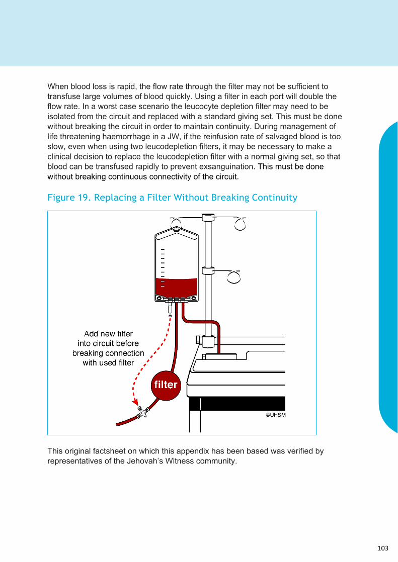
103
When blood loss is rapid, the flow rate through the filter may not be sufficient to transfuse large volumes of blood quickly. Using a filter in each port will double the flow rate. In a worst case scenario the leucocyte depletion filter may need to be isolated from the circuit and replaced with a standard giving set. This must be done without breaking the circuit in order to maintain continuity. During management of life threatening haemorrhage in a JW, if the reinfusion rate of salvaged blood is too slow, even when using two leucodepletion filters, it may be necessary to make a clinical decision to replace the leucodepletion filter with a normal giving set, so that blood can be transfused rapidly to prevent exsanguination. This must be done without breaking continuous connectivity of the circuit. Figure 19. Replacing a Filter Without Breaking Continuity
This original factsheet on which this appendix has been based was verified by representatives of the Jehovah’s Witness community.

104
Appendix 3
Autologous Transfusion Label The autologous transfusion label (Figure 20) has been developed by the UK Cell Salvage Action Group to standardise labeling for autologous blood. The labels are supplied by the manufacturers of the Intraoperative Cell Salvage equipment. Figure 20. Autologous Transfusion Label
Front of Autologous
transfusion label
Back of Autologous
transfusion label

105
Appendix 4
Intraoperative Cell Salvage Data Collection Form The following (Figure 21) is an example of a data collection form used for Intraoperative Cell Salvage.
Figure 21. Example of an Intraoperative Cell Salvage Data Collection Form

106
This training resource has been funded by NHS Blood and Transplant, a Special Health Authority within the NHS (England).
This resource can be reproduced in any form for education purposes without prior consent.
However, for all other purposes, consent from the UK Cell Salvage Action Group is required before use.

107
UK CELL SALVAGE ACTION GROUP
EDUCATION WORKBOOK