intraocular lenses information booklet - lasik md · hyperopia have some degree of astigmatism. in...
TRANSCRIPT

Intraocular LensesInformation
Booklet
lasikmd.com
MLIB-IOL-EN-3 1-866-366-2020 Page 1
The information contained herein is subject to change without prior notice. Please refer to our Patient Care Centre for further information.
TABLE OF CONTENTS
WELCOME TO LASIK MD ............................................................................................................................................... 3
About LASIK MD ......................................................................................................................................................... 3
Eye care professionals .............................................................................................................................................. 3
HOW THE EYE WORKS ................................................................................................................................................... 4
COMMON EYE AND VISION CONDITIONS .............................................................................................................. 5
Myopia (Nearsightedness) ...................................................................................................................................... 5
Hyperopia (Farsightedness) ................................................................................................................................... 5
Astigmatism ................................................................................................................................................................. 5
ABOUT CATARACTS ....................................................................................................................................................... 6
How do cataracts develop? .................................................................................................................................... 6
Are there other types of cataracts? ...................................................................................................................... 7
Who is at risk for developing cataracts? ............................................................................................................. 7
What are the symptoms of a cataract? ................................................................................................................ 7
How are cataracts detected? .................................................................................................................................. 8
OUR PROCEDURES ......................................................................................................................................................... 9
Cataract surgery (CAT) and refractive cataract surgery (RCS)* ................................................................... 9
Refractive lens exchange (RLE) .............................................................................................................................. 9
Phakic intraocular lens implantation (PIOL) ................................................................................................... 10
WHO IS ELIGIBLE FOR A PROCEDURE? ................................................................................................................... 11
WHAT HAPPENS DURING THE PROCEDURE? ...................................................................................................... 12
Cataract surgery (CAT, RCS) and refractive lens exchange (RLE) ............................................................. 12
Phakic intraocular lens implantation (PIOL) ................................................................................................... 13
OUR TREATMENTS ........................................................................................................................................................ 14
Artificial monofocal intraocular lenses ............................................................................................................. 14
Monovision with monofocal artificial intraocular lenses ........................................................................... 14
Lens PresbyVisionTM ................................................................................................................................................ 15
Phakic intraocular lenses ....................................................................................................................................... 16
LIMITS TO CORRECTION ............................................................................................................................................. 17
Amblyopia .................................................................................................................................................................. 17
Strabismus .................................................................................................................................................................. 17
lasikmd.com
MLIB-IOL-EN-3 1-866-366-2020 Page 2
The information contained herein is subject to change without prior notice. Please refer to our Patient Care Centre for further information.
Presbyopia .................................................................................................................................................................. 17
HOW WILL SURGERY IMPROVE MY LIFE? .............................................................................................................. 18
Reduced dependence on glasses and contacts ............................................................................................ 18
POTENTIAL COMPLICATIONS ................................................................................................................................... 19
THE PATIENT CARE PROCESS AND PROCEDURE ................................................................................................ 24
Step 1—Preparing for your pre-operative assessment .............................................................................. 24
Step 2—Day of surgery .......................................................................................................................................... 25
Step 3—Post-operative care ................................................................................................................................ 27
FINANCIAL RESPONSIBILITY ...................................................................................................................................... 28
INFORMED CONSENT .................................................................................................................................................. 28
Steps of the consent process ............................................................................................................................... 28
OUR COMMITMENT TO YOUR VISION .................................................................................................................... 29
lasikmd.com
MLIB-IOL-EN-3 1-866-366-2020 Page 3
The information contained herein is subject to change without prior notice. Please refer to our Patient Care Centre for further information.
WELCOME TO LASIK MD Thank you for expressing interest in LASIK MD. We know that the decision to undergo vision correction is an important one—and we want to make sure that you have all the facts before taking the leap. Like most of our patients, you may be feeling excited about reducing the need for wearing glasses or contacts, but you probably have questions about the procedure beforehand. In this booklet, you will find answers to some of the questions you may have about this life-changing procedure, along with information detailing the benefits, potential complications and the steps you will take along your path to clear vision. About LASIK MD LASIK MD provides high-quality vision correction and personalized care. Our mission is to help our patients live life to the fullest, using the highest surgical standards and the latest technology at an affordable price.
Eye care professionals At LASIK MD, our eye care professionals are experienced in the pre-operative, operative and post-operative management of vision correction procedures. Throughout your LASIK MD experience, you will interact with our highly trained staff, including our experienced optometrists and surgeons.
Optometrist Your optometrist has completed four years of optometry school, has obtained a doctor of optometry (OD) degree, is trained in diagnosing and treating refractive errors by non-surgical means, and has experience in providing post-operative care following vision correction procedures. Your optometrist, who will collaborate closely with your surgeon to ensure the best possible surgical result, may be able to assume responsibility for your care as early as the day following surgery.
Surgeon Your surgeon has a doctor of medicine (MD) degree and is experienced in the medical and surgical management of refractive errors and eye diseases. In addition to four years of university, the surgeon has spent four years in medical school, followed by a five-year residency in ophthalmology.
lasikmd.com
MLIB-IOL-EN-3 1-866-366-2020 Page 4
The information contained herein is subject to change without prior notice. Please refer to our Patient Care Centre for further information.
LASIK MD surgeons have performed over one million vision correction procedures (including 425,000 in Quebec alone), offering LASIK (laser-assisted in-situ keratomileusis), PRK (photorefractive keratectomy), refractive lens exchange (RLE), cataract surgery (CAT and RCS), and phakic intraocular lens implantation (PIOL). RLE, CAT, RCS and PIOL will be referred to, collectively, as the “procedure” in the following materials, and will be briefly described in the following sections. Please read all of the material in this package carefully. Remember that we provide this package in addition to, but not as a replacement for, direct discussions with your eye care professional. You may also find it helpful to consult our website, at lasikmd.com or to contact one of our consultants at 1-866-366-2020 if you have any other questions. Also, please keep in mind that our patient care representatives are not trained to give a medical diagnosis or to determine the specific price of a surgery. Pricing will vary depending on your prescription and the condition of your eyes. Specific pricing will be given to you following a series of tests at your pre-operative consultation.
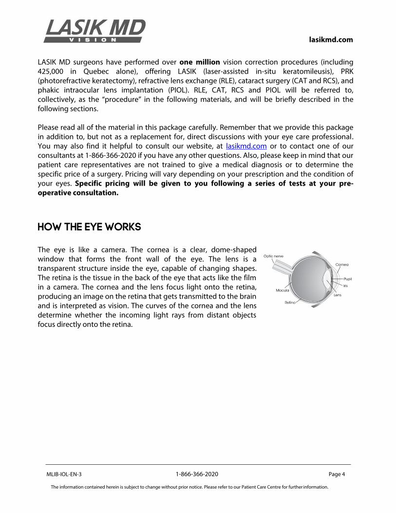
HOW THE EYE WORKS The eye is like a camera. The cornea is a clear, dome-shaped window that forms the front wall of the eye. The lens is a transparent structure inside the eye, capable of changing shapes. The retina is the tissue in the back of the eye that acts like the film in a camera. The cornea and the lens focus light onto the retina, producing an image on the retina that gets transmitted to the brain and is interpreted as vision. The curves of the cornea and the lens determine whether the incoming light rays from distant objects focus directly onto the retina.
lasikmd.com
MLIB-IOL-EN-3 1-866-366-2020 Page 5
The information contained herein is subject to change without prior notice. Please refer to our Patient Care Centre for further information.
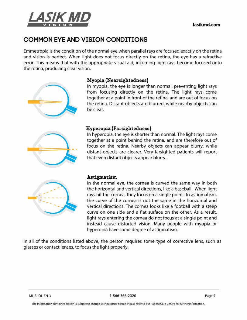
COMMON EYE AND VISION CONDITIONS
Emmetropia is the condition of the normal eye when parallel rays are focused exactly on the retina and vision is perfect. When light does not focus directly on the retina, the eye has a refractive error. This means that with the appropriate visual aid, incoming light rays become focused onto the retina, producing clear vision.
Myopia (Nearsightedness) In myopia, the eye is longer than normal, preventing light rays from focusing directly on the retina. The light rays come together at a point in front of the retina, and are out of focus on the retina. Distant objects are blurred, while nearby objects can be clear.
Hyperopia (Farsightedness) In hyperopia, the eye is shorter than normal. The light rays come together at a point behind the retina, and are therefore out of focus on the retina. Nearby objects can appear blurry, while distant objects are clearer. Very farsighted patients will report that even distant objects appear blurry.
Astigmatism
In the normal eye, the cornea is curved the same way in both the horizontal and vertical directions, like a baseball. When light rays hit the cornea, they focus on a single point. In astigmatism, the curve of the cornea is not the same in the horizontal and vertical directions. The cornea looks like a football with a steep curve on one side and a flat surface on the other. As a result, light rays entering the cornea do not focus at a single point and instead cause distorted vision. Many people with myopia or hyperopia have some degree of astigmatism.
In all of the conditions listed above, the person requires some type of corrective lens, such as glasses or contact lenses, to focus the light properly.
lasikmd.com
MLIB-IOL-EN-3 1-866-366-2020 Page 6
The information contained herein is subject to change without prior notice. Please refer to our Patient Care Centre for further information.
ABOUT CATARACTS Most cataracts are age-related. Cataracts are very common in older people. By age 80, more than half of all North Americans either have a cataract or have had cataract surgery. A cataract can appear in just one or both eyes. It cannot spread from one eye to the other. How do cataracts develop?
Age-related cataracts develop in two ways:
1. Clumps of protein reduce the sharpness of the image reaching the retina.
The lens consists mostly of water and protein. When the protein clumps up, it clouds the lens and reduces the light that reaches the retina. The clouding may become severe enough to cause blurred vision. Most age-related cataracts develop from protein clusters. When a cataract is small, the cloudiness affects only a small part of the lens. You may not notice any changes in your vision. Cataracts tend to "grow" slowly, so vision gradually worsens. Over time, the cloudy area in the lens may grow and the cataract may increase in size, thus making seeing clearly more difficult. Your vision may get duller or blurrier.
2. The clear lens slowly changes to a yellowish/brownish colour, adding a brownish tint to vision.
As the clear lens slowly colours with age, your vision gradually may acquire a brownish shade. At first, the amount of tinting may be minimal, and won’t necessarily cause a vision-related problem. Over time, increased tinting may make it more difficult to read and perform other routine activities. This gradual change in the amount of tinting does not affect the sharpness of the image transmitted to the retina.
If you have advanced lens discolouration, you may not be able to identify blues and purples. For example, you may be wearing what you believe to be a pair of black socks, only to find out from friends that you are wearing purple socks.

lasikmd.com
MLIB-IOL-EN-3 1-866-366-2020 Page 7
The information contained herein is subject to change without prior notice. Please refer to our Patient Care Centre for further information.
Are there other types of cataracts?
Yes. While most cataracts are related to aging, there are other types of cataracts:
• Secondary cataracts: Cataracts can form after surgery for other eye problems, such as glaucoma. Cataracts can also develop in people who have other health problems, such as diabetes. Cataracts are sometimes linked to steroid use.
• Traumatic cataracts: Cataracts can develop after an eye injury, sometimes years later.
• Congenital cataracts: Some babies are born with cataracts, or develop them in childhood, often in both eyes. These cataracts may be so small that they do not affect vision. If they do affect vision, then cataract surgery is recommended.
• Radiation cataracts: Cataracts can develop after exposure to some types of radiation.
Who is at risk for developing cataracts?
The risk of cataracts can increase with age. Other risk factors for cataracts include:
• Certain diseases (such as diabetes); • Personal behaviour (smoking or alcohol use); • Environment (prolonged exposure to ultraviolet sunlight).
What are the symptoms of a cataract?
• Cloudy or blurry vision; • Colours appear faded; • Glare: headlights, lamps, or sunlight may appear too bright; halos may appear around
lights; • Poor night vision; • Double vision or multiple images in one eye (this symptom may clear up as the
cataract grow); • Frequent prescription changes in your eyeglasses or contact lenses.
These symptoms can also be a sign of other eye problems. If you are experiencing any of these symptoms, check with your ophthalmologist.
lasikmd.com
MLIB-IOL-EN-3 1-866-366-2020 Page 8
The information contained herein is subject to change without prior notice. Please refer to our Patient Care Centre for further information.
How are cataracts detected?
Cataracts are detected through a comprehensive eye exam that consists of various tests:
• Visual acuity testing: This eye chart test measures how well you see at various distances.
• Dilated eye exam: Drops are placed in your eyes to widen—or dilate—the pupils. Your eye care professional uses a special magnifying lens to examine the diverse parts of your eye for signs of damage and other eye problems. After the exam, your near vision may remain blurred for several hours.
• Tonometry: Completed with an instrument that measures the pressure inside the eye. Numbing drops may be applied to your eye for this test.
• Biometry: Completed with a device that measures the curvature and also different parts of the eye in order to calculate the best intraocular lens implant power needed.
Your ophthalmologist may also perform other tests to learn more about the structure and health of your eye.
lasikmd.com
MLIB-IOL-EN-3 1-866-366-2020 Page 9
The information contained herein is subject to change without prior notice. Please refer to our Patient Care Centre for further information.
OUR PROCEDURES More and more people are choosing intraocular lens surgery for improved quality of life. Worldwide, over 50 million intraocular lenses (IOL) have been implanted in patients. Cataract surgery (CAT) and refractive cataract surgery (RCS)* *Please note that cataract surgery is not available in all LASIK MD clinics.
A cataract is removed when vision loss interferes with everyday activities, such as driving, reading, or watching TV. The final decision for cataract surgery is made by the patient and depends on how much the decreased vision from the cataract is bothersome. Undergoing cataract surgery at an earlier stage allows for a quicker recovery and surgery less complicated procedure. Waiting until vision is very poor can often make surgery and recovery more difficult. Sometimes a cataract should be removed even if it does not cause problems with your vision. For example, a cataract should be removed if it prevents examination or treatment of another eye problem, such as age-related macular degeneration or diabetic retinopathy. The symptoms of early cataracts may be improved with new eyeglasses, brighter lighting, anti-glare sunglasses, or magnifying lenses. But if these measures do not help, surgery may be the only effective treatment.
Refractive lens exchange (RLE) Refractive lens exchange (RLE) is a safe, effective and proven method of vision correction designed to treat refractive errors such as myopia, hyperopia, astigmatism, and presbyopia. Your surgeon will recommend the RLE procedure if your prescription can’t be corrected with LASIK, or if the curvature of your eye does not allow for full vision correction with LASIK. With RLE, there is no limit to the prescription that can be corrected. RLE offers patients stable, long-term results. Once RLE is performed, cataract surgery will not be required in the future since the natural lens is removed and replaced with a permanent artificial lens. This can minimize the amount of eye surgery necessary throughout your lifetime. Most patients who undergo RLE only need one single surgery to correct their vision. You may have been presented with the choice between getting LASIK or RLE. Rest assured: if both options have been offered to you, you can expect excellent results with either procedure. We will help guide you in making the decision best suited to you. Refractive lens exchange is an elective surgical procedure. There is no medical condition or emergency condition requiring that you have any RLE procedures. We cannot guarantee that RLE will improve your vision, or that it will eliminate your need for glasses or contact lenses. After the procedure, you may still need glasses or contact lenses for some purposes, either immediately after the procedure or years later.
lasikmd.com
MLIB-IOL-EN-3 1-866-366-2020 Page 10
The information contained herein is subject to change without prior notice. Please refer to our Patient Care Centre for further information.
Phakic intraocular lens implantation (PIOL) Phakic intraocular lens implantation (PIOL) is a great alternative for patients under 45 years of age with either high hyperopic or myopic prescriptions who are non-candidates for laser refractive surgery. It is called "phakic" because the eye's natural lens is left untouched. This is in contrast to cataract (CAT and RCS) and refractive lens exchange (RLE) procedures which remove and replace the eye's natural lens. Phakic intraocular lenses function like contact lenses to correct both hyperopia (farsightedness) and myopia (nearsightedness). The difference is that they work from within your eye instead of sitting on the surface of it. Unlike contact lenses, you cannot feel an intraocular lens in your eye and, apart from regular eye exams, these lenses typically do not require any maintenance. PIOL implantation can permanently correct your vision, yet no natural tissue is removed in any way during the procedure. The procedure is typically quick (generally, surgery takes just 10 to 15 minutes per eye, sometimes less, to complete) and most people are able to resume daily activities in just a few short days with clearer vision. Phakic intraocular lenses are completely removable, allowing patients, as well as their ophthalmologist, to make changes whenever applicable.
lasikmd.com
MLIB-IOL-EN-3 1-866-366-2020 Page 11
The information contained herein is subject to change without prior notice. Please refer to our Patient Care Centre for further information.
WHO IS ELIGIBLE FOR A PROCEDURE? RLE is recommended for patients over 45 years of age, while CAT and RCS can be performed when vision loss starts to interfere with everyday activities like driving, reading, or watching TV. PIOL can be performed on patients under 45 years of age who have either a high hyperopic or myopic prescription and are non-candidates for laser refractive surgery.. Other factors, such as the general health of your eye, will be examined at the pre-operative assessment. Certain conditions may mean you might impede your eligibility for the procedure. These conditions can also potentially lead to additional risks or complications. If you have been or are currently at risk of the conditions listed below, we suggest that you discuss them thoroughly with your optometrist and your surgeon as they might interfere with the healing process and require additional care. Those conditions include, but are not limited to:
• Eye inflammation or infection;
• Severely dry eyes; • Corneal and retinal degenerative diseases;
• Excessive corneal scarring or inadequate corneal tissue; • Certain rheumatological conditions (such as lupus or rheumatoid arthritis, for example);
• Diabetes with advanced retinal disease; • Pregnancy.

lasikmd.com
MLIB-IOL-EN-3 1-866-366-2020 Page 12
The information contained herein is subject to change without prior notice. Please refer to our Patient Care Centre for further information.
WHAT HAPPENS DURING THE PROCEDURE?
Cataract surgery (CAT, RCS) and refractive lens exchange (RLE) Cataract surgeries (CAT and RCS) and refractive lens exchange (RLE) are surgical procedures that replace the natural lens of the eye with an artificial one of a preselected strength. The procedure is performed on an outpatient basis. It generally takes under 10 to 15 minutes per eye, but the length may vary according to the hardness of your natural lens. At your request, we will offer you a mild sedative to help you relax. Ask your clinical counsellor about this possibility during your pre-operative assessment. Before your CAT, RCS or RLE procedure begins, different eye drops will be used to dilate the pupil and numb your eye. The surgeon starts by entering the anterior chamber of the eye through a microscopic port incision in the cornea. The surgeon then uses a specialized and precise instrument, called a phacoemulsifier, to remove the lens of the eye. The lens is then replaced with an artificial one with a strength calculated based on the pre-operative determination of the strength of your eye. Within minutes, natural forces seal the microincision to the cornea. Intraocular lenses used with CAT, RCS, and RLE can correct nearsightedness (myopia), farsightedness (hyperopia), astigmatism and presbyopia. The RLE, CAT, and RCS procedures offer extremely fast recovery: within hours of the surgery, the incision begins to heal. Most patients are able to resume day-to-day activities just 24 hours after the surgery. Your surgeon may prescribe eye drops for three weeks after surgery.
lasikmd.com
MLIB-IOL-EN-3 1-866-366-2020 Page 13
The information contained herein is subject to change without prior notice. Please refer to our Patient Care Centre for further information.
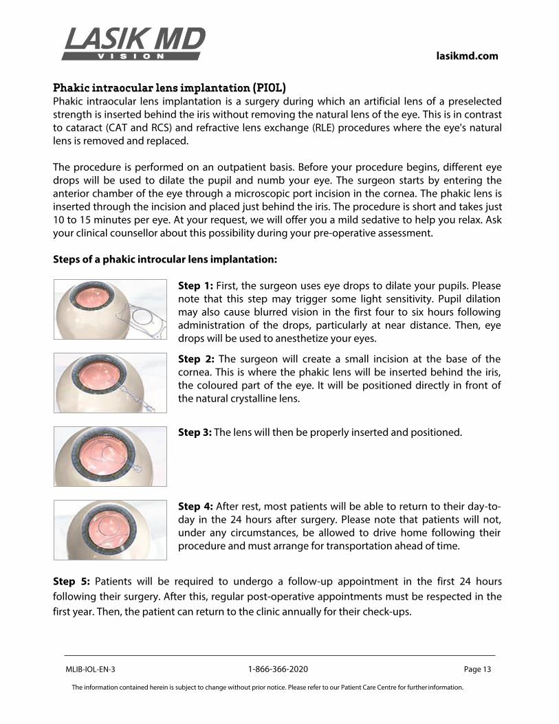
Phakic intraocular lens implantation (PIOL) Phakic intraocular lens implantation is a surgery during which an artificial lens of a preselected strength is inserted behind the iris without removing the natural lens of the eye. This is in contrast to cataract (CAT and RCS) and refractive lens exchange (RLE) procedures where the eye's natural lens is removed and replaced. The procedure is performed on an outpatient basis. Before your procedure begins, different eye drops will be used to dilate the pupil and numb your eye. The surgeon starts by entering the anterior chamber of the eye through a microscopic port incision in the cornea. The phakic lens is inserted through the incision and placed just behind the iris. The procedure is short and takes just 10 to 15 minutes per eye. At your request, we will offer you a mild sedative to help you relax. Ask your clinical counsellor about this possibility during your pre-operative assessment. Steps of a phakic introcular lens implantation:
Step 1: First, the surgeon uses eye drops to dilate your pupils. Please note that this step may trigger some light sensitivity. Pupil dilation may also cause blurred vision in the first four to six hours following administration of the drops, particularly at near distance. Then, eye drops will be used to anesthetize your eyes.
Step 2: The surgeon will create a small incision at the base of the cornea. This is where the phakic lens will be inserted behind the iris, the coloured part of the eye. It will be positioned directly in front of the natural crystalline lens.
Step 3: The lens will then be properly inserted and positioned.
Step 4: After rest, most patients will be able to return to their day-to-day in the 24 hours after surgery. Please note that patients will not, under any circumstances, be allowed to drive home following their procedure and must arrange for transportation ahead of time.
Step 5: Patients will be required to undergo a follow-up appointment in the first 24 hours
following their surgery. After this, regular post-operative appointments must be respected in the
first year. Then, the patient can return to the clinic annually for their check-ups.
lasikmd.com
MLIB-IOL-EN-3 1-866-366-2020 Page 14
The information contained herein is subject to change without prior notice. Please refer to our Patient Care Centre for further information.
OUR TREATMENTS
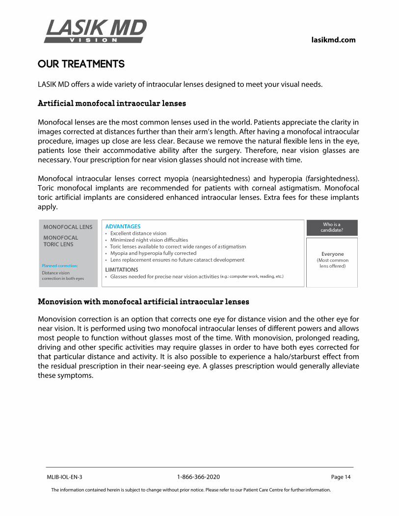
LASIK MD offers a wide variety of intraocular lenses designed to meet your visual needs. Artificial monofocal intraocular lenses Monofocal lenses are the most common lenses used in the world. Patients appreciate the clarity in images corrected at distances further than their arm’s length. After having a monofocal intraocular procedure, images up close are less clear. Because we remove the natural flexible lens in the eye, patients lose their accommodative ability after the surgery. Therefore, near vision glasses are necessary. Your prescription for near vision glasses should not increase with time. Monofocal intraocular lenses correct myopia (nearsightedness) and hyperopia (farsightedness). Toric monofocal implants are recommended for patients with corneal astigmatism. Monofocal toric artificial implants are considered enhanced intraocular lenses. Extra fees for these implants apply.
Monovision with monofocal artificial intraocular lenses
Monovision correction is an option that corrects one eye for distance vision and the other eye for near vision. It is performed using two monofocal intraocular lenses of different powers and allows most people to function without glasses most of the time. With monovision, prolonged reading, driving and other specific activities may require glasses in order to have both eyes corrected for that particular distance and activity. It is also possible to experience a halo/starburst effect from the residual prescription in their near-seeing eye. A glasses prescription would generally alleviate these symptoms.
lasikmd.com
MLIB-IOL-EN-3 1-866-366-2020 Page 15
The information contained herein is subject to change without prior notice. Please refer to our Patient Care Centre for further information.
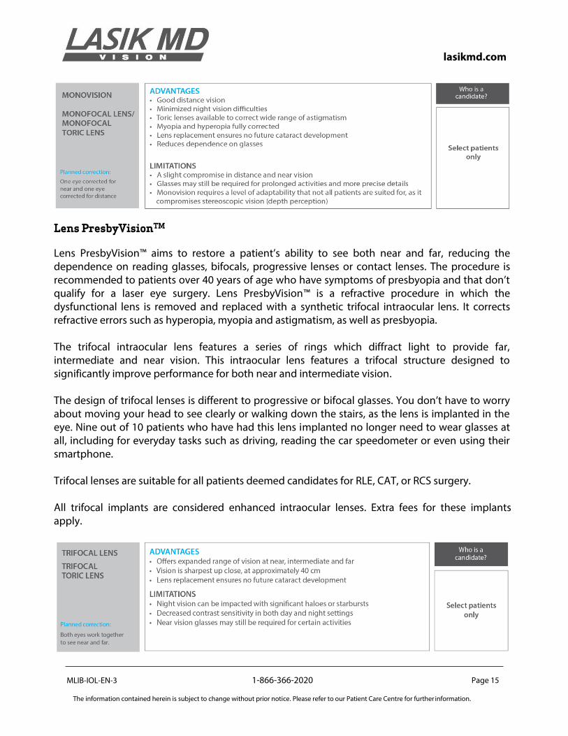
Lens PresbyVisionTM
Lens PresbyVision™ aims to restore a patient’s ability to see both near and far, reducing the dependence on reading glasses, bifocals, progressive lenses or contact lenses. The procedure is recommended to patients over 40 years of age who have symptoms of presbyopia and that don’t qualify for a laser eye surgery. Lens PresbyVision™ is a refractive procedure in which the dysfunctional lens is removed and replaced with a synthetic trifocal intraocular lens. It corrects refractive errors such as hyperopia, myopia and astigmatism, as well as presbyopia. The trifocal intraocular lens features a series of rings which diffract light to provide far, intermediate and near vision. This intraocular lens features a trifocal structure designed to significantly improve performance for both near and intermediate vision. The design of trifocal lenses is different to progressive or bifocal glasses. You don’t have to worry about moving your head to see clearly or walking down the stairs, as the lens is implanted in the eye. Nine out of 10 patients who have had this lens implanted no longer need to wear glasses at all, including for everyday tasks such as driving, reading the car speedometer or even using their smartphone. Trifocal lenses are suitable for all patients deemed candidates for RLE, CAT, or RCS surgery. All trifocal implants are considered enhanced intraocular lenses. Extra fees for these implants apply.
lasikmd.com
MLIB-IOL-EN-3 1-866-366-2020 Page 16
The information contained herein is subject to change without prior notice. Please refer to our Patient Care Centre for further information.
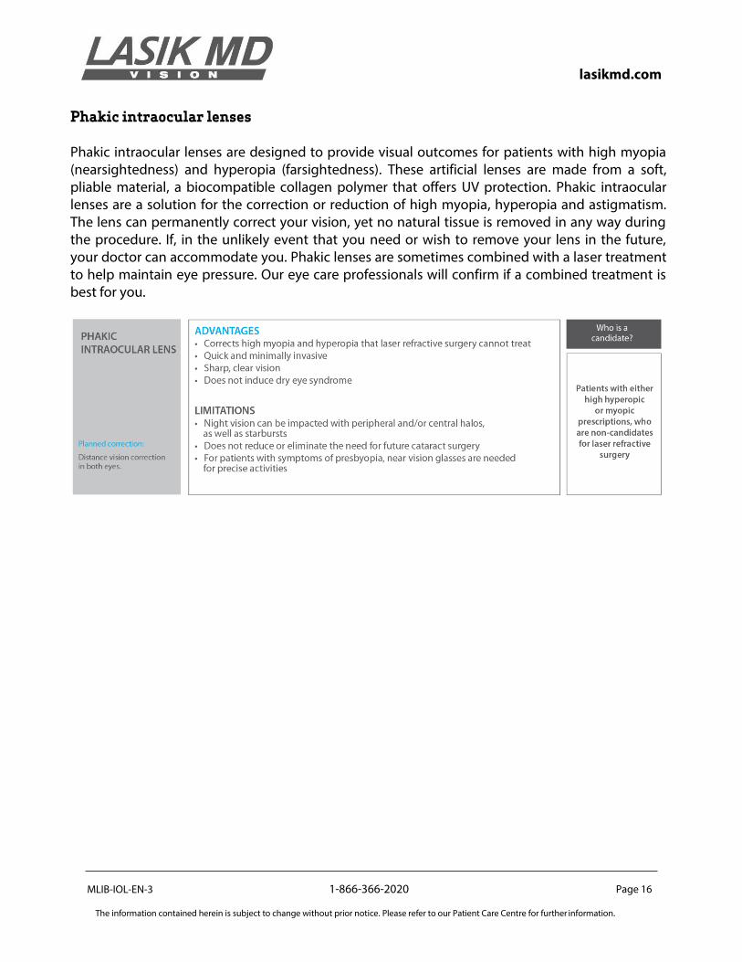
Phakic intraocular lenses Phakic intraocular lenses are designed to provide visual outcomes for patients with high myopia (nearsightedness) and hyperopia (farsightedness). These artificial lenses are made from a soft, pliable material, a biocompatible collagen polymer that offers UV protection. Phakic intraocular lenses are a solution for the correction or reduction of high myopia, hyperopia and astigmatism. The lens can permanently correct your vision, yet no natural tissue is removed in any way during the procedure. If, in the unlikely event that you need or wish to remove your lens in the future, your doctor can accommodate you. Phakic lenses are sometimes combined with a laser treatment to help maintain eye pressure. Our eye care professionals will confirm if a combined treatment is best for you.
lasikmd.com
MLIB-IOL-EN-3 1-866-366-2020 Page 17
The information contained herein is subject to change without prior notice. Please refer to our Patient Care Centre for further information.
LIMITS TO CORRECTION These procedures are unable to correct vision defects, like those listed below, which do not arise from refractive errors. Patients with such conditions may be subject to additional risks and additional side effects and should discuss their condition(s) with the eye care professionals before deciding whether to have the procedure. Amblyopia
Amblyopia, or “lazy eye”, is a medical condition that develops in early childhood in which a person with reduced vision in one eye relies on the other eye to focus. Refractive lens exchange and cataract procedures will not reduce or eliminate amblyopia, and will not improve vision in the amblyopic eye. If the patient experiences side effects or complications from the procedure in the eye that is able to focus, he or she could experience a loss of vision because that eye would no longer be able to compensate for the other. Strabismus
Strabismus is an eye disorder caused by a weakness in the eye muscles in which the eyes may not be aligned properly. Refractive lens exchange or cataract procedures will not correct, reduce, eliminate or prevent strabismus. Patients with strabismus may develop double vision as a result of or as a side effect of the procedure. Presbyopia
Presbyopia occurs when the crystalline lens of the eye loses the ability to change shape and focus on near objects, a process known as accommodation. Blurred vision from this condition typically occurs in people aged 40 and over. This progressive loss of function is caused by the stiffening of the lens, which results in reduced clarity. RLE,RCS and CAT will result in presbyopia immediately post-operatively unless a special trifocal type of intraocular lens is placed in the eye.
lasikmd.com
MLIB-IOL-EN-3 1-866-366-2020 Page 18
The information contained herein is subject to change without prior notice. Please refer to our Patient Care Centre for further information.
HOW WILL SURGERY IMPROVE MY LIFE? Reduced dependence on glasses and contacts
There are numerous potential benefits for patients who undergo intraocular surgery. Almost all of these advantages are associated with reduced dependence on eyeglasses and/or contact lenses. While the use of glasses or contact lenses can be an effective method of correcting a refractive error, it is also a method that can place restrictions on normal, everyday activities.
Reduced dependence on corrective lenses can result in more freedom for patients with active lifestyles. Many recreational activities, such as water sports or contact sports, tend to be much more enjoyable when the necessity of wearing glasses or contacts is removed. In some cases, patients choose intraocular surgery for professional purposes, rather than recreational ones. Additionally, corrective lenses are also not permitted in certain fields of employment. For contact lens wearers, intraocular surgery can also eliminate the time and effort involved in cleaning, removing and replacing lenses. In addition, over time, the costs associated with maintaining and replacing corrective lenses can be prohibitive. Many eyeglass wearers also cite cosmetic or aesthetic reasons for wanting to undergo the procedure. The reasons for undergoing intraocular refractive surgery will be different for every individual. For those who have required corrective lenses throughout most of their lives, the simple prospect of being able to drive without wearing glasses or contacts, or of being able to wake up and see without putting on glasses or contacts, may be sufficient reason in itself. The potential benefits, as well as the potential complications, can vary, and should be considered carefully. The patient is the only person who can decide whether the benefits of intraocular surgery outweigh the risks.
lasikmd.com
MLIB-IOL-EN-3 1-866-366-2020 Page 19
The information contained herein is subject to change without prior notice. Please refer to our Patient Care Centre for further information.
POTENTIAL COMPLICATIONS
Like any surgical procedure, RLE, CAT, RCS and PIOL involve risks of unsuccessful results, complications, or serious injury from unknown and unforeseen causes. Although the vast majority of our patients experience a significant improvement in their vision, neither your surgeon, the clinic or the staff, can promise or guarantee that the procedure will be 100% effective or make your vision better than it was before the procedure.
There is a slight possibility that the procedure or a complication arising from it can cause your vision to be blurred, doubled, distorted, or to have halos or other disturbances, and that these would not be easily corrected with glasses or contact lenses. In the event that this should occur, your surgeon will discuss and offer you advice on further treatment, which may involve medications or more surgical procedures. The outcome can usually be corrected by medications, lens exchange or external surface corneal surgery.
During your pre-operative examination, the likely outcomes will be shared with you based on your particular situation. Although it is not possible to list every potential risk or complication that may result from the procedure, the most important ones are described below. Please note that serious complications are very rare and that the vast majority of our patients are highly satisfied with the results of their procedure. Halos, starbursts, glares and ghosting After a procedure, some patients may experience an optical effect called halos or starbursts, especially around lights at nighttime or in dim light. These symptoms usually arise from optical aberrations induced by the intraocular lens implanted in the eye. Glares and haloes may be permanent in 1-2% of patients. The possibility of functioning difficultly at night is very rare, with only 0.1% of cases reporting this issue post-operatively. Floaters New floaters can occur after CAT, RCS or RLE surgery due to a condition called posterior vitreous detachment. The back part of the eye is filled with gel. In younger patients, this gel is attached to the back wall of the eye. As one ages, this gel becomes more liquid and can detach from the back wall of the eye. During this process, floaters can develop as small bits of the vitreous gel float around the eye more than they previously did. Intraocular surgery can sometimes accelerate this process of posterior vitreous detachment, and thus, more floaters become evident post-surgery. Also, floaters that were already present in the eye often become more visible after the cloudy cataract is removed from the eye. Floaters typically diminish with time, though it can take weeks or months for them to become less noticeable. This condition is benign and does not require corrective surgery. In some rare instances, a sudden shower of new floaters can occur, causing a tear in the retina or even, in worst cases, early retinal detachment.
lasikmd.com
MLIB-IOL-EN-3 1-866-366-2020 Page 20
The information contained herein is subject to change without prior notice. Please refer to our Patient Care Centre for further information.
High intraocular pressure High intraocular pressure can occur for several reasons after the procedure. Immediately after the procedure, some eyes react to the gels used during the operation, causing a temporary pressure spike. This can be treated with pills and drops. The steroid drops used during the first week after surgery may, on rare occasions, result in increased pressure to the eye in certain individuals. Finally, retained lens fragments and/or inflammation in the eye can increase pressure. This increase typically drops to normal levels upon cessation of steroid therapy. If the pressure is significantly elevated, it will need to be closely monitored and may require additional topical and/or oral medications. It is important for you to attend scheduled follow-up visits to allow your eye care professional to monitor your eye pressure in order to modify the medication schedule as needed. Posterior capsule opacification (PCO) After CAT, RCS or RLE surgery, the capsular bag in which the IOL is placed may cloud, resulting in a decrease in vision. This complication is called posterior capsule opacification (or PCO). This “after-cataract” is usually easily corrected using a laser procedure called a YAG capsulotomy which creates a hole in the posterior capsule, thereby allowing vision to be restored again. There is a 10%-15% possibility of a PCO occurring. Inflammation Some patients may develop temporary inflammation such as iritis or uveitis. Inflammation is easily treated with steroid drops. Infection In less than 0.07% of cases, a severe bacterial infection and inflammation of the eye can occur in the first few days or weeks after surgery. This condition is called endophthalmitis. If not addressed quickly, it can cause loss of vision or in rare instances, even loss of an eye. Patients receive very powerful antibiotic drops in the weeks after the surgery to help guard against severe infections. If endophthalmitis does occur, additional antibiotics are usually injected into the eye to help clear the infection. Unusual pain or loss of vision during the first week following the procedure may be symptoms of an infection. If you are experiencing any of these symptoms, contact LASIK MD immediately. Undercorrection/Overcorrection In spite of sophisticated equipment and modern surgical techniques, refractive regressions can occur after undergoing a surgery. These are more likely to happen in very high myopic and hyperopic patients. Residual refractive errors are easily corrected within the first year by performing laser eye surgery.
lasikmd.com
MLIB-IOL-EN-3 1-866-366-2020 Page 21
The information contained herein is subject to change without prior notice. Please refer to our Patient Care Centre for further information.
Capsular rupture and vitreous leak An extremely thin membrane, called the capsule, holds the natural lens of the eye in place. During CAT, RCS and RLE surgery, the natural lens of the eye is removed, but the capsule is left in place to hold the new lens implant in the eye in the appropriate position. If the capsule is perforated during the surgery, a capsular rupture is said to have occurred. This may allow vitreous gel from the back of the eye to come forward into the front of the eye through the broken capsular membrane. The surgeon may then need to perform an anterior vitrectomy, in which the prolapsed vitreous is removed from the front part of the eye. A plastic lens implant can usually be safely placed in the eye after capsular rupture has occurred. The risk of a vitreous leak or loss occurring is 0.5%, and less that 0.1% of cases lead to serious vision loss. Retained lens fragment When a capsular rupture occurs, part of the eye’s natural lens risks falling into the back part of the eye. This complication is called a retained lens fragment and is usually handled as a capsular rupture (see above). In some cases, the patient may be referred to an external specialist for further care. The possibility of this complication occurring is 0.2%. Only 0.025% of cases risk leading to serious vision loss. Retinal detachment Retinal detachment is more common in patients with pre-existing myopia and there is a 5% risk of it occurring, depending upon the patient’s age and severity of their myopia. There is only a 1% risk of this occurring in hyperopic patients. If the pre-existing myopia is already quite strong in the patient, it can lead to a higher chance of visual loss and—in worst cases—blindness of the eye. This is when the retina peels off the back of the eye. Precautionary steps may be taken to prevent the risk of this complication from occurring in patients with high degrees of myopia (including laser treatment to the periphery of the retina). If the complication is caught and treated early, this condition will not lead to any serious loss of vision. Retinal swelling Properly known as cystoid macular edema (CME), this generally innocuous complication occurs in approximately 3% of cases and involves temporary swelling of the central retina, leading to some visual distortion. However, it can be treated with additional eye drops and usually can be completely resolved. Only in rare circumstances does this condition cause a permanent decrease in visual acuity. The risk for CME is higher in patients with high hyperopia. Bleeding Severe bleeding inside or around the eye, called a hemorrhage, very rarely happens during routine surgery. However, patients with fragile blood vessels in the back of the eye tend to be more at risk. The blood vessels may rupture due to a sudden drop in pressure during surgery. This can result in loss of vision and possibly permanent blindness of the eye (a 0.005% risk).
lasikmd.com
MLIB-IOL-EN-3 1-866-366-2020 Page 22
The information contained herein is subject to change without prior notice. Please refer to our Patient Care Centre for further information.
Loss of visual acuity
While extremely rare, all refractive procedures can result in damage to the eye, including the loss
of visual acuity—including, in most severe cases, a loss of functional vision.
Wound leak In rare circumstances, the microincision created in the cornea during surgery may leak post-operatively. Often, all that is necessary to treat this is to place a contact lens on the eye until it seals. Rarely does it require returning to the operating room to stitch the wound. The risk of this occurring is 0.1%. Displaced lens During CAT, RCS and RLE surgery, a new intraocular lens is placed inside the capsular bag. If the intraocular lens becomes dislocated severely while inside the bag, your visual acuity may decrease substantially. A weakness in the fibres that hold the bag as well as any additional trauma to the eye can dislocate a lens. This may require surgical intervention to reposition the lens, or possibly glasses to correct the change in your prescription. There is a 0.1% possibility of this occurring. Optical imbalance If the surgeon performs the procedure on each eye on different days, the eyes may not be able to balance and focus properly until the procedure is performed on both eyes. Iris trauma/Prolapse The iris is rarely impacted during surgery. In the case that it is, however, the iris can become floppy and slip out of the corneal incision (called a prolapse). This may require further interventions. Post-operatively, it may result in a poorly functioning iris or pupil, and c a n a l s o l e a v e a hole in the iris, leading to minor visual disturbances. The risk of mild iris trauma occurring is approximately 0.1%. For more serious occurrences, however, the risk is 0.01%. Corneal burn In rare instances, the edge of the cornea where the phacoemulsification probe enters the eye may become overheated and mildly burn. This is usually self-limited and heals with time. The risk of this occurring is approximately in 0.1% of treatments. Corneal clouding Properly known as a corneal edema, this condition occurs as a result of the cornea’s inability to pump out water due to damage of the inside layer, called the endothelium. As we age, the endothelium of the cornea becomes less effective in pumping out water, and intraocular surgery can speed up this natural-weakening of the corneal endothelium to the point where the cornea begins to retain water. As a result, it swells up and becomes opaque, thereby leading to cloudy vision. If mild, this condition is treated with some special eye drops, and if severe, it can be treated with a corneal transplant. Risk of severe corneal edema is approximately 0.1%.
lasikmd.com
MLIB-IOL-EN-3 1-866-366-2020 Page 23
The information contained herein is subject to change without prior notice. Please refer to our Patient Care Centre for further information.
Eyelid droop The eyelids have a natural tendency to droop with age. The eyelid speculum used during surgery may hasten this process slightly.
Complications related to phakic intraocular lens implantation (PIOL)
As is the case with any type of surgical procedure, PIOL can lead to certain complications, though these are rare. Some examples of complications can include an increase in intraocular pressure, over- or under-correction, central and peripheral halos, night glare, loss of visual acuity, infections, and inflammation. These complications are detailed in the previous pages of this booklet. Additionally, PIOL can cause damage to the crystalline lens. Since the lens is placed inside the eye, there is potential risk in touching the eye's natural lens. While occurring in less than 1.5% of patients, any damage to the natural lens may lead to the development of cataract. In the most serious case, it may require cataract surgery. Our eye care professionals will explain these uncommon risks at your pre-operative appointment. In the event of any sort of complication arising, our staff is well-trained to provide the necessary treatment and care.
lasikmd.com
MLIB-IOL-EN-3 1-866-366-2020 Page 24
The information contained herein is subject to change without prior notice. Please refer to our Patient Care Centre for further information.
THE PATIENT CARE PROCESS AND PROCEDURE
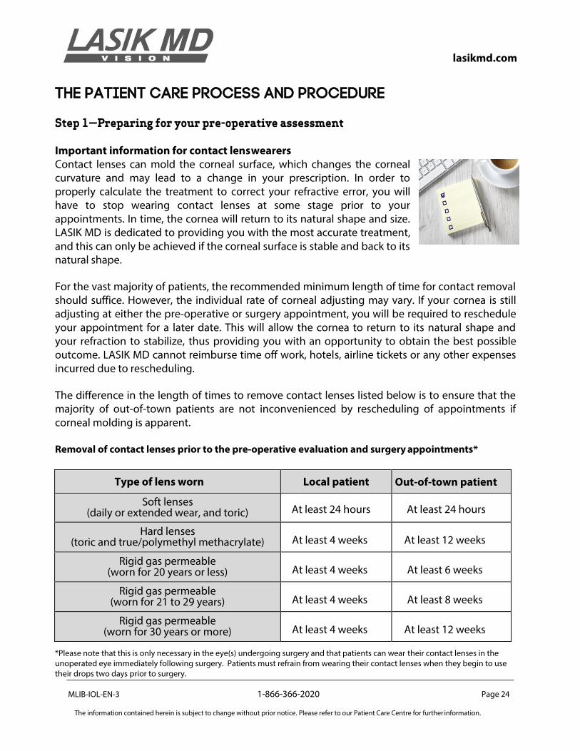
Step 1—Preparing for your pre-operative assessment Important information for contact lens wearers Contact lenses can mold the corneal surface, which changes the corneal curvature and may lead to a change in your prescription. In order to properly calculate the treatment to correct your refractive error, you will have to stop wearing contact lenses at some stage prior to your appointments. In time, the cornea will return to its natural shape and size. LASIK MD is dedicated to providing you with the most accurate treatment, and this can only be achieved if the corneal surface is stable and back to its natural shape.
For the vast majority of patients, the recommended minimum length of time for contact removal should suffice. However, the individual rate of corneal adjusting may vary. If your cornea is still adjusting at either the pre-operative or surgery appointment, you will be required to reschedule your appointment for a later date. This will allow the cornea to return to its natural shape and your refraction to stabilize, thus providing you with an opportunity to obtain the best possible outcome. LASIK MD cannot reimburse time off work, hotels, airline tickets or any other expenses incurred due to rescheduling.
The difference in the length of times to remove contact lenses listed below is to ensure that the majority of out-of-town patients are not inconvenienced by rescheduling of appointments if corneal molding is apparent. Removal of contact lenses prior to the pre-operative evaluation and surgery appointments*
Type of lens worn Local patient Out-of-town patient
Soft lenses (daily or extended wear, and toric) At least 24 hours At least 24 hours
Hard lenses (toric and true/polymethyl methacrylate) At least 4 weeks At least 12 weeks
Rigid gas permeable (worn for 20 years or less) At least 4 weeks At least 6 weeks
Rigid gas permeable (worn for 21 to 29 years) At least 4 weeks At least 8 weeks
Rigid gas permeable (worn for 30 years or more) At least 4 weeks At least 12 weeks
*Please note that this is only necessary in the eye(s) undergoing surgery and that patients can wear their contact lenses in the unoperated eye immediately following surgery. Patients must refrain from wearing their contact lenses when they begin to use their drops two days prior to surgery.
MLIB-IOL-EN-3
1-866-366-2020 Page 25
The information contained herein is subject to change without prior notice. Please refer to our Patient Care Centre for further information.
lasikmd.com
How to prepare for your pre-operative assessment
• Consult the Contact Lens Policy to determine the minimum amount of time prior to your
procedure that your contact lenses must not be worn.
• Pupil dilation will be performed; therefore you will experience blurred vision anywhere from 4–7 hours after.
• You may not be able to drive or to return to work after the evaluation.
• Your eyes may be sensitive to light, so we recommend that you bring sunglasses.
• Out of respect for other patients and to ensure your visit is as comfortable as possible, please do not bring children with you. The duration of your stay will be approximately two to three hours.
• LASIK MD will not be held responsible for any costs incurred for travel and/or accommodation, lost employment income or any additional expenses incurred due to the patient being deemed a non-candidate, requiring re-treatments, rescheduling, or delays.
Step 2—Day of surgery What happens before surgery?
• If they has been prescribed by your eye care specialist, two (2) days before surgery, start using the drops. You must purchase these at the pharmacy. Please contact the clinic if you have not received a prescription. Please note that this is not applicable in Quebec.
• If you wear contact lenses, they should be removed before surgery in the eye(s) to be operated on. Patients must refrain from wearing their contact lenses when they begin to use their drops two days prior to surgery.
• If you have surgery scheduled on your second eye at a later date, you may wear a contact lens in the non-operated eye up until two days prior to surgery on your second eye. Do not wear eye makeup the day of surgery.
• If travelling from outside Canada, please remember to carry proper identification, such as your passport and/or other proof of citizenship.
MLIB-IOL-EN-3
1-866-366-2020 Page 26
The information contained herein is subject to change without prior notice. Please refer to our Patient Care Centre for further information.
lasikmd.com
The day of surgery
• You can expect to feel nervous, anxious and/or excited prior to your procedure. This is a
completely natural, normal response.
• As you are not allowed to drive following your surgery, please arrange for transportation after the procedure.
• Please note that your eyes will be irritated and light-sensitive following the procedure. This usually diminishes within 24 hours of surgery.
• Do not use any makeup, alcohol-based or scented products the day of surgery.
• We recommend avoiding alcohol 24 hours prior to and 24 hours after your surgery, as it tends to dehydrate the tissues and can delay the healing process.
• Wear comfortable clothing on your surgery day. Please do not wear clothing, such as wool or fleece, which can lead to lint in the surgical suite.
• Out of respect for others and to ensure that your visit is as comfortable as possible, we ask that you do not bring children with you. The duration of your visit will take, on average, two hours.
After the procedure Please remember that your follow-up care is as important as the actual procedure.
• Follow the eye drop regimen recommended by the surgeon.
• Your first mandatory post-operative appointment will take place at our clinic within the first two weeks following your surgery.
• Following this visit you are required to attend at least two additional post-operative appointments.
MLIB-IOL-EN-3
1-866-366-2020 Page 27
The information contained herein is subject to change without prior notice. Please refer to our Patient Care Centre for further information.
lasikmd.com
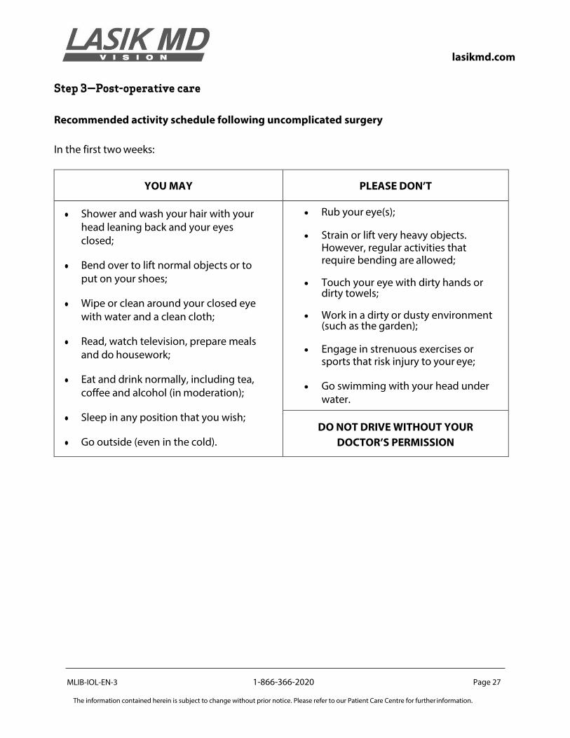
Step 3—Post-operative care
Recommended activity schedule following uncomplicated surgery
In the first two weeks:
YOU MAY
PLEASE DON’T
● Shower and wash your hair with your head leaning back and your eyes closed;
● Bend over to lift normal objects or to put on your shoes;
● Wipe or clean around your closed eye with water and a clean cloth;
● Read, watch television, prepare meals and do housework;
● Eat and drink normally, including tea, coffee and alcohol (in moderation);
● Sleep in any position that you wish;
● Go outside (even in the cold).
• Rub your eye(s);
• Strain or lift very heavy objects. However, regular activities that require bending are allowed;
• Touch your eye with dirty hands or dirty towels;
• Work in a dirty or dusty environment (such as the garden);
• Engage in strenuous exercises or sports that risk injury to your eye;
• Go swimming with your head under water.
DO NOT DRIVE WITHOUT YOUR
DOCTOR’S PERMISSION
MLIB-IOL-EN-3
1-866-366-2020 Page 28
The information contained herein is subject to change without prior notice. Please refer to our Patient Care Centre for further information.
lasikmd.com
FINANCIAL RESPONSIBILITY
Our clinic charges a single combined fee for its services. The price of your surgery also comprises post-operative care, including a same-day and/or 24-hour follow-up exam as well as subsequent exams in the first three months. Please note that you will be responsible for paying the remaining balance for surgery on the day of the procedure. For your convenience, payment may be made by Visa, MasterCard, debit card, certified cheque or cash. We do not accept personal cheques. LASIK MD offers financing to its Canadian patients. Should you require financing, this must be done before the day of surgery. Ask your clinical counsellor about the different options available to you. The procedure fee does not cover the cost of glasses, contact lenses, certain medications, costs associated with completing additional post-operative appointments, or services provided at other facilities.
INFORMED CONSENT
You have the right to consent to or to refuse any treatment or procedure at any time prior to its performance. Consent is a process that involves many steps, involving the patient, the surgeon and LASIK MD’s staff. Please remember that the staff and the surgeon at LASIK MD are available to help address your concerns, so do not hesitate to ask questions.
Steps of the consent process Eye exam During your pre-operative evaluation, we will examine your eyes to determine if you are a candidate for intraocular surgery according to criteria established by the surgeon. We will then provide you with an explanation of the procedure, the risks, complications and expected benefits, the alternatives, if any, and any particular conditions that might affect your decision to undergo the procedure.
Surgical counselling Before your surgery, we will ensure that you have a copy of the Surgical Information Package and the Consent Form(s). We will ask that you review these documents while we are present to address any questions that you have. After this, we will complete much of the information on the consent form(s) with you in preparation for the signing and witnessing of your signature.

MLIB-IOL-EN-3
1-866-366-2020 Page 29
The information contained herein is subject to change without prior notice. Please refer to our Patient Care Centre for further information.
lasikmd.com
Surgeon meeting To assist you in making an informed decision, your surgeon will review, with you, the risks and complications that are specific to your case. Please notify your surgeon if you have unanswered questions or if you aren’t sure about something. You will also be given a specific post-operative plan, for which you will also need to provide consent. Your surgeon is not required to explain risks that are extremely unlikely, or those that your surgeon does not know about, even if these become known at a later time. Your surgeon will provide you with information and materials considered necessary for a person in your position to use in deciding whether or not to undergo the procedure.
Patient consent form If, after reading this material and speaking with the counsellor, optometrist, eye care professional, and your surgeon, you decide to undergo the procedure, you will need to sign the Patient Consent Form(s). The Patient Consent Form(s) will indicate to us that you have been made aware of the nature of the procedure along with any risks or benefits associated with it. In signing this, you have also been made aware of any alternatives to this procedure, and you are thereby making an informed decision to undergo this procedure. You may request a copy of your Consent Form(s) at any time.
OUR COMMITMENT TO YOUR VISION
At LASIK MD, we understand that undergoing intraocular surgery is a very important decision. We
are devoted to helping you and making you feel at ease throughout the entire process. Our clinics
are equipped with new generation technology and our doctors are among the most experienced
in the industry.
If you have any questions, or if you would like to schedule an appointment, please contact our Patient Care Centre at 1-866-366-2020. A Patient Care Representative is available to assist you seven days a week, and will be more than happy to help you.
Improve your vision today and see how life begins with LASIK MD.

lasikmd.com facebook.com/lasikmdvisiontwitter.com/lasikmd