intramolecular donor-acceptor systems: structures of novel linked porphyrin- phenolphthalein...
TRANSCRIPT

Photochemistry and Photobiology Vol. 51, No. 3, pp. 285-291, 1990 Printed in Great Britain. All rights reserved
0031-8655190 $03.0()+0.00 Copyright 0 1990 Pergamon Press plc
INTRAMOLECULAR DONOR-ACCEPTOR SYSTEMS: STRUCTURES OF NOVEL LINKED PORPHYRIN-
PHENOLPHTHALEIN MOLECULAR SYSTEMS FRANCIS D’SOUZA and V. K R I S H N A N ~
Department of Inorganic and Physical Chemistry, Indian Institute of Science, Bangalore-560 012, India
(Received 8 June 1989; accepted 28 Auguyi 1989)
Abstract-Intramolecular donor-acceptor systems bearing porphyrins covalently linked to phenol- phthalein at various distances and at different orientations have been synthesized. Optical absorption and emission spectra and magnetic resonance studies reveal interaction between the porphyrin unit and lactoneisemiquinone forms of phenolphthalein. The charge-transfer quenching of fluoresence in these systems is found to be more pronounced compared to the intermolecular systems. The lifetime studies are interpreted in terms of intramolecular electron transfer.
IN T R 0 DUCT I 0 N
The primary events in plant and bacterial photosyn- thesis are ultrafast electronic excitation energy transfer processes accompanied by electron transfer reactions which yield stable charge-separation states. The initial charge separation occurs within a few picoseconds. This is followed by charge migration to neighboring quinone molecules within a few nanoseconds. It is known that in the reaction center of photosynthetic bacteria the charge separ- ation proceeds by electron transfer from an excited donor to a quinone through the intermediacy of a bacteriopheophytin molecule (Barber 1988; Deisen- hofer et al., 1985; Fleming et al., 1988). Elegant molecular models have been synthesized to contrib- ute to the understanding of these molecular events, and it has been possible to partially reproduce the long lived charge separation (Gust et al., 1988). Model studies of systems comprised of a porphyrin donor covalently linked to an acceptor provide important information on the molecular mechanism involved in these processes. A choice of different molecular covalent linkages provides ample scope for positioning the donor and acceptor at desired distances and orientations.
Many model compounds with porphyrin donors linked to acceptors have been reported in the litera- ture, viz., quinones (Schmidt et al., 1988), nitroar- omatics (Maiya and Krishnan, 1985), methylviol- ogen (Harriman et al., 1984; Blondeed et al., 1985) and pyromellitic anhydride (Cowan et al., 1987, 1985). The quenching of fluorescence in these sys- tems has been explained on the basis of intramolecu- lar electron transfer. The porphyrin-quinone sys- tems assume special significance in the investigation
*To whom correspondence should be addressed. ?Abbreviations: Phen, [3,3-bis(4-hydroxyphenyl)-l-(3H)-
isobenzofuranone], phenolphthalein; ZnTPP. 5,10.15,20- tetraphenylporphyrinato-zinc(II), CuTPP, 5,10,15,20- tetraphenylporphyrinato copper(I1).
of photoinduced intramolecular electron transfer reactions in view of the widespread occurrence of quinones in biological electron transfer reactions. We describe here novel porphyrin-phenolphthalein systems and report their structural and photochem- ical properties. The choice of phenolphthalein is based on the following: phenolphthalein in the base form is endowed with two potential acceptor semiquinone entities while the lactone form (in acidic media) is relatively a weak acceptor. Thus, different interesting structural and photochemical behavior is anticipated in the study of porphyrin- phenolphthalein in acid and base media.
We report here the synthesis of a few porphyrin-phenolphthalein systems in which the porphyrin unit is covalently attached to the phenol- phthalein entity through different numbers of -O-CH2-CH2- bridges (Fig. 1). The ortho, rneta and para 5-hydroxyphenyl-l0,15,20-triphenylpor- phyrins, are linked to one of the hydroxy groups of the phenolphthalein moiety in order to introduce orientational features. The zinc(I1) and copper(I1) derivatives of these porphyrin-phenolphthalein sys- tems are chosen as structural probes to investigate the optical spectral and magnetic resonance fea- tures.
MATERIALS AND METHODS
The porphyrin-phenolphthalein systems were synthes- ized by the condensation reaction of phenolphthalein with the functionalized porphyrins bearing an alkyl or alkoxy bromide side chain on one of the meso aryl groups of tetraphenylporphyrin (Fig. I ) . The starting materials for the preparation of the derivatized porphryins were, 5 4 0 , m or p)hydroxyphenyl-l0,15,20-triphenylporphryin, 2,2’- dibromodiethyl ether, and 1,2 dibromoethane. All the chemicals employed in this study were procured from BDH Chemicals, Bombay, India. The 2,2’-dibromodiethyl ether was prepared from the reaction of 2,2’-dihydroxydie- thy1 ether and PBr, according to the published procedure (Pedersen, 1967). The 5-(0, m or p)hydroxy phenyl- 10,15,20-triphenylporphyrins were synthesized from the reaction of ortho, mela or para hydroxybenzaldehyde
285
286 FRANCIS D’SOUZA and V. KRISHNAN
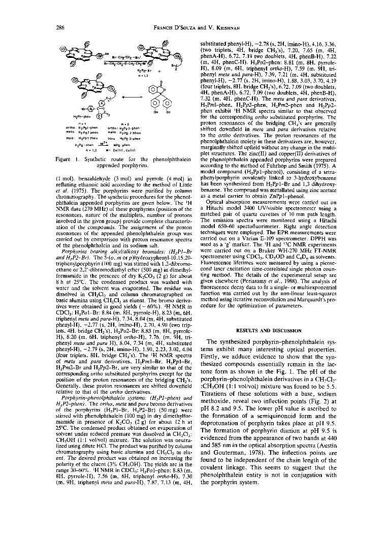
n = 1 n : 2 Ortho H I F ‘ Q - P ~ ~ ~ Ortho n2Pp2-phon m*ta H2Pm1-phen W e l a H2m 2.ph.n
PO^ H ~ P ~ I - P M ~ para H Z P ~ 2-phrn
HZPg-phen M P p - p h n
n = 1,2 M = Z n l l l ) , Cul l l )
Figure 1. Synthetic route for the phenolphthalein appended porphyrins.
(1 mol), benzaldehyde (3 mol) and pyrrole (4 mol) in refluxing ethanoic acid according to the method of Little et al. (1975). The porphyrins were purified by column chromatography. The synthetic procedures for the phenol- phthalein appended porphyrins are given below. The ‘H NMR data (270 MHz) of these porphyrins (position of the resonances, nature of the multiplets, number of protons involved in the given group) provide complete characteriz- ation of the compounds. The assignment of the proton resonances of the appended phenolphthalein group was carried out by comparison with proton resonance spectra of the phenolphthalein and its sodium salt.
Porphyrins bearing alkylialkoxy bromides: (H,PI-Br and H2P2-Br). The 5-(0, m or p)hydroxyphenyl-l0.15,20- triphenylporphyrin (100 mg) was stirred with 1,2-dibromo- ethane or 2,2’-dibromodiethyl ether (500 mg) in dimethyl- formamide in the presence of dry K,CO, (2 g) for about 8 h at 25°C. The condensed product was washed with water and the solvent was evaporated. The residue was dissolved in CH,CI, and column chromatographed on basic alumina using CHzClz as eluent. The bromo deriva- tives were obtained in good yields (- 60%). ’H NMR in CDCI,: H,Pol-Br: 8.84 (m, 8H, pyrrole-H), 8.23 (m, 6H, triphenyl meta and para-H), 7.34,8.04 (m, 4H, substituted phenyl-H), -2.77 (s, 2H, imino-H), 2.70, 4.90 (two trip- lets, 4H, bridge CH,’s), H2Po2-Br: 8.83 (m, 8H, pyrrole- H), 8.20 (m. 6H, triphenyl ortho-H), 7.76, (m, 9H, tri- phenyl meta and para H), 8.04, 7.34 (m, 4H, substituted phenyl-H), -2.79 (s, 2H, imino-H), 1.90, 2.23, 3.02, 4.04 (four triplets, 8H, bridge CH,’s). The ‘H NMR spectra of mefa and para derivatives, H,Pml-Br, H,Ppl-Br, H2Pm2-Br and HZPp2-Br, are very similar to that of the corresponding ortho substituted porphyrins except for the position of the proton resonances of the bridging CH,’s. Generally, these proton resonances are shifted downfield relative to that of the ortho derivatives.
Porphyrin-phenolphthalein systems: (H,Pl-phen) and H2P2-phen). The ortho, meta and para bromo derivatives of the porphyrins (H,Pl-Br, H2P2-Br) (50 mg) were stirred with phenolphthalein (100 mg) in dry dimethylfor- mamide in presence of K,CO, (2 g) for about 12 h at 25°C. The condensed product obtained on evaporation of solvent under reduced pressure was dissolved in CH2Cl2: CH,OH (1:l volivol) mixture. The solution was neutra- lized using dilute HCI. The product was purified by column chromatography using basic alumina and CHzClz as elu- ent. The desired product was obtained on increasing the polarity of the eluent (3% CH,OH). The yields are in the range 30-60%. ‘H NMR in CDC13: H,Pol-phen: 8.83 (m, 8H, pyrrole-H), 7.56 (m, 6H, triphenyl ortho-H), 7.30 (m, 9H, triphenyl meta and para-H), 7.87, 7.13 (m, 4H,
substituted phenyl-H), -2.78 (s, 2H, imino-H), 4.16,3.36, (two triplets, 4H, bridge CH,’s), 7.20, 7.65 (m, 4H, phenA-H), 6.72, 7.19 two doublets, 4H, phenB-H), 7.22 (m, 4H, phenC-H). H2P02-phen: 8.81 (m, 8H, pyrrole- H), 8.09 (m, 6H, triphenyl ortho-H), 7.59 (m, 9H, tri- phenyl meta and para-H), 7.39, 7.21 (m, 4H, substituted phenyl-H), -2.77 (s, 2H, imino-H), 1.88, 3.03, 3.70, 4.19 (four triplets, 8H, bridge CH,’s), 6.72,7.09 (two doublets, 4H, phenA-H), 6.72, 7.09 (two doublets, 4H, phenB-H), 7.32 (m, 4H, phenC-H). The meta and para derivatives, H,Pml-phen, HZPp2-phen, H2Pm2-phen and H,Pp2- phen exhibit ‘H NMR spectra similar to that observed for the corresponding ortho substituted porphyrins. The proton resonances of the bridging CH,’s are generally shifted downfield in meta and para derivatives relative to the ortho derivatives. The proton resonances of the phenolphthalein moiety in these derivatives are, however, marginally shifted upfield without any change in the multi- plet structures. The zinc(I1) and copper(I1) derivatives of the phenolphthalein appended porphyrins were prepared according to the method of Fuhrhop and Smith (1975). A model compound (H,Ppl-phenol), consisting of a tetra- phenylporphyrin covalently linked to 3-hydroxybenzene has been synthesized from H,Ppl-Br and 1,3 dihydroxy- benzene. The compound was metallated using zinc acetate as a metal carrier to obtain ZnPpl-phenol.
Optical absorption measurements were carried out on a Hitachi model 3400 UVlvisible spectrometer using a matched pair of quartz cuvettes of 10 mm path length. The emission spectra were monitored using a Hitachi model 650-60 spectofluorimeter. Right angle detection techniques were employed. The EPR measurements were carried out on a Varian E-109 spectrometer. DPPH was used as a ‘g’ marker. The ’H and I3C NMR experiments were carried out on a Bruker WH-270 MHz FT-NMR spectrometer using CDCI,, CD,OD and C,D, as solvents. Fluorescence lifetimes were measured by using a picose- cond laser excitation time-correlated single photon coun- ting method. The details of the experimental setup are given elsewhere (Periasamy et al., 1988). The analysis of fluorescence decay data to fit a single- or multiexponential function was carried out by the non-linear least-squares method using iterative reconvolution and Marquardt’s pro- cedure for the optimization of parameters.
RESULTS AND DISCUSSION
The synthesized porphyrin-phenolphthalein sys- tems exhibit many interesting optical properties. Firstly, we adduce evidence to show that the syn- thesized compounds essentially remain in the lac- tone form as shown in the Fig. 1. The pH of the porphyrin-phenolphthalein derivatives in a CH2C12- :CH30H (1:l vol/vol) mixture was found to be 5.5. Titrations of these solutions with a base, sodium methoxide, reveal two inflection points (Fig. 2) at p H 8.2 and 9.5. The lower p H value is ascribed to the formation of a semiquinonoid form and the deprotonation of porphyrin takes place at pH 9.5. The formation of porphyrin dianion at p H 9.5 is evidenced from the appearance of two bands at 440 and 585 nm in the optical absorption spectra (Austin and Gouterman, 1978). The inflection points are found to be independent of the chain length of the covalent linkage. This seems to suggest that the phenolphthalein entity is not in conjugation with the porphyrin system.

Novel linked porphyrin-phenolphthalein systems 287
11
10
f 9 - PH
8 -
7 -
6 -
I
-
I I I I I
0 9 7
W v c 0 L 0 VI
n
n a
h (nm) Figure 3. Optical absorption spectra of (a) ZnPol-phen (---) and (b) H2Pol-phen in CH,CI, (-). The concen-
tration of porphyrins is 0.01 mM.
The optical absorption spectra of two representa- tive porphyrin-phenolphthalein derivatives in CHzClz are shown in Fig. 3 and the data are summa- rized in Table 1. In all these compounds, the pres- ence of the phenolphthalein moiety is shown by the appearance of absorption band around 275 nm. The visible absorption spectra of the porphryins show a typical etio type of spectrum. It is clear from Table 1 that though the band positions of the porphyrin- phenolphthalein system are not significantly changed in comparison to the unappended porphy- rins, the intensity (e values) of these bands shows a decrease. It is interesting to observe that the decrease in the intensities vary with (a) the chain length of the covalent linkage ZnP1-phen < ZnP2-phen and (b) the nature of the substitution on the porphyrin entity as ortho < meta < para in a given system. Moreover, the normalized spectra of a representative zinc(I1) derivative in the Soret region (Fig. 4) reveal that the FWHM values mar- ginally decrease in comparison with the unappended ZnTPPt . Typically, the FWHM values of ZnPo2- phen is found to be 645 cm-' compared to 651 cm-' of ZnTPP. These observations can be interpreted in terms of weak interaction between the phenol- phthalein and porphyrin moieties. Phenolphthalein in basic solutions exhibits absorption bands at 275 and 550 nm.. The latter transition arises from the TI + TI* transition of the semiquinone form. The absorption spectra of the porphyrin-phenolphthalein derivatives in basic solution reveal bands in the same region as that observed for the lactone deriva- tives. However, the intensity of the 275 nm band in these derivatives shows a decrease in basic solutions and it is difficult to locate the 550 nm band of phenolphthalein. This is ascribed to (a) the intrinsi- cally small E values of the 550 nm band of phenol-
Table 1. Optical absorption data of porphyrin-phenolphthalein systems in CH,CI,: CH,OH (1:l volivol) at 298 K
Compound Q I Q2 Q.3 Q4
H,TPP HzPol-phen H,Pml-phen H,PpI-phen H2P02-phen H2pm2-phen HzPp2-phen ZnTPP ZnPol-phen ZnPm 1-phen ZnPpl-phen ZnPo2-phen ZnPm2-phen ZnPp2-phen CUTPP CuPol-phen CuPml-phen CuPpl-phen CuPo2-phen CuPm2-phen CuPp2-phen
646 (3.52) 645 (3.21) 646 (3.22) 647 (3.20)
644 (3.16) 647 (3.20)
644 (3.03)
592 (3.74) 588 (3.48) 588 (3.40)
588 (3.40)
592 (3.38) 589 (3.21)
593 (3.28) 596 596 597 596 596 596 598
549 (3.72) 549 (3.62) 549 (3.68) 550 (3.60) 549 (3.40) 552 (3.60) 553 (3.45) 556 (4.30) 556 (3.67) 556 (3.81) 556 (3.98) 560 (4.01) 557 (3.98)
538 (4.37) 537 (4.18)
557 (4.02)
537 (4.20) 538 (4.27) 536 (4.23)
538 (4.23) 537 (4.16)
513 (4.16) 514 (4.08) 514 (4.10) 515 (3.90) 516 (3.80) 516 (3.84) 517 (3.95)
- ~ ~ - 81
417 (5.76) 418 (5.35) 418 (5.42) 419 (5.48) 418 (5.28) 418 (5.30) 419 (5.39) 420 (5.82) 422 (5.20)
421 (5.42)
422 (5.27) 422 (5.34) 411 (5.73)
412 (5.47) 412 (5.53) 413 (5.31) 412 (5.36) 413 (5.49)
422 (5.36)
422 (5.20)
412 (5.36)
Phenolphthalein UV band
~~ ~
- 279 (4.20) 279 (4.37) 279 (4.25) 277 (4.22) 277 (4.20) 276 (4.27)
277 (3.98)
278 (4.20) 273 (3.90) 279 (4.10) 279 (4.42)
279 (4.12) 279 (4.15) 279 (4.21) 277 (4.18) 279 (4.20) 279 (4.17)
280 (4.12)
PA? 5 1 3 4

FRANCIS D’SOUZA and V. KRISHNAN 288
0 0
2
0 U C
n
n 4
L 0 v)
h (nm)
Figure 4. Normalized absorption spectral profiles in the Soret region of ZnTPP, (-); ZnPo2-phen, (. . .);
ZnPol-phen, (---) in CH2CI, at 298 K.
M: 2H, Zn LII)
Figure 5 . The positions of I T NMR resonance signals of porphyrin-phenolphthalein system in neutral and basic conditions. The values of I7C resonance signals of only
the carbons of phenolphthalein are given.
phthalein (semiquinonoid form) in addition to the appearance of intense bands of porphyrin in this region and (b) the restricted resonance of the semiquinonoid form in the appended system (in contrast to the free phenolphthalein) which may also reduce the E values. We are unable to detect the interaction of the semiquinonoid form with the metal ions (in the porphyrin center) by the optical absorption spectral method. However, the existence of the semiquinonoid form in basic media is clearly evident from the I3C NMR spectral data. The lac- tone form in CD30D:C6D6 (1:1 vol/vol) exhibits I3C resonances at 134, 171 and 93 ppm originating respectively from the C,, Ch and C, carbons (Fig. 5). These resonances are comparable to that observed for the free phenolphthalein in the lactone
form (C, 134, Cb 172 and C, 94 ppm). Addition of a base to this solution induces the formation of a semiquinonoid form with a characteristic shift in the resonance of the C, and C, carbons. The two resonances at 117 and 134 ppm in the semiqui- nonoid form are due, respectively, to the aromatic carbon of the semiquinone aryl group and the car- bon atom to which porphyrin is attached through ether linkages. Moreover, the C, carbon resonance is shifted upfield due to aromatization. The I3C resonance of keto carbonyl group in the metal derivatives does not show any shift in the peak position indicating the absence of the coordinative interaction of the keto carbonyl to the metal center in the porphyrin unit.
The singlet emission spectra of the free-base and zinc(I1) derivatives in CH2C12:CH30H (1:l vol/vol) reveal two bands at 654 and 720 nm, and 604 and 652 nm, respectively, corresponding to the Q(0,O) and Q( 1,O) transitions. Phenolphthalein was found to be nonfluorescent both in the lactone and semiquinone forms. It is interesting to note that the fluorescence intensity of the derivatives varies with the nature of the substitution (ortho > meta >para) indicating that the interaction is a maximum with the ortho substitution of the phenolphthalein entity (Fig. 6a). It is reasonable to expect that the phenol- phthalein unit is closer to the porphyrin ring relative to para substitution. It is found that the emission yields of these derivatives are not significantly alt- ered in the basic media. However, the free base porphyrin derivatives are deprotonated in the basic media as seen from the appearance of a fluorescence band at 690 nm corresponding to the dianion. We
725 700 675 650 6 hnm-
675 625 575 h n m -
Figure 6 . Fluorescence emission spectra of (a) ( i ) H,TPP, (i i ) H,Ppl-phen, (iii) H,Prnl-phen, (iv) H,Pol-phen and (b) (i) ZnTPP, (ii) ZnTPP and phenolphthalein (100 fold) in CH,CI, at room temperature. The concentration of
porphyrins employed is 1 p M .

Novel linked porphyrin-phenolphthalein systems 289
h also investigated the intermolecular complexation of phenolphthalein and its sodium salt (semiquinonoid form) with ZnTPP. It was seen that the fluorescence intensity of ZnTPP emission was not quenched when a 1 : l molar mixture of the porphyrin and phenolphthalein (or its sodium salt) was employed. However, an increase in the phenolphthalein con- centration (100 fold) decreased the intensity by about 10% (Fig. 6b). These observations indicate that the CT interaction is more pronounced in the intramolecular system than the intermolecular sys- tem. This observation is similar to that observed in the literature (Wasielewski and Niemezyk, 1984; Maiya and Krishnan, 1985). The possibility of exci- tation energy transfer in these systems is ruled out because phenolphthalein both in acidic and basic media does not exhibit absorption bands in the region (600-720 nm) of porphyrin emission. It may be noted that the 3-hydroxybenzene appended por- phyrin, ZnPpl-phenol, exhibits an emission spec- trum identical to that observed for ZnTPP indicating absence of any quenching due to electron transfer.
The fluorescence decay data of the zinc(I1) deriva- tives in CH2CI2:CH30H (1:l vol/vol) mixture are shown in Fig. 7 along with the results of fitting and reveal the existence of two components (short and long). The lifetimes calculated from the decay data are shown in Table 2. It can be seen that though all the zinc(I1) derivatives exhibit two components, the weighted average lifetime of the species for these derivatives is - 1.6 ns. The lifetime of ZnTPP is CH2C12:CH30H (1:l vol/vol) mixture is found to be 1.8 ns. Preliminary photophysical measurements on the model compound ZnPpl-phenol, reveal a lifetime of 1.76 ns. It may be noted that the lifetime of the long component of the phenolphthalein appended derivatives is almost coincident with the lifetime observed for the model compound (ZnPpl-phenol) lacking electron transfer. Hence, it is suggested that the short lifetime is associated with electron transfer reaction and the electron transfer rate (k,.) was calculated using the expression
k,, == 7;' - 7;' (1)
r 3 V 0 s
CHANNEL- CHANNEL --+
II Id I l l b l
t 2 z 0
0 s
CHANNEL- C H A N N E L +
Figure 7. Fluorescence decay profiles for the zinc(I1) porphyrin-phenolphthalein derivatives obtained from
M solutions at room temperature in CH,CI,: CHzOH (1:l vol/vol) (excitation wavelength 420 nm) (a) ZnPol- phen and (b) on addition of KOH II(a) ZnPo2-phen and
(b) on addition of KOH.
where T, and T, are the lifetime of the short and long components.
The values of k,, (Table 2) reveal the following features: (i) the values of k,, observed in the present study are of the same order of magnitude as those observed for some of the linked porphyrin-quinone molecules (Siemiarczuk et al., 1983); (ii) the elec- tron transfer rate (k,.) decreases with an increase in the length of the covalent linkage; (iii) the k,, values vary with the nature of substitution as ortho > para; (iv) the k,, values of the zinc derivatives in the semiquinonoid form are generally lower except in the case of ZnPo2-phen. The reason for this is not clear. It is worth mentioning here that the electrochemical redox potential of these derivatives measured in CH2C12 reveal estimates for the energy of the C T states ZnPol-phenlZnPfo1-phen- and ZnPo2-phenlZnpto2-phen- to be 1.78 and 1.75 eV,
Table 2. Electron transfer rate constants for porphyrin-phenolphthalein systems in CHZCI,: CH,OH (1: 1 volivol) at 25°C
Lifetime Amplitudes k, , x l o x ~~~~~~~~~~~~~ ~ ~ . . ~~~~~
Compound T, (ns) T , (ns) A1 AZ X 2 (s- -~~ .... ~~~~~~~~~~
ZnPol-phen 0.83 1.68 ZnPol-phen + KOH 1.49 2.20 ZnPpl-phen 1.02 1.78 ZnPpl-phen + KOH 1.11 1.69 ZnPo2-phen 1.19 1.92 ZnPo2-phen + KOH 0.91 1.70 ZnPp2-phen 1.47 2.35 ZnPp2-phen + KOH 1.46 2.11
0.31 0.69 1.21 6.09 0.52 0.36 1.21 2.16 0.30 0.66 1.28 4.18 0.43 0.52 1.28 3.09 0.56 0.45 1.19 3.19 0.26 0.68 1. 19 5.10 0.87 0.13 1.21 2.88 0.84 0.16 1.21 2.10

290 FRANCIS D'SOUZA and V. KRISHNAN
respectively. The algebraic sum of the CT energies and the excited singlet energies yields a value of free energy change for electron transfer reaction AGet. The small exergonicity of AGet (- -0.30 eV) calculated from the data are consistent with the presence of singlet excited state electron transfer in these systems.
It is generally expected that the singly linked donor-acceptor systems should exhibit several con- formations. The two limiting cases would be (i) where the phenolphthalein moiety is close to the porphyrin unit and (ii) the phenolphthalein moiety is away from the porphyrin entity. The lifetime data, however, reveal two components which may correspond to two extreme conformers. It was of interest to see whether the presence of closed con- formers could be probed by an E P R active copper porphyrin-phenolphthalein system. With this in view the EPR spectra of Cu(I1) derivatives were recorded in toluene at 130 K. A representative spec- trum is shown in Fig. 8. All the spectra were simu- lated using the Gaussiann quadrature method, and the EPR g and A tensors were calculated from the spectral features assuming a spin Hamiltonian for axial symmetry (Manoharan and Rogers, 1969). The data are given in Table 3. The EPR parameters d o not show any significant change in acidic and basic
1OOG -
Figure 8. EPR spectrum of (a) CuPol-phen in toluene at 130 K and (b) the simulated spectra using Gaussian
quadrature method.
form. However, the gll and gl values for the porphyrin-phenolphthalein derivatives decreased considerably relative to those observed for CuTPP. A decrease in the values of AT and A:" and an increase in the A1 and AY implies an interaction between the Cu(I1) ion and the covalently linked phenolphthalein unit. It is possible to obtain the bonding parameter (aZ), a measure of covalency of the inplane wbonding of the Cu-N bond in these derivatives, using the method described by Kievel- son et al. (1961). The difference in the magnitudes of a' values (Aa') between the CuTPP and Cu(I1) derivatives of porphyrin-phenolphthalein systems gives a measure of the covalent factor arising from CT interaction. The ha2 values of the phenol- phthalein derivatives varies with the nature of the substitution (ortho > meta > para) and also with the chain length. The decrease in a2 values for Cu(I1) derivatives of porphyrin-phenolphthalein signifies an increase in covalency of Cu-N bonds in these systems. This is ascribed to the n-electron donation of the copper porphryin to the phenol- phthalein unit which increases Cu-N IJ covalency (Chandrashekar and Krishnan, 198l).This could happen only if the phenolphthalein unit is pos- itioned close to the porphyrin unit. Thus the EPR data provide indirect evidence for the possible exist- ence of a closed conformer.
The results of the present study illustrate the presence of intramolecular electron transfer in the porphyrin systems. The k,, values are only margin- ally different in the lactone and semiquinone form of phenolphthalein. This can be rationalized from molecular model calculations using the C A R T pro- gramme (Maiya and Krishnan, 1983). In this study we employed X-ray crystallographic data for the porphyrins (Silvers and Tulinsky, 1967) and the phenolphthalein entity was considered planar in the semiquinone form and tetrahedral in the lactone form. The ethylene oxide bridges were assumed to be in the cis form (Hejashema et al . , 1981). The molecular geometry of the porphyrin-phenol- phthalein systems was found to be a folded confor- mation. It was found that the distance between the porphyrin unit (center) and the phenolphthalein C, (in the lactone form) (- 10 A) is not significantly
Table 3. EPR parameters of Cu(I1) porphyrin-phenolphthalein systems in toluene at 130 K
Compound
( X lo4 cm-I) _ _ _ _ ~ ~ _ _ _ _ _ _ _ _ _ .~
CuTPP 2.185 2.038 201.0 32.76 14.8 16.20 - CuPol-phen 2.176 2.029 197.0 32.60 14.6 16.21 0.0246 CuPml-phen 2.177 2.030 198.1 32.72 14.8 16.20 0.0205 CuPpl-phen 2.177 2.030 198.0 32.70 14.8 16.21 0.0205 CuPo2-phen 2.175 2.028 197.1 32.58 14.9 16.25 0.0265 CuPmZphen 2.176 2.029 198.0 32.60 14.8 16.21 0.0220 CuPp2-phen 2.178 2.031 198.1 32.70 14.8 16.20 0.0201
Novel linked porphyrin-phenolphthalein systems 29 1
different from the distance between the porphyrin entity and the center of the aryl entity phenB of phenolphthalein (semiquinone form). It is difficult to perceive the distinct conformations in these sys- tems in view of the unknown structure of the lactone and semiquinone forms of phenolphthalein.
Acknowledgements-The authors are grateful to the Department of Science and Technology, and Department of Non-Conventional Energy Sources, Government of India, for their support. The authors are grateful to the Tata Institute of Fundamental Research, Bombay for the picosecond facility.
REFERENCES Austin, E. and M. Gouterman (1978) Absorption and
emission of weak complexes with acids, bases and salts. Bioinorg. Chem. 9, 281-298.
Barber, J. (1988) Photosynthesis, electron transfer theory in question. Nature 333, 114-1 15.
Blondeed, G., D. De Keuleeleire, A. Harriman and L. R. Milgrom (1985) Fluorescence of covalently bound zinc porphyrin-viologen complexes. Chem. Phy. Lett.
Chandrashekar, T. K. and V. Krishnan (1981) Donor properties of metallomacrocylic tetrapyrrole pigments with syn-trinitrobenzene. Inorg. Chem. 20, 2782-2786.
Cowan, J. A. and J. M. K. Sanders (1985) Pyromellitim- ide-bridged porphyrins as model photosynthetic sys- tems: synthesis and steady state fluorescence properties. J . Chem. SOC. Perkin Trans. I , 243552437’,
Cowan, J . A,, J . K. M. Sanders, G. S. Beddard and R. J . Harrison (1987) Modelling of photosynthetic reaction centre: photoinduced electron transfer in a pyromellit- imide-bridged ‘special pair’ porphyrin dimer. J . Chem. SOC. Chem. Commun, 55-58.
Deisenhofer, J., 0. Epp, K. Miki, R. Huber and H. Mickel (1985) Structure of the protein subunits in the photosynthetic reaction centre of R. viridis at 3 A resol- ution. Nature 318, 618-624.
Fleming, G. R., J . L. Martin and J. Breton (1988) Rates of primary electron transfer in photosynthetic reaction centres and their mechanic implications. Nature 337, 190- 192.
Fuhrhop, J. H. and K. M. Smith (1975) In Porphyrins and Metalloporphyrins (Edited by K. M. Smith), Chap. 9, Elsevier, Amsterdam.
Gust, D., T. A. Moore. D. Barret, L. 0. Harding, L. R. Making, P. A. Liddel, R. C. De Schryver, M. V. Auweraer, R.V. Bensasson and R. Rougee (1988) Pho- toinduced charge separation in a Carotenoid-
118, 77-82.
porphyrin-diquinone tetrad: enhanced quantum yields via multistep electron transfer. J . Am. Chem. SOC. 110, 321-323 and references cited therein.
Harriman, A,, G. Porter and A. Wilowsha (1984) Photore- dox properties of zinc porphyrin-violegen complexes. J . Chem. SOC. Faraday Trans. 2, 80, 191-204.
Hejashema, Y . , K . Kanetsuki, J . Shiokawa and N . Tanaka (1981) Structure of the complex of neodymium nitrate with pentaethylene glycol. Bull. Chem. Soc. Jpn 54, 1567.
Kievelson, D. and R. Neiman (1961) ESR studies on the bonding in copper complexes. J . Chem. Phys. 35,
Little, R. G., P. A. Anton and I. J . A . Loach (1975) The synthesis of some substituted tetraphenyl porphyrins. J . Heterocyclic Chem. 12. 343-348.
Maiya, G. B. and V. Krishnan (1983) Porphyrins appended with podand side arm. Evidence for the existence of folded conformer. Inorg. Chim. Acfa 77, L13-Ll5.
Maiya, G. B. and V. Krishnan (1983) Porphyrins appended with podand side arm. Evidence for the exis- tence of folded conformer. Inorg. Chim. Acfa 77, LlFL15.
Manoharan, P. T. and M. T. Rogers (1969) In Electron Spin Resonance of Metal Complexes (Edited by T. F. Yen). Plenum Press, New York.
Pedersen, C. J . (1967) Cyclic polyethers and their com- plexes with metal salts. J. Am. Chem. Soc. 89,
Periasamy, N . , S. Doraiswamy, G. B. Maiya and B. Ven- kataraman (1988) Quenching of cationic dyes by ionic quenchers. J . Chem. Phys. 88, 1638-1651.
Schmidt, J . A , , A. R. McIntosh, A. C. Weedon, J. R. Bolton, J . C. Connolly, J. K. Hurley and M. R. Wasi- elewski (1988) Intramolecular photochemical electron transfer. Singlet and triplet mechanism of electron trans- fer in a covalently linked porphyrin-amide-quinone molecule. J . Am. Chem. SOC. 110, 1733-1740.
Siemiarczuk, A . , A. R. McIntosh, Ho Te-Fu, M. J . Still- man, K. J . Roach, A. C. weedon, J. R. Bolton and J. s. Connolly (1983) Intramolecular photochemical electron transfer. Fluorescence studies of linked porphyrin- quinone compounds. J . Am. Chern. Soc. 105. 7’224-7230.
Silvers, S. T. and A. Tulinsky (1967) The crystal structure of triclinic tetraphenylporphyrin. J . Am. Chem. SOC. 89,
Wasielewski, M. R. and M. P. Niemezyk (1984) Photoin- duced electron transfer in meso-tetraphenyl triptycenylporphyrin-quinone. Restricted donor- acceptor distances and orientations. J . Am. Chem. SOC. 106, 5043-5045.
149-156.
1707-1 736.
333 1-3337.