intralesional administration of allogeneic bone marrow
TRANSCRIPT
Summary. When a severe neurological lesion occurs asa consequence of intracerebral bleeding, no effectivetreatment for improving the outcome is currentlyavailable. In the present study, intracerebral hemorrhage(ICH) was induced by stereotactic injection of 0.5 U ofcollagenase type IV in the striatum of adult Wistar rats,and three days later, intralesional administration of2x106 allogeneic bone marrow stromal cells (BMSC) insaline (n:10), or saline only (n:10), was performed. Inthe following 30 days, functional outcome was evaluatedin each animal by rotarod and the modified neurologicalseverity score (mNSS) test. Progressive and functionalimprovement was observed in BMSC-transplanted ratscompared with controls, together with morphologicalimages suggesting that intracerebral administration ofBMSC increases endogenous neurogenesis at the levelof subventricular zone (SVZ). These findings suggestthat local administration of allogeneic BMSC could beuseful to reduce the neurological deficits caused byintracerebral hemorrhage. Key words: Bone marrow stromal cells, Intracerebralhemorrhage, Endogenous neurogenesis, Cell therapy
Introduction
Intracerebral hemorrhage (ICH) is one of the mostdevastating forms of stroke and the third cause of deathin developed countries. 38% of patients survive the firstyear, but ICH leads to severe neurological deficits, andcurrently no effective treatment for improving the
outcome is available (Ferro, 2006; Andres et al., 2008).In the last years, cell therapy using adult stem cellsoffers new strategies for the treatment of neurologicaldiseases, and some experimental studies suggested thatlocal or systemic administration of bone marrow-derivedstem cells can reduce the neurological deficits caused byintracerebral bleeding (Andres et al., 2008).Nevertheless, nowadays it is difficult to find argumentsfavouring the superiority of bone marrow stromal cells(BMSC) or bone marrow containing hematopoietic stemcells, when the repair of injured central nervous tissue isneeded (Vaquero and Zurita, 2009). On the other hand,we must still obtain better knowledge of the mechanismsby which adult stem cells carry out their beneficialeffects. Keeping in mind the advantage of the lowantigenicity of BMSC, the results obtained after the useof these cells for spinal cord repair (Zurita and Vaquero,2004, 2006; Zurita et al., 2008), and the absence ofprevious studies showing the potential utility of BMSCin ICH, we studied here whether intracerebraladministration of allogeneic stromal cells, obtained frombone marrow, can restore neurological functionspreviously suppressed by intracerebral bleeding.Material and methods
In the present study, care of the animals compliedwith that stipulated by the principles of AnimalLaboratory Care and the Guide for the Care and Uses ofLaboratory Animals issued by the American NationalSociety for medical research. Experimental model
Female adult Wistar rats weighing 200 to 250 g weresubjected to an experimental model of intracranialhemorrhage (ICH). The animals were anesthetized with
Intralesional administration of allogeneic bone marrow stromal cells reduces functional deficits after intracerebral hemorrhage L. Otero1, C. Bonilla1, C. Aguayo1, M. Zurita1 and J. Vaquero1,21Neuroscience Research Unit and 2Service of Neurosurgery, Puerta de Hierro Hospital, Majadahonda, Madrid, Spain
Histol Histopathol (2010) 25: 453-461
Offprint requests to: Jesús Vaquero, M.D, Ph.D. Servicio deNeurocirugía, Hospital Universitario Puerta de Hierro-Majadahonda,Joaquín Rodrigo, 2, 28222-Majadahonda, Madrid, Spain. e-mail:[email protected]
http://www.hh.um.esHistology andHistopathology
Cellular and Molecular Biology
Sevorane 3% using a face mask with oxygen flow at 3l/min. After subcutaneous injection of Meloxicam (2mg/kg) and Morphine (2.5 mg/kg), the animals wereplaced in a stereotactic frame. Craniotomy wasperformed adjacent to bregma and intrastriatalhemorrhage was induced by administration of bacterialcollagenase type IV (Sigma-Aldrich, Madrid, Spain).Using a microinjector pump (mod 310 Stoelting Co.,Wood Dale, IL,USA), 2 µl of saline containig 0.5 U ofcollagenase were injected into the striatum over a periodof 5 minutes (coordenates: 0.04 mm posterior, 3.5 mmlateral, 6 mm ventral). At this moment, the animals wererandomly divided into two groups. Three days later, tenanimals (treated group) were subjected to a stereotacticinjection of 10 µl of saline containing 2x10⁶ BMSC, intothe injured zone. In the other ten animals (control group)10 µml of saline without BMSC were administered.Additionally, five adult female rats without any lesiontype were studied to compare the functional resultsobtained in our experimental groups and to study thesubventricular zone (SVZ).BMSC isolation and characterization
Bone marrow stromal cells were obtained from adultmale donor Wistar rats. Using a 1-ml syringe and a 21-gauge needle, fresh whole bone marrow was harvestedaseptically from tibias and femurs. Both ends of thebones were cut and the marrow was extruded with 5 mlof alpha-MEM medium (Gibco BRL Co. Ltd, USA).Bone marrow was mechanically dissociated to obtain ahomogeneous cell suspension. The cell suspension wasfiltered through a 70-mm mesh nylon strainer and placedin a 75-cm2 flask for tissue culture with 12 ml alpha-MEM medium containing 20% fetal bovine serum(FBS), 2 mM L-glutamine, 100 units/ml penicillin, 100µg/ml streptomycin and 25 ng/ml amphotericin B. Thecells were incubated at 37°C in 5% CO2 for three days.At this time, non-adherent cells were removed byreplacing the medium. The culture medium was replaced3 times per week. After the cultures reached confluency,they were lifted by incubation in a solution containing0.25% trypsin and 1 mM EDTA for 5 min at 37°C. Flowcytometry experiments were performed on a flowcytometer (Cytomic FC 500-MPL, Beckman CoulterInc., Fullerton, CA, USA) for the characterization ofBMSC. Antibodies used were unconjugated CD31(Becton Dickinson, Franklin Lakes, NJ, USA), PE-labeled CD45 (Becton Dickinson, Franklin Lakes, NJ,USA), FITC-labeled CD105 (Southern Biotech,Birmingham, Alabama, USA) and APC-labeled CD133(Miltenyl Biotec, Bergisch Gladbach, Germany). BMSCwere strongly positive for CD105 and negative forCD31, CD45 and CD133. The BMSC obtained, whichshowed viability higher than 95%, were injected into thelesion zone. In five animals, previous to intracerebraladministration, BMSC were labeled with 5-bromo-2’-deoxiuridine (BrdU). For BrdU-labeling, BMSC werecultured with 10 µM BrdU (Sigma-Aldrich, Madrid,
Spain) for 48 h. They were rinsed three times with alfa-MEM without serum and the culture was rinsed with0.25% trypsin and 1 mM EDTA for 5 min at 37°C. Behavioral tests
Rotarod test was used to evaluate motorcoordination, and the modified neurological severityscores. The mNSS test previously described (Li et al.,2001) was used in order to measure the sensitive andmotor deficits. In the Rotarod test the rats were placedon the rotarod cylinder, and the time the animalsremained on the Rotarod was measured. The speed wasslowly increased from 4 to 40 rpm within a period of 1minute. The mNSS test is a composite of motor, sensory,balance and reflex tests, and neurological function wasgraded on a scale of 0 to 18. A score of 0 is associatedwth normal neurological function, and a score of 18represents the maximal functional deficit. In the severityscores of injury, 1 point is awarded for a specificabnormal behavior or for the lack of a tested reflex; thus,the higher score is the most severe injury.
The animals were training in both behavioral testsfor 10 days before ICH, and basal data are recorded asthe mean of the last two days of training. Aftertransplantation, the animals were evaluated twice perweek, for four weeks, and data for each group wererecorded as mean ± standard deviation (SD).Kolmogorov-Smirnow test was performed to studywhether the data were normally distributed, and we useda repeated measures ANOVA test for a comparison ofthe mean scores within each group, at the different timepoints of follow-up. The statistical analysis wasperformed by means of the InStat statistical system (v1.01, GraphPad Software Inc., San Diego, CA), withp<0.05 considered as statistically significant. Histological studies
One month after BMSC administration, the rats weresacrificed for histological studies. For sacrifice, theanimals were anesthetized with 8% sevoflurane in acontinuous oxygen flow of 3 l/min, and perfusedtranscardially with 20 ml heparinized saline followed by60 ml of 4% paraformaldehyde in 0.1M PBS (pH 7.4).The brains were then dissected, post-fixed in 4%formalin for 1-2 days at room temperature, and for eachrat, a block containing the zone of the ICH wasprocessed for paraffin sectioning. A series of 5-µm thicksections were cut with a microtome through each blockand mounted on glass slides for histological observationsafter hematoxylin-eosin staining. For immunohisto-chemical studies, adjacent slides were placed in a boiledcitrate buffer (pH 6) in a microwave oven (650-720 w).After rinsing in PBS, the sections were exposed to 3%H2O2 for 30 min to quench endogenous peroxidaseactivity. Before incubation of primary antibodies, non-specific binding was blocked for one hour with 3%normal serum from the species in which the secondary
454Cell therapy for intracerebral hemorrhage
antibody was raised. Primary antibodies used weredirected against: BrdU monoclonal antibody (1:100,Chemicon International, Inc. Temecula, CA.), nestinmonoclonal antibody (1:100, Chemicon InternationalInc., Temecula, CA, USA), Ki-67 monoclonal antibody(1:200 Master Diagnostica) and doublecortin policlonalantibody (1:300, Santa Cruz Biothecnology). Thesections were incubated with secondary antibodiesconjugated to biotin, 1:200 (Vector Inc, CA, USA).Subsequently, the sections were washed in PBS, andincubated with avidin-biotin-horseradish peroxidasecomplex (Vector Inc, CA, USA). 3,3’-diaminobenzidine(DAB) was used as a chromogen. Control slices, lackingprimary or secondary antibodies, were analyzed witheach series. The sections were studied using lightmicroscopy.
For identification of male donor cells, in situhybridization studies were performed. For this, 5-µmthick sections were dewaxed and rehydrated with xileneand graded ethanol and subsequently digested withproteinase K (30 µg /ml) for 15 minutes at 37°C. Weused a biotinilated-DNA probe for sequence specificityfor murine Sry gene, the sex-determining region of Y-chromosome. Hybridization was performed in ahybridization mixture consisting of 50% deionizedformamide, 2% salmon test DNA, 10% dextran sulphate,10% 50X Denhardt’s solution and 400 ng biotinilated-labeled probe at 50°C overnight. For conventionalinmunohistochemistry to visualize the biotinilated-labeled probe, a mouse anti-biotin mouse monoclonalantibody (1:100, Jackson ImmunoResearch Laboratories,Inc., Baltimore Pike, USA), secondary antibodies anti-mouse biotin-conjugated antibody (5 µg/ml, VectorLaboratories Inc, Burlingame, CA, USA) and incubatedwith avidin–biotin–horseradish peroxidase complex(Vector Inc, CA, USA) were used. DAB was used as achromogen to visualize under conventional microscopy.For double stain, the secondary antibody used wasRhodamine(TRITC)-conjugated anti-mouse antibody(1:200, Jackson ImmunoResearch Laboratories, Inc.,Baltimore Pike, USA). The primary antibodies used afterwashing in PBS were anti-NeuN monoclonal antibody(1:500 Chemicon International, Inc. Temecula, CA.) andanti-Glial Fibrillary Acidic Protein (PGFA) (1 µl/ml, LabVision Corporation). The secondary antibody used wasCyTM 2-conjugated anti-mouse antibody (1:200,Jackson ImmunoResearch Laboratories, Inc., BaltimorePike, USA). After washing in PBS the sections wereincubated in Dapi and mounted in Glicerol medium. Theslices were visualized by fluorescence microscopy.
In order to analyze a possible increase in theproliferative activity following BMSC transplantation,ten sections corresponding to the lesion zone wereselected at random from each animal and the number ofKi-67-positive cells, at least in ten differentmicroscopical fields, at 400x, was recorded from eachhistological section. These recordings were made byimage analysis morphometry (Optimas, 6.2 softwarepackage, Optimas Corporation, Bothell, WA) using a
macro application, and conducted by two investigatorstrained in morphometric determinations, with noknowledge of the experimental group from which eachsample had been obtained. The means recorded by theseinvestigators were regarded as final values. For eachexperimental group the total number of immunopositivecells was averaged and values were expressed as countmeans ± standard deviations. We used the unpairedStudent’s t test, for a comparison of the number ofimmunopositive cells in the two experimental groups. Results
In our present study, all the animals showedsignificant neurological dysfunction after ICH, measuredwith rotarod and mNSS tests, but a clear improvementwas recorded in the course of follow-up in the group ofanimals that received intralesional BMSC.
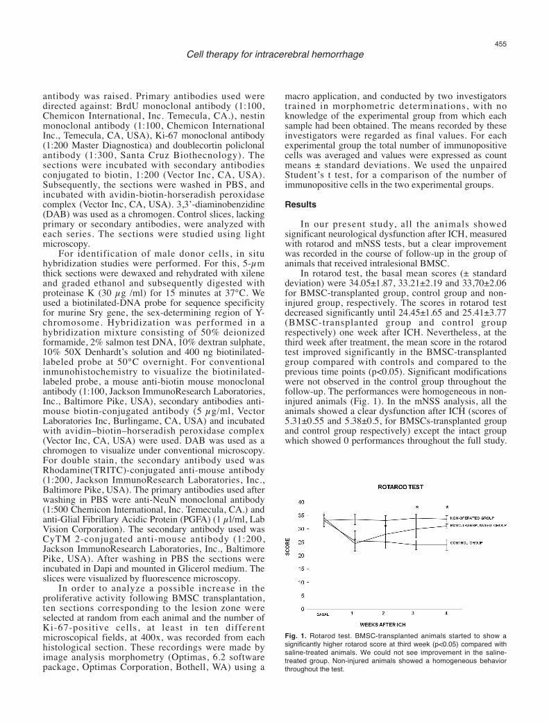
In rotarod test, the basal mean scores (± standarddeviation) were 34.05±1.87, 33.21±2.19 and 33,70±2.06for BMSC-transplanted group, control group and non-injured group, respectively. The scores in rotarod testdecreased significantly until 24.45±1.65 and 25.41±3.77(BMSC-transplanted group and control grouprespectively) one week after ICH. Nevertheless, at thethird week after treatment, the mean score in the rotarodtest improved significantly in the BMSC-transplantedgroup compared with controls and compared to theprevious time points (p<0.05). Significant modificationswere not observed in the control group throughout thefollow-up. The performances were homogeneous in non-injured animals (Fig. 1). In the mNSS analysis, all theanimals showed a clear dysfunction after ICH (scores of5.31±0.55 and 5.38±0.5, for BMSCs-transplanted groupand control group respectively) except the intact groupwhich showed 0 performances throughout the full study.
455Cell therapy for intracerebral hemorrhage
Fig. 1. Rotarod test. BMSC-transplanted animals started to show asignificantly higher rotarod score at third week (p<0.05) compared withsaline-treated animals. We could not see improvement in the saline-treated group. Non-injured animals showed a homogeneous behaviorthroughout the test.
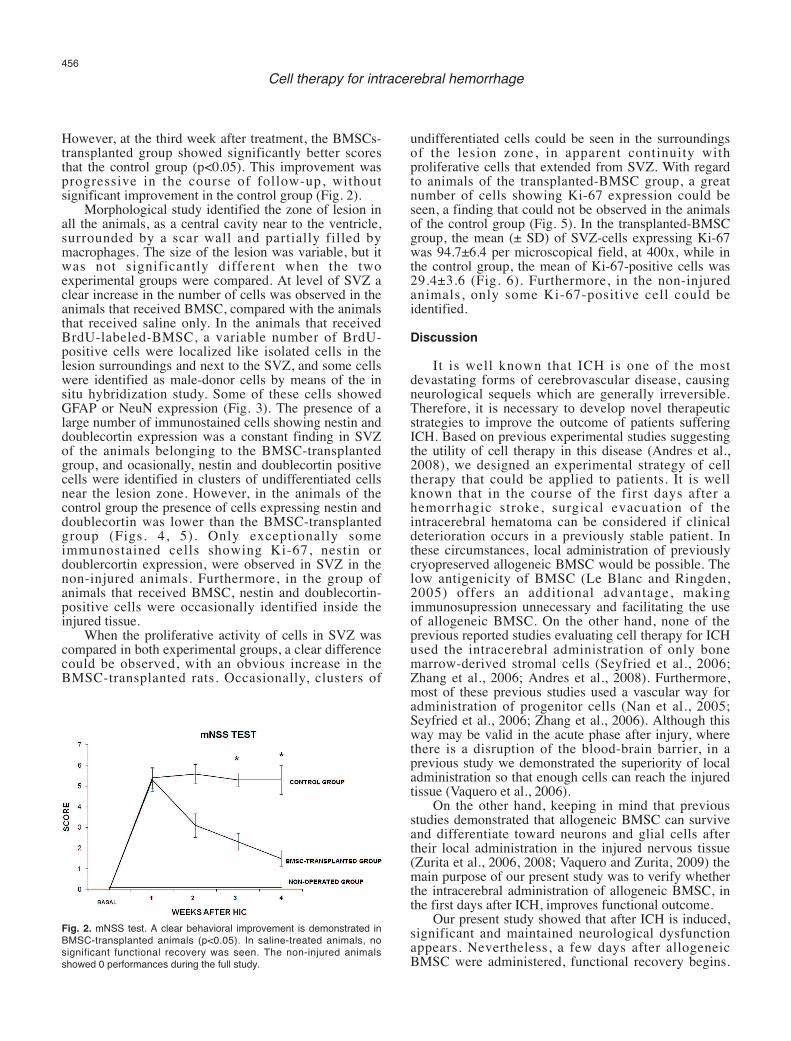
However, at the third week after treatment, the BMSCs-transplanted group showed significantly better scoresthat the control group (p<0.05). This improvement wasprogressive in the course of follow-up, withoutsignificant improvement in the control group (Fig. 2).
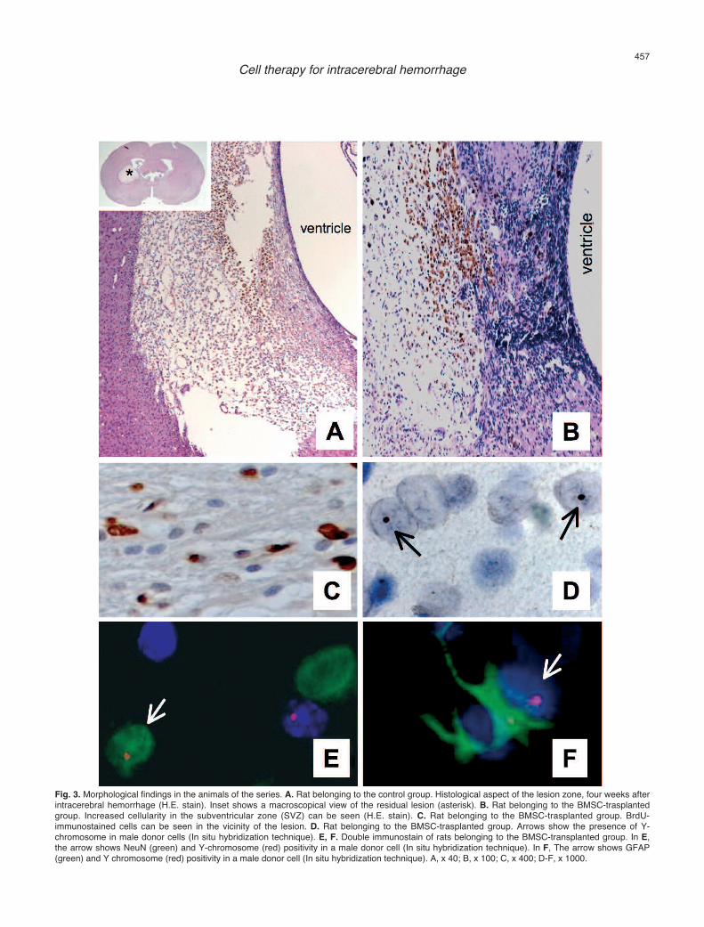
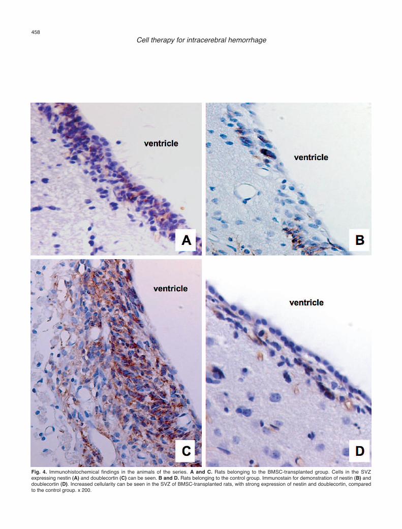
Morphological study identified the zone of lesion inall the animals, as a central cavity near to the ventricle,surrounded by a scar wall and partially filled bymacrophages. The size of the lesion was variable, but itwas not significantly different when the twoexperimental groups were compared. At level of SVZ aclear increase in the number of cells was observed in theanimals that received BMSC, compared with the animalsthat received saline only. In the animals that receivedBrdU-labeled-BMSC, a variable number of BrdU-positive cells were localized like isolated cells in thelesion surroundings and next to the SVZ, and some cellswere identified as male-donor cells by means of the insitu hybridization study. Some of these cells showedGFAP or NeuN expression (Fig. 3). The presence of alarge number of immunostained cells showing nestin anddoublecortin expression was a constant finding in SVZof the animals belonging to the BMSC-transplantedgroup, and ocasionally, nestin and doublecortin positivecells were identified in clusters of undifferentiated cellsnear the lesion zone. However, in the animals of thecontrol group the presence of cells expressing nestin anddoublecortin was lower than the BMSC-transplantedgroup (Figs. 4, 5). Only exceptionally someimmunostained cells showing Ki-67, nestin ordoublercortin expression, were observed in SVZ in thenon-injured animals. Furthermore, in the group ofanimals that received BMSC, nestin and doublecortin-positive cells were occasionally identified inside theinjured tissue.
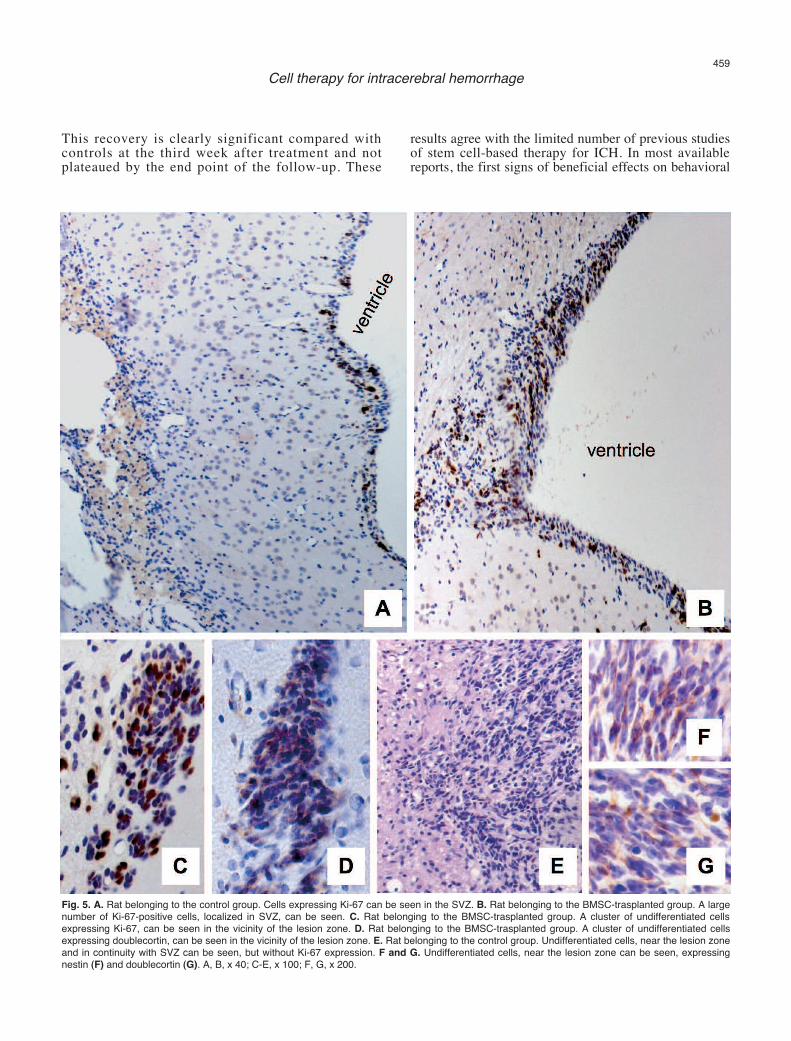
When the proliferative activity of cells in SVZ wascompared in both experimental groups, a clear differencecould be observed, with an obvious increase in theBMSC-transplanted rats. Occasionally, clusters of
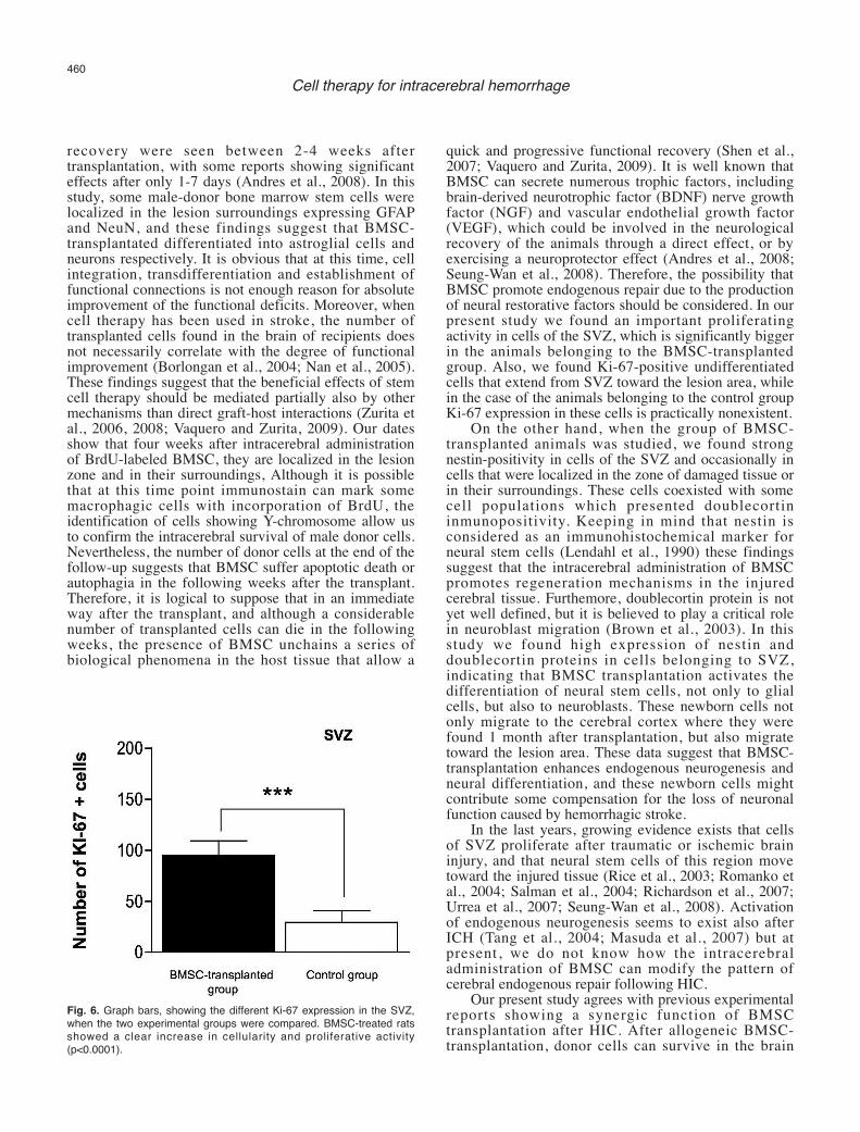
undifferentiated cells could be seen in the surroundingsof the lesion zone, in apparent continuity withproliferative cells that extended from SVZ. With regardto animals of the transplanted-BMSC group, a greatnumber of cells showing Ki-67 expression could beseen, a finding that could not be observed in the animalsof the control group (Fig. 5). In the transplanted-BMSCgroup, the mean (± SD) of SVZ-cells expressing Ki-67was 94.7±6.4 per microscopical field, at 400x, while inthe control group, the mean of Ki-67-positive cells was29.4±3.6 (Fig. 6). Furthermore, in the non-injuredanimals, only some Ki-67-positive cell could beidentified. Discussion
It is well known that ICH is one of the mostdevastating forms of cerebrovascular disease, causingneurological sequels which are generally irreversible.Therefore, it is necessary to develop novel therapeuticstrategies to improve the outcome of patients sufferingICH. Based on previous experimental studies suggestingthe utility of cell therapy in this disease (Andres et al.,2008), we designed an experimental strategy of celltherapy that could be applied to patients. It is wellknown that in the course of the first days after ahemorrhagic stroke, surgical evacuation of theintracerebral hematoma can be considered if clinicaldeterioration occurs in a previously stable patient. Inthese circumstances, local administration of previouslycryopreserved allogeneic BMSC would be possible. Thelow antigenicity of BMSC (Le Blanc and Ringden,2005) offers an additional advantage, makingimmunosupression unnecessary and facilitating the useof allogeneic BMSC. On the other hand, none of theprevious reported studies evaluating cell therapy for ICHused the intracerebral administration of only bonemarrow-derived stromal cells (Seyfried et al., 2006;Zhang et al., 2006; Andres et al., 2008). Furthermore,most of these previous studies used a vascular way foradministration of progenitor cells (Nan et al., 2005;Seyfried et al., 2006; Zhang et al., 2006). Although thisway may be valid in the acute phase after injury, wherethere is a disruption of the blood-brain barrier, in aprevious study we demonstrated the superiority of localadministration so that enough cells can reach the injuredtissue (Vaquero et al., 2006).
On the other hand, keeping in mind that previousstudies demonstrated that allogeneic BMSC can surviveand differentiate toward neurons and glial cells aftertheir local administration in the injured nervous tissue(Zurita et al., 2006, 2008; Vaquero and Zurita, 2009) themain purpose of our present study was to verify whetherthe intracerebral administration of allogeneic BMSC, inthe first days after ICH, improves functional outcome.
Our present study showed that after ICH is induced,significant and maintained neurological dysfunctionappears. Nevertheless, a few days after allogeneicBMSC were administered, functional recovery begins.
456Cell therapy for intracerebral hemorrhage
Fig. 2. mNSS test. A clear behavioral improvement is demonstrated inBMSC-transplanted animals (p<0.05). In saline-treated animals, nosignificant functional recovery was seen. The non-injured animalsshowed 0 performances during the full study.
457Cell therapy for intracerebral hemorrhage
Fig. 3. Morphological findings in the animals of the series. A. Rat belonging to the control group. Histological aspect of the lesion zone, four weeks afterintracerebral hemorrhage (H.E. stain). Inset shows a macroscopical view of the residual lesion (asterisk). B. Rat belonging to the BMSC-trasplantedgroup. Increased cellularity in the subventricular zone (SVZ) can be seen (H.E. stain). C. Rat belonging to the BMSC-trasplanted group. BrdU-immunostained cells can be seen in the vicinity of the lesion. D. Rat belonging to the BMSC-trasplanted group. Arrows show the presence of Y-chromosome in male donor cells (In situ hybridization technique). E, F. Double immunostain of rats belonging to the BMSC-transplanted group. In E,the arrow shows NeuN (green) and Y-chromosome (red) positivity in a male donor cell (In situ hybridization technique). In F, The arrow shows GFAP(green) and Y chromosome (red) positivity in a male donor cell (In situ hybridization technique). A, x 40; B, x 100; C, x 400; D-F, x 1000.
458Cell therapy for intracerebral hemorrhage
Fig. 4. Immunohistochemical findings in the animals of the series. A and C. Rats belonging to the BMSC-transplanted group. Cells in the SVZexpressing nestin (A) and doublecortin (C) can be seen. B and D. Rats belonging to the control group. Immunostain for demonstration of nestin (B) anddoublecortin (D). Increased cellularity can be seen in the SVZ of BMSC-transplanted rats, with strong expression of nestin and doublecortin, comparedto the control group. x 200.
This recovery is clearly significant compared withcontrols at the third week after treatment and notplateaued by the end point of the follow-up. These
results agree with the limited number of previous studiesof stem cell-based therapy for ICH. In most availablereports, the first signs of beneficial effects on behavioral
459Cell therapy for intracerebral hemorrhage
Fig. 5. A. Rat belonging to the control group. Cells expressing Ki-67 can be seen in the SVZ. B. Rat belonging to the BMSC-trasplanted group. A largenumber of Ki-67-positive cells, localized in SVZ, can be seen. C. Rat belonging to the BMSC-trasplanted group. A cluster of undifferentiated cellsexpressing Ki-67, can be seen in the vicinity of the lesion zone. D. Rat belonging to the BMSC-trasplanted group. A cluster of undifferentiated cellsexpressing doublecortin, can be seen in the vicinity of the lesion zone. E. Rat belonging to the control group. Undifferentiated cells, near the lesion zoneand in continuity with SVZ can be seen, but without Ki-67 expression. F and G. Undifferentiated cells, near the lesion zone can be seen, expressingnestin (F) and doublecortin (G). A, B, x 40; C-E, x 100; F, G, x 200.
recovery were seen between 2-4 weeks aftertransplantation, with some reports showing significanteffects after only 1-7 days (Andres et al., 2008). In thisstudy, some male-donor bone marrow stem cells werelocalized in the lesion surroundings expressing GFAPand NeuN, and these findings suggest that BMSC-transplantated differentiated into astroglial cells andneurons respectively. It is obvious that at this time, cellintegration, transdifferentiation and establishment offunctional connections is not enough reason for absoluteimprovement of the functional deficits. Moreover, whencell therapy has been used in stroke, the number oftransplanted cells found in the brain of recipients doesnot necessarily correlate with the degree of functionalimprovement (Borlongan et al., 2004; Nan et al., 2005).These findings suggest that the beneficial effects of stemcell therapy should be mediated partially also by othermechanisms than direct graft-host interactions (Zurita etal., 2006, 2008; Vaquero and Zurita, 2009). Our datesshow that four weeks after intracerebral administrationof BrdU-labeled BMSC, they are localized in the lesionzone and in their surroundings, Although it is possiblethat at this time point immunostain can mark somemacrophagic cells with incorporation of BrdU, theidentification of cells showing Y-chromosome allow usto confirm the intracerebral survival of male donor cells.Nevertheless, the number of donor cells at the end of thefollow-up suggests that BMSC suffer apoptotic death orautophagia in the following weeks after the transplant.Therefore, it is logical to suppose that in an immediateway after the transplant, and although a considerablenumber of transplanted cells can die in the followingweeks, the presence of BMSC unchains a series ofbiological phenomena in the host tissue that allow a
quick and progressive functional recovery (Shen et al.,2007; Vaquero and Zurita, 2009). It is well known thatBMSC can secrete numerous trophic factors, includingbrain-derived neurotrophic factor (BDNF) nerve growthfactor (NGF) and vascular endothelial growth factor(VEGF), which could be involved in the neurologicalrecovery of the animals through a direct effect, or byexercising a neuroprotector effect (Andres et al., 2008;Seung-Wan et al., 2008). Therefore, the possibility thatBMSC promote endogenous repair due to the productionof neural restorative factors should be considered. In ourpresent study we found an important proliferatingactivity in cells of the SVZ, which is significantly biggerin the animals belonging to the BMSC-transplantedgroup. Also, we found Ki-67-positive undifferentiatedcells that extend from SVZ toward the lesion area, whilein the case of the animals belonging to the control groupKi-67 expression in these cells is practically nonexistent.
On the other hand, when the group of BMSC-transplanted animals was studied, we found strongnestin-positivity in cells of the SVZ and occasionally incells that were localized in the zone of damaged tissue orin their surroundings. These cells coexisted with somecell populations which presented doublecortininmunopositivity. Keeping in mind that nestin isconsidered as an immunohistochemical marker forneural stem cells (Lendahl et al., 1990) these findingssuggest that the intracerebral administration of BMSCpromotes regeneration mechanisms in the injuredcerebral tissue. Furthemore, doublecortin protein is notyet well defined, but it is believed to play a critical rolein neuroblast migration (Brown et al., 2003). In thisstudy we found high expression of nestin anddoublecortin proteins in cells belonging to SVZ,indicating that BMSC transplantation activates thedifferentiation of neural stem cells, not only to glialcells, but also to neuroblasts. These newborn cells notonly migrate to the cerebral cortex where they werefound 1 month after transplantation, but also migratetoward the lesion area. These data suggest that BMSC-transplantation enhances endogenous neurogenesis andneural differentiation, and these newborn cells mightcontribute some compensation for the loss of neuronalfunction caused by hemorrhagic stroke.
In the last years, growing evidence exists that cellsof SVZ proliferate after traumatic or ischemic braininjury, and that neural stem cells of this region movetoward the injured tissue (Rice et al., 2003; Romanko etal., 2004; Salman et al., 2004; Richardson et al., 2007;Urrea et al., 2007; Seung-Wan et al., 2008). Activationof endogenous neurogenesis seems to exist also afterICH (Tang et al., 2004; Masuda et al., 2007) but atpresent, we do not know how the intracerebraladministration of BMSC can modify the pattern ofcerebral endogenous repair following HIC.
Our present study agrees with previous experimentalreports showing a synergic function of BMSCtransplantation after HIC. After allogeneic BMSC-transplantation, donor cells can survive in the brain
460Cell therapy for intracerebral hemorrhage
Fig. 6. Graph bars, showing the different Ki-67 expression in the SVZ,when the two experimental groups were compared. BMSC-treated ratsshowed a clear increase in cellularity and proliferative activity(p<0.0001).
tissue, showing morphological evidence oftransdifferentiation to neurons and astroglial cells.Furthemore, BMSC-transplantation enhancesendogenous neurogenesis that usually exists afterintracerebral hemorrhage. Both mechanisms cancontribute to compensate the loss of neural tissue and ofneurological function. On the other hand, theobservation that after this type of cell therapy quick andprogressive neurological recovery can be obtainedsuggests that the beneficial effect of BMSC can beattributed mainly to a stimulation of neuronal plasticity.Although our results should be extrapolated to thehuman disease with caution, it is obvious that the use ofallogeneic BMSC offers great promise for developingnovel and efficacious strategies in patients sufferingICH.Acknowledgements This work was supported by grants from Fina-Biotech S.L.
References
Andres R.H., Guzman R., Ducray A.D., Mordasini P., Gera A., Barth A.,Widmer H.R. and Steinberg G.K. (2008). Cell replacement therapyfor intracerebral hemorrhage. Neurosurg. Focus 24, E15.
Borlongan C.V., Hadma M., Sanberg C.D. and Sanberg P.R. (2004).Central nervouus system entry of peripherally injected umbilical cordblood cells is not required for neuroprotection in stroke. Stroke 35,2385-2389.
Brown J.P., Couillard-Despres S., Cooper-Kuhn C.M., Winkler J., AignerL. and Kunh H.G. (2003). Transient expression of doublecortinduring adult neurogenesis. J. Comp. Neurol. 467, 1-10.
Ferro J.M. (2006). Update on intracerebral haemorrhage. J. Neurol. 253,985-999.
Le Blanc K. and Ringdén O. (2005). Immunobiology of humanmesenchymal stem cells and future use in hematopioetic stem celltransplantation. Biol. Blood Marrow. Transplant. 11, 321-334.
Lendahl U., Zimmerman L.B. and McKay R.D.G. (1990). CNS stem cellsexpress a new class of intermediate filament protein. Cell 60, 585-595.
Li Y., Chen J. and Chopp M. (2001). Adult bone marrow transplantationafter stroke in adult rats. Cell Transplant.10, 31-40.
Masuda T., Isobe Y., Aihara N., Furuyama F., Misumi S., Kim T.,Nishino H. and Hida H. (2007). Increase in neurogenesis andneuroblast migration after a small intracerebral hemorrhage in rats.Neurosci. Lett. 425, 114-119.
Nan Z., Grande A., Sanberg C.D., Sanberg P.R. and Low W.C. (2005).Infusion of human umbilcal cord blood ameliorates neurologicdeficits in rats with hemorrhagic brain injury. Ann. N.Y. Acad. Sci.1049, 84-96.
Rice A.C., Khaldi A., Harvey H.B., Salman N.J., White F., Fillmore H.and Bullock M.R. (2003). Proliferation and neuronal differentiation of
mitotically active cells following traumatic brain injury. Exp. Neurol.183, 406-417.
Richardson R.M., Sun D. and Bullock M.R. (2007). Neurogenesis aftertraumatic brain injury. Neurosurg. Clin. N. Am. 18, 169-181.
Romanko M.J., Rola R., Fike J.R., Szele F.G., Dizon M.L., Felling R.J.,Brazel C.Y. and Levison S.W. (2004). Roles of the mammaliansubventricular zone in cell replacement after brain injury. Progr.Neurobiol. 74, 77-99.
Salman H., Gosh P. and Kernie S.G. (2004). Subventricular zone neuralstem cells remodel the brain following traumatic injury in adult mice.J. Neurotrauma 21, 283-292.
Seung-Wan Y., Sung-Soo K., Soo-Yeol L., Hey-Sun L., Hyung-Soo K.,Young-Don L. and Suh-Kim H. (2008). Mesenchymal stem cellspromote proliferation of endogenous neural stem cells and survivalof newborn cells in a rat stroke model. Exp. Mol. Med. 40, 387-397.
Seyfried D., Ding J., Han Y., Li Y., Chen J. and Chopp M. (2006).Effects of intravenous administration of human bone marrow stromalcells after cerebral hemorrhage in rats. J. Neurosurg. 104, 313-318.
Shen L.H., Li Y., Chen J., Cui Y., Zhang C., Kapke A., Lu M., Savant-Bhonsale S. and Chopp M. (2007). One-year follow-up after bonemarrow stromal cell treatment in middle-aged female rats withstroke. Stroke 38, 2150-2156.
Tang T., Li X.Q., Wu H., Luo J.K., Zhang H.X. and Luo T.L. (2004).Activation of endogenous neural stem cells in experimentalintracerebral hemorrhagic rat brains. Chin. Med. J. (Engl) 117, 1342-1347.
Urrea C., Castellanos D.A., Sagen J., Tsoulfas P., Bramlett H.M. andDietrich W.D. (2007). Widespread cellular proliferation and focalneurogenesis after traumatic brain injury in the rat. Restor. Neurol.Neurosc. 25, 65-76.
Vaquero J. and Zurita M. (2009). Bone marrow stromal cells for spinalcord repair: a challenge for contemporary neurobiology. Histol.Histopathol. 24, 107-116.
Vaquero J., Zurita M., Oya S. and Santos M. (2006). Cell therapy usingbone marrow stromal cells in chronic paraplegic rats: systemic orlocal administration? Neurosci. Lett. 398, 129-134.
Zhang H., Huang Z., Xu Y. and Zhang S. (2006). Differentiation andneurological benefit of the mesenchymal stem cells transplanted intothe rat brain following intracerebral hemorrhage. Neurol. Res. 28,104-112.
Zurita M. and Vaquero J. (2004). Functional recovery in chronicparaplegia after bone marrow stromal cells transplantation.Neuroreport. 15, 1105-1108.
Zurita M. and Vaquero J. (2006). Bone marrow stromal cells canachieve cure of chronic paraplegic rats: functional andmorphological outcome one year after transplantation. Neurosci.Lett. 402, 51-56.
Zurita M., Vaquero J., Bonilla C., Santos M., De Haro J., Oya S. andAguayo C. (2008). Functional recovery of chronic paraplegic pigsafter autologous transplantation of bone marrow stromal cells.Transplantation 86, 845-853.
Accepted November 2, 2009
461Cell therapy for intracerebral hemorrhage