intestinal pregnane x receptor links xenobiotic exposure ... endocrinol-pxr.pdf · intestinal...
TRANSCRIPT
Intestinal Pregnane X Receptor Links XenobioticExposure and Hypercholesterolemia
Yipeng Sui,* Robert N. Helsley,* Se-Hyung Park, Xiulong Song, Zun Liu,and Changcheng Zhou
Department of Pharmacology and Nutritional Sciences (Y.S., R.N.H., S.-H.P., X.S., Z.L., C.Z.) and SahaCardiovascular Research Center (C.Z.), University of Kentucky, Lexington, Kentucky 40506
Recent studies have associated endocrine-disrupting chemical (EDC) exposure with the increasedrisk of cardiovascular disease in humans, but the underlying mechanisms responsible for theseassociations remain elusive. Many EDCs have been implicated in activation of the nuclear receptorpregnane X receptor (PXR), which acts as a xenobiotic sensor to regulate xenobiotic metabolismin the liver and intestine. Here we report an important role of intestinal PXR in linking xenobioticexposure and hyperlipidemia. We identified tributyl citrate (TBC), one of a large group of Foodand Drug Administration–approved plasticizers for pharmaceutical or food applications, as apotent and selective PXR agonist. TBC efficiently activated PXR and induced PXR target geneexpression in vitro and in vivo. Interestingly, TBC activated intestinal PXR but did not affect hepaticPXR activity. Exposure to TBC increased plasma total cholesterol and atherogenic low-densitylipoprotein cholesterol levels in wild-type mice, but not in PXR-deficient mice. TBC-mediated PXRactivation stimulated the expression of an essential cholesterol transporter, Niemann-Pick C1-like1 (NPC1L1), in the intestine. Promoter analysis revealed a DR-4 type of PXR response element in thehuman NPC1L1 promoter, and TBC promoted PXR recruitment onto the NPC1L1 promoter. Con-sistently, TBC treatment significantly increased lipid uptake by human and murine intestinal cellsand deficiency of PXR inhibited TBC-elicited lipid uptake. These findings provide critical mecha-nistic insight for understanding the impact of EDC-mediated PXR activation on lipid homeostasisand demonstrate a potential role of PXR in mediating the adverse effects of EDCs on cardiovas-cular disease risk in humans. (Molecular Endocrinology 29: 765–776, 2015)
Influences of the chemical environment on human healthhave recently become the subject of intense interest.
Mounting evidence shows that endocrine-disruptingchemicals (EDCs) can interfere with complex endocrinesignaling mechanisms and result in adverse consequencesin humans and wildlife (1, 2). Recent findings have impli-cated exposure to EDCs in the etiology of cardiovasculardisease (CVD) and metabolic disorders (1–6). For in-stance, higher bisphenol A (BPA) exposure has been con-sistently associated with CVD in multiple large-scale hu-man population studies (4, 5, 7). Exposure to certainpolychlorinated biphenyls (PCBs) induces hypercholes-terolemia and promotes atherosclerosis in animals (8, 9).Circulating PCB levels have been associated with athero-
sclerotic plaques in elderly individuals (10). High circu-lating levels of phthalates are also associated with carotidatherosclerosis (11). However, the underlying mecha-nisms responsible for these associations remain largelyunknown, which continues to hamper rational assess-ment of the health risks of EDC exposure.
Many EDCs such as phthalates, PCBs, and BPA and itsanalogs have been implicated in the activation of the preg-nane X receptor (PXR) (also known as steroid and xeno-biotic receptor) (12–15). PXR is a nuclear receptor acti-vated by numerous endogenous hormones, dietarysteroids, pharmaceutical agents, and xenobiotic chemi-cals (15–17). PXR functions as a xenobiotic sensor thatinduces expression of genes required for xenobiotic me-
ISSN Print 0888-8809 ISSN Online 1944-9917Printed in U.S.A.Copyright © 2015 by the Endocrine SocietyReceived November 6, 2014. Accepted March 23, 2015.First Published Online March 26, 2015
* Y.S. and R.N.H. contributed equally to the study.Abbreviations: ATBC, acetyl tributyl citrate; ATEC, acetyl triethyl citrate; BPA, bisphe-nol A; BW, body weight; ChIP, chromatin immunoprecipitation; CVD, cardiovasculardisease; CYP, cytochrome P450; DEHP, di(2-ethylhexyl) phthalate; DEP, diethyl phtha-late; DiBP, di-isobutyl phthalate; DiNP, diisononyl phthalate; DMSO, dimethyl sulfoxide;
O R I G I N A L R E S E A R C H
doi: 10.1210/me.2014-1355 Mol Endocrinol, May 2015, 29(5):765–776 mend.endojournals.org 765
The Endocrine Society. Downloaded from press.endocrine.org by [${individualUser.displayName}] on 01 May 2015. at 06:58 For personal use only. No other uses without permission. . All rights reserved.

tabolism in the liver and intestine, including cytochromesP450 (CYPs), conjugating enzymes (eg, glutathione trans-ferase), and ABC family transporters (eg, multidrug resis-tance 1 [MDR1]) (15, 18). In the past decade, the role ofPXR as a xenobiotic sensor has been well established (15).However, the role of PXR in mediating the pathophysio-logical effects of EDCs in humans and animals remainselusive.
The identification of PXR as a xenobiotic sensor pro-vided an important tool for the study of new mechanismsthrough which xenobiotic exposure affects diseases. Re-cent evidence indicates that PXR may also play an impor-tant role in the regulation of lipid homeostasis (19–24).For instance, it is well-known that many clinically rele-vant PXR ligands (eg, rifampicin and ritonavir) can ele-vate plasma lipid levels in patients and increase their CVDrisk (25–28). A meta-analysis of 7 genome-wide associa-tion studies indicated that common genetic variants inPXR can affect plasma lipid levels in humans and 19 PXRsingle nucleotide polymorphisms were identified to sig-nificantly affect plasma low-density lipoprotein (LDL)cholesterol levels (29). We have recently demonstratedthat chronic activation of PXR elicited by feeding mice themouse PXR ligand pregnane 16�-carbonitrile (PCN) ledto increased levels of plasma total cholesterol and theatherogenic lipoproteins LDL and very low-density lipo-protein (VLDL) in wild-type (WT) mice, but not in PXR-deficient (PXR�/�) mice (19). Activation of PXR also in-creased plasma total cholesterol and VLDL levels inapolipoprotein E *3-Leiden mice, which exhibit a human-like lipoprotein distribution on a cholesterol-rich diet(20). Very recently, we identified amprenavir, a widelyused antiretroviral drug, as a potent PXR-selective ago-nist (24). Exposure to amprenavir significantly increasedplasma total cholesterol and LDL cholesterol levels in WTmice, but not in PXR�/� mice (24).
Although emerging evidence is consistent with the hy-pothesis that modulation of PXR activity alters lipid ho-meostasis, the mechanisms underlying PXR ligand–elic-ited hyperlipidemia remain largely unknown. PXR isexpressed at high levels in the liver and intestine, twoorgans that play a central role in whole-body lipid ho-meostasis. PXR has been reported to regulate several key
hepatic lipogenic genes that promote dyslipidemia andhepatic steatosis, including CD36, SCD-1, lipin-1, In-sig-1, and S14 (21, 30–32); however, the role of intestinalPXR in the regulation of lipid homeostasis remainselusive. Here we report that intestinal PXR plays an im-portant role in linking EDC exposure and hypercholes-terolemia. We identified several phthalate substitute plas-ticizers widely used in food packaging materials, medicaldevices, cosmetics, and pharmaceutical drugs as agonistsof PXR. Tributyl citrate (TBC), one of a large group ofFood and Drug Administration (FDA)–approved phar-maceutical plasticizers, is a potent and selective PXR ag-onist but does not activate other nuclear receptors. Inter-estingly, TBC activated intestinal PXR but did not affecthepatic PXR activity. Short-term TBC exposure increasedplasma total cholesterol and atherogenic LDL cholesterollevels in WT mice, but not in PXR�/� mice. We found thatTBC-mediated PXR activation stimulated the expressionof the intestinal cholesterol transporter Niemann-PickC1-like 1 (NPC1L1) and significantly increased lipid up-take by human and murine intestinal cells. We also iden-tified a PXR-binding site in the human NPC1L1 pro-moter, indicating that NPC1L1 is a bona fide PXR targetgene. These findings provide critical mechanistic insightfor understanding the impact of EDC-mediated PXR ac-tivation on lipid homeostasis and demonstrate a potentialrole of PXR in mediating the adverse effects of EDCs onCVD risk in humans.
Materials and Methods
Reagents and plasmidsTBC, acetyl tributyl citrate (ATBC), di(2-ethylhexyl) phtha-
late (DEHP), acetyltriethyl citrate(ATEC), diisononyl phthalate(DiNP), triethyl citrate (TEC), di-n-butyl phthalate (DnBP), di-isobutyl phthalate (DiBP), diethyl phthalate (DEP), PCN, andrifampicin (RIF) were purchased from Sigma-Aldrich. All of thechemicals were dissolved in dimethyl sulfoxide (DMSO). Hu-man (h) and mouse (m) PXR expression vectors, GAL4 DNA-binding domain (DBD)-linked nuclear receptor ligand-bindingdomain vectors (GAL4-hPXR, GAL4-mPXR, GAL4-rat PXR,GAL4-retinoid acid receptor [RAR] �, GAL4-retinoid X recep-tor [RXR], GAL4-farnesoid X receptor [FXR], GAL4-liver Xreceptor [LXR], GAL4-peroxisome proliferator–activated re-ceptor [PPAR] �, GAL4-PPAR�, and GAL4-vitamin D receptor[VDR]), and CMX-�-galactosidase expression vectors were de-scribed before (12, 33, 34). VP16-PXR, GAL4-nuclear receptorcorepressor (NCoR), GAL4-silencing mediator of retinoid andthyroid hormone (SMRT), GAL4-steroid receptor coactivator-1(SRC1), GAL4-PPAR binding protein (PBP), PXR-dependentCYP3A4 promoter reporter (CYP3A4XREM-luciferase),CYP3A2 promoter reporter [(CYP3A2)3-luciferase], and GAL4reporter (MH100-luciferase) were described previously (12, 33,
DBD, DNA-binding domain; DBP, dibutyl phthalate; EDC, endocrine-disrupting chemicals;EMSA, electrophoretic mobility shift assay; FDA, Food and Drug Administration; FXR,farnesoid X receptor; HDL, high-density lipoprotein; LDL, low-density lipoprotein; m,mouse; LXR, liver X receptor; MDR1, multidrug resistance 1; NCoR, nuclear receptorcorepressor; NPC1L1, Niemann-Pick C1-like 1; PBP, peroxisome proliferator–activatedreceptor binding protein; PCB, polychlorinated biphenyl; PCN, pregnane 16�-carbonitrile;PPAR, peroxisome proliferator–activated receptor; PXR, pregnane X receptor; qPCR,quantitative real-time PCR; RAR, retinoid acid receptor; RIF, rifampin; RXR, retinoid Xreceptor; siRNA, small interfering RNA; SMRT, silencing mediator of retinoid and thyroidhormone; TBC, tributyl citrate; TEC, triethyl citrate; VLDL, very low-density lipoprotein;VDR, vitamin D receptor; WT, wild type.
766 Sui et al Role of Intestinal PXR in Lipid Homeostasis Mol Endocrinol, May 2015, 29(5):765–776
The Endocrine Society. Downloaded from press.endocrine.org by [${individualUser.displayName}] on 01 May 2015. at 06:58 For personal use only. No other uses without permission. . All rights reserved.
35). For the human NPC1L1/DR-4 reporter, 4 copies of theDR-4 element (�9590 to �9574 bp) were synthesized and in-serted into a pGL3 promoter vector (Promega) by annealingcomplementary oligonucleotides 5�-GCAGATCACTTGAG-GTCAGG-3� containing cohesive ends of the restriction enzymesites, KpnI and MluI. The 2 mutant constructs were generated ina similar manner by using oligonucleotides 5�-GCAGAACACTT-GAGAACAGG-3� and 5�-GCAGATCTCTTGAGATCTGG-3�for DR-4m1 and DR-4m2, respectively.
Cell culture and transfectionsThe human intestine epithelial cell line LS180 and hepatic
cell line HepG2 were obtained from the American Type CultureCollection. The human hepatoma HepaRG cells were purchasedfrom Life Technologies. Transfection assays were performed asdescribed previously (12, 24). The cells were transfected withvarious expression plasmids as well as the corresponding lu-ciferase reporter plasmids, together with CMX-�-galactosidasecontrol plasmids using FuGENE 6 (Roche Diagnostics). Thecells were then incubated with the corresponding ligands asindicated in the figure legends for 24 hours, and �-galactosidaseand luciferase assays were performed as described previously(12, 33). Fold activation was calculated relative to solvent con-trols. Each data point represents the average of triplicate exper-iments � SEM and was replicated in 3 to 5 independent exper-iments. For the mammalian 2-hybrid assays, LS180 cells weretransfected with the GAL4 reporter, VP16-hPXR, and GAL-SRC1, GAL-PBP, GAL-NCoR, and GAL-SMRT (12, 33). Thecells were then treated with compounds at the indicatedconcentrations.
AnimalsC57BL/6 WT mice were purchased from The Jackson Labo-
ratory. PXR-deficient mice (PXR�/�) on a C57BL/6 back-ground were used as described previously (19). All of the ani-mals were housed in the Division of Laboratory AnimalResources, University of Kentucky, as approved by the Institu-tional Animal Care and Use Committee in a specific pathogen–free environment with a light-dark cycle. Eight-week-old maleWT or PXR�/� mice were fed a semisynthetic low-fat AIN76diet containing 0.02% cholesterol (Research Diets) (19, 36, 37)and treated by oral gavage with vehicle (corn oil) or 10 mg/kgbody weight (BW) TBC daily for 7 days. In addition, WT micewere also treated with vehicle control or 10 mg/kg/BW TBCdaily by ip injection. On the day of euthanization, mice werefasted for 6 hours after the dark phase (feeding cycle) (19, 37).Mice were then anesthetized by ip injection with ketamine (FortDodge Animal Health). Mice were exsanguinated by left ven-tricular puncture, and blood was collected into EDTA-contain-ing syringes. Plasma was prepared by spinning at 16 000 g for10 minutes. The circulation was flushed with PBS, and intestinaland liver tissues were collected and stored in RNAlater solution(Life Technologies). Mouse primary hepatocytes and entero-cytes were isolated as described previously (38, 39). Primarycells were cultured in multiwell plates and treated with the in-dicated compounds before being harvested for quantitative real-time PCR (qPCR) and Western blot analysis or cholesterol up-take assay.
Plasma analysisPlasma total cholesterol and triglyceride concentrations were
determined enzymatically by colorimetric methods as describedpreviously (Roche) (24, 40). Lipoprotein fractions were isolatedby spinning 60 �L of plasma in a TL-100 ultracentrifuge (Beck-man Coulter) at its own density (1.006 g/mL) at 70 000 rpm for3 hours to harvest the supernatant and then after adjustment ofthe infranatant with solid KBr to a density of 1.063 g/mL spin-ning it for 70 000 rpm for 18 hours to harvest the supernatant(40). The cholesterol content of each supernatant and the finalinfranatant were measured and taken to be VLDL (�1.006g/mL), LDL (1.006 � d � 1.063 g/mL), and high-density lipo-protein (HDL) (d � 1.063 g/mL) cholesterol, respectively. Cho-lesterol concentrations in all 3 fractions were then determinedenzymatically by a colorimetric method (Roche). Plasma cho-lesterol levels were determined enzymatically in the originalplasma sample.
Cholesterol uptake assayA cholesterol uptake assay was performed as described pre-
viously (41). In brief, micelles were prepared by mixing 9.7 mMtaurocholate (Sigma-Aldrich), 6.47 mM egg yolk L-�-phos-phatidylcholine (Sigma-Aldrich), and 1.5 mM cholesterol (Sig-ma-Aldrich), together with 1 �Ci of [1,2-3H(N)]cholesterol(PerkinElmer)/�mol of cholesterol and evaporated under a mildstream of argon. The lipid film was hydrated in serum-freeMEM containing 0.5% fatty acid–free BSA (Sigma-Aldrich) andincubated at 37°C in a rotating incubator. Solutions were fil-tered through a 0.45-�m surfactant-free cellulose acetate filter(Corning) and used to incubate the LS180 cells or primary en-terocytes. After extensive PBS washing, the signals of the cellswere counted by a �-counter.
Electrophoretic mobility shift assay (EMSA)EMSA was performed as described previously (38, 42). In
brief, NPC1L1/DR-4 and CYP3A4/ER-6 probes were createdby annealing the oligonucleotides 5�-GGGCAGATCACTT-GAGGTCAGGAG-3�(NPC1L1/DR-4),5�-GGGCAGAACACTTGAGAACAGGAG-3� (NPC1L1/DR-4m1), 5�-GGGCAGATCTCTTGAGaTCTGGAG-3� (NPC1L1/DR-4m2), or 5�-ATATGAACTCAAAGGAGGTCAGTG-3� (CYP3A4/ER6) tothe complementary strand. Double-stranded oligonucleotideswere end labeled using T4 polynucleotide kinase (New EnglandBiosciences) and �-[32P]ATP (PerkinElmer). Then 5 �L of invitro–translated PXR or RXR protein was incubated with 2 �gof poly d(I-C) (Promega), 2 �L of bandshift buffer (50 mMMgCl2 and 340 mM KCl), and 6 �L of delta buffer (0.1 mMEDTA, 40 mM KCl, 25 mM HEPES [pH 7.6], 8% Ficoll 400,and 1 mM dithiothreitol) on ice for 10 minutes. 32P-labeleddouble-stranded oligonucleotide probe (100 000 cpm) was thenadded, and the reaction was incubated for another 30 minuteson ice. For the supershift assays, proteins were incubated with 2�g of goat anti-PXR (sc-7739; Santa Cruz Biotechnology) orrabbit anti-PXR (sc-25381; Santa Cruz Biotechnology) antibod-ies for 1 hour before the addition of 32P-labeled probe. Thebinding complexes were subjected to electrophoresis in a 6%nondenaturing polyacrylamide gel containing 0.5� Tris-borate-EDTA (TBE). The gels were dried and visualized by exposure toX-ray film.
doi: 10.1210/me.2014-1355 mend.endojournals.org 767
The Endocrine Society. Downloaded from press.endocrine.org by [${individualUser.displayName}] on 01 May 2015. at 06:58 For personal use only. No other uses without permission. . All rights reserved.
Chromatin immunoprecipitation (ChIP)ChIP analysis was performed by using an antibody against
PXR (sc-25381; Santa Cruz Biotechnology) and a SimpleChIPEnzymatic Chromatin IP Kit (Cell Signaling). The precipitatedgenomic DNA relative to inputs was analyzed by semiquantita-tive PCR using specific primers and DNA polymerase (Takara).The sequences of primer sets used for PCR are listed in Supple-mental Table 1.
RNA isolation and qPCR analysisTotal RNA was isolated from mouse tissues and intestinal
LS180, HepaRG, and primary cells using TRIzol reagent (LifeTechnologies) per the manufacturer-supplied protocol. qPCRwas performed using gene-specific primers and the SYBR GreenPCR kit (Life Technologies) as described previously (12, 24).The primer sets used in this study are listed in SupplementalTable 1.
Western blot analysisWestern blot analysis was performed as described previously
(42). Proteins were isolated from cells or mouse tissues by ho-mogenization in radioimmunoprecipitation assay buffer withcomplete mini protease inhibitor cocktail (Roche). Protein con-centrations were determined by the Pierce BCA protein assay kit(Thermo Scientific). Anti-PXR antibodies were purchased fromSanta Cruz Biotechnology (sc-7739), anti-NPC1L1 antibodieswere purchased from Novus Biologicals (NB400–128), and an-ti-�-actin antibodies were purchased from Sigma-Aldrich(A2066).
Statistical analysisAll data are expressed as means � SEM unless otherwise
noted. Statistical analysis was performed using a two-sample,two-tailed Student t test unless otherwise noted, with P � .05regarded as significant. One-way ANOVA was used when mul-tiple comparisons were made, followed by the Dunnett t test formultiple comparisons to a control.
Results
FDA-approved phthalate substitute plasticizers canactivate PXR
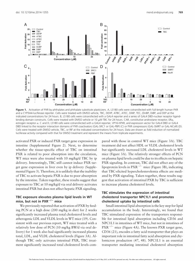
Based on previous findings that several plastic-associ-ated chemicals including BPA and phthalates can activatePXR (12, 43), we examined several widely used phtha-lates and phthalate substitute plasticizers for PXR activa-tion in transient transfection assays. Consistent with pre-vious reports (44, 45), DEHP, DiNP, and ATBC canactivate PXR and induce PXR-mediated CYP3A4-lu-ciferase reporter activities (Figure 1A). TBC, an FDA-approved phthalate substitute plasticizer used in foodcontact substances and as a pharmaceutical excipient,was a more potent PXR agonist than any of the othertested plasticizers and induced reporter gene activity in adose-dependent manner (Figure 1A). We then tested theability of TBC to activate a panel of other nuclear recep-
tors, including mouse PXR, human RAR�, RXR, FXR,LXR�, PPAR�, PPAR�, VDR, constitutive androstanereceptor, and estrogen receptor � (Figure 1B). TBC canactivate both human and mouse PXR but was unable toactivate any of the other nuclear receptors. Thus, TBC isa PXR-selective agonist.
Nuclear receptor coregulators play critical roles in nu-clear receptor signaling. We used a mammalian 2-hybridassay to evaluate the effects TBC on PXR coregulatorinteraction. TBC promoted the specific interactions be-tween PXR and the coactivators SRC-1 and PBP (Figure1C). Consistent with our previous report (12, 33), unli-ganded PXR interacted with corepressors, NCoR andSMRT (Figure 1D), and TBC disrupted this interaction asdid the potent human PXR ligand RIF (Figure 1D). Thus,binding of TBC to PXR inhibits PXR-corepressor inter-action and promotes PXR-coactivator recruitment,thereby inducing PXR transcriptional activation in a con-centration-dependent manner.
TBC activates intestinal PXR but does not affecthepatic PXR activity
We next used human hepatoma HepaRG cells (46) andintestinal LS180 cells (24, 33) to test the effects of TBCexposure on PXR activity and target gene expression. Theknown human PXR ligand RIF induced the expression ofbona fide PXR target genes involved in phase I (CYP3A4),phase II (UGT1A1), and phase III (MDR1) metabolism inboth cell lines. Interestingly, TBC stimulated PXR targetgene expression in intestinal LS180 cells but not inHepaRG cells (Figure 2A). We then isolated mouse pri-mary hepatocytes and enterocytes to confirm these find-ings. As expected, the mouse PXR ligand PCN inducedPXR target gene expression in both hepatocytes and en-terocytes (Figure 2B). However, TBC was only able toinduce PXR target gene expression in enterocytes but notin hepatocytes (Figure 2B). We also performed transfec-tion assays in LS180 and HepG2 cell lines. Consistently,TBC did not activate PXR in HepG2 cells but was able toinduce PXR reporter activity in LS180 cells after beingtreated for as few as 3 hours (Supplemental Figure 1).
To further investigate the tissue-specific effects of TBCon PXR activity in vivo, WT mice were treated with ve-hicle (corn oil), 10 mg/kg BW TBC, or PCN daily by oralgavage for 1 week. Consistent with our previous report(38), PCN activated PXR and induced target gene expres-sion in both liver and intestine (Figure 2C). However,TBC only stimulated expression of known PXR targetgenes in intestine but not in liver (Figure 2C). To examinewhether lower TBC doses can activate intestinal PXR invivo, we also treated mice with 2.5 or 5 mg/kg/d TBC byoral gavage for 1 week. Neither 5 nor 2.5 mg/kg/d TBC
768 Sui et al Role of Intestinal PXR in Lipid Homeostasis Mol Endocrinol, May 2015, 29(5):765–776
The Endocrine Society. Downloaded from press.endocrine.org by [${individualUser.displayName}] on 01 May 2015. at 06:58 For personal use only. No other uses without permission. . All rights reserved.
activated PXR or induced PXR target gene expression inintestine (Supplemental Figure 2). Next, to determinewhether the tissue-specific effect of TBC on intestinalPXR is related to poor absorption into the circulation,WT mice were also treated with 10 mg/kg/d TBC by ipdelivery. Interestingly, TBC still cannot induce PXR tar-get gene expression in liver even by ip delivery (Supple-mental Figure 3). Therefore, it is unlikely that the inabilityof TBC to activate hepatic PXR is due to poor absorptionby the intestine. Taken together, these results suggest thatexposure to TBC at 10 mg/kg/d via oral delivery activatesintestinal PXR but does not affect hepatic PXR signaling.
TBC exposure elevates plasma lipid levels in WTmice, but not in PXR�/� mice
We previously reported that activation of PXR by feed-ing PCN at a high dose (200 mg/kg in diet) for 2 weekssignificantly increased plasma total cholesterol levels andatherogenic LDL and VLDL levels in WT mice (19). Con-sistent with our previous report, WT mice treated with arelatively low dose of PCN (10 mg/kg BW/d via oral de-livery) for 1 week also had significantly increased plasmatotal, LDL, and VLDL cholesterol levels (Figure 3A). Al-though TBC only activates intestinal PXR, TBC treat-ment significantly increased total cholesterol levels com-
pared with those in control WT mice (Figure 3A). TBCtreatment did not affect HDL or VLDL cholesterol levelsbut significantly increased LDL cholesterol levels in WTmice (Figure 3A). The relatively stronger effects of PCNon plasma lipid levels could be due to its effects on hepaticPXR signaling. In contrast, TBC did not affect any of thelipoprotein levels in PXR�/� mice (Figure 3B), indicatingthat TBC-elicited hypercholesterolemia effects are medi-ated by PXR signaling. Taken together, these results sug-gest that activation of intestinal PXR by TBC is sufficientto increase plasma cholesterol levels.
TBC stimulates the expression of intestinalcholesterol transporter NPC1L1 and promotescholesterol uptake by intestinal cells
Small intestinal lipid absorption is the key step for lipidaccumulation in the body. Interestingly, we found thatTBC stimulated expression of the transporters responsi-ble for intestinal lipid absorption including CD36 andNPC1L1 in intestines of WT mice, but not in intestines ofPXR�/� mice (Figure 4A). The known PXR target gene,CD36 (21), encodes a fatty acid transporter that plays animportant role in intestinal fatty acid absorption and chy-lomicron production (47, 48). NPC1L1 is an essentialtransporter mediating intestinal cholesterol absorption
Cont
rol
hPXR
mPX
R αRA
R RXR
FXR α
LXR
αPP
AR
γPP
AR VDR
CAR α
ER
0
10
20
30
40
50
60
70ControlTBC
Fold
act
ivat
ion
0
5
10
15
20
25
DEHP
ATECDiNPTECDnBP
TBC
DiBPDEP
0 5 10 20Concentration (μM)
ATBC
Control
Fold
Act
ivat
ion
A B
Concentration (μM)
TBC - 5 10 20 - - -RIF - - - - 5 10 20
0
2
4
6
8 ControlSRC1PBP
Fold
Act
ivat
ion
Concentration (μM)
0
2
4
6 ControlSMRTNcoR
TBC - 5 10 20 - - -RIF - - - - 5 10 20
Fold
Act
ivat
ion
C D
Figure 1. Activation of PXR by phthalates and phthalate substitute plasticizers. A, LS180 cells were cotransfected with full-length human PXRand a CYP3A4-luciferase reporter. Cells were treated with DMSO vehicle, TBC, DEHP, ATBC, ATEC, DiNP, TEC, (DnBP, DiBP, and DEP at theindicated concentrations for 24 hours. B, LS180 cells were cotransfected with a GAL4 reporter and a series of GAL4 DBD-nuclear receptor ligand-binding domain constructs. Cells were treated with DMSO vehicle or 10 �M TBC for 24 hours. CAR, constitutive androstane receptor; ER�,estrogen receptor �. C and D, LS180 cells were cotransfected with a GAL4 reporter, VP16-hPXR, and expression vector for GAL4 DBD or GAL4DBD linked to the receptor interaction domains of PXR coactivators (GAL-SRC1 or GAL-PBP) (C) or PXR corepressors (GAL-SMRT or GAL-NCoR) (D).Cells were treated with DMSO vehicle, TBC, or RIF at the indicated concentrations for 24 hours. Data are shown as fold induction of normalizedluciferase activity compared with that for DMSO treatment and represent the means from triplicate experiments.
doi: 10.1210/me.2014-1355 mend.endojournals.org 769
The Endocrine Society. Downloaded from press.endocrine.org by [${individualUser.displayName}] on 01 May 2015. at 06:58 For personal use only. No other uses without permission. . All rights reserved.

(49–51). Western blot analysis also confirmed that TBCinduced intestinal NPC1L1 protein levels in WT mice butnot in PXR�/� mice (Figure 4B). Consistent with a previ-ous report (52), PXR activation did not affect expressionlevels of the cholesterol efflux transporters ABCG5 andABCG8. In addition, the known PXR ligand PCN wasalso able to induce NPC1L1 expression in intestine (Sup-plemental Figure 4), indicating that NPC1L1 is a down-stream target of PXR.
To further investigate the effects of TBC on PXR ac-tivity and NPC1L1 expression, we isolated primary en-terocytes from WT and PXR�/� mice. Consistent with invivo results, TBC treatment increased NPC1L1 mRNAand protein levels in WT enterocytes (Figure 4, C and D).TBC-mediated NPC1L1 induction was abolished in PXR-deficient enterocytes. Because NPC1L1 plays a key role incholesterol uptake by intestinal cells, we then performedcholesterol uptake assays using primary enterocytes. Asexpected, TBC treatment increased [3H]cholesterol up-take by primary enterocytes of WT mice but did not affectcholesterol uptake by PXR-deficient enterocytes (Figure4E). Taken together, these results indicate a previously
unrecognized role of PXR in the regulation of intestinalcell cholesterol uptake.
Activation of PXR by TBC transcriptionallyregulates NPC1L1 expression and increasescholesterol uptake by human intestinal cells
To determine the impact of TBC-mediated PXR acti-vation on NPC1L1 regulation and cholesterol uptake inhuman intestinal cells, we used small interfering RNA(siRNA) to successfully reduce PXR expression in humanLS180 cells (Figure 5A). As expected, TBC treatment in-duced NPC1L1 mRNA levels in control LS180 cells andsiRNA-mediated PXR knockdown decreased TBC-in-duced NPC1L1 induction (Figure 5B). In addition, TBCwas also able to significantly increase [3H]cholesterol up-take by LS180 cells (Figure 5C). LS180 cells transfectedwith siRNA against PXR can still take up [3H]cholesterolupon TBC treatment, which may be due to incompleteinhibition of endogenous PXR expression by siRNA.However, TBC-mediated cholesterol update was signifi-cantly reduced by siRNA-mediated PXR knockdown.These results confirm that TBC-mediated PXR activation
CYP3A4
UGT1A1
MDR1
CYP3A4
UGT1A1
MDR102468
10
15
20 ControlTBCRIF
HepaRG LS180
***
** ***
***
***
****mR
NA
exp
ress
ion
** ***
CYP3A11
GSTA1
MDR1a
CYP3A11
GSTA1
MDR1a0
1
2
3
45678 Control
TBCPCN
Hepatocyte Enterocyte
**
***
* *****
mR
NA
exp
ress
ion
* **
A
B CYP3A11
Contro
lTB
CPCN
02468
1030
40
50
60
mR
NA
exp
ress
ion
*
*
GSTA1
Contro
lTB
CPCN
0
2
4
6
8
mR
NA
exp
ress
ion
*
*
MDR1a
Contro
lTB
CPCN
0
1
2
3
4
5
mR
NA
exp
ress
ion
*
*
CYP3A11
Contro
lTB
CPCN
0
1
21015202530
mR
NA
exp
ress
ion **
GSTA1
Contro
lTB
CPCN
0
1
2
3
4
mR
NA
exp
ress
ion
**
MDR1a
Contro
lTB
CPCN
0
1
2
3
mR
NA
exp
ress
ion ***
Liver
Intestine
C
Figure 2. TBC activates intestinal PXR but does not affect hepatic PXR activity. A, Human HepaRG hepatoma cells and LS180 intestinal cellswere treated with control medium or medium containing 10 �M TBC or RIF. Total RNA was isolated, and gene expression levels of human PXRtarget genes were analyzed by qPCR (n � 3; **, P � .01; ***, P � .001). B, Primary hepatocytes and enterocytes were isolated from WT mice andwere incubated with control medium or medium containing 10 �M TBC or PCN. Total RNA was isolated, and gene expression levels of mouse PXRtarget genes were analyzed by qPCR (n � 3; *P � .05; **P � .01; ***P � .001). C, Eight-week-old WT mice were treated with vehicle or 10 mg/kg BW TBC or PCN daily by oral gavage for 1 week. Total RNA was isolated from liver and small intestine. Expression levels of PXR target genes,CYP3A11, GSTA1, and MDR1a, were measured by qPCR (n � 4–5; *, P � .05; **, P � .01; and ***, P � .001 compared with the control group).
770 Sui et al Role of Intestinal PXR in Lipid Homeostasis Mol Endocrinol, May 2015, 29(5):765–776
The Endocrine Society. Downloaded from press.endocrine.org by [${individualUser.displayName}] on 01 May 2015. at 06:58 For personal use only. No other uses without permission. . All rights reserved.

can increase NPC1L1 expression and cholesterol uptakein human intestinal cells.
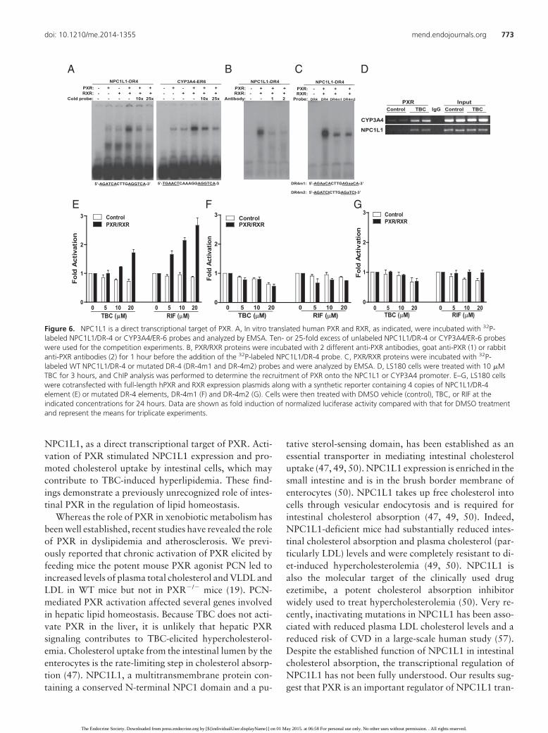
We next analyzed the promoter of the human NPC1L1gene and identified a DR-4-type (direct repeat spaced by 4nucleotides) of nuclear receptor response element (AG-ATCACTTGAGGTCA), similar to DR-4 elements foundin other PXR target genes (16). EMSA confirmed that thePXR and RXR heterodimer was able to bind to this DR-4element as well as the positive control, the ER-6 (evertedrepeat spaced by 6 nucleotides) element in the CYP3A4promoter (Figure 6A). The binding of NPC1L1/DR-4 orCYP3A4/ER-6 by PXR-RXR is specific as excess coldprobe decreased PXR-RXR binding to those elements(Figure 6A). In addition, 2 different anti-PXR antibodiesdisrupted the protein-DNA complex (Figure 6B), suggest-ing that PXR is a component of the protein complex thatbinds to the NPC1L1/DR4 element. Further, mutations ofthe DR-4 elements were also able to abolish the binding ofPXR-RXR dimers to both mutant DR-4 sites (Figure 6C).
Next, ChIP analysis demonstrated that TBC can pro-mote the recruitment of PXR onto the NPC1L1 promoterregion containing the DR-4 element (Figure 6D). ChIP onthe CYP3A4 promoter region containing the ER-6 ele-ment was included as a positive control. Last, 4 copies ofthe NPC1L1/DR-4 or mutated DR-4 elements were syn-thesized and inserted into a luciferase reporter vector.Transfection assays were performed to determinewhether the DR-4 motif is necessary and sufficient formediating PXR transactivation. Indeed, both TBC and
RIF can activate PXR and increase the NPC1L1/DR-4reporter gene activity in a dose-dependent manner (Figure6E). In contrast, TBC and RIF had no effect on activity of2 mutated DR-4 reporters (Figure 6, F and G). Collec-tively, activation of PXR by TBC transcriptionally regu-lates NPC1L1 expression and increases cholesterol up-take by human intestinal cells.
Discussion
Because of their variety and low costs, plastics are funda-mental in modern life, and plastic production exceeded300 million tons in 2010 (2). Plastic-associated chemicalsare produced in high volume for use in the production ofplastics, including the base chemical BPA and numerousplasticizers. The adverse effects of BPA and several phtha-late plasticizers (eg, DEHP) on human health have at-tracted considerable attention and engendered contro-versy in the past few decades, partly due to theirendocrine-disrupting properties. We and others haveidentified BPA, BPA analogs (eg, bisphenol B and bisphe-nol AF), and the widely used phthalate plasticizer DEHPas potent PXR agonists (12, 13, 43). In addition to thesewell-known EDCs, there are many phthalate substituteplasticizers that have not been tested for endocrine dis-ruption. For example, citrate esters, including TBC,ATBC, and TEC, represent a large group of plasticizersthat have been approved by the FDA to be used in food
Control TBC PCN0
50
100
150
****
Total Cholesterolm
g/dL
Control TBC PCN0
10
20
30
40
50
*
***
LDL
mg/
dL
Control TBC PCN0
2
4
6
8
10
*
VLDL
mg/
dL
Control TBC PCN0
20
40
60
80 HDL
mg/
dL
Control TBC0
50
100
150 Total Cholesterol
mg/
dL
Control TBC0
10
20
30
40
50 LDL
mg/
dL
Control TBC0
2
4
6
8
10 VLDL
mg/
dL
Control TBC0
20
40
60
80 HDL
mg/
dL
B
A
Figure 3. Exposure to TBC induces hyperlipidemia in WT but not in PXR�/� mice. A, Eight-week-old male WT mice were treated with vehicle or10 mg/kg BW TBC or PCN daily by oral gavage for 1 week. Plasma total cholesterol levels and lipoprotein levels (LDL, VLDL, and HDL) weremeasured (n � 6–10; *, P � .05 and ***, P � .001 compared with the control group). B, Eight-week-old male PXR�/� mice were treated withvehicle or 10 mg/kg BW TBC daily by oral gavage for 1 week. Plasma total cholesterol levels and lipoprotein levels (LDL, VLDL, and HDL) weremeasured (n � 4–5).
doi: 10.1210/me.2014-1355 mend.endojournals.org 771
The Endocrine Society. Downloaded from press.endocrine.org by [${individualUser.displayName}] on 01 May 2015. at 06:58 For personal use only. No other uses without permission. . All rights reserved.

packaging materials, vinyl toys, medical devices, cosmet-ics, and pharmaceutical drugs (53–56). ATBC has re-cently been shown to activate PXR and induce PXR targetgene expression (45). However, it was unclear whetherATBC or other phthalate substitute plasticizers have anyadverse effects on the development of complex diseases.In the current study, we demonstrate that TBC is a more
potent PXR agonist than ATBC in our assays. Similar toATBC, TBC activated intestinal PXR and induced PXRtarget gene expression but did not affect hepatic PXRactivity. Nevertheless, TBC-mediated intestinal PXR ac-tivation was sufficient to increase plasma cholesterol lev-els, especially atherogenic LDL levels in WT mice. Wethen identified a key intestinal cholesterol transporter,
CYP3A11
GSTA1
MDR1aCD36
NPC1L1
ABCG5
ABCG8
CYP3A11
GSTA1
MDR1aCD36
NPC1L1
ABCG5
ABCG80
2
4
6
8 ControlTBC
*
*
* *
*
*
WT PXR-/-
mR
NA
expr
essi
on
β-actin
WT PXR-/-
NPC1L1
Control TBC Control TBC
WT PXR-/-0.0
0.5
1.0
1.5
2.0
2.5TBCControl
***
NP
C1L
1mR
NA
exp
ress
ion
WT PXR
NPC1L1
Control TBC Control TBC
WT PXR-/-0
2
4
6
8
*TBCControl
[3H
]-ch
oles
tero
l upt
ake
(x10
3cp
m)
**
A B
C D E-/-
β-actin
Figure 4. Activation of PXR by TBC stimulates the expression of the intestinal cholesterol transporter NPC1L1 in mice and increases cholesteroluptake by murine intestinal cells. A, WT and PXR�/� mice were treated with vehicle or 10 mg/kg BW TBC daily for 1 week. Total RNA was isolatedfrom small intestine, and the expression levels of indicated genes were measured by qPCR (n � 5–6 per group; *, P � .05). B, Western blotanalysis of intestinal NPC1L1 protein levels in control or TBC-treated WT and PXR�/� mice. The top band indicates glycosylated NPC1L1. C and D,Primary enterocytes isolated from WT and PXR�/� mice were treated with vehicle control or 10 �M TBC for 3 hours. NPC1L1 mRNA levels weremeasured by qPCR (n � 3; ***, P � .001), and protein levels were analyzed by Western blot (D). E, Primary enterocytes isolated from WT andPXR�/� mice were treated with vehicle control or 10 �M TBC for 2 hours, followed by incubation with [3H]cholesterol and TBC for 1 hour. Thecellular cholesterol uptake was then measured (n � 3; *, P � .05; and **, P � .01).
PXR siRNA - +
PXR
Control siPXR0.0
0.5
1.0
1.5
2.0
Control TBC** *
[3 H] c
hole
ster
ol u
ptak
e(x
103
cpm
)
*
Control siPXR0
1
2
3
4 ControlTBC
***
NPC
1L1
mR
NA
expr
essi
on
A CB
β-actin
Figure 5. TBC promotes cholesterol uptake by human intestinal cells. A, Western blot analysis of PXR levels in human intestinal LS180 cellstransfected with control siRNA or siRNA against PXR (siPXR). B, Control or siPXR LS180 cells were treated with 10 �M TBC for 3 hours, andNPC1L1 expression was analyzed by qPCR (n � 3; ***, P � .001). C, Control or siPXR LS180 cells were treated with 10 �M TBC for 2 hoursfollowed by incubation with [3H]cholesterol and TBC for an additional 1 hour. The cellular cholesterol uptake was then measured (n � 3; *, P �.05; and **, P � .01).
772 Sui et al Role of Intestinal PXR in Lipid Homeostasis Mol Endocrinol, May 2015, 29(5):765–776
The Endocrine Society. Downloaded from press.endocrine.org by [${individualUser.displayName}] on 01 May 2015. at 06:58 For personal use only. No other uses without permission. . All rights reserved.
NPC1L1, as a direct transcriptional target of PXR. Acti-vation of PXR stimulated NPC1L1 expression and pro-moted cholesterol uptake by intestinal cells, which maycontribute to TBC-induced hyperlipidemia. These find-ings demonstrate a previously unrecognized role of intes-tinal PXR in the regulation of lipid homeostasis.
Whereas the role of PXR in xenobiotic metabolism hasbeen well established, recent studies have revealed the roleof PXR in dyslipidemia and atherosclerosis. We previ-ously reported that chronic activation of PXR elicited byfeeding mice the potent mouse PXR agonist PCN led toincreased levels of plasma total cholesterol and VLDL andLDL in WT mice but not in PXR�/� mice (19). PCN-mediated PXR activation affected several genes involvedin hepatic lipid homeostasis. Because TBC does not acti-vate PXR in the liver, it is unlikely that hepatic PXRsignaling contributes to TBC-elicited hypercholesterol-emia. Cholesterol uptake from the intestinal lumen by theenterocytes is the rate-limiting step in cholesterol absorp-tion (47). NPC1L1, a multitransmembrane protein con-taining a conserved N-terminal NPC1 domain and a pu-
tative sterol-sensing domain, has been established as anessential transporter in mediating intestinal cholesteroluptake (47, 49, 50). NPC1L1 expression is enriched in thesmall intestine and is in the brush border membrane ofenterocytes (50). NPC1L1 takes up free cholesterol intocells through vesicular endocytosis and is required forintestinal cholesterol absorption (47, 49, 50). Indeed,NPC1L1-deficient mice had substantially reduced intes-tinal cholesterol absorption and plasma cholesterol (par-ticularly LDL) levels and were completely resistant to di-et-induced hypercholesterolemia (49, 50). NPC1L1 isalso the molecular target of the clinically used drugezetimibe, a potent cholesterol absorption inhibitorwidely used to treat hypercholesterolemia (50). Very re-cently, inactivating mutations in NPC1L1 has been asso-ciated with reduced plasma LDL cholesterol levels and areduced risk of CVD in a large-scale human study (57).Despite the established function of NPC1L1 in intestinalcholesterol absorption, the transcriptional regulation ofNPC1L1 has not been fully understood. Our results sug-gest that PXR is an important regulator of NPC1L1 tran-
NPC1L1
CYP3A4
Control TBC IgG Control TBC PXR Input
5’-AGATCACTTGAGGTCA-3’ 5’-TGAACTCAAAGGAGGTCA-3’
NPC1L1-DR4PXR: - + - + + + - + - + + + RXR: - - + + + + - - + + + +
CYP3A4-ER6
Cold probe: - - - - 10x 25x - - - - 10x 25x
PXR: - + + + RXR: - + + +
Antibody: - - 1 2
DR4m1: 5’-AGAaCACTTGAGaaCA-3’
DR4m2: 5’-AGATCtCTTGAGaTCt-3’
NPC1L1-DR4PXR: - + + + RXR: - + + +
Probe: DR4 DR4 DR4m1 DR4m2
NPC1L1-DR4
0 5 10 20 0 5 10 200
1
2
3 ControlPXR/RXR
TBC (μμM) RIF (μM)
Fold
Act
ivat
ion
0 5 10 20 0 5 10 200
1
2
3 ControlPXR/RXR
Fold
Act
ivat
ion
TBC (μM) RIF (μM)0 5 10 20 0 5 10 20
0
1
2
3 ControlPXR/RXR
Fold
Act
ivat
ion
TBC (μM) RIF (μM)
A B C D
E F G
Figure 6. NPC1L1 is a direct transcriptional target of PXR. A, In vitro translated human PXR and RXR, as indicated, were incubated with 32P-labeled NPC1L1/DR-4 or CYP3A4/ER-6 probes and analyzed by EMSA. Ten- or 25-fold excess of unlabeled NPC1L1/DR-4 or CYP3A4/ER-6 probeswere used for the competition experiments. B, PXR/RXR proteins were incubated with 2 different anti-PXR antibodies, goat anti-PXR (1) or rabbitanti-PXR antibodies (2) for 1 hour before the addition of the 32P-labeled NPC1L1/DR-4 probe. C, PXR/RXR proteins were incubated with 32P-labeled WT NPC1L1/DR-4 or mutated DR-4 (DR-4m1 and DR-4m2) probes and were analyzed by EMSA. D, LS180 cells were treated with 10 �MTBC for 3 hours, and ChIP analysis was performed to determine the recruitment of PXR onto the NPC1L1 or CYP3A4 promoter. E–G, LS180 cellswere cotransfected with full-length hPXR and RXR expression plasmids along with a synthetic reporter containing 4 copies of NPC1L1/DR-4element (E) or mutated DR-4 elements, DR-4m1 (F) and DR-4m2 (G). Cells were then treated with DMSO vehicle (control), TBC, or RIF at theindicated concentrations for 24 hours. Data are shown as fold induction of normalized luciferase activity compared with that for DMSO treatmentand represent the means for triplicate experiments.
doi: 10.1210/me.2014-1355 mend.endojournals.org 773
The Endocrine Society. Downloaded from press.endocrine.org by [${individualUser.displayName}] on 01 May 2015. at 06:58 For personal use only. No other uses without permission. . All rights reserved.

scription. PXR can directly bind to a DR-4 motif in hu-man NPC1L1 promoter and stimulate NPC1L1expression upon ligand activation. Thus, PXR-mediatedNPC1L1 up-regulation may contribute to TBC and otherligand-induced hypercholesterolemia.
Whereas NPC1L1 plays an essential role in intestinalcholesterol absorption, another PXR-regulated trans-porter, CD36, mediates enterocyte uptake of fatty acids,which are then converted to triglycerides for transportinto chylomicrons (47, 48). We and others previouslyreported that activation of PXR induces CD36 expressionand increases lipid accumulation in the liver (21), intes-tine (23), and macrophage (19). In addition, activation ofintestinal PXR can also induce the expression levels ofseveral enzymes involved in intestinal lipid transportationand chylomicron secretion including diacylglycerol acyl-transferase 1 and 2 (23). Therefore, the functions of PXRin intestinal lipid homeostasis are complex, and furtherstudies are needed to define the precise mechanismsthrough which intestinal PXR modulates lipid homeosta-sis in animal models as well as in humans.
It is intriguing that TBC can only activate intestinalPXR but not hepatic PXR. Several tissue-specific PXRligands have been identified (35, 58, 59). For example,rifaximin, a nonsynthetic antibiotic, has been shown to bea potent intestine-specific PXR ligand (58). The tissue-specific effect of rifaximin on intestinal PXR was proba-bly related to its poor absorption as rifaximin accumu-lated in the intestine after oral treatment. However, thereason that TBC cannot activate hepatic PXR is unlikelydue to poor absorption by the intestine as TBC was un-able to active hepatic PXR even after ip delivery. Further,TBC treatment did not affect PXR activity in HepaRGand HepG2 cells in vitro. It is possible that hepatic cellscan rapidly catalyze TBC. Further studies, including de-velopment of appropriate HPLC-tandem mass spectrom-etry assays and examination of TBC metabolites in vivoand in vitro, will be required to elucidate the detailedmechanisms underlying the tissue-specific effects of TBCand similar chemicals. In addition, we previously demon-strated that tocotrienols, members of the vitamin E fam-ily, show tissue-specific induction of PXR target genes,particularly CYP3A4 (35). Tocotrienols can up-regulateexpression of CYP3A4 in human primary hepatocytes butnot in LS180 cells. Nuclear receptor coregulators playcritical roles in nuclear receptor activation and are alsoinvolved in the mechanisms underlying the divergent ac-tivities of selective estrogen receptor modulators (60, 61).We found that NCoR is expressed at different levels inintestinal LS180 cells and primary hepatocytes and thattocotrienols can only partially disrupt the interaction ofunliganded PXR and NCoR in LS180 cells (35). There-
fore, it is plausible that the coregulator difference betweenliver and intestine may contribute to the tissue-specificeffects of TBC.
A fundamental question about all EDC studies iswhether low-dose exposure to EDCs can influence humanendocrine functions and cause adverse effects. Currently,there is little information about human exposure to TBC.The dose of 10 mg/kg/d TBC used in the present study totreat animals was based on human exposure to other plas-ticizers such as DEHP, DnBP, and ATBC. A retrospectivehuman biomonitoring study of German adults aged 20 to29 years showed median daily intakes for DnBP andDEHP of 7 and 4 mg/kg BW/d, respectively. Fourteenpercent of subjects showed DnBP intakes above the tol-erable daily intake value of 10 mg/kg BW/d set by theEuropean Food Safety Authority (62, 63). Patients takingdrugs containing another citrate ester plasticizer, ATBC,may be exposed to as much as 20 mg of ATBC daily (45).The 10 mg/kg BW/d TBC dose used in our study was alsosignificantly lower than concentrations experimentallyused for DEHP (1000 mg/kg BW/d) and DnBP (2000mg/kg BW/d) to treat mice in some studies (2, 64). Wehave also examined whether lower TBC concentrationscan activate PXR in vivo by testing 2 lower doses, 2.5 and5 mg/kg/d, but neither dose activated PXR. Therefore, the10 mg/kg/d dose used in this study is close to the lowerlimit of the TBC concentration that activates PXR in vivo.Considering the wide use of TBC in consumer products(eg, plastic wrap, toys, and drugs), we believe that thedose, which was sufficient to activate PXR activity andcause adverse effects in vivo, was reasonable. It would beinteresting to measure the internal TBC concentrations inhuman samples and correlate internal TBC concentra-tions to plasma lipid levels in the future. Such informationwould provide important insights about the relationshipof EDC exposure and disease outcomes in humans.
In addition to TBC, we and others have identifiedmany other environmentally significant chemicals (eg,BPA, bisphenol B, and DEHP) as PXR agonists. Further,we recently demonstrated that certain EDCs can synergis-tically activate human PXR (12). The synergism betweendifferent EDCs supports the need to include mixtures forfuture in vitro and in vivo studies, which may have im-portant implications for environmental chemical risk as-sessment. In addition to CVD, activation of PXR has beenshown to induce tumor aggressiveness in humans andmice (65). Further studies are needed to investigatewhether TBC-mediated intestinal PXR activation can in-duce tumorigenesis in animal models.
In summary, we have demonstrated that the phthalatesubstitute plasticizer TBC is a potent agonist of PXR. Ofparticular interest is the finding that TBC activated intes-
774 Sui et al Role of Intestinal PXR in Lipid Homeostasis Mol Endocrinol, May 2015, 29(5):765–776
The Endocrine Society. Downloaded from press.endocrine.org by [${individualUser.displayName}] on 01 May 2015. at 06:58 For personal use only. No other uses without permission. . All rights reserved.
tinal PXR but did not affect hepatic PXR activity. Nev-ertheless, activation of intestinal PXR by TBC was suffi-cient to increase plasma cholesterol levels in mice. Wethen identified the intestinal cholesterol transporterNPC1L1 as a direct transcriptional target of PXR andfound that activation of PXR increased cholesterol up-take by intestinal cells. These findings demonstrate a pre-viously unrecognized role of PXR in the regulation ofintestinal lipid homeostasis. Findings from this study willhopefully stimulate further investigations of phthalatesubstitute plasticizers, in particular, the mechanisms bywhich TBC and other chemicals activate PXR, the tissue-specific TBC activation of PXR, and PXR regulation ofintestinal lipid homeostasis. Activation of PXR should betaken into consideration for future risk assessment ofphthalate substitute plasticizers and related environmen-tal chemicals.
Acknowledgments
Address all correspondence and requests for reprints to:Changcheng Zhou, PhD, Department of Pharmacology and Nu-tritional Sciences, University of Kentucky, 900 South LimestoneStreet, 517 Wethington Building, Lexington, KY 40536. E-mail:[email protected].
This work was supported in part by the National Institutes ofHealth (Grants R01ES023470, R21ES022745, P20GM103527,and T32DK007778) and the American Heart Association (Grant14POST18740064).
Disclosure Summary: The authors have nothing to disclose.
References
1. Diamanti-Kandarakis E, Bourguignon JP, Giudice LC, et al. Endo-crine-disrupting chemicals: an Endocrine Society scientific state-ment. Endocr Rev. 2009;30:293–342.
2. Casals-Casas C, Desvergne B. Endocrine disruptors: from endo-crine to metabolic disruption. Annu Rev Physiol. 2011;73:135–162.
3. vom Saal FS, Myers JP. Bisphenol a and risk of metabolic disorders.JAMA. 2008;300:1353–1355.
4. Lang IA, Galloway TS, Scarlett A, et al. Association of urinarybisphenol a concentration with medical disorders and laboratoryabnormalities in adults. JAMA. 2008;300:1303–1310.
5. Melzer D, Osborne NJ, Henley WE, et al. Urinary bisphenol aconcentration and risk of future coronary artery disease in appar-ently healthy men and women. Circulation. 2012;125:1482–1490.
6. Lind L, Lind PM. Can persistent organic pollutants and plastic-associated chemicals cause cardiovascular disease? J Intern Med.2012;271:537–553.
7. Melzer D, Rice NE, Lewis C, Henley WE, Galloway TS. Associationof urinary bisphenol a concentration with heart disease: evidencefrom NHANES 2003/06. PLoS One. 2010;5:e8673.
8. Nagaoka S, Miyazaki H, Aoyama Y, Yoshida A. Effects of dietarypolychlorinated biphenyls on cholesterol catabolism in rats. Br JNutr. 1990;64:161–169.
9. Arsenescu V, Arsenescu RI, King V, Swanson H, Cassis LA. Poly-chlorinated biphenyl-77 induces adipocyte differentiation and pro-inflammatory adipokines and promotes obesity and atherosclero-sis. Environ Health Perspect. 2008;116:761–768.
10. Lind PM, van Bavel B, Salihovic S, Lind L. Circulating levels ofpersistent organic pollutants (POPs) and carotid atherosclerosis inthe elderly. Environ Health Perspect. 2012;120:38–43.
11. Lind PM, Lind L. Circulating levels of bisphenol A and phthalatesare related to carotid atherosclerosis in the elderly. Atherosclerosis.2011;218:207–213.
12. Sui Y, Ai N, Park SH, et al. Bisphenol A and its analogues activatehuman pregnane X receptor. Environ Health Perspect. 2012;120:399–405.
13. Tabb MM, Kholodovych V, Grün F, Zhou C, Welsh WJ, BlumbergB. Highly chlorinated PCBs inhibit the human xenobiotic responsemediated by the steroid and xenobiotic receptor (SXR). EnvironHealth Perspect. 2004;112:163–169.
14. Masuyama H, Hiramatsu Y, Kunitomi M, Kudo T, MacDonaldPN. Endocrine disrupting chemicals, phthalic acid and nonylphe-nol, activate pregnane X receptor-mediated transcription. Mol En-docrinol. 2000;14:421–428.
15. Zhou C, Verma S, Blumberg B. The steroid and xenobiotic receptor(SXR), beyond xenobiotic metabolism. Nucl Recept Signal. 2009;7:e001.
16. Blumberg B, Sabbagh W Jr, Juguilon H, et al. SXR, a novel steroidand xenobiotic-sensing nuclear receptor. Genes Dev. 1998;12:3195–3205.
17. Kliewer SA, Moore JT, Wade L, et al. An orphan nuclear receptoractivated by pregnanes defines a novel steroid signaling pathway.Cell. 1998;92:73–82.
18. Kliewer SA, Goodwin B, Willson TM. The nuclear pregnane Xreceptor: a key regulator of xenobiotic metabolism. Endocr Rev.2002;23:687–702.
19. Zhou C, King N, Chen KY, Breslow JL. Activation of PXR induceshypercholesterolemia in wild-type and accelerates atherosclerosisin apoE deficient mice. J Lipid Res. 2009;50:2004–2013.
20. de Haan W, de Vries-van der Weij J, Mol IM, et al. PXR agonismdecreases plasma HDL levels in ApoE3-Leiden.CETP mice.Biochim Biophys Acta. 2009;1791:191–197.
21. Zhou J, Zhai Y, Mu Y, et al. A novel pregnane X receptor-mediatedand sterol regulatory element-binding protein-independent lipo-genic pathway. J Biol Chem. 2006;281:15013–15020.
22. Ricketts ML, Boekschoten MV, Kreeft AJ, et al. The cholesterol-raising factor from coffee beans, cafestol, as an agonist ligand forthe farnesoid and pregnane X receptors. Mol Endocrinol. 2007;21:1603–1616.
23. Cheng J, Krausz KW, Tanaka N, Gonzalez FJ. Chronic exposure torifaximin causes hepatic steatosis in pregnane X receptor-human-ized mice. Toxicol Sci. 2012;129:456–468.
24. Helsley RN, Sui Y, Ai N, Park SH, Welsh WJ, Zhou C. Pregnane Xreceptor mediates dyslipidemia induced by the HIV protease inhib-itor amprenavir in mice. Mol Pharmacol. 2013;83:1190–1199.
25. Khogali AM, Chazan BI, Metcalf VJ, Ramsay JH. Hyperlipidaemiaas a complication of rifampicin treatment. Tubercle. 1974;55:231–233.
26. Carr A, Samaras K, Chisholm DJ, Cooper DA. Pathogenesis ofHIV-1-protease inhibitor-associated peripheral lipodystrophy, hy-perlipidaemia, and insulin resistance. Lancet. 1998;351:1881–1883.
27. Shafran SD, Mashinter LD, Roberts SE. The effect of low-doseritonavir monotherapy on fasting serum lipid concentrations. HIVMed. 2005;6:421–425.
28. Eirís JM, Lojo S, Del Río MC, et al. Effects of long-term treatmentwith antiepileptic drugs on serum lipid levels in children with epi-lepsy. Neurology. 1995;45:1155–1157.
29. Lu Y, Feskens EJ, Boer JM, Müller M. The potential influence ofgenetic variants in genes along bile acid and bile metabolic pathway
doi: 10.1210/me.2014-1355 mend.endojournals.org 775
The Endocrine Society. Downloaded from press.endocrine.org by [${individualUser.displayName}] on 01 May 2015. at 06:58 For personal use only. No other uses without permission. . All rights reserved.

on blood cholesterol levels in the population. Atherosclerosis.2010;210:14–27.
30. He J, Gao J, Xu M, et al. PXR ablation alleviates diet-induced andgenetic obesity and insulin resistance in mice. Diabetes. 2013;62:1876–1887.
31. Moreau A, Téruel C, Beylot M, et al. A novel pregnane X receptorand S14-mediated lipogenic pathway in human hepatocyte. Hepa-tology. 2009;49:2068–2079.
32. Roth A, Looser R, Kaufmann M, et al. Regulatory cross-talk be-tween drug metabolism and lipid homeostasis: constitutive andro-stane receptor and pregnane X receptor increase Insig-1 expression.Mol Pharmacol. 2008;73:1282–1289.
33. Zhou C, Poulton EJ, Grün F, et al. The dietary isothiocyanatesulforaphane is an antagonist of the human steroid and xenobioticnuclear receptor. Mol Pharmacol. 2007;71:220–229.
34. Zhou C, Tabb MM, Nelson EL, et al. Mutual repression betweensteroid and xenobiotic receptor and NF-�B signaling pathwayslinks xenobiotic metabolism and inflammation. J Clin Invest. 2006;116:2280–2289.
35. Zhou C, Tabb MM, Sadatrafiei A, Grün F, Blumberg B. Tocotrien-ols activate the steroid and xenobiotic receptor, SXR, and selec-tively regulate expression of its target genes. Drug Metab Dispos.2004;32:1075–1082.
36. Teupser D, Persky AD, Breslow JL. Induction of atherosclerosis bylow-fat, semisynthetic diets in LDL receptor-deficient C57BL/6Jand FVB/NJ mice: comparison of lesions of the aortic root, brachio-cephalic artery, and whole aorta (en face measurement). Arterio-scler Thromb Vasc Biol. 2003;23:1907–1913.
37. Sui Y, Xu J, Rios-Pilier J, Zhou C. Deficiency of PXR decreasesatherosclerosis in apoE-deficient mice. J Lipid Res. 2011;52:1652–1659.
38. Zhou C, Assem M, Tay JC, et al. Steroid and xenobiotic receptorand vitamin D receptor crosstalk mediates CYP24 expression anddrug-induced osteomalacia. J Clin Invest. 2006;116:1703–1712.
39. Nassir F, Wilson B, Han X, Gross RW, Abumrad NA. CD36 isimportant for fatty acid and cholesterol uptake by the proximal butnot distal intestine. J Biol Chem. 2007;282:19493–19501.
40. Zhou C, Pridgen B, King N, Xu J, Breslow JL. HyperglycemicIns2AkitaLdlr�/� mice show severely elevated lipid levels and in-creased atherosclerosis: a model of type 1 diabetic macrovasculardisease. J Lipid Res. 2011;52:1483–1493.
41. Cai L, Eckhardt ER, Shi W, et al. Scavenger receptor class B type Ireduces cholesterol absorption in cultured enterocyte CaCo-2 cells.J Lipid Res. 2004;45:253–262.
42. Sui Y, Park SH, Xu J, et al. IKK� links vascular inflammation toobesity and atherosclerosis. J Exp Med. 2014;211:869–886.
43. Takeshita A, Koibuchi N, Oka J, Taguchi M, Shishiba Y, Ozawa Y.Bisphenol-A, an environmental estrogen, activates the human or-phan nuclear receptor, steroid and xenobiotic receptor-mediatedtranscription. Eur J Endocrinol. 2001;145:513–517.
44. DeKeyser JG, Laurenzana EM, Peterson EC, Chen T, OmiecinskiCJ. Selective phthalate activation of naturally occurring humanconstitutive androstane receptor splice variants and the pregnane Xreceptor. Toxicol Sci. 2011;120:381–391.
45. Takeshita A, Igarashi-Migitaka J, Nishiyama K, Takahashi H,Takeuchi Y, Koibuchi N. Acetyl tributyl citrate, the most widelyused phthalate substitute plasticizer, induces cytochrome p450 3athrough steroid and xenobiotic receptor. Toxicol Sci. 2011;123:460–470.
46. Kanebratt KP, Andersson TB. Evaluation of HepaRG cells as an in
vitro model for human drug metabolism studies. Drug Metab Dis-pos. 2008;36:1444–1452.
47. Abumrad NA, Davidson NO. Role of the gut in lipid homeostasis.Physiol Rev. 2012;92:1061–1085.
48. Nauli AM, Nassir F, Zheng S, et al. CD36 is important for chylo-micron formation and secretion and may mediate cholesterol up-take in the proximal intestine. Gastroenterology. 2006;131:1197–1207.
49. Davis HR Jr, Zhu LJ, Hoos LM, et al. Niemann-Pick C1 like 1(NPC1L1) is the intestinal phytosterol and cholesterol transporterand a key modulator of whole-body cholesterol homeostasis. J BiolChem. 2004;279:33586–33592.
50. Altmann SW, Davis HR Jr, Zhu LJ, et al. Niemann-Pick C1 like 1protein is critical for intestinal cholesterol absorption. Science.2004;303:1201–1204.
51. Jia L, Betters JL, Yu L. Niemann-pick C1-like 1 (NPC1L1) proteinin intestinal and hepatic cholesterol transport. Annu Rev Physiol.2011;73:239–259.
52. Yu L, Gupta S, Xu F, et al. Expression of ABCG5 and ABCG8 isrequired for regulation of biliary cholesterol secretion. J Biol Chem.2005;280:8742–8747.
53. Finkelstein M, Gold H. Toxicology of the citric acid esters: tributylcitrate, acetyl tributyl citrate, triethyl citrate, and acetyl triethylcitrate. Toxicology. 1959;1:283–298.
54. Meyers DB, Autian J, Guess WL. Toxicity of plastics used in med-ical practice. II. Toxicity of citric acid esters used as plasticizers.J Pharm Sci. 1964;53:774–777.
55. El-Gendy NA. Pharmaceutical plasticizers for drug delivery sys-tems. Curr Drug Deliv. 2012;9:148–163.
56. Hirata-Koizumi M, Takahashi M, Matsumoto M, Kawamura T,Ono A, Hirose A. Toxicity effects of phthalate substitute plasticiz-ers used in toys (in Japanese). Kokuritsu Iyakuhin Shokuhin EiseiKenkyusho Hokoku. 2012;130:31–42.
57. Myocardial Infarction Genetics Consortium I, Stitziel NO, WonHH, et al. Inactivating mutations in NPC1L1 and protection fromcoronary heart disease. N Engl J Med. 2014;371:2072–2082.
58. Ma X, Shah YM, Guo GL, et al. Rifaximin is a gut-specific humanpregnane X receptor activator. J Pharmacol Exp Ther. 2007;322:391–398.
59. Cheng J, Shah YM, Ma X, et al. Therapeutic role of rifaximin ininflammatory bowel disease: clinical implication of human preg-nane X receptor activation. J Pharmacol Exp Ther. 2010;335:32– 41.
60. Glass CK, Rosenfeld MG. The coregulator exchange in transcrip-tional functions of nuclear receptors. Genes Dev. 2000;14:121–141.
61. McDonnell DP, Connor CE, Wijayaratne A, Chang CY, Norris JD.Definition of the molecular and cellular mechanisms underlying thetissue-selective agonist/antagonist activities of selective estrogen re-ceptor modulators. Recent Prog Horm Res. 2002;57:295–316.
62. Halden RU. Plastics and health risks. Annu Rev Public Health.2010;31:179–194.
63. Wittassek M, Wiesmüller GA, Koch HM, et al. Internal phthalateexposure over the last two decades—a retrospective humanbiomonitoring study. Int J Hyg Environ Health. 2007;210:319–333.
64. Lapinskas PJ, Brown S, Leesnitzer LM, et al. Role of PPAR� inmediating the effects of phthalates and metabolites in the liver.Toxicology. 2005;207:149–163.
65. Wang H, Venkatesh M, Li H, et al. Pregnane X receptor activationinduces FGF19-dependent tumor aggressiveness in humans andmice. J Clin Invest. 2011;121:3220–3232.
776 Sui et al Role of Intestinal PXR in Lipid Homeostasis Mol Endocrinol, May 2015, 29(5):765–776
The Endocrine Society. Downloaded from press.endocrine.org by [${individualUser.displayName}] on 01 May 2015. at 06:58 For personal use only. No other uses without permission. . All rights reserved.