intestinal hexose absorption: transcellular or paracellular fluxes

1
Journal of Physiology Intestinal hexose absorption: transcellular or paracellular fluxes Chris I. Cheeseman Membrane Protein Group, Department of Physiology, University of Alberta, Edmonton, AB, Canada T6G 2H7 Email: [email protected] The currently accepted model of intestinal hexose transport (Fig. 1) has entry across the brush-border membrane (BBM) mediated by the sodium-dependent glucose–galactose transporter (SGLT1) and by the facilitated fructose transporter GLUT5. Exit of all three hexoses across the basolateral membrane (BLM) into the blood stream is mediated by another facilitated transporter, GLUT2. The absorption of hexoses by the small intestine in mammalian omnivores is a very efficient process which has a safety factor to ensure that hexoses do not spill over into the colon. The arrival of sugars in the lower bowel can produce diarrhoea resulting from an osmotically induced back-flux of water into the lumen or as the consequence of the increased nutrients driving a bacterial over- growth. One mechanism designed to avoid this situation is the ability of the small intestine to increase its absorptive capacity for hexoses when their dietary load increases. If alterations in dietary carbohydrate are sustained for three or more days, expression of the transport proteins responsible for hexose absorption are altered in parallel (Ferraris, 2001). In addition, there is recent evidence to indicate that some transporter activity is regulated within the time course of a single meal (Cheeseman, 1997). However, these transcellular pathways for the intestinal absorption of hexoses have been calculated to have a limited capacity and appear to provide a rather low safety margin for the complete absorption of dietary carbohydrate (Lam et al. 2002). Madara & Pappenheimer (1987) proposed that additional transepithelial fluxes could be accounted for by paracellular solute movement carried by solvent drag, i.e. diffusion. This proposal generated a debate over the actual substrate concentrations achieved in the intestinal lumen during a carbohydrate rich meal, with a suggested range from 50 to 250 mM. However, this did little to settle the actual issue of whether the major proportion of hexoses cross the enterocytes mediated by transporter proteins or whether passage between the cells is nutritionally significant. It is clear that, when glucose transport is measured in vivo over a wide range of luminal concentrations, uptake has both a saturable, carrier-mediated com- ponent and an almost linear component which could represent diffusion. Recently, a new model for hexose absorption has been proposed which could explain the majority of these experimental observations and which would indicate that hexose movement is almost exclusively transcellular. In this issue of The Journal of Physiology, Helliwell & Kellett (2002) provide compelling evidence that the majority of glucose absorption is carrier mediated. However, entry into the epithelium involves not only SGLT1 but also GLUT2, the low affinity facilitated hexose transporter previously believed to be only expressed in the BLM. Apparently, the expression of GLUT2 in the apical membrane is highly regulated through the activation of PKC bII when SGLT1 transports glucose. GLUT2 in the BBM would provide a high capacity, low affinity pathway for the entry of glucose, galactose and fructose into the epithelium, i.e. the so-called diffusional pathway. Thus, at the early stages of a meal, when the luminal concentrations of hexoses are high, entry would be rapid and obviate any need for non-specific paracellular fluxes. Helliwell & Kellett (2002) argue that this additional pathway has been frequently overlooked because of the sensitivity of apical GLUT2 expression to tissue stress such as luminal distention or poor oxygenation. Many classical techniques used in the past to investigate hexose absorption have employed high luminal perfusion rates, which cause distention, or involve significant handling of the tissue to gain access to the luminal surface. Such conditions induce the internalisation of apical GLUT2, down-regulating this entry pathway. Additional support for this new model comes from two sets of independent observations. The increased presence of GLUT2 has been detected in the rat jejunal BBM using immuno- histochemistry (Au et al. 2002) in response to luminal glucose and/or vascular glucagon- like peptide 2. Also, the majority of glucose absorption in the dog small intestine has been shown to be carrier mediated and can be blocked by transport inhibitors (Pencek et al. 2002). Thus, as summarised in Fig. 2, it appears there are at least three paths of entry for hexoses into the intestinal epithelium, SGLT1, GLUT5 and GLUT2. Both SGLT1 and GLUT2 are highly regulated by the insertion or removal of transporter protein from the BBM in response to the presence of hexoses in the lumen. While I have retained a paracellular route, evidence would suggest that this may account for only 1–2 % of the total hexose fluxes during a meal. Also, the previously determined safety factors for hexose absorption need to be reassessed given that the total carrier-mediated capacity appears to have been significantly underestimated. These new ideas regarding the routes and regulation of hexose absorption will provide a fertile new area of investigation on how hexose transport proteins are targeted and trafficked to and from the intestinal BBM. The signalling pathways for these events also need to be defined in more detail and may provide useful loci for modulating absorption rates under a variety of conditions. AU, A., GUPTA, A., SCHEMBRI, P. & CHEESEMAN, C. I. (2002). Biochemical Journal (in the Press) (July 3rd epub ahead of print). CHEESEMAN, C. I. (1997). American Journal of Physiology 273, R1965–1971. FERRARIS, R. P. (2001). Biochemical Journal 360, 265–276. HELLIWELL, P. A. & KELLETT, G. L. (2002). Journal of Physiology 544, 579–589. LAM, M. M., O’CONNOR, T. P. & DIAMOND, J. (2002). Journal of Physiology 542, 493–500. MADARA, J. L. & PAPPENHEIMER, J. R. (1987). Journal of Membrane Biology 100, 149–164. PENCEK, R. R., KOYAMA, Y., LACY, D. B., JAMES, F. D., FUEGER, P. T., JABBOUR, K., WILLIAMS, P. E. & WASSERMAN, D. H. (2002). Journal of Nutrition 132, 1929–1934. PERSPECTIVES Journal of Physiology (2002), 544.2, p. 336 DOI: 10.1113/jphysiol.2002.029850 © The Physiological Society 2002 www.jphysiol.org Figure 1 Figure 2

Upload: chris-i-cheeseman

Post on 03-Aug-2016

213 views

Category:

Documents


1 download

TRANSCRIPT

Jou

rnal

of P

hysi

olog

y

Intestinal hexose absorption:transcellular or paracellularfluxesChris I. Cheeseman

Membrane Protein Group, Departmentof Physiology, University of Alberta,Edmonton, AB, Canada T6G 2H7

Email: [email protected]

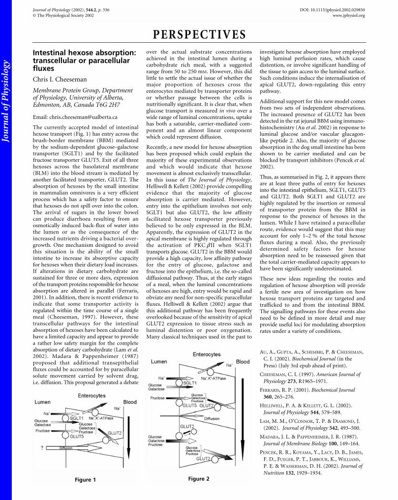
The currently accepted model of intestinalhexose transport (Fig. 1) has entry across thebrush-border membrane (BBM) mediatedby the sodium-dependent glucose–galactosetransporter (SGLT1) and by the facilitatedfructose transporter GLUT5. Exit of all threehexoses across the basolateral membrane(BLM) into the blood stream is mediated byanother facilitated transporter, GLUT2. Theabsorption of hexoses by the small intestinein mammalian omnivores is a very efficientprocess which has a safety factor to ensurethat hexoses do not spill over into the colon.The arrival of sugars in the lower bowelcan produce diarrhoea resulting from anosmotically induced back-flux of water intothe lumen or as the consequence of theincreased nutrients driving a bacterial over-growth. One mechanism designed to avoidthis situation is the ability of the smallintestine to increase its absorptive capacityfor hexoses when their dietary load increases.If alterations in dietary carbohydrate aresustained for three or more days, expressionof the transport proteins responsible for hexoseabsorption are altered in parallel (Ferraris,2001). In addition, there is recent evidence toindicate that some transporter activity isregulated within the time course of a singlemeal (Cheeseman, 1997). However, thesetranscellular pathways for the intestinalabsorption of hexoses have been calculated tohave a limited capacity and appear to providea rather low safety margin for the completeabsorption of dietary carbohydrate (Lam et al.2002). Madara & Pappenheimer (1987)proposed that additional transepithelialfluxes could be accounted for by paracellularsolute movement carried by solvent drag,i.e. diffusion. This proposal generated a debate

over the actual substrate concentrationsachieved in the intestinal lumen during acarbohydrate rich meal, with a suggestedrange from 50 to 250 mM. However, this didlittle to settle the actual issue of whether themajor proportion of hexoses cross theenterocytes mediated by transporter proteinsor whether passage between the cells isnutritionally significant. It is clear that, whenglucose transport is measured in vivo over awide range of luminal concentrations, uptakehas both a saturable, carrier-mediated com-ponent and an almost linear componentwhich could represent diffusion.

Recently, a new model for hexose absorptionhas been proposed which could explain themajority of these experimental observationsand which would indicate that hexosemovement is almost exclusively transcellular.In this issue of The Journal of Physiology,Helliwell & Kellett (2002) provide compellingevidence that the majority of glucoseabsorption is carrier mediated. However,entry into the epithelium involves not onlySGLT1 but also GLUT2, the low affinityfacilitated hexose transporter previouslybelieved to be only expressed in the BLM.Apparently, the expression of GLUT2 in theapical membrane is highly regulated throughthe activation of PKC bII when SGLT1transports glucose. GLUT2 in the BBM wouldprovide a high capacity, low affinity pathwayfor the entry of glucose, galactose andfructose into the epithelium, i.e. the so-calleddiffusional pathway. Thus, at the early stagesof a meal, when the luminal concentrationsof hexoses are high, entry would be rapid andobviate any need for non-specific paracellularfluxes. Helliwell & Kellett (2002) argue thatthis additional pathway has been frequentlyoverlooked because of the sensitivity of apicalGLUT2 expression to tissue stress such asluminal distention or poor oxygenation.Many classical techniques used in the past to

investigate hexose absorption have employedhigh luminal perfusion rates, which causedistention, or involve significant handling ofthe tissue to gain access to the luminal surface.Such conditions induce the internalisation ofapical GLUT2, down-regulating this entrypathway.

Additional support for this new model comesfrom two sets of independent observations.The increased presence of GLUT2 has beendetected in the rat jejunal BBM using immuno-histochemistry (Au et al. 2002) in response toluminal glucose and/or vascular glucagon-like peptide 2. Also, the majority of glucoseabsorption in the dog small intestine has beenshown to be carrier mediated and can beblocked by transport inhibitors (Pencek et al.2002).

Thus, as summarised in Fig. 2, it appears thereare at least three paths of entry for hexosesinto the intestinal epithelium, SGLT1, GLUT5and GLUT2. Both SGLT1 and GLUT2 arehighly regulated by the insertion or removalof transporter protein from the BBM inresponse to the presence of hexoses in thelumen. While I have retained a paracellularroute, evidence would suggest that this mayaccount for only 1–2 % of the total hexosefluxes during a meal. Also, the previouslydetermined safety factors for hexoseabsorption need to be reassessed given thatthe total carrier-mediated capacity appears tohave been significantly underestimated.

These new ideas regarding the routes andregulation of hexose absorption will providea fertile new area of investigation on howhexose transport proteins are targeted andtrafficked to and from the intestinal BBM.The signalling pathways for these events alsoneed to be defined in more detail and mayprovide useful loci for modulating absorptionrates under a variety of conditions.

AU, A., GUPTA, A., SCHEMBRI, P. & CHEESEMAN,

C. I. (2002). Biochemical Journal (in the

Press) (July 3rd epub ahead of print).

CHEESEMAN, C. I. (1997). American Journal ofPhysiology 273, R1965–1971.

FERRARIS, R. P. (2001). Biochemical Journal360, 265–276.

HELLIWELL, P. A. & KELLETT, G. L. (2002).

Journal of Physiology 544, 579–589.

LAM, M. M., O’CONNOR, T. P. & DIAMOND, J.

(2002). Journal of Physiology 542, 493–500.

MADARA, J. L. & PAPPENHEIMER, J. R. (1987).

Journal of Membrane Biology 100, 149–164.

PENCEK, R. R., KOYAMA, Y., LACY, D. B., JAMES,

F. D., FUEGER, P. T., JABBOUR, K., WILLIAMS,

P. E. & WASSERMAN, D. H. (2002). Journal ofNutrition 132, 1929–1934.

PERSPECTIVES

Journal of Physiology (2002), 544.2, p. 336 DOI: 10.1113/jphysiol.2002.029850

© The Physiological Society 2002 www.jphysiol.org

Figure 1 Figure 2