international journal of scientific research and reviews · received mime therapy along with...
TRANSCRIPT
Sharvani Belle Praveen Kumar et al., IJSRR 2018, 7(1), 427–441
IJSRR, 7(1) Jan –March. 2018 Page 427
Research article Available online www.ijsrr.org ISSN: 2279–0543
International Journal of Scientific Research and Reviews
Efficacy of Mime Therapy and Conventional Therapy Versus Conventional Therapy in Improving the Facial Functions of Bell’s Palsy Patients
Sharvani Belle Praveen Kumar* and Thomas Annie
Physiotherapy department, JSSCPT,
JSS hospital campus, Ramanuja Road, Mysuru, Karnataka, India-570004 Email id:[email protected], Phone no: 9964709215
ABSTRACT
Bell’s palsy is the peripheral paresis of the facial nerve. It is a lower motor neuron disease of the facial nerve characterized by acute unilateral peripheral facial muscle weakness. Physiotherapy can be beneficial with Bell’s palsy and it is important to be implemented prior to recovery in order to help and prevent permanent contractures of the paralyzed facial muscles. Mime therapy is a performance art to improve the symmetry of facial features and increases the voluntary power of muscle to help patients regain expression and control of their facial muscles. Conventional therapy is a method of treatment which includes electrical stimulation, tapping techniques and heat modalities. Combination of mime and physiotherapy aims to promote symmetry of the face at rest and during movement to control Synkinesis. Hence accurate treatment of Bells palsies is important and this study intended to analyze the efficacy of Mime therapy along with conventional therapy versus conventional therapy on Bell’s palsy. 30 subjects with Bell’s palsy, age ranging from 20-40years, equally into two groups of namely Group A (15) and Group B (15), pre and post treatment score was checked using sunny brook facial grading scale. Group A received first with Mime therapy followed by Electrical stimulation .Group B received only conventional therapy. The study concluded that there is a statistically significant improvement in facial function within the groups and patient who received Mime therapy along with Conventional therapy in Bells palsy. KEY WORDS: Bell’s palsy, facial function, facial symmetry, Mime therapy, Conventional therapy, Sunny brook facial grading scale, home programmes.
*Corresponding Author
Belle Sharvani Praveen Kumar Physiotherapy department, JSSCPT,
JSS hospital campus, Ramanuja Road, Mysuru, Karnataka, India-570004
Email id: [email protected]
Phone no: 9964709215

Sharvani Belle Praveen Kumar et al., IJSRR 2018, 7(1), 427–441
IJSRR, 7(1) Jan –March. 2018 Page 428
INTRODUCTION In our body face is considered as the image of soul. Face has a unique feature of attraction
which is due to the symmetry and it implies the good health.1,2 It has an influence on interpersonal
relationship. Face has the function of expression which helps for communication. This is due to the
coordinated functions of neuro-musculoskeletal system .If there is any disturbance in this
coordination, can result in facial paralysis and cause facial asymmetry. The facial asymmetry can
disturb individual’s quality of life as well as there may be a psychological impact of skewed facial
appearance.2
The two common forms of facial asymmetry or facial paralysis are Bells palsy and facial
palsy which is due to the lesion of facial nerve. Bell’s palsy is named after Sir Charles Bell, who
has long been considered to be the first to describe idiopathic facial paralysis in the early
19thcentury.It is a lower motor nerve lesion of facial nerve results in unilateral facial muscle
weakness. The causes can be compression of facial nerve along the nerve course, herpes infections,
exposure to cold air, middle ear infections, traumatic injuries or post surgeries of dental and Ear,
nose or throat.3 The lesion in the facial nerve leads to decrease conduction of impulses to the muscles
supplied having the function of facial expressions. It affects the muscles of forehead, face and neck.
The bells palsy is common in both male and females; it is seen between the age group of 15-40 years.
Symptoms of Bell's palsy usually begin suddenly and reach their peak within 48 hours.4 Symptoms
vary from person to person and can range in severity from mild weakness to total paralysis .The
common features are loss of forehead wrinkles and inability to frown, Droopy eyebrow and inability
to raise eyebrow, Inability to close the eye fully or blink ,Watery eye (crocodile tears),Inability to
squint, Painful eye with symptoms of grittiness or irritation, Sensitivity to light, Drooling from the
weak corner of your mouth, Excess or reduced salivation (dry mouth),Inability to flare nostril, Loss
of taste in the anterior 2/3rd of the tongue, Hyperacusis. it is generally believed that spontaneous
recovery is seen within nine months after bell’s palsy. The complete recovery of facial functions
seen 70% of people within three months, and sometimes around 30% continue to suffer with facial
asymmetry at rest and during movements which is termed as synkinesis.5 There is evidence that early
treatment (within 3 days after the onset) is necessary for therapy to be effective.
The physiotherapy treatment for Bell’s palsy includes Electrical stimulation of affected facial
muscle, facial exercises and massage. 6 Around 1970s, Mime therapy was developed in the
Netherlands by a mime actor named Jan Bronk and an otolaryngologist named Pieter Devriese
specifically for people with facial nerve paresis through collaboration between medical clinicians and
other mime-actors. Two decades of positive experiences in several Dutch university medical centres

Sharvani Belle Praveen Kumar et al., IJSRR 2018, 7(1), 427–441
IJSRR, 7(1) Jan –March. 2018 Page 429
have shaped mime therapy into its present form .Mime therapy is a type of physiotherapy. It was
created to help patients who had limited or restricted facial movement or a lack of facial muscle
control. Conventionally speaking, miming is a performance art that relies on expression and body
movement to communicate without speaking. Miming demands a highly-refined sense of body and
muscle control. During mime therapy, patients perform a series of mime-like Active facial exercises
to increase the voluntary power of muscle. The most common facial expression exercises include
brow raise, eye closure, snarl, smile, and pucker and pout in particular sequences. The goal of mime
therapy is to improve the symmetry of facial features and to help patients regain expression and
control of their facial muscles. Mime therapy consists of auto massage (stroking, effleurage, finger
kneading), stretching with facilitation exercises, relaxation, inhibition of Synkinesis and co-
ordination and emotional expression exercises. 7, 8 These exercise help by series of pattern of changes
in facial movement for facial rehabilitation illustrates the plasticity of the facial neuromotor system.
The brain learns to assign new roles to neurons, reducing abnormal patterns of movement and
restoring appropriate patterns of facial muscle activity for intended facial actions.9 Some of the
studies indicates that mime therapy reduces facial asymmetry both at rest and during voluntary
movements and thereby it reduces synkinesis.
Conventional therapy includes electrical stimulation is most commonly used treatment for
Bell’s palsy. The other additional treatments are tapping techniques, heat modalities and exercises. In
electrical stimulation, the stimulation is given to the paralysed muscles which are dennervated and to
the nerve trunks or branches until the voluntary movement returns. The nerve impulses to the
muscles are blocked due to the lesion in facial nerve, by giving electrical stimulation the external
stimulus can produce electric impulses which helps in restoring the muscle properties.10 By this the
recovery can complete within two to three months.6 But, if continuous external electrical stimulation
is given, at one point the muscles will become over stimulated. The result is short, tight, stiff muscles
that cannot move. Tight muscles become painful and may spasm or twitch. It may also cause
unwanted movement in the muscles on the affected side of your face, known as Synkinesis.11
There are several outcome measures to see the recovery in Bell’s palsy. One of the measures is,
Sunnybrook Facial Grading System (SFGS) which was developed in Toronto by Ross in 1992.12 It is
a regional scale that measures also Synkinesis. The regional scores are weighted for the composite
score. It is based on the evaluation of resting symmetry, degree of voluntary excursion of facial
muscles, and degree of Synkinesis associated with specified voluntary movement to form one single
composite score from 0 to 100. Different regions of the face are examined separately using five
standard expressions. All items of the grading system are evaluated on ordinal scales. Firstly, the
physician assesses the symmetry of the eye, cheek (nasolabial fold) and mouth at rest. Choices under
Sharvani Belle Praveen Kumar et al., IJSRR 2018, 7(1), 427–441
IJSRR, 7(1) Jan –March. 2018 Page 430
each item are provided to be assigned a value of 0–2, and the sum is assigned a weighted factor of 5.
Secondly, the physician is asked to rate facial movements during five standard facial expressions on
a scale of 1–5 (1 = no movement, 2 = slight movement, 3 = mild excursion, 4 = near normal
movement, and 5 = normal movement). The values are added together and multiplied by 4. In the
third step, the physician is to grade the severity of synkinesis on a four–point scale (1 = none, 2 =
mild, 3 = moderate, and 4 = severe) during the five expressions as in the second step. The sum of
synkinesis score is given the weighted factor of 1. An overall score is totaled and the dimension
combined to obtain one overall composite score. The scores are weighted to result in a composite
score of 100 for normal facial function and a score of zero for complete facial paralysis.9, 13
There are various Physiotherapeutic managements are in practice for the treatment of Bells Palsy
but lacks strong evidence for being a best treatment procedure. Hence this is an effort taken to know
the effectiveness of Mime Therapy along with conventional therapy or the effectiveness of only
conventional therapy in improving the facial functions of Bells palsy patients. There are a few
research studies on mime therapy along with conventional therapy on improving unilateral muscle
weakness of face, and very limited researches on effect of Mime therapy in Bell’s palsy are
available. So this study intended to analyze the efficacy of Mime therapy along with conventional
therapy versus conventional therapy on Bell’s palsy by using the tool Sunnybrook Facial Grading
System (SFGS)
EXPERIMENTAL SECTION The study design used is pre-post experimental design by using convenience sample method.
The sample size is of 30 and divided into two groups, Group A and Group Beach group consists of
15 individuals suffering from bell’s palsy. The ethical clearance and informed consent was taken
prior to the study. The intervention was given for four week, 6 sessions per week .The inclusion
criteria for the selection of study subjects were Patients clinically diagnosed with Bells Palsy
(unilateral peripheral facial nerve palsy) and duration of more than 3 days from onset, Age group
between 20 to 40 years, both male and female gender, both right and left side. And the exclusion
criteria includes Subjects with the history of surgical intervention for facial nerve palsy, Subjects
with other form of neurological impairments or sensory loss over face, Subjects with pain of any
other origin, Subjects with any deformity or disability requiring medical attention, Subjects with age
less than 20 or greater than 40 years, Patient with history of immunodeficiency syndromes, Patient
with metal implants. The outcome measure used was Sunny Brook Facial Grading Scale. the
materials used for treatment procedure were Treatment Couch, Electrical stimulator, Mirror, Tapping

Sharvani Belle Praveen Kumar et al., IJSRR 2018, 7(1), 427–441
IJSRR, 7(1) Jan –March. 2018 Page 431
tape, Powder, Pen and Paper, Kidney tray, Lint pad or plate electrodes and pen electrodes, two leads,
Straps, Cotton, Gel, Water bowl.
Subjects willing to participate in the study were briefed about the study and the intervention.
The assessment was performed and the initial facial symmetry was measured using the Sunny Brook
Facial Grading Scale. The subjects were randomly allocated and assigned to either Group A, Group
B with 15 subjects in each group respectively, Group A received Conventional therapy (electrical
stimulation) along with Mime therapy and Group B received conventional therapy (electrical
stimulator) .Subjects were blinded on either type of intervention and to which group they were
belonged. Throughout the treatment sessions, subjects from both the groups were not allowed to have
any interaction to each other and the subjects were not aware of what kind of treatment they received
and its effects.
Assessment was taken on the day of admission and at the end of intervention; follow up was
done after 2 weeks to check for recurrence. Patient were given treatment until recovery and were
excluded from the study if more than 4 weeks of treatment.
Procedure of intervention for Group A: Mime therapy and Conventional therapy
In this group, 15 patients were screened and did baseline evaluation. The intervention was
given Mime therapy followed by Electrical stimulation. Mime therapy which included stretching
exercise, massage, specific facial expression exercises like vowels and consonants, relaxation
techniques and active assisted range of motion exercises. 6 days a week for four weeks and exercise
were made to do under supervision as follows:
Mime Therapy Exercise includes
1. Stretching exercises-to relieve muscles involved in synkinesis.
2. Massage for face and neck daily for 10-15 minutes
3. Facial expression exercises-5 repetitions three times daily.
4. Relaxation technique was taught to patient which is a combination of marked asymmetry of
facial posture at rest (as for movement control category) with spontaneous twitching and
facial muscle spasms
Electrical Stimulation exercises are:
1. Faradic stimulation using 0.1 – 1 ms duration pulse delivered at a frequency of 1 – 2 pulses/s
or more.

Sharvani Belle Praveen Kumar et al., IJSRR 2018, 7(1), 427–441
IJSRR, 7(1) Jan –March. 2018 Page 432
2. This was given for 50 – 200 contractions, 3 sessions week until recovery.
3. For stimulating muscles which is completely de-innervated, interrupted galvanic stimulation
of (IGS) of 100 ms triangular pulses was given at a rate of 1 pulse/s for 30 – 100
contractions/sessions.
4. During each session electrical stimulation may be stopped once muscle fatigue occurs
Procedure of intervention for Group B: Electrical stimulation (conventional therapy)
In this group, 15 patients were screened and these patients received only electrical stimulation
followed by home programme for 6 days a week up to 4 weeks duration.The Parameters followed for
the treatment was: Interrupted direct current (I.D.C.) or Galvanic current was used to stimulate the
facial muscles with pulse duration of100ms, given to the muscles of the face and faradic current was
used for each facial nerve trunks. 90 contractions were given to each muscle in three sessions and ten
contractions were given to each facial nerve trunk. The intensity was increased until minimal visible
contractions of the muscle were obtained. Electrical stimulation was given to patients once daily for
six days a week for a period of four weeks. Treatment was given for the duration of 30 to 40 min per
secession14, 15. The patients were provided with a list of Home programme and safety tips.16
Outcome measurement Subjects of both groups participated in the study were evaluated for outcome measurement such as
facial Synkinesis, facial function prior to the treatment and after 4 weeks of intervention and again
asked to review for reoccurrence. The patients were treated till maximal recovery but were not
considered for statistical analysis.
The Sunnybrook Facial Grading Scale (FGS) was used which measures different regions like:
Resting symmetry
Symmetry of voluntary movement
Synkinesis
FGS domain scores are combined to form a weighted composite score from 0 (complete flaccid
paralysis) to 100 (normal function).
Procedure for measuring Resting Symmetry (RS) Patient is explained about the procedure and then assessment of symmetry of the eye, cheek
(nasolabial fold)and mouth at rest is checked and scored respectively based on SFGS i.e. Choices

Sharvani Belle Praveen Kumar et al., IJSRR 2018, 7(1), 427–441
IJSRR, 7(1) Jan –March. 2018 Page 433
under each item are provided to be assigned a value of 0-2, and then the sum of all the three is
multiplied by 5, the score known as Resting symmetry score
Procedure for measuring Symmetry of Voluntary Movement (SMV) In this step the rating of facial movements is done by using five standard facial expressions
such as (forehead wrinkle {FRO}, gentle eye closure {OCS}, open mouth smile{ZYG/RIS}, snarl
{LLA/LLS}, and lip pucker {OOS/OOI}) on a scale of 1-5(1 = no movement, 2 = slight
movement, 3 = mild excursion, 4 = near normal movement, and 5 = normal movement). The values
are added together and multiplied by 4, the score obtained is known as Voluntary movement score.
Procedure for measuring Synkinesis (S)
This step includes grading the severity of Synkinesis on a four-point scale(1 = none, 2 =
mild, 3 = moderate, and 4 = severe)during the five expressions as in the second step. The sum of
Synkinesis score is multiplied by 1 and the score is known as Synkinesis score.
Procedure for total SFGS score An overall score is calculated by subtracting all three parts as voluntary movement score –
resting symmetry score -Synkinesis score.
The data (facial function measures) recorded prior to the intervention (pre test) and at the end of
fourth week (post test) is analyzed statistically.
RESULTS
The results was analysed by using the soft ware SPSS 16.0.comparative analysis was done for
the outcome measure Sunnybrook Facial Grading Scale by keeping the significance level at
p<0.005.Pearson Chi-Square test and has been used to analyze the significant of basic characteristic
of gender, age and side distribution of the subjects studied. Wilcoxon test has been used to analyze
the comparison between Pre and Post treatment of the subjects with Bell’s palsy. Mann Whitney ‘Q’
test has been used to analysis the comparison between the two group, Group A and Group B
variables pre-intervention to post-intervention with calculation of percentage of change.

Sharvani Belle Praveen Kumar et al., IJSRR 2018, 7(1), 427–441
IJSRR, 7(1) Jan –March. 2018 Page 434
Table 1: Mean and standard deviation of age in Group A and Group B
Groups Mean &standard deviation of age
Group A(n=15) 29.5± 4.95
Group B(n=15) 31±5.81
The Table 1 results shows that mean age of group A was 29.5±4.95 and group B was 31±5.81
Table 2: the side of facial paralysis in Group A and Group B
Left Right
Group A(n=15) 6(40%) 9 (60%)
Group B(n=15) 9 (60%) 6(40%)
The Table 2 results shows that the number of subjects affected with left side was 6 in group A and 9 in group B, right
side was 9 in group A and 6 in group B.
Table 3.Gender difference between Group A and Group B
Male Female
Group A(n=15) 7(46.6%) 8(53.4%)
Group B(n=15) 5(33.33%) 10(66.67%)
The Table 3 results shows that male participant were 7 in group A and 5 in group B, whereas female participants are 8 in
group A and 10 in group B.
Table-4: Mean and SD of Resting Symmetry (RS) in Group A and Group B
N Mean and Std. Deviation
Group-A Pre RS 15 15.33±1.29
Post RS 15 15.00±.00
Group-B Pre RS 15 15.33±1.29
Post RS 15 15.00±.00
The Table 4 results shows that mean and standard deviation of resting symmetry in group A and group B for pre -
intervention was 15.33±1.29 and post intervention was15.00±.00.
Sharvani Belle Praveen Kumar et al., IJSRR 2018, 7(1), 427–441
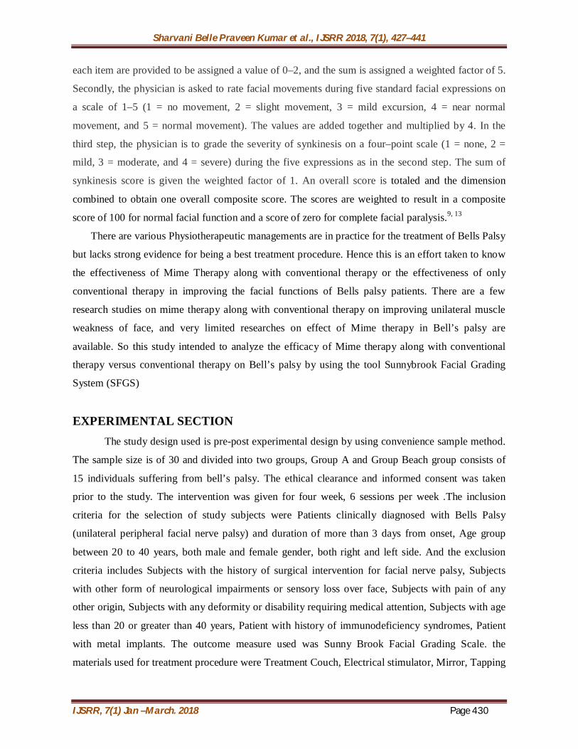
Table-5: Mean and SD of Symmetry of Voluntary Movement (SVM)
The table 5 shows the symmetry of voluntary movement prior to the intervention was 66.93±12.33 in group A and after
intervention the results shows as 83.47±10, where as in group B it was 75.27±10.44 prior to the intervention and post
intervention the results shows as 86.4±7.37.
Graph-1 Mean and SD of symmetry of voluntary movement
Table-6: Pre- post comparison in SVM
Average
improvement
z-value p-value Result
Group-A Pre SVM – post SVM 16.53333 3.464 0.001 P<0.05 sig
Group-B Pre SVM – post SVM 11.13333 3.413 0.001 P<0.05 sig
The Table 6 shows that symmetry of voluntary movement in Group A and B shows significance difference before and
after intervention, p value is 0.001.
0102030405060708090
pre post pre post
Group-A Group-B
66.9333
83.466775.2667
86.4
SVM N Mean and Std.
Deviation
Std. Error Mean
Group-A Pre SVM 15 66.93±12.33 3.18
Post SVM 15 83.47±10.24 2.64
Group-B pre SVM 15 75.27±10.44 2.69
Post SVM 15 86.40±7.38 1.90
Sharvani Belle Praveen Kumar et al., IJSRR 2018, 7(1), 427–441
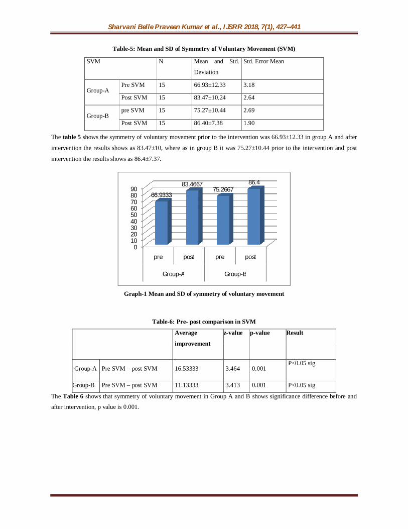
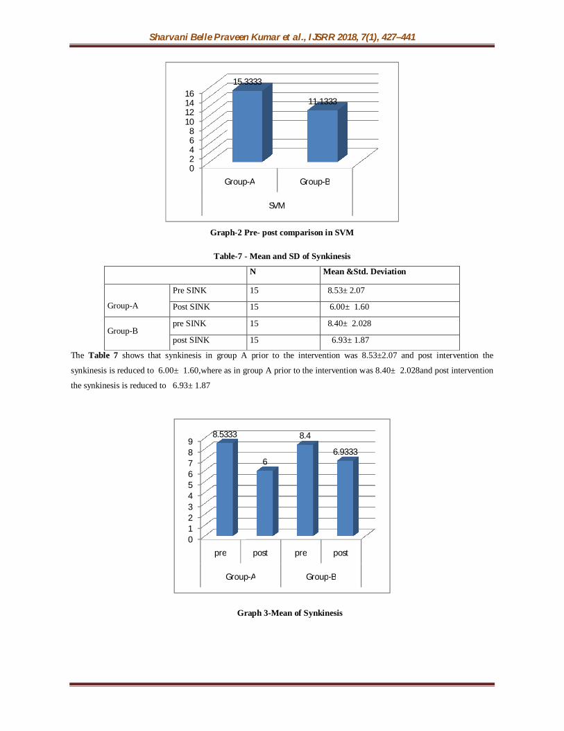
Graph-2 Pre- post comparison in SVM
Table-7 - Mean and SD of Synkinesis
N Mean &Std. Deviation
Group-A
Pre SINK 15 8.53± 2.07
Post SINK 15 6.00± 1.60
Group-B pre SINK 15 8.40± 2.028
post SINK 15 6.93± 1.87
The Table 7 shows that synkinesis in group A prior to the intervention was 8.53±2.07 and post intervention the
synkinesis is reduced to 6.00± 1.60,where as in group A prior to the intervention was 8.40± 2.028and post intervention
the synkinesis is reduced to 6.93± 1.87
Graph 3-Mean of Synkinesis
02468
10121416
Group-A Group-B
SVM
15.3333
11.1333
0123456789
pre post pre post
Group-A Group-B
8.5333
6
8.4
6.9333

Sharvani Belle Praveen Kumar et al., IJSRR 2018, 7(1), 427–441
Table-8 - Pre post comparison of Synkinesis
Average improvement z- value p-value
Group A Pre SINK-post SINK 2.5333 3.214 0.001
Group B Pre SINK-post SINK 1.46667 3.326 0.001
The table 8 shows the results of synkinesis in group A and group B, where the P value is 0.001 which indicates the
significant reduction in synkinesis.
The Table 9 shows the mean and standard deviation of SFGS prior to intervention in group A was 44.60±14.74 and post
Table 9-Mean and standard deviation of SFGS
intervention was 62.87±11.34, whereas in group B prior to intervention was 57.40±7.41 and post intervention
was66.53±8.37.
Table-10 - pre post comparison of SFGS
Average
improvement
z-value p-value result
Group- A Pre SFGS – post SFGS -18.26667 3.416 0.001 P<0.05 sig
Group-B Pre SFGS – post SFGS -9.13333 3.299 0.001 P<0.05 sig
The Table 10 shows the average of pre and post SFGS
Graph – 4-Average improvement in SFGS
0
5
10
15
20
GROUP-A GROUP-B
SFGS
18.266
9.133
N Mean and Std.
Deviation
Std. Error Mean
Group-A Pre SFGS 15 44.60±14.74 3.81
Post SFGS 15 62.87±11.34 2.93
Group-B Pre SFGS 15 57.40±7.41 1.91
Post SFGS 15 66.53±8.37 2.16

Sharvani Belle Praveen Kumar et al., IJSRR 2018, 7(1), 427–441
Graph-5: Shows pre post comparison between group A and group B of SVM, Synkinesis and SFGS
Since both the groups are showing significant improvement, we need to find which is better.
Therefore between groups comparison is done, result as follows
Comparison between A and B in SVM shows P<0.005, average improvement is more in
group A. Hence Group A is better than Group B.
Comparison between A and B in Synkinesis shows P<0.05, means there is significant
difference between group A and B. Average improvement is more in group A.
Same as SFGS shows average improvement is more in group A
DISCUSSION The present clinical trial was conducted to study the Efficacy of Mime therapy along with
conventional therapy in improving facial function on patients with acute Bell’s palsy. The SBFGS
used to evaluate severity of facial nerve paresis, included three components resting symmetry,
voluntary movements and Synkinesis. In the present study asymmetry has reduced in both the groups
but more in a Mime therapy along with conventional therapy group than others. This improvement
may be because massage improves circulation and maintains muscle properties. Visual feedback has
shown to control muscle activities in facial muscle. Also miming demands highly refined sense of
body and muscle control. Earlier studies proved that mime therapy has shown to create new growth
0
2
4
6
8
10
12
14
16
18
20
Gro
up-A
Gro
up-B
Gro
up-A
Gro
up-B
Gro
up-A
Gro
up-B
SVM SINK SFGS
15.3333
11.1333
2.53331.4667
18.2667
9.1333
Sharvani Belle Praveen Kumar et al., IJSRR 2018, 7(1), 427–441
IJSRR, 7(1) Jan –March. 2018 Page 439
and increase production of collagen and connective tissue in facial muscles and restore facial muscle
action.
In group A, improvement in facial function could be because of Mime therapy and
conventional therapy which is a combination of an active movement with simultaneous passive
stimulation which helps in rapid restoration of facial function. Mime therapy found to be effective by
neuro physiological mechanism of production of highly refined sense of muscle control. This
treatment technique produces a total and immediate recovery of facial function. Another Study say
that mime therapy has an effect on patients with long term facial nerve paresis and it shows that it
improves facial symmetry.
In accordance with a study of Cronin and Steenerson (2003) biofeedback by surface
electromyography results revealed improvement in facial symmetry. Ahmad SJ and Rather AH
(2012) did a prospective Study of physical therapy in facial nerve paralysis and found that
physiotherapy in the form of electrotherapy and facial exercises has an effective role in the early
management of peripheral facial paralysis. Because the effectiveness of therapeutic changes differs
greatly among patients, it is essential that effectiveness of trials directly measure patient related
improvement and satisfaction with treatment.
In group B the improvement in facial function could be because of electrical stimulation
which is aimed to increase the passive activation of muscle by firing the motor point and nerve root.
Its passive rhythmic movement re-establishes the function of muscle and gain control thereby
improving facial function.
When the improvement in facial function of group A was compared with group B subjects
there was no significant difference, however group A subjects showed greater percentage of
improvement in facial function. This could be due to added effect of active movement along with
simultaneous passive stimulation in Group A which is lacking in Group B. Mime therapy along with
Conventional therapy was better in improving facial function as it has the additional benefit which
may engage additional muscle control. The difference in improvement can be variable as pre
intervention comparison of means between Group A and Group B found that there is no statistically
significant difference in RS, SVM, and SYN between the groups. Therefore this may also interfere
with the post intervention means. SFGS average improvement showed in Group A.
The findings in this study are based on the subjects with age of 20 to 40 years having bells
palsy. Therefore effects cannot be generalised with other age groups.
Hence based on the analysis and findings, the present study found that with 4 week of Mime therapy
along with conventional therapy showed better result with P value <0.05. So mime therapy along
with conventional therapy is a good choice of treatment for people with Bell’s palsy. Thus Mime

Sharvani Belle Praveen Kumar et al., IJSRR 2018, 7(1), 427–441
IJSRR, 7(1) Jan –March. 2018 Page 440
therapy can be used in the treatment of people with acute Bell’s palsy to get improvement in Facial
asymmetry within a shorter period of time.
CONCLUSION The present study concluded that, Mime therapy and conventional therapy together are shown
to have effect on improving facial function and reduction of facial Synkinesis in Bell’s palsy
patients. Conventional therapy alone also has effect on improving facial function and reduction of
facial Synkinesis in Bell’s palsy patients. The combination of Mime therapy and Conventional
therapy showed more effect on improving facial function and reduction of facial Synkinesis in Bell’s
palsy patients. Hence it is recommended that combination of both Mime therapies along with
Conventional therapy is clinically beneficial on improving facial function and reduction of facial
Synkinesis for Bell’s palsy patient. The limitationswas the subjects with small range group between
20 to 40 years of age were considered for the study, thus results cannot be generalized to individual
age. It is a short duration study in which follow up was not done, therefore long term effects were not
known. Further recommendations for the future research is considering large sample, to find the
added effect of conventional therapy along with other techniques and follow up for long term effects.
REFERENCES
1. Fink B and Penton-Voak. “Evolutionary psychology of facial attractiveness”. Current
Directions of Psychological Science. 2002; 1:154-158.
2. Heymans PG. “The impact of facial paresis: Psychological mechanism”. In Buerskens CHG
et al (Eds) The Facial Palsies, Utrecht: Lemma. 2005; 335-356.
3. Peitersen E. Bell’s Palsy; The spontaneous course of 2,500 peripheral facial nerve palsies of
different etiologies”.Actaotolaryngolsuppl. 2002; 549: 4-30.
4. Greco A, et al, Bell's palsy and autoimmunity, Autoimmune Rev (2012),
doi:10.1016/j.autrev.2012;05:008
5. Lewis P Rowland, Mielkie. Merritt's Textbook of Neurology .9th Edition . ISBN-13: 978-
0812117127.
6. Jagmohan Singh, Textbook of Electrotherapy, 1st Edition. Jaypee brothers medical
publishers, 2005; 1; 104- 6.
7. Mistry Gopi S, Sheth Megha S, Vyas Neeta J: Int J Med Res Health Sci. DOI:
10.5958/j.2319-5886.3.1.026 www.ijmrhs.com, 2014;3(1):133-136.

Sharvani Belle Praveen Kumar et al., IJSRR 2018, 7(1), 427–441
IJSRR, 7(1) Jan –March. 2018 Page 441
8. Beurskens CH, Heymans PG. "Mime therapy improves facial symmetry in people with long-
term facial nerve paresis: a randomized controlled trial.” Aust J Physiotherapy. 2006; 52 (3):
177–83.
9. Coulson S, Croxon G, Adams R and O’Dwyer N, Reliability of the ‘Sydney’, ‘Sunnybrook’
and ‘House Brackmann’ facial grading systems to assess voluntary movement and
Synkinesis following facial nerve paralysis. Otolaryngology–Head and Neck Surgery, 2005;
132: 543–549.
10. Nelson RM, Currier DP. Clinical Electrotherapy. 2nd ed. New Delhi: Prentice Hall of India
Private Limited; 1991; 97-104.
11. Mehta RP, WernickRobinson M, Hadlock TA. "Validation of the Synkinesis Assessment
Questionnaire.” Laryngoscope. 2007; 117 (5): 923–6.
12. Berg T. Medical Acta Universitatis upsaliensis Digital Comrehensive Summaries of Uppsala
Dissertations from the Faculty of Medicine.Uppsala.ISBN978-91-554-7541-3. 2009; 460:47
13. Wei-Li Hu, Brenda Ross, and Julian Nedzelski: Department of Otolaryngology, University
of Toronto Faculty of Medicine, Sunnybrook & Women's College Health Science Centre,
Toronto, Ontario: Reliability of the Sunnybrook Facial Grading System by Novice Users
The Journal of Otolaryngology Volume 30, Issue 04, August 2001;208-209.
14. Modlin m, Forsinger E, Hoffer C et.all. Electrical stimulation pf denervated muscle. Artif
Organs. Mar 2005; 29(3):203-6.
15. Shorde LW. Treatment of facial muscles affected by Bell’s palsy with high voltage electrical
muscle stimulation. J manipulative physiology ther. Jun 1993; 16(5):347-52.
16. V. Parkas, K. Hariohm, P. Vijayakumar and D. Thangjam Bindiya Functional Training in the
Management of Chronic Facial Paralysis: Received November 23, 2010.Accepted December
12, 2011.© 2012 American Physical Therapy Association.