international journal of pharmaceutics - kinampark.comkinampark.com/kpyear/files/2016 lim,...
TRANSCRIPT

International Journal of Pharmaceutics 514 (2016) 41–51
Combinatorial nanodiamond in pharmaceutical and biomedicalapplications
Dae Gon Lima, Racelly Ena Prima, Ki Hyun Kima, Eunah Kangb,*, Kinam Parkc,Seong Hoon Jeonga,**aCollege of Pharmacy, Dongguk University-Seoul, Gyeonggi, Republic of Koreab School of Chemical Engineering and Material Science, Chung-Ang University, 221 Heukseok-Dong, Dongjak-Gu, Seoul, Republic of KoreacDepartments of Pharmaceutics and Biomedical Engineering, Purdue University, West Lafayette, IN 47907, United States
A R T I C L E I N F O
Article history:Received 8 May 2016Received in revised form 2 June 2016Accepted 3 June 2016
Keywords:NanodiamondDrug deliverySurfaceComplex formation
A B S T R A C T
One of the newly emerging carbon materials, nanodiamond (ND), has been exploited for use in traditionalelectric materials and this has extended into biomedical and pharmaceutical applications. Recently, NDshave attained significant interests as a multifunctional and combinational drug delivery system. NDstudies have provided insights into granting new potentials with their wide ranging surface chemistry,complex formation with biopolymers, and combination with biomolecules. The studies that have provedND inertness, biocompatibility, and low toxicity have made NDs much more feasible for use in real in vivoapplications. This review gives an understanding of NDs in biomedical engineering and pharmaceuticals,focusing on the classified introduction of ND/drug complexes. In addition, the diverse potentialapplications that can be obtained with chemical modification are presented.
ã 2016 Elsevier B.V. All rights reserved.
Contents lists available at ScienceDirect
International Journal of Pharmaceutics
journal homepage: www.elsev ier .com/locate / i jpharm
1. Introduction
Nanodiamonds (NDs) have been developed as electric materialssince they have promising properties such as energy absorbance,thermal diffusivity, and high capacity (Branson et al., 2013; Limet al., 2013; Sundar et al., 2014). NDs also have applications inlubricants, composites, electromagnetic shielding, and specialcatalysts similar to other carbon materials (Mochalin et al., 2012).Recently, NDs have emerged as a new platform for nano/biomaterials due to their interfacial integration with a variety ofbiomolecules, including proteins, polymers, chemical drugs, andgenes. In detail, exploring the biomedical applications of versatilepotential ND via chemical modification or physical absorption hasbeen proposed for many different applications including photo-acoustic imaging agents, polymer composites as a dental resin(Mochalin et al., 2011), gene carrier (Bertrand et al., 2015; Zhanget al., 2009), drug reservoir (Chen et al., 2009), and fluorescencemarker (Schrand et al., 2011). The easy access of NDs to biomedicalapplications relies on the moderate condition of the surface
* Corresponding author.** Corresponding author at: College of Pharmacy, Dongguk University-Seoul,Goyang, Gyeonggi 410-820, Republic of Korea.
E-mail addresses: [email protected] (E. Kang), [email protected](S.H. Jeong).
http://dx.doi.org/10.1016/j.ijpharm.2016.06.0040378-5173/ã 2016 Elsevier B.V. All rights reserved.
modifying capability in charges and other functional groups on theND substrate. Moreover, it has been observed that ND particlesuptaken by cells are minimally cytotoxic and biocompatible, whichmeans they do not affect mitochondrial function or ATP productionat the cellular level (Schrand et al., 2007a); however, thebiocompatibility of ND varies depending on its material properties.
The nature of promising NDs depends mainly on their chemicalproduction and purification procedures. NDs can be produced viadetonation method (Mochalin et al., 2012), chemical vapordeposition (Liu and Dandy, 1995), high-energy ion irradiation ofgraphite (Daulton et al., 2001), and high-energy ball milling ofdiamond microcrystals (Boudou et al., 2009). Different productionmethods, treatment conditions, and processing techniques resultin diverse types of NDs that vary in size, shape, structure, and evensurface chemistry (Kulakova, 2004; Paci et al., 2013; Sabirov andOsawa, 2015). This creates distinct surface properties making itpossible to use as an extensively good platform for other potentialdiscoveries (Paci et al., 2013). However, this is also why identifyingthe physical and chemical properties of NDs and carrying outquantitative analysis of its surrounding chemistries remain achallenge (Mochalin et al., 2012).
NDs contain a core diamond crystalline structure and possess aunique surface structure. They have a large specific surface area,high adsorption capacity, and chemical inertness (Kulakova, 2004;Mochalin et al., 2012). Various functional groups in the ND surface

Fig. 1. Summary of the functional groups present on the nanodiamond surface.
42 D.G. Lim et al. / International Journal of Pharmaceutics 514 (2016) 41–51
have been revealed saturation of the reactive surface of carbonatoms (Kulakova, 2004) and existence of dangling bonds forvarious covalent bonding (Krueger, 2008). These dangling bondsreact with the surrounding media to trigger the functionalizationof the ND particles (Krueger, 2008).
The ND surface is covered with amorphous carbon and mainlyoxygen-containing functional groups [6]. These functional groupsare analyzed using Fourier Transform Infrared Spectroscopy (FT-IR), which can detect functional groups and changes in the surfacechemistry of functionalized NDs (Chen et al., 2009; Mochalin et al.,2012). Measurements confirm that there is significant presence ofoxygen-containing groups on the ND surface (Chen et al., 2009).Small amounts of nitrogen-containing groups, methyl andmethylene groups, sulfone, and other groups are also present(Kulakova, 2004). Fig. 1 shows a summary of the functional groupspresent on the ND surface.
The ND retains amorphous carbon, graphitic shells, and the sp3phase of carbon on the diamond surface (Gaebel et al., 2012),which is available for surface functionalization. The physicochem-ical properties of the ND may be altered via surface modification(Sabirov and Osawa, 2015). Functional groups can be replaced byother groups, but they always remain attached within the surfaceof the ND (Kulakova, 2004). They can also be the binding site for thecovalent integration of ND into polymer structures and helpimprove the dispersibility of ND powders in common solvents (Liuet al., 2004). Various groups are compatible with ND surface
Fig. 2. Schematic description of ND surface modification. Carboxylated ND (ND–COOH) iwith gas at high-temperature condition (red area) or wet chemistry modification at ambiCopyright 2012 Macmillan Publishers Ltd.). (For interpretation of the references to col
chemistries, allowing for radical surface functionalization, andthus present a distinct characteristic of ND compared to othernanoparticles (Mochalin et al., 2012; Wahab et al., 2015); ND alsoexhibits high surface reactivity compared to other carbon nano-structures. ND is chemically functionalized in many ways, but theoutcome depends on the purity and uniformity of their surfacechemistry (Fig. 2) (Mochalin et al., 2012; Sabirov and Osawa, 2015;Wahab et al., 2015). Surface functionalization was also said to affectthe stability of ND surfaces (Mochalin et al., 2012). Thefunctionality of the ND surface is responsible for its drug bindingability and imaging, and is also a determining factor for its otherapplications (Paci et al., 2013).
With these specific potentials and capabilities, ND complexesthat are formed either by physical adsorption or chemicalconjugation have the benefits of reducing the multidrug resistanceof anticancer drugs, enhancing delivery efficacy with convectivediffusion. This property of ND complexes may possibly be useful fordiverse smart designs (Wang et al., 2014). Small ND of 5 nmdiameter can perform as a carrier platform by holding drugmolecules within an intracellular compartment due to their largesurface area. Moreover, ambiguous surface functional groups andtheir density on the ND have dependence on versatile sources. Thisreview summarized the versatile chemical modifications of NDsrequired to develop its desirable properties, the potentials of ND asa drug delivery carrier platform, and the fate of ND within cells andother in vivo applications. Future applications of ND were alsodiscussed with the objective of creating pharmaceutical applica-tions.
2. Molecular dynamics
Possible concepts of versatile and novel ND applications havebeen disclosed in several recent studies, which have made a strongdriving force towards analogous goals. However, different com-mercial sources of NDs provide ambiguous and veiled surfacecharacteristics that require fundamental understanding andmethodology standardization in ND experiments (Lai and Barnard,2014). Understanding the faceted ND surface is a critical step intaking advantage of the high surface area to volume ratio forbiomedical and pharmaceutical applications. Furthermore,
s a common starting material. The surface of ND–COOH can be modified by reactionent-temperature condition (blue area). (Adapted by permission from Mochalin et al.,our in this figure legend, the reader is referred to the web version of this article.)

D.G. Lim et al. / International Journal of Pharmaceutics 514 (2016) 41–51 43
stoichiometric adsorption of biomolecules on ND surface needs tobe clarified based on the understanding of its properties.
Regarding this aspect, several computer simulations andmolecular dynamics have given fundamental insights into bareNDs. The adsorption strength of bare NDs was investigated withregard to the distribution of basic functional groups such as COOH,OH, O, and H at the atomic level (Lai and Barnard, 2014). It is saidthat the surface reconstruction by functional groups specifies facetorientation and that ultimate particle shape determines theconcentrated adsorbents on ND surfaces. Facet (111) providesthe crowded density of COOH groups, resulting in weak adsorption.It is interesting that the size and shape of facets are also one of thefactors for determining the surface properties of NDs, resulting inits varied adsorption ability. Barnard et al. extensively studied theeffects of shape and size on NDs (Barnard and Per, 2014; Barnardand Sternberg, 2005). The electro-potentials of the surface becomedifferent depending on the shape, which might be a critical factorin determining the ND properties in an aqueous solution. Thedegree of protonation in the edges and corners of ND facets mayvary depending on the size of the facets.
ND itself is influenced by the factors of its exterior environmentsuch as the temperature and pH (Adnan et al., 2011; Lai andBarnard, 2012). Together with the basic understanding of ND suchas its molecular dynamics, the behavior of ND complexes withdrugs has also been investigated. One study simulated the pHdependent interaction between doxorubicin (Dox) hydrochlorideand faceted ND (Guan et al., 2010). Molecular dynamics suggeststhat the manner in which Dox adsorption on ND depends on the
Fig. 3. pH-dependent doxorubicin (Dox) adsorption on the ND surface. Magnified ND–DDox are close to minimum at low pH, while their strong electrostatic interactions lead tmodeling with eight NDs at (c) low pH and (d) high pH (modified with permission fro
pH. In ND/Dox complexes, ND was integrated with 30% functionalgroups at different pH levels. As shown in Fig. 3, it was noted thatDox was located at 15 Å from the ND surface at low pH, maintaininga stable separation. Dox was bound to the ND surface at high pH,indicating that the electrostatic interaction was strong enough(Adnan et al., 2011). The pH dependent behavior of ND mightchange depending on the major surface functionality. In addition topH adjustment, van der Waals forces, hydrogen bonding, and facet-dependent dipole moments are also factors in determining NDaggregation and drug adsorption. It is critical to understandcompletely how the relationships between diverse factors influ-ence ND agglomeration and drug adsorption.
In an advanced study, a multiscale simulation of siRNA genedelivery was carried out with polyethyleneimine (PEI)-based NDcomplexes (Kim et al., 2012). From the first layer of PEI on ND, thesimulation estimated the number of PEI to the ND and the effectiveratio of siRNA adhering to the PEI/ND surface. It was noted that theapproach of the large molecule, PEI, was limited depending on thefacet size and facet direction.
3. Nanodiamond in biomedical imaging
3.1. Fluorescence nanodiamond with nitrogen-vacancy sites
Laser-irradiated ND with nitrogen-vacancy color center isknown to possess fluorescence and magnetism characteristics,which make it useful for smart applications in nano-scaledbiolabeling and bioimaging (Faklaris et al., 2009; Havlik et al.,
ox complex at (a) low pH and (b) high pH. The electrostatic interactions of ND withhe Dox molecules to be adsorbed on the ND surface at high pH. Schematic view ofm Adnan et al., Copyright 2011 American Chemical Society).
44 D.G. Lim et al. / International Journal of Pharmaceutics 514 (2016) 41–51
2013). Since the first direct observation of 5 nm fluorescencediamond (FND) was made, ND has garnered growing interests as apromising material (Bradac et al., 2010; Wrachtrup, 2010). Thelong-lasting optical properties of laser-irradiated ND withnitrogen vacancy sites were enhanced with the addition of asilica shell coating (Prabhakar et al., 2013). This nanocompositematerial with ND provided continuous cellular tracking due to itsstable photoluminescence. The PEG-PEI-coated ND providedimproved cellular uptake, showing localized FND at subcellularregions (Prabhakar et al., 2013). Moreover, the long term stabilityand biocompatibility of laser-irradiated fluorescence ND in vivowas examined. The development of the intrinsic properties of FNDwas also investigated with photostability, imaging resolution, andfluorescence intensity (Vaijayanthimala et al., 2012). However,FND with a nitrogen-vacancy color center is not widely used due toits price and limited production. The FND production methodvaries depending on the ND source, input energy, and ion species(Nagl et al., 2015). The accurate set-up of FND production and thebiomedical applications of FND still have a lot of areas to beexamined.
3.2. Fluorescence diamond with extrinsic dyes
Fluorescence dyes as a model drug were complexed with NDvia physical adsorption (Chang et al., 2008; Lien et al., 2012).The studies provide insights about novel ND nanocomposites asa tool for bioimaging and drug delivery platforms. Since it is noteasy to access ND with nitrogen vacancy sites due to the highcost of the equipment and unfamiliarity with the methods ofproduction, alternative approaches to introduce extrinsicfluorescence dyes have been achieved via conjugation ontothe ND surface (Hens et al., 2008; Schrand et al., 2011). CoupledND complexes between aminated ND and reactive N-hydrox-ysuccinimide (NHS) functionalized TAMRA were utilized fortemporal and mechanistic cell tracking (Schrand et al., 2011). Co-localizations, early endosomes, or lysosomes in N2A cells weretraced over time. The destination of ND localization intorespective cell compartments may enhance the targeted site-specific drug delivery of genes or proteins. TAMRA conjugatedwith ND plays a role as a bright biomarker with highfluorescence intensity, probably due to the high reactive sitesof ND.
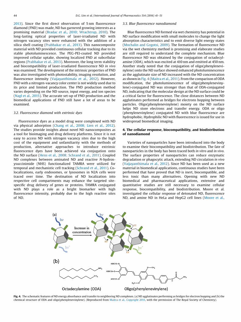
Fig. 4. The schematic features of ND energy absorbance and transfer to neighboring ND cochemical structure of ODA and oligo(phenylenevinylene). (Reproduced from Maitra et
3.3. Blue fluorescence nanodiamond
Blue fluorescence ND formed via wet chemistry has potential inND surface modification with small molecules to change the lightabsorption characteristics and to emit diverse light energy states(Mochalin and Gogotsi, 2009). The formation of fluorescence NDvia the wet chemistry method is promising and elaborate studiesare still required to understand the complete mechanism. Bluefluorescence ND was obtained by the conjugation of octadecyl-amine (ODA), which was excited at 410 nm and emitted at 450 nm.Another study noted that the conjugation of oligo(phenylenevi-nylene) onto the ND surface showed enhanced photoluminescenceas the agglutinate size of ND increased with the ND concentrationas shown in Fig. 4 (Maitra et al., 2011). From the comparison of ODAmodification, the photoluminescence of oligo(phenyleneviny-lene)-conjugated ND was stronger than that of ODA-conjugatedND, indicating that the molecular design at the ND surface could bea critical factor for fluorescence. The close distances between NDagglutinates performed as bridges for electrons hopping betweenparticles. Oligo(phenylenevinylene) moiety on the ND surfacehelps ND store electrons and transfer energy. ODA or oligo(phenylenevinylene) conjugated ND with blue fluorescence arehydrophobic. Hydrophilic ND with fluorescence is issued for use inwidespread biomedical imaging.
4. The cellular response, biocompatibility, and biodistributionof nanodiamond
Varieties of nanoparticles have been introduced into the bodyto examine their biocompatibility and biodistribution. The fate ofnanoparticles in the body has been traced both in vitro and in vivo.The surface properties of nanoparticles can reduce enzymaticdegradation or phagocytic attack, extending ND circulation in vivo(Vaijayanthimala et al., 2012). Since ND has been used as a newmaterial in biomedical applications, continuous studies have beenperformed that have proved that ND is inert, biocompatible, andless toxic than many alternatives. Opening with new NDbiomedical and pharmaceutical applications, extensive andquantitative studies are still necessary to examine cellularresponse, biocompatibility, and biodistribution. Moore et al.investigated the cellular response of detonated ND, fluorescenceND, and amine ND in HeLa and HepG2 cell lines (Moore et al.,
mplexes. (a) ND agglutinates performing as bridges for electron hopping and (b) theal., Copyright 2011, with the permission of The Royal Society of Chemistry).

D.G. Lim et al. / International Journal of Pharmaceutics 514 (2016) 41–51 45
2014). ND at 25 mg/mL concentration did not show promotedapoptosis, an inflammatory response, or inhibited proliferation atthe level of the transcriptional response. Moreover, ND/daunoru-bicin complexes at a ratio of 5:1 showed less toxicity at anequivalent dose of the free drug for metabolic activity, cell deathfrom lactate dehydrogenase release, and initial apoptosis fromcaspase activity (Man et al., 2014). Simple physical drug adsorptiononto the ND and subsequent cellular treatment in vitro showedpotentially promising results as a new drug reservoir. However, itshould be noted that the stability of the ND complexes was notexamined over time in biological media and with appropriateexposure to ND or ND/drug complexes into cells.
Several studies have shown that bare ND is less toxic comparedto other carbon materials such as carbon nanotubes, graphenes,and graphites (Paget et al., 2014; Schrand et al., 2007b; Zhu et al.,2012). Fluorescent dye-conjugated ND was injected to mice forbiodistribution study. ND was accumulated in the lung, liver,spleen, and kidney in a day and its clearance lasted over 10 days.Moreover, fluorescence signal was also observed in the bladder(Chow et al., 2011). When ND, graphite, and graphene oxidenanoparticles were injected to rats intraperitoneally, ND showedsignificantly less aggregates in mesentery, connective, andabdominal lipid tissue compared to graphite and graphene oxidenanoparticles (Kurantowicz et al., 2015). The biocompatibility andbiodistribution of ND still have critical issues for further realisticapplications. Inert bare ND and heparin/polyarginine-coated NDshowed dramatically improved hemocompatibility compared tothe group of graphene (Li et al., 2013). Liu et al. observed the
Fig. 5. Cellular trafficking of ND agglutinates in cell cycles and division. (a) The trackininterphase and mitotic phase (prophase, metaphase, and telophase), (b) Schematic picCopyright 2009, with permission from Elsevier).
cellular tracking of fluorescence-labeled ND in an A549 cell (Liuet al., 2009). It was noted that ND agglutinates were localized in theinterphase and mitotic phase (prophase, metaphase, and telo-phase). ND was also separated and localized into two daughtercells. ND was shown in all cell cycle phases, while not disturbingthe spindle formation or chromosome segregation. The studyproved that endocytic ND agglutinates were not cytotoxic in celldivision and differentiation over 10 days of long term observation.One study by Marcon et al. also examined the cytotoxicity of NDswith different surface functionalities (–OH, –NH2, –COOH) inHEK293 cells and Xenopus laevis embryos (Marcon et al., 2010). Nocytotoxicity was present in up to 50 mg/mL of ND in HEK293 cells,while the embryotoxicity for carboxylated ND was shown for bothgastrulation and neurulation. Moreover, Rojas et al. studied in vivoND biodistribution using radioactive 18F-labeled ND (Fig. 5) (Rojaset al., 2011). The individual particle sizes of surface modified NDwas approximately 7 nm, while aggregates in aqueous solutionwere approximately 680 nm, as measured by light scattering thatrequired surfactants in their dispersion solvent of up to 5% (Rojaset al., 2011). Interestingly, prefiltered NDs showed a highlyexcreted distribution into the urinary tract, proving that stableNDs aggregates in size are critical for biodistribution and excretion.The long term toxicity study of ND in rats was also investigated(Vaijayanthimala et al., 2012). Fluorescence ND was administeredwith subcutaneous, intradermal, and intraperitoneal injections fora dose per week, and rats were sacrificed for the tissue morphologyafter 12 weeks. Dark carbon-laden cells were observed in thedermis with no tissue damage, inflammation, or necrosis in the
g of ND agglutinate at each cellular phase. ND agglutinates were localized in theture of the suggested mechanism of ND localization. (Reproduced from Liu et al.,

46 D.G. Lim et al. / International Journal of Pharmaceutics 514 (2016) 41–51
surrounding cells. Promising results of ND applications in vivo havebeen shown in a way, yet further extensive studies should beperformed for broader and more realistic ND applications.
5. Nanodiamond dispersion
Unusually strong aggregates of ND lead to elaborating thedeagglomeration steps of grinding, milling with salts (Pentecostet al., 2010), sonication with the addition of salt, and theaddition of surfactant (Xu and Zhao, 2012; Xu et al., 2005).Without impairing the intrinsic properties of NDs, the efforts toprepare well-dispersed NDs in an aqueous solution have beenperformed. Detonated ND is approximately 5 nm in size, butoften forms named aggregates or agglutinates in the range ofseveral hundred nanometers in aqueous solution (Chang et al.,2011). At present, ND can be classified according to itsagglutinins and disintegrated aggregates. Agglutinin ND isbound with covalent C��C bonds between internanocrystals,forming unusually strong tight aggregates. Even under strongultrasonication, agglutinates are stable without disintegration.Disintegrated NDs are composed of agglutinate and smallaggregates interconnected by van der Waals forces andelectrostatic interaction. NDs have been heavily functionalizedwith hydrophilic groups on its surface including phenylated,carboxylated, and sulfonylated NDs to produce water-dispers-ible NDs. These functional groups help in the production of astable dispersion of NDs in aqueous solution (�100 mg/mL) viamild ultrasonification (Huang et al., 2007). It was hypothesizedand confirmed that the carboxyl group on the ND helped theND interface ND/drug complexes through physiosorption andelectrostatic interactions (Chen et al., 2009). The dependence ofthe ND functional group on ND/drug complexes has beencharacterized with ND/drug imaging and UV–vis analysis, andvarious ND surface modifications grant specific functionalities.However, ND aggregation (cluster formation) in biologicalsolutions limits its wider applications.
One simulation suggests that a ND with a hydroxyl group ismore thermodynamically stable than a ND with a carboxyl or anamine group (Krueger and Lang, 2012), and the size of agglutinatesis greatly dependent on Coulombs law by the van der Waalsinteraction rather than electrostatic interaction (Chang et al.,2011). It was reported that a varied facet size originates the dipolemoments, determining the final size of the ND agglutinates (Laiand Barnard, 2014). It was also reported that ND aggregates cancause DNA damage in embryonic stem cells (Xing et al., 2011); theresults implied that the cellular damage caused by the ND wasprobably due to the unacceptable agglutinate size in theintracellular compartment. Thus, for ND to be exploited inbiomedical and pharmaceutical areas as a drug delivery platform,the stable dispersion of ND in aqueous and biological solution iscritical. Surface modified ND with high purity also showed anarrow distribution in an aqueous solution (Kuznetsov et al., 2012).Ultimately, chemical surface modification may be required todisperse the NDs evenly in aqueous solution and be furthercirculated when in vivo.
Niu et al. showed promising potentials, proving that thephotoluminescence of the ND itself could be controlled by laserchemistry (Niu et al., 2011). Laser irradiation on the ND resulted inthe ablation of the amorphous carbon layer and a disconnectionbetween ND agglutinates. Unusually strong ND agglutinates couldbe controlled by laser irradiation with the addition of a PEGsurfactant. The ND size distribution was critically varied, resultingin unique light absorption and photoluminescence. The studyshowed the relevant importance of ND surface modification anddispersion.
6. Nanodiamond drug delivery
6.1. Physical adsorption of small molecular drugs
The most important advantages of ND as a drug deliveryplatform arise from its strong physical adsorption. The intrinsicproperties of ND adsorption originated from its high surfacearea to volume ratio and its facet-dependent dipole moment.Even the organic impurities were absorbed within the ND, orthe ND contained bound and even absorbed water at itsmolecular level. The absorbed water is not easily removed, evenduring drying procedures (at 393 K), because it is absorbedinside the ND pores that form upon aggregation (Kulakova,2004). The strong capability of physical absorption is known tobe controlled by the ND surface functional groups that can alsobe modified via oxidation such as through simple acid-washing(Krueger and Lang, 2012).
The chemical conjugation of prodrug onto the ND surface bymicrowave-assisted paclitaxel conjugation has been suggested tofacilitate the functionality of the ND surface and increase the drugloading efficiency; the chemical conjugation of paclitaxel doubledthe loading efficiency (Hsieh et al., 2015). It was also reported thatpolyglycerol was conjugated on the ND surface as a linker (Zhaoet al., 2014a). Arg-Gly-Asp (RGD) cell penetrating peptide sequenceand Dox were conjugated at the end of the polyglycerol.ND–polyglycerol with Dox in the respective cell line showedselective effects with minimal uptake and toxicity in a macro-phage, while being highly sensitive to the drug in cancer cells.
In fact, rather than the chemical conjugation of prodrug,ND/drug delivery via physical adsorption was intensively reflectedin diverse studies due to its simple process. Oxidized and bare NDhas often been investigated for its sorption of heavy metal, dyes,proteins, and chemical drugs (Chernysheva et al., 2015; Chu et al.,2014; Manus et al., 2009; Wang et al., 2012). To understand the NDfundamentals as a drug delivery carrier, Mochalin et al. studied NDagglomeration/deagglomeration and the adsorption capacity ofdifferent drugs (Mochalin et al., 2013). The surface functionalgroup on ND with amine, carboxyl, and hydrogen groups showedvaried adsorption levels for Dox and polymyxin B. ND dispersionand the drug solution being dependent on the pH condition requireclarification, since the solubility of Dox is also changed, accordingto pH. The ionized and charged surface functional groups on NDs atadapted pHs also alter the drug adsorption capacity. Salaam et al.studied the ND–Dox complexes formed by physical adsorption(Salaam et al., 2014a; Salaam et al., 2014b). The release of thosecomplexes was investigated as the pH changed. Since Dox wassolubilized at low pH, accelerated Dox release was expected at lowpH. It is not evident that pH controlled release is mainlyattributable to the ND surface reservoir ability or the intrinsicproperties of Dox. The promising desorption control of a drug,which is an important property of therapeutics, is shown in thisstudy. Rye et al. also developed ND/Dox complexes, particularlyusing microfluidic devices (Ryu et al., 2015). The simple process ofmicrofluidics controlled the size of the ND/Dox agglutinate, whichfacilitated cellular uptake. Approximately 50 nm of the NDagglutinate was shown to have high cellular uptake. Moreover,ND/Dox complexes were investigated for their ability to inhibit thelung metastasis of breast cancer (Xiao et al., 2013). Drug loadingwas carried out with a 5:1 weight ratio of ND to Dox. To prolong thein vivo circulation, 5% PEGylated positively charged amine lipidmolecules were also used to coat the ND/drug dispersion. Thoughthe ND/Dox lipid vesicles showed low cytotoxicity and therapeuticpotential, it was ambiguous whether the enhanced anti-metastaticeffect resulted from the ND/drug complexes or the aid of lipidvesicle formulation.

D.G. Lim et al. / International Journal of Pharmaceutics 514 (2016) 41–51 47
The study to investigate in vivo biodistribution also showed that1,2-distearoyl-sn-glycero-3-phosphoethanolamine (DSPE)-PEGlipid–coated NDs and poorly water soluble sorafenib complexesenhanced the circulation time in vivo (Zhang et al., 2014). Theresidual drug concentration in tissue was improved 15-fold,compared to bare ND/sorafenib complexes without lipid coating,and inhibited tumor growth in tumor xenograft models. ND usedfor the physical adsorption of mitoxantrone possessed highlypositive zeta potential and probably hydrogenated ND (Toh et al.,2014). Mitoxantrone adsorption resulted in a slight decrease ofpositive zeta potential. It can be suggested that the p–pinteraction and hydrogen bonding with the ND surface are majorinteracting forces with mitoxantrone, imposed from the structureof mitoxantrone. The release of mitoxantrone was accelerated atlow pH, indicating that the physical adsorption of the drug on NDshas potential for use in controlling the manner of drug release.
Anticancer drug delivery with specific targeting has beenexamined, mainly focusing on overcoming multi-drug resistance(MDR) with cancer cells. Surprisingly, ND showed increasedanticancer drug retention and thus antitumor activity. NDcomplexes with Dox showed increased acute apoptotic responsesand enhanced drug retention compared to treatment with Doxalone in MDCK-MDR drug resistant cells (Chow et al., 2011). ND-enhanced delivery and decreased drug efflux was not only definedfor solid tumors but also effective in leukemia cancer cells. ND–daunorubicin (DNR) complex showed lower IC50 values andefficacy against drug resistant K562 leukemia cells (Man et al.,2014). The pH dependent release of drugs was exampled usingDNR in leukemia that was bound via simple physical adsorption(Man et al., 2014). pH-dependent ND/DNR complexes showedsignificantly reduced gene expression of three major effluxproteins in the ATP-binding cassette (ABC) transporter. ND/DNRcomplexes were hypothesized in which NDs played a role as stabledrug-releasing platform and bypassed the efflux pump, elevatingthe drug concentration within cells and thus enhancing the drug’stherapeutic efficacy. It was suggested that the ND/drug deliveryplatform assisted the reduction of multidrug resistance in cancercell lines. Active drug efflux transporters such as ABC transporterscause anticancer drug resistance against anthracyclines such asDox and DNR (Schinkel and Jonker, 2003). Drug conjugates withNDs might evade drug efflux transporters by entering the cellthrough endocytosis (Zeng et al., 2014). The therapeutic effect ofepirubicin-adsorbed ND was investigated against chemoresistanthepatic cancer in vivo (Fig. 6) (Wang et al., 2014). The epirubicin–ND complex also showed improved antitumor activity and reducedmulti-drug resistance effects when compared to epirubicin alone
Fig. 6. Schematic description of epirubicin–ND complex formation and the tumor-insecondary allograft tumor formation compared to only epirubicin treated case at enhainjection (Reprinted with permission from Wang et al., Copyright 2014 American Chem
and an ND-treated group. The complex formation betweenepirubicin and ND prevents the efflux of epirubicin via ABCtransporters due to the change of molecular size and structure(Wang et al., 2014).
Enhanced convective drug delivery associated with the drugand NDs prevents drug efflux via the ABC transporter. Theextended drug retention within cells elevates the possibility totarget passively in the intracellular compartment, and thusfacilitates the inhibition of initial cancer stem cells mediated withtumor growth. NDs not only reduce drug resistance but alsoprevent the side effects associated with a high drug concentration.Dox showed myelosuppression, systemic immune response, andliver toxicity at a high concentration. ND/drug complexes might bean alternative delivery platform, replacing the small molecules ofan ABC transporter inhibitor. Though ND/drug complexes providedhigh loading efficiency as a capable drug reservoir, specificformulations seemed to be demanded for ultimately long lastingin vivo circulation and targeting. Surely, developing the method ofND use in optimal pharmaceutical formulations is required in thenear future to approach realistic therapeutic agents. Table 1 listsvarious examples of nanodiamond/drug complexes.
6.2. Adsorption of biomolecules onto nanodiamonds
Together with the adsorption of small molecular drugs, theadsorption of relatively large biomolecules onto the ND surface hasbecome an emerging interest as a new therapeutic deliveryplatform. A fundamental understanding of ND/biomolecule(proteins, siRNA, and DNA) complexes grants insight into thebiomolecular array on NDs. The protein size, surface charge, andcomplex ratio of protein to NDs are the determining factors thatform the biomolecule layer on the ND.
Protein adsorption was examined with different surface chargefunctionalities on NDs (Aramesh et al., 2015). The small proteinlysozyme formed multilayers with significant conformationalchanges, while the large protein albumin formed monolayerswith minimal conformational changes on the ND surfaces.Depending on the surface charges on ND, the protein conformationand array between proteins become distinct. One particular studyinvestigated the quantitative analysis of ND/protein adsorption byratio (Lin et al., 2015). A ND with 3.7 nm in diameter possessed asaturated surface forming 1:1 myoglobin/ND complexes of theprotein myoglobin with a molecular weight of 17 kDa. Anotherstudy reported that the physical adsorption and desorption ofinsulin onto NDs could be controlled in modulated pH environ-ment (Shimkunas et al., 2009). Insulin was desorbed from the ND
itiation evaluation of epirubicin–ND complex. Epirubicin-ND complex preventednced tumor-initiation evaluation in allografts when seeded at 300 tumor cells perical Society).

Table 1The physical properties of nanodiamond/drug complexes.
Mechanism ND-Source Drug Diameter (nm) Surface charge (mV) Ref.
Covalent linkage ND-COOH Recombinant growth hormone 9.3 N.D. Chu et al. (2014)ND-COOH a2b1 integrin-binding peptide 303 17 Knapinska et al. (2015)ND-PEG Cisplatin 66.9 N.D. Zhao et al. (2014b)Pristine ND Paclitaxel 10 N.D. Liu et al. (2010)
Physical adsorption ND-COOH Purvalanol A 556 27.2 Chen et al. (2009)ND-COOH Hydroxytamoxifen 278.9 25.7 Chen et al. (2009)ND-COOH Dexamethasone 77.55 21.9 Chen et al. (2009)ND-COOH Transferrin/doxorubicin 235 N.D. Wang et al. (2015)ND-COOH Daunorubicin 93.1 N.D. Man et al. (2014)ND-DGEA Doxorubicin 89 21 Salaam et al. (2014a)ND-Gd Epirubicin 89.2 19.6 Wang et al. (2014)ND-H siRNA 30 �15 Bertrand et al. (2015)ND-NH2 Plasmid DNA (GFP) 100 35 Zhang et al. (2009)ND-PEG Doxorubicin 187.7 �27.8 Wang et al. (2013)ND-PEI600 Plasmid DNA (GFP) 250 45 Zhang et al. (2009)Pristine ND Bovine Insulin 1690 �12 Shimkunas et al. (2009)Pristine ND Myramistin 150 29 Chernysheva et al. (2015)Pristine ND Sorafenib tosylate/DSPE-PEG 127.6 N.D. Zhang et al. (2014)Pristine ND Mitoxantrone 54.6 47.8 Toh et al. (2014)Pristine ND Bone morphogenetic proteins 120 N.D. Moore et al. (2013)Pristine ND Alginate/Cisplatin 486.9 �14.8 Cui et al. (2016)Pristine ND Amoxicilin 143.1 46.08 Lee et al. (2015)
48 D.G. Lim et al. / International Journal of Pharmaceutics 514 (2016) 41–51
and released into the cell at high pH. However, it should be notedthat the hydrodynamic diameters of NDs and insulin/NDcomplexes range over 1 mm; a simple pH adjustment mightmodulate the exterior charge of the protein and the NDaggregation. Protein stability and dispersion stability need to beclarified at each pH, forming stable ND complexes with stablecolloidal properties.
siRNA, which is smaller than proteins, might be moreapplicable in ND complexation. Hydrogenated ND with a highlycationic positive charge (50 mV of zeta potential) have beeneffective at binding therapeutic siRNA (Alhaddad et al., 2011),which was successfully targeted against the Ewing sarcomajunction and oncogenes, strongly inhibiting its expression incells. The cellular tracking of ND supported that ND in denseaggregate forms were localized in multivesicular cell bodies andproteins in late endosomes. Due to strong cationic charges,hydrogenated ND was an effective siRNA delivery platform. Inthe case of oxidized NDs with negative charges, ND withcationic polymers such as PEI and polyallyamine were layeredfor the vectorization of siRNA (Alhaddad et al., 2011). Theysuggested that cationic polymeric layers on NDs can modulatethe internalization pathways. PAH-coated ND/siRNA complexespromoted internalization through predominant clathrin-medi-ated endocytosis, while PEI-coated ND/siRNA facilitated cla-thrin-mediated endocytosis and macropynocytosis.
Charged peptide biomolecules were directly delivered on NDsurface. Protamine, a biodegradable and positively charged peptidepolymer, adsorbed onto the NDs through hydrogen bonding orelectrostatic interaction (Cao et al., 2013). Protamine is mainlycomposed of positive arginine peptides, which are known torestore downregulated MiR-203 in tumors such as esophageal,gastric, and colorectal cancers. The restoration of MiR-203expression by the delivery of micro-RNA ND/protamine complexesinhibits tumor cell proliferation, migration, and invasion. It isnoted that micro-RNA/ND/protamine complexes are also success-fully vectorized in the intracellular compartment, showing that aND is effective as a gene vector for using large molecules asdelivery platforms and therapeutics. Highly dense positive chargeson the ND surface provided stable micro-RNA residence duringvectorization into the cellular compartment.
6.3. Complexes with biopolymers
The diverse chemical modifications of the ND surface are easilyaccessed due to the abundant surface modality. The chemical endgroups on the ND surfaces have been conjugated with extendedfunctional chemical designs including dopamine derivatives(Barras et al., 2011), versatile biopolymers (Barras et al., 2011;Lee et al., 2013), functional groups including biotin (Krueger et al.,2008), and fluorescence in small molecules (Hens et al., 2008).
Together with abundant chemically modulated ND surfaces,biopolymers with reactive groups have been combined with ND,augmenting intense layering. One particular study investigatedNDs encapsulated in a PEI/chitosan nanogel (Kim et al., 2014).The ND-embedded PEI/chitosan nanogel provided a sustainedrelease of timolol from the hydrogel lens, as lysozyme activatesthe enzymatic degradation of chitosan. The loading of ND-embedded PEI/chitosan nanogel into a poly (2-hydroxyethylmethacrylate) (polyHEMA) lens also improved the mechanicalproperties of Young’s modulus and the tensile strength. NDconjugated with thermosensitive poly(N-isopropylacrylamide)(polyNIPAM)/dopamine derivatives preserved the properties ofthermosensitive polyNIPAM and presented the reversible tem-perature-dependent aggregation of ND complexes, which prob-ably resulted from the hydrophobic/hydrophilic alignments ofpolyNIPAM (Barras et al., 2011).
Poly(ethylene glycol)-b-poly(2-(dimethylamino)ethyl methac-rylate-co-butyl methacrylate) (PEG-b-P(DMAEMA-co-BMA)) wasused for non-covalent coating on the ND (Lee et al., 2013). Theblock copolymer was composed of positive charged moieties toattract negatively charged carboxylic moieties on the ND. At thesame time, hydrophobic moieties could interact with thehydrophobic ND surface (Lee et al., 2013). Moreover, the PEGblock formed hydrophilic shell to prevent the aggregation of ND.The study suggested that the stable dispersion of the ND might becompromised by the interactions between hydrophobic forces,electrostatic interaction, and the repelling force of the polymer. Itwas noted that the design of the polymer and the colloidalproperties of ND made it critical for stable dispersions. Surface-modified NDs that are compatible in an aqueous solution willsurely provide a platform for many biomedical applications.

D.G. Lim et al. / International Journal of Pharmaceutics 514 (2016) 41–51 49
ND was also embedded in biopolymeric matrixes with astimulating protein to induce tissue regeneration. There have beensignificant efforts to develop a biocompatible substrate that cansupport biomimetic matrices and perform a controlled release ofdrugs (Huang et al., 2008; Mansoorianfar et al., 2013; Xing et al.,2013). ND was incorporated into the structure of the naturalextracellular matrix (ECM), and promoted the adsorption of celladhesion–mediating molecules in an advantageous geometricalconformation (Bacakova et al., 2014). In addition, ND has beenshown to increase the surface area of scaffolds, allowing fornumerous non-covalent interactions between the scaffold surfaceand proteins (Tsapikouni and Missirlis, 2008). For example, the NDsurface was functionalized with bone morphogenetic protein-2(BMP-2), to induce localized bone regeneration (Kloss et al., 2008).BMP-2 adsorbed onto ND/poly(L-lactide)-co-(e-caprolactone) co-polymer scaffolds promoted de novo bone formation in in vitrohuman mesenchymal stem cells and in vivo mandibular defectedrats, demonstrating the potential of integrating ND into tissue-engineering disease applications (Suliman et al., 2015).
7. Pharmaceutical concerns and remarks
The biomedical applications of carbon materials includingcarbon nanotubes, graphite, graphite platelets, and graphenenanosheets and dots have been widely used as multifunctionalcarriers. The recently developed material, nanodiamond, isgarnering increasing interest as a multifunctional drug deliveryplatform. The ND has been proven to possess low toxicity in cells,and high in vivo biocompatibility and biodistribution. In addition,the UV protection (Shenderova et al., 2007; Wu et al., 2015) andantibacterial abilities (Chatterjee et al., 2015; Wehling et al., 2014)of ND has been reported. ND could attenuate UV radiation throughabsorption and scattering, a phenomenon dependent on factorssuch as the ND particle size and nitrogen defects (Shenderova et al.,2007). ND as an energy absorber is applicable to sunscreen and aprotective agent for photo-sensitive drugs. Wu et al. studied theUVB-blocking efficiencies of ND; ND showed 94% UVB-blockingefficiency at a nanomaterial concentration of 2 mg/cm2 andprotected keratinocytes, fibroblasts, and C57BL/6J mouse skinfrom UVB-induced inflammation (Wu et al., 2015). In addition, NDinhibited the growth of gram-negative bacteria via attachment tothe bacterial cell wall (Chatterjee et al., 2015). Oxidized ND showedstrong bactericidal activity in not only gram-negative bacteria, butalso gram-positive (Wehling et al., 2014). The bactericidal activityof ND could be applicable in preservatives and improve the shelf-life of pharmaceuticals. ND can be functionalized in many waysdue to its large, accessible, and tailorable external surface(Mochalin and Gogotsi, 2015). The functionalization of ND enablessophisticated surface functionalization without hindering theuseful properties of the ND core. ND could improve not onlyphysical properties (Mochalin et al., 2011; Sundar et al., 2014), butalso various interesting properties leading to uses such as targeteddrug delivery (Bertrand et al., 2015), sustained drug release (Cuiet al., 2016), fluorescence (Bumb et al., 2013; Maitra et al., 2011),antioxidant (Adach et al., 2015), and pH-mediated drug delivery(Shimkunas et al., 2009; Zhao et al., 2014b).
Though injection formulation was the primarily researcheddrug delivery system, various formulations using ND has beensought. ND increased the photo-stability of eugenol and improvedthe in vitro skin permeation of eugenol by maintaining a high drugconcentration through drug adsorption (Lim et al., 2016); thisproperty is applicable for topical drug delivery. An ND-embeddednanogel showed a sustained release of glaucoma drugs from thehydrogel lens and proved the possibility of using NDs as aneffective vehicle for the ocular delivery of bioactive molecules (Kimet al., 2014). A ND-conjugated scaffold showed induced bone
formation in both in vitro and in vivo experiments (Suliman et al.,2015). NDs provide enhanced mechanical properties and a largesurface area, which are favorable for implanted devices. Theextended uses of ND as exogenous material are the main concernsof real applications in pharmaceuticals, together with the accurateunderstanding between the ND and drugs.
Overall, regarding newly emerging exogenous materials, NDwas introduced as a drug delivery platform and its otherbiomedical applications were summarized in this review. Mainlyby using the tools of physical adsorption and surface modification,the studies were extensively applied for diverse objectives.Together with the investigation of new applications, there areavenues of research to be examined for pharmaceutical formula-tion and the optimal design of the drug delivery systems, includingtargeting, smart activity, and organ-specific delivery. As ND isknown for their inertness and their physical adsorption capability,their energy absorbance may also impact their employment inbiomedical engineering and pharmaceutical applications.
Acknowledgment
This research was supported by the Bio & Medical TechnologyDevelopment Program of the NRF funded by the Korean govern-ment, MSIP (NRF-2014M3A9A9073811) and the Basic ScienceResearch Program through the National Research Foundation ofKorea (NRF) funded by the Ministry of Science, ICT & FuturePlanning (NRF-2015R1C1A2A01053307).
References
Adach, K., Fijalkowski, M., Skolimowski, J., 2015. Antioxidant effect of hydroxylateddiamond nanoparticles measured in soybean oil. Fuller. Nanotub. CarbonNanostruct. 23, 1024–1032.
Adnan, A., Lam, R., Chen, H., Lee, J., Schaffer, D.J., Barnard, A.S., Schatz, G.C., Ho, D.,Liu, W.K., 2011. Atomistic simulation and measurement of pH dependent cancertherapeutic interactions with nanodiamond carrier. Mol. Pharm. 8, 368–374.
Alhaddad, A., Adam, M.P., Botsoa, J., Dantelle, G., Perruchas, S., Gacoin, T., Mansuy, C.,Lavielle, S., Malvy, C., Treussart, F., Bertrand, J.R., 2011. Nanodiamond as a vectorfor siRNA delivery to Ewing sarcoma cells. Small 7, 3087–3095.
Aramesh, M., Shimoni, O., Ostrikov, K., Prawer, S., Cervenka, J., 2015. Surface chargeeffects in protein adsorption on nanodiamonds. Nanoscale 7, 5726–5736.
Bacakova, L., Kopova, I., Stankova, L., Liskova, J., Vacik, J., Lavrentiev, V., Kromka, A.,Potocky, S., Stranska, D., 2014. Bone cells in cultures on nanocarbon-basedmaterials for potential bone tissue engineering: a review. Phys. Status Solidi A211, 2688–2702.
Barnard, A.S., Per, M.C., 2014. Size and shape dependent deprotonation potential andproton affinity of nanodiamond. Nanotechnology 25, 445702.
Barnard, A.S., Sternberg, M., 2005. Substitutional nitrogen in nanodiamond andbucky-diamond particles. J. Phys. Chem. B 109, 17107–17112.
Barras, A., Lyskawa, J., Szunerits, S., Woisel, P., Boukherroub, R., 2011. Directfunctionalization of nanodiamond particles using dopamine derivatives.Langmuir 27, 12451–12457.
Bertrand, J.-R., Pioche-Durieu, C., Ayala, J., Petit, T., Girard, H.A., Malvy, C.P., Le Cam,E., Treussart, F., Arnault, J.-C., 2015. Plasma hydrogenated cationic detonationnanodiamonds efficiently deliver to human cells in culture functional siRNAtargeting the Ewing sarcoma junction oncogene. Biomaterials 45, 93–98.
Boudou, J.-P., Curmi, P.A., Jelezko, F., Wrachtrup, J., Aubert, P., Sennour, M.,Balasubramanian, G., Reuter, R., Thorel, A., Gaffet, E., 2009. High yield fabricationof fluorescent nanodiamonds. Nanotechnology 20, 235602.
Bradac, C., Gaebel, T., Naidoo, N., Sellars, M.J., Twamley, J., Brown, L.J., Barnard, A.S.,Plakhotnik, T., Zvyagin, A.V., Rabeau, J.R., 2010. Observation and control ofblinking nitrogen-vacancy centres in discrete nanodiamonds. Nat. Nanotechnol.5, 345–349.
Branson, B.T., Beauchamp, P.S., Beam, J.C., Lukehart, C.M., Davidson, J.L., 2013.Nanodiamond nanofluids for enhanced thermal conductivity. ACS Nano 7,3183–3189.
Bumb, A., Sarkar, S.K., Billington, N., Brechbiel, M.W., Neuman, K.C., 2013. Silicaencapsulation of fluorescent nanodiamonds for colloidal stability and facilesurface functionalization. J. Am. Chem. Soc. 135, 7815–7818.
Cao, M., Deng, X., Su, S., Zhang, F., Xiao, X., Hu, Q., Fu, Y., Yang, B.B., Wu, Y., Sheng, W.,Zeng, Y., 2013. Protamine sulfate-nanodiamond hybrid nanoparticles as a vectorfor MiR-203 restoration in esophageal carcinoma cells. Nanoscale 5, 12120–12125.
Chang, I.P., Hwang, K.C., Chiang, C.-S., 2008. Preparation of fluorescent magneticnanodiamonds and cellular imaging. J. Am. Chem. Soc. 130, 15476–15481.
Chang, L.-Y., Osawa, E., Barnard, A.S., 2011. Confirmation of the electrostatic self-assembly of nanodiamonds. Nanoscale 3, 958–962.

50 D.G. Lim et al. / International Journal of Pharmaceutics 514 (2016) 41–51
Chatterjee, A., Perevedentseva, E., Jani, M., Cheng, C.-Y., Ye, Y.-S., Chung, P.-H., Cheng,C.-L., 2015. Antibacterial effect of ultrafine nanodiamond against gram-negativebacteria Escherichia coli. J. Biomed. Opt. 20 051014–051014.
Chen, M., Pierstorff, E.D., Lam, R., Li, S.Y., Huang, H., Osawa, E., Ho, D., 2009.Nanodiamond-mediated delivery of water-insoluble therapeutics. ACS Nano 3,2016–2022.
Chernysheva, M.G., Myasnikov, I.Y., Badun, G.A., 2015. Myramistin adsorption ondetonation nanodiamonds in the development of drug delivery platforms.Diam. Relat. Mater. 55, 45–51.
Chow, E.K., Zhang, X.-Q., Chen, M., Lam, R., Robinson, E., Huang, H., Schaffer, D.,Osawa, E., Goga, A., Ho, D., 2011. Nanodiamond therapeutic delivery agentsmediate enhanced chemoresistant tumor treatment. Sci. Transl. Med. 3 73ra21–73ra21.
Chu, H.L., Chen, H.W., Tseng, S.H., Hsu, M.H., Ho, L.P., Chou, F.H., Li, M., Chang, Y.C.,Chen, P.H., Tsai, L.Y., 2014. Development of a growth-Hormone-Conjugatednanodiamond complex for cancer therapy. ChemMedChem 9, 1023–1029.
Cui, Z., Zhang, Y., Zhang, J., Kong, H., Tang, X., Pan, L., Xia, K., Aldalbahi, A., Li, A., Tai, R.,2016. Sodium alginate-functionalized nanodiamonds as sustainedchemotherapeutic drug-release vectors. Carbon 97, 78–86.
Daulton, T., Kirk, M., Lewis, R., Rehn, L., 2001. Production of nanodiamonds by high-energy ion irradiation of graphite at room temperature. Nucl. Instrum. Meth. B175, 12–20.
Faklaris, O., Joshi, V., Irinopoulou, T., Tauc, P., Sennour, M., Girard, H., Gesset, C.,Arnault, J.C., Thorel, A., Boudou, J.P., Curmi, P.A., Treussart, F., 2009.Photoluminescent diamond nanoparticles for cell labeling: study of the uptakemechanism in mammalian cells. ACS Nano 3, 3955–3962.
Gaebel, T., Bradac, C., Chen, J., Say, J., Brown, L., Hemmer, P., Rabeau, J., 2012.Size-reduction of nanodiamonds via air oxidation. Diam. Relat. Mater 21,28–32.
Guan, B., Zou, F., Zhi, J., 2010. Nanodiamond as the pH-responsive vehicle for ananticancer drug. Small 6, 1514–1519.
Havlik, J., Petrakova, V., Rehor, I., Petrak, V., Gulka, M., Stursa, J., Kucka, J., Ralis, J.,Rendler, T., Lee, S.Y., Reuter, R., Wrachtrup, J., Ledvina, M., Nesladek, M., Cigler, P.,2013. Boosting nanodiamond fluorescence: towards development of brighterprobes. Nanoscale 5, 3208–3211.
Hens, S.C., Cunningham, G., Tyler, T., Moseenkov, S., Kuznetsov, V., Shenderova, O.,2008. Nanodiamond bioconjugate probes and their collection byelectrophoresis. Diam. Relat. Mater. 17, 1858–1866.
Hsieh, Y.H., Liu, K.K., Sulake, R.S., Chao, J.I., Chen, C., 2015. Microwave-assistedefficient conjugation of nanodiamond and paclitaxel. Bioorg. Med. Chem. Lett25, 2074–2077.
Huang, H., Pierstorff, E., Osawa, E., Ho, D., 2007. Active nanodiamond hydrogels forchemotherapeutic delivery. Nano Lett. 7, 3305–3314.
Huang, H., Pierstorff, E., Osawa, E., Ho, D., 2008. Protein-mediated assembly ofnanodiamond hydrogels into a biocompatible and biofunctional multilayernanofilm. ACS Nano 2, 203–212.
Kim, H., Man, H.B., Saha, B., Kopacz, A.M., Lee, O.S., Schatz, G.C., Ho, D., Liu, W.K.,2012. Multiscale simulation as a framework for the enhanced design ofnanodiamond-polyethylenimine-based gene delivery. J. Phys. Chem. Lett. 3,3791–3797.
Kim, H.J., Zhang, K., Moore, L., Ho, D., 2014. Diamond nanogel-embedded contactlenses mediate lysozyme-dependent therapeutic release. ACS Nano 8, 2998–3005.
Kloss, F.R., Gassner, R., Preiner, J., Ebner, A., Larsson, K., Hächl, O., Tuli, T., Rasse, M.,Moser, D., Laimer, K., 2008. The role of oxygen termination of nanocrystallinediamond on immobilisation of BMP-2 and subsequent bone formation.Biomaterials 29, 2433–2442.
Knapinska, A.M., Tokmina-Roszyk, D., Amar, S., Tokmina-Roszyk, M., Mochalin, V.N.,Gogotsi, Y., Cosme, P., Terentis, A.C., Fields, G.B., 2015. Solid-phase synthesischaracterization, and cellular activities of collagen-model nanodiamond-peptide conjugates. Peptide Sci. 104, 186–195.
Krueger, A., Lang, D., 2012. Functionality is key: recent progress in the surfacemodification of nanodiamond. Adv. Funct. Mater 22, 890–906.
Krueger, A., Stegk, J., Liang, Y., Lu, L., Jarre, G., 2008. Biotinylated nanodiamond:simple and efficient functionalization of detonation diamond. Langmuir 24,4200–4204.
Krueger, A., 2008. New carbon materials: biological applications of functionalizednanodiamond materials. Chem.-Eur. J. 14, 1382–1390.
Kulakova, I., 2004. Surface chemistry of nanodiamonds. Phys. Status Solidi A 46,636–643.
Kurantowicz, N., Strojny, B., Sawosz, E., Jaworski, S., Kutwin, M., Grodzik, M.,Wierzbicki, M., Lipi�nska, L., Mitura, K., Chwalibog, A., 2015. Biodistribution of ahigh dose of diamond, graphite, and graphene oxide nanoparticles aftermultiple intraperitoneal injections in rats. Nanoscale Res. Lett. 10, 1–14.
Kuznetsov, O., Sun, Y., Thaner, R., Bratt, A., Shenoy, V., Wong, M.S., Jones, J., Billups,W.E., 2012. Water-soluble nanodiamond. Langmuir 28, 5243–5248.
Lai, L., Barnard, A.S., 2012. Nanodiamond for hydrogen storage: temperature-dependent hydrogenation and charge-induced dehydrogenation. Nanoscale 4,1130–1137.
Lai, L., Barnard, A.S., 2014. Anisotropic adsorption and distribution of immobilizedcarboxyl on nanodiamond. Nanoscale 6, 14185–14189.
Lee, J.W., Lee, S., Jang, S., Han, K.Y., Kim, Y., Hyun, J., Kim, S.K., Lee, Y., 2013.Preparation of non-aggregated fluorescent nanodiamonds (FNDs) by non-covalent coating with a block copolymer and proteins for enhancement ofintracellular uptake. Mol. Biosyst. 9, 1004–1011.
Lee, D.-K., Kim, S.V., Limansubroto, A.N., Yen, A., Soundia, A., Wang, C.-Y., Shi, W.,Hong, C., Tetradis, S., Kim, Y., 2015. Nanodiamond–Gutta Percha compositebiomaterials for root canal therapy. ACS Nano 9, 11490–11501.
Li, H.C., Hsieh, F.J., Chen, C.P., Chang, M.Y., Hsieh, P.C., Chen, C.C., Hung, S.U., Wu, C.C.,Chang, H.C., 2013. The hemocompatibility of oxidized diamond nanocrystals forbiomedical applications. Sci. Rep. 3, 3044.
Lien, Z.-Y., Hsu, T.-C., Liu, K.-K., Liao, W.-S., Hwang, K.-C., Chao, J.-I., 2012. Cancer celllabeling and tracking using fluorescent and magnetic nanodiamond.Biomaterials 33, 6172–6185.
Lim, C.H., Sorkin, A., Bao, Q., Li, A., Zhang, K., Nesladek, M., Loh, K.P., 2013. Ahydrothermal anvil made of graphene nanobubbles on diamond. Nat. Commun.4, 1556.
Lim, D.G., Kim, K.H., Kang, E., Lim, S.H., Ricci, J., Sung, S.K., Kwon, M.T., Jeong, S.H.,2016. Comprehensive evaluation of carboxylated nanodiamond as a topicaldrug delivery system. Int. J. Nanomed. 11, 2381–2395.
Lin, C.L., Lin, C.H., Chang, H.C., Su, M.C., 2015. Protein attachment on nanodiamonds.J. Phys. Chem. A 119, 7704–7711.
Liu, H., Dandy, D.S., 1995. Studies on nucleation process in diamond CVD: anoverview of recent developments. Diam. Relat. Mater. 4, 1173–1188.
Liu, Y., Gu, Z., Margrave, J.L., Khabashesku, V.N., 2004. Functionalization of nanoscalediamond powder: fluoro- alkyl-, amino-, and amino acid-nanodiamondderivatives. Chem. Mater. 16, 3924–3930.
Liu, K.K., Wang, C.C., Cheng, C.L., Chao, J.I., 2009. Endocytic carboxylatednanodiamond for the labeling and tracking of cell division and differentiation incancer and stem cells. Biomaterials 30, 4249–4259.
Liu, K.-K., Zheng, W.-W., Wang, C.-C., Chiu, Y.-C., Cheng, C.-L., Lo, Y.-S., Chen, C., Chao,J.-I., 2010. Covalent linkage of nanodiamond-paclitaxel for drug delivery andcancer therapy. Nanotechnology 21, 315106.
Maitra, U., Jain, A., George, S.J., Rao, C.N., 2011. Tunable fluorescence inchromophore-functionalized nanodiamond induced by energy transfer.Nanoscale 3, 3192–3197.
Man, H.B., Kim, H., Kim, H.J., Robinson, E., Liu, W.K., Chow, E.K., Ho, D., 2014.Synthesis of nanodiamond-daunorubicin conjugates to overcome multidrugchemoresistance in leukemia. Nanomed.-Nanotechnol 10, 359–369.
Mansoorianfar, M., Shokrgozar, M.A., Mehrjoo, M., Tamjid, E., Simchi, A., 2013.Nanodiamonds for surface engineering of orthopedic implants: enhancedbiocompatibility in human osteosarcoma cell culture. Diam. Relat. Mater. 40,107–114.
Manus, L.M., Mastarone, D.J., Waters, E.A., Zhang, X.-Q., Schultz-Sikma, E.A.,MacRenaris, K.W., Ho, D., Meade, T.J., 2009. Gd(III)-nanodiamond conjugates forMRI contrast enhancement. Nano Lett. 10, 484–489.
Marcon, L., Riquet, F., Vicogne, D., Szunerits, S., Bodart, J.F., Boukherroub, R., 2010.Cellular and in vivo toxicity of functionalized nanodiamond in Xenopusembryos. J. Mater. Chem. 20, 8064–8069.
Mochalin, V.N., Gogotsi, Y., 2009. Wet chemistry route to hydrophobic bluefluorescent nanodiamond. J. Am. Chem. Soc. 131, 4594–4595.
Mochalin, V.N., Gogotsi, Y., 2015. Nanodiamond–polymer composites. Diam. Relat.Mater. 58, 161–171.
Mochalin, V.N., Neitzel, I., Etzold, B.J., Peterson, A., Palmese, G., Gogotsi, Y., 2011.Covalent incorporation of aminated nanodiamond into an epoxy polymernetwork. ACS Nano 5, 7494–7502.
Mochalin, V.N., Shenderova, O., Ho, D., Gogotsi, Y., 2012. The properties andapplications of nanodiamonds. Nat. Nanotechnol. 7, 11–23.
Mochalin, V.N., Pentecost, A., Li, X.M., Neitzel, I., Nelson, M., Wei, C., He, T., Guo, F.,Gogotsi, Y., 2013. Adsorption of drugs on nanodiamond: toward development ofa drug delivery platform. Mol. Pharm. 10, 3728–3735.
Moore, L., Gatica, M., Kim, H., Osawa, E., Ho, D., 2013. Multi-protein delivery bynanodiamonds promotes bone formation. J. Dent. Res. (0022034513504952).
Moore, L., Grobarova, V., Shen, H., Man, H.B., Micova, J., Ledvina, M., Stursa, J.,Nesladek, M., Fiserova, A., Ho, D., 2014. Comprehensive interrogation of thecellular response to fluorescent, detonation and functionalized nanodiamonds.Nanoscale 6, 11712–11721.
Nagl, A., Hemelaar, S.R., Schirhagl, R., 2015. Improving surface and defect centerchemistry of fluorescent nanodiamonds for imaging purposes-a review. Anal.Bioanal. Chem..
Niu, K.Y., Zheng, H.M., Li, Z.Q., Yang, J., Sun, J., Du, X.W., 2011. Laser dispersion ofdetonation nanodiamonds. Angew. Chem. Int. Ed. Engl. 50, 4099–4102.
Paci, J.T., Man, H.B., Saha, B., Ho, D., Schatz, G.C., 2013. Understanding the surfaces ofnanodiamonds. J. Phys. Chem. C 117, 17256–17267.
Paget, V., Sergent, J., Grall, R., Altmeyer-Morel, S., Girard, H., Petit, T., Gesset, C.,Mermoux, M., Bergonzo, P., Arnault, J., 2014. Carboxylated nanodiamonds areneither cytotoxic nor genotoxic on liver, kidney, intestine and lung human celllines. Nanotoxicology 8, 46–56.
Pentecost, A., Gour, S., Mochalin, V., Knoke, I., Gogotsi, Y., 2010. Deaggregation ofnanodiamond powders using salt- and sugar-assisted milling. ACS Appl. Mater.Interfaces 2, 3289–3294.
Prabhakar, N., Nareoja, T., von Haartman, E., Sen Karaman, D., Jiang, H., Koho, S.,Dolenko, T.A., Hanninen, P.E., Vlasov, D.I., Ralchenko, V.G., Hosomi, S., Vlasov, I.I.,Sahlgren, C., Rosenholm, J.M., 2013. Core-shell designs of photoluminescentnanodiamonds with porous silica coatings for bioimaging and drug delivery II:application. Nanoscale 5, 3713–3722.
Rojas, S., Gispert, J.D., Martin, R., Abad, S., Menchon, C., Pareto, D., Victor, V.M.,Alvaro, M., Garcia, H., Herance, J.R., 2011. Biodistribution of amino-functionalized diamond nanoparticles. In vivo studies based on 18 Fradionuclide emission. ACS Nano 5, 5552–5559.

D.G. Lim et al. / International Journal of Pharmaceutics 514 (2016) 41–51 51
Ryu, T.K., Lee, G.J., Rhee, C.K., Choi, S.W., 2015. Cellular uptake behavior ofdoxorubicin-conjugated nanodiamond clusters for efficient cancer therapy.Macromol. Biosci..
Sabirov, D.S., Osawa, E., 2015. Dipole polarizability of nanodiamonds and relatedstructures. Diam. Relat. Mater. 55, 64–69.
Salaam, A.D., Hwang, P., McIntosh, R., Green, H.N., Jun, H.W., Dean, D., 2014a.Nanodiamond-DGEA peptide conjugates for enhanced delivery of doxorubicinto prostate cancer. Beilstein. J. Nanotechnol. 5, 937–945.
Salaam, A.D., Hwang, P.T., Poonawalla, A., Green, H.N., Jun, H.W., Dean, D., 2014b.Nanodiamonds enhance therapeutic efficacy of doxorubicin in treatingmetastatic hormone-refractory prostate cancer. Nanotechnology 25, 425103.
Schinkel, A.H., Jonker, J.W., 2003. Mammalian drug efflux transporters of the ATPbinding cassette (ABC) family: an overview. Adv. Drug Deliver. Rev. 55, 3–29.
Schrand, A.M., Dai, L., Schlager, J.J., Hussain, S.M., Osawa, E., 2007a. Differentialbiocompatibility of carbon nanotubes and nanodiamonds. Diam. Relat. Mater.16, 2118–2123.
Schrand, A.M., Huang, H., Carlson, C., Schlager, J.J., Osawa, E., Hussain, S.M., Dai, L.,2007b. Are diamond nanoparticles cytotoxic? J. Phys. Chem. B 111, 2–7.
Schrand, A.M., Lin, J.B., Hens, S.C., Hussain, S.M., 2011. Temporal and mechanistictracking of cellular uptake dynamics with novel surface fluorophore-boundnanodiamonds. Nanoscale 3, 435–445.
Shenderova, O., Grichko, V., Hens, S., Walch, J., 2007. Detonation nanodiamonds asUV radiation filter. Diam. Relat. Mater. 16, 2003–2008.
Shimkunas, R.A., Robinson, E., Lam, R., Lu, S., Xu, X., Zhang, X.Q., Huang, H., Osawa, E.,Ho, D., 2009. Nanodiamond-insulin complexes as pH-dependent proteindelivery vehicles. Biomaterials 30, 5720–5728.
Suliman, S., Xing, Z., Wu, X., Xue, Y., Pedersen, T.O., Sun, Y., Døskeland, A.P., Nickel, J.,Waag, T., Lygre, H., 2015. Release and bioactivity of bone morphogeneticprotein-2 are affected by scaffold binding techniques in vitro and in vivo. J.Control. Release 197, 148–157.
Sundar, L.S., Singh, M.K., Ramana, E.V., Singh, B., Gracio, J., Sousa, A.C., 2014.Enhanced thermal conductivity and viscosity of nanodiamond-nickelnanocomposite nanofluids. Sci. Rep. 4, 4039.
Toh, T.B., Lee, D.K., Hou, W., Abdullah, L.N., Nguyen, J., Ho, D., Chow, E.K., 2014.Nanodiamond-mitoxantrone complexes enhance drug retention inchemoresistant breast cancer cells. Mol. Pharm 11, 2683–2691.
Tsapikouni, T.S., Missirlis, Y.F., 2008. Protein–material interactions: from micro-to-nano scale. Mater. Sci. Eng. B-Adv. 152, 2–7.
Vaijayanthimala, V., Cheng, P.Y., Yeh, S.H., Liu, K.K., Hsiao, C.H., Chao, J.I., Chang, H.C.,2012. The long-term stability and biocompatibility of fluorescent nanodiamondas an in vivo contrast agent. Biomaterials 33, 7794–7802.
Wahab, Z., Foley, E.A., Pellechia, P.J., Anneaux, B.L., Ploehn, H.J., 2015. Surfacefunctionalization of nanodiamond with phenylphosphonate. J. Coll. Interf. Sci.450, 301–309.
Wang, H.D., Yang, Q.Q., Niu, C.H., Badea, I., 2012. Adsorption of azo dye ontonanodiamond surface. Diam. Relat. Mater. 26, 1–6.
Wang, D., Tong, Y., Li, Y., Tian, Z., Cao, R., Yang, B., 2013. PEGylated nanodiamond forchemotherapeutic drug delivery. Diam. Relat. Mater. 36, 26–34.
Wang, X., Low, X.C., Hou, W., Abdullah, L.N., Toh, T.B., Mohd Abdul Rashid, M., Ho, D.,Chow, E.K., 2014. Epirubicin-adsorbed nanodiamonds kill chemoresistanthepatic cancer stem cells. ACS Nano 8, 12151–12166.
Wang, Z., Tian, Z., Dong, Y., Li, L., Tian, L., Li, Y., Yang, B., 2015. Nanodiamond-conjugated transferrin as chemotherapeutic drug delivery. Diam. Relat. Mater58, 84–93.
Wehling, J., Dringen, R., Zare, R.N., Maas, M., Rezwan, K., 2014. Bactericidal activity ofpartially oxidized nanodiamonds. ACS Nano 8, 6475–6483.
Wrachtrup, J., 2010. Nanoparticles: switching blinking on and off. Nat. Nanotechnol.5, 314–315.
Wu, M.-S., Sun, D.-S., Lin, Y.-C., Cheng, C.-L., Hung, S.-C., Chen, P.-K., Yang, J.-H.,Chang, H.-H., 2015. Nanodiamonds protect skin from ultraviolet B-induceddamage in mice. J. Nanobiotechnol. 13, 35.
Xiao, J., Duan, X., Yin, Q., Zhang, Z., Yu, H., Li, Y., 2013. Nanodiamonds-mediateddoxorubicin nuclear delivery to inhibit lung metastasis of breast cancer.Biomaterials 34, 9648–9656.
Xing, Y., Xiong, W., Zhu, L., Osawa, E., Hussin, S., Dai, L.M., 2011. DNA damage inembryonic stem cells caused by nanodiamonds. ACS Nano 5, 2376–2384.
Xing, Z., Pedersen, T.O., Wu, X., Xue, Y., Sun, Y., Finne-Wistrand, A., Kloss, F.R., Waag,T., Krueger, A., Steinmüller-Nethl, D., 2013. Biological effects of functionalizingcopolymer scaffolds with nanodiamond particles. Tissue Eng. Pt, A 19, 1783–1791.
Xu, Q., Zhao, X., 2012. Electrostatic interactions versus van der Waals interactions inthe self-assembly of dispersed nanodiamonds. J. Mater. Chem. 22, 16416–16421.
Xu, X.Y., Zhu, Y.W., Wang, B.C., Yu, Z.M., Xie, S.Z., 2005. Mechanochemical dispersionof nanodiamond aggregates in aqueous media\. J. Mater. Sci. Technol. 21, 109–112.
Zeng, X., Morgenstern, R., Nyström, A.M., 2014. Nanoparticle-directed sub-cellularlocalization of doxorubicin and the sensitization breast cancer cells bycircumventing GST-mediated drug resistance. Biomaterials 35, 1227–1239.
Zhang, X.-Q., Chen, M., Lam, R., Xu, X., Osawa, E., Ho, D., 2009. Polymer-functionalized nanodiamond platforms as vehicles for gene delivery. ACS Nano3, 2609–2616.
Zhang, Z., Niu, B., Chen, J., He, X., Bao, X., Zhu, J., Yu, H., Li, Y., 2014. The use of lipid-coated nanodiamond to improve bioavailability and efficacy of sorafenib inresisting metastasis of gastric cancer. Biomaterials 35, 4565–4572.
Zhao, L., Xu, Y.H., Akasaka, T., Abe, S., Komatsu, N., Watari, F., Chen, X., 2014a.Polyglycerol-coated nanodiamond as a macrophage-evading platform forselective drug delivery in cancer cells. Biomaterials 35, 5393–5406.
Zhao, L., Xu, Y.H., Qin, H., Abe, S., Akasaka, T., Chano, T., Watari, F., Kimura, T.,Komatsu, N., Chen, X., 2014b. Platinum on nanodiamond: a promising prodrugconjugated with stealth polyglycerol: targeting peptide and acid-responsiveantitumor drug. Adv. Funct. Mater. 24, 5348–5357.
Zhu, Y., Li, J., Li, W., Zhang, Y., Yang, X., Chen, N., Sun, Y., Zhao, Y., Fan, C., Huang, Q.,2012. The biocompatibility of nanodiamonds and their application in drugdelivery systems. Theranostics 2, 302–312.