international journal of anatomy and research, original ... · utilization of formalin embalmed...
TRANSCRIPT
Int J Anat Res 2015, 3(2):1111-1113. ISSN 2321-4287 1111
Original Article
UTILIZATION OF FORMALIN EMBALMED SPECIMENS UNDER ECO-FRIENDLY CONDITIONS BY ADVANCED PLASTINATION TECHNIQUER. Menaka*, S. Chaurasia.
ABSTRACT
Address for Correspondence: Dr. R. Menaka, Assistant Professor, Department of VeterinaryAnatomy, Vanbandhu College of Veterinary Sciences & Animal Husbandry, Navsari Agriculturaluniversity, Navsari-396 450 (Gujarat), India. E-Mail: [email protected]
Assistant Professor, Department of Veterinary Anatomy, Vanbandhu College of Veterinary Sciences& Animal Husbandry, Navsari Agricultural university, Navsari-396 450 (Gujarat), India.
Preparation of anatomical models and teaching aids is a challenging task in the medical, veterinary andparamedical sciences as like as life form. The successful preservation of conventional methods by embalmedcadavers/ corpse’s are routinely practiced for educational/research purposes. The existing form of preservationtechnique is not promising to meet the current challenges in the teaching and learning of human/veterinaryanatomy. The embalming fluid causes potential health hazards with continuous exposure of formalin fumes.The study was conducted on dissected cadaverous embalmed specimens by using advanced plastinationtechnique. The 10% formalin fixed and preserved specimens of buffalo head and horse limb were subjected todehydration, impregnation and hardening with clearing, dehydrating and curing agents. Plastinationmethodology consists of slowly replacing tissue fluids, lipids with a dehydrating agent and replaced withpolymer under force impregnation. In these processes, water and lipids in biological tissues are replaced bycurable polymers. The yielded specimens are pleasant to handle, non toxic, pliable, dried and don’t smell ordecay. These plastinates are well utilized in routine practical demonstrations of gross anatomical observationsin institutional teaching as well as learning. The plastinated specimens are today’s milestone in medicaleducation and become an ideal teaching tool not only in anatomy but also in pathology, obstetrics, radiologyand surgery. Hence, any methodology or technique that would decrease the level of exposure to formaldehydeshould be explored. Plastinates offer this excellent alternative as it lowers the risk of undue exposure toformaldehyde with higher health and safety regulations in our country.KEY WORDS: Embalmed specimens, anatomical techniques, plastinates, formaldehyde and impregnation.
INTRODUCTION
International Journal of Anatomy and Research,Int J Anat Res 2015, Vol 3(2):1111-13. ISSN 2321- 4287
DOI: http://dx.doi.org/10.16965/ijar.2015.167
Access this Article online
Quick Response code Web site:
Received: 30 Mar 2015 Accepted: 21 May 2015Peer Review: 30 Mar 2015 Published (O):30 Jun 2015Revised: None Published (P):30 Jun 2015
International Journal of Anatomy and ResearchISSN 2321-4287
www.ijmhr.org/ijar.htm
DOI: 10.16965/ijar.2015.167
Many preservative methods have been in placeof thousand years to help with the decompo-sition of the body. Mummification is the one ofthe oldest preservative technique and popularlyknown as mummies were prepared andpreserved in pyramids by Egyptians. In 1896formalin was introduced for cadaver preservation.
There after, scientists were developed colorpreserving embalming fluids/solutions topreserve life like color appearance and flexibilityto aid in the study of the body. In 1925, theembedding medium paraffin wax impregnationwas introduced. However, 1960’s the organs/human bodies were prepared by plasticpolymers/or polyester resins. In recent

Int J Anat Res 2015, 3(2):1111-1113. ISSN 2321-4287 1112
R. Menaka, S.Chaurasia. UTILIZATION OF FORMALIN EMBALMED SPECIMENS UNDER ECO-FRIENDLY CONDITIONS BY ADVANCEDPLASTINATION TECHNIQUE.
past years, the whole human bodies/cadaverswere preserved by cryo-preservation methodwhich involves the cooling of the body to verylow temperature for preserving the body/organs.The most challenging eco-friendly anatomicaltechnique is plastination and more thanhundreds of laboratories in world wide adaptedfor preparation and preservation of anatomicalteaching aids or museum models in the 21st
century [1]. The cadavers are the only source ofteaching tools in the human and veterinaryanatomy and handled regularly by the staff andstudents in the anatomical laboratory. Theembalming fluid causes potential health hazardswith continuous exposure [2]. The anatomistsare try to find out new alternatives to overcomethe various health hazards during handling ofembalmed cadavers in institutional teaching ofmedical and veterinary sciences. The cadaversshould be eco-friendly nature duringdemonstration and handling with safety levelsof individual health.Principles of Plastination: To follow thestandard anatomical dissection procedure, thisincludes the removal of the skin, fatty andconnective tissues. Individual anatomicalstructures to be prepared by using standardanatomical dissection tools in order to removeskin, fatty connective tissues. Further decayingprocess should be halt and prevent by pumpingembalming solution formalin in to the bodythrough the arteries. The dissected and formalinfixed anatomical specimens to be placed in thesolvent chemical bath like acetone to removethe water and fat soluble fats form the tissues.Further, the anatomical specimens are placedin vacuum chamber and also immersed incurable reactive polymers like silicone rubber.During forced impregnation the curable reactivepolymers replaces the vaporized acetone undervacuum conditions and aids to polymers perme-ate in to the cell. Slowly, the body cells havebeen saturated with reactive /curable plastics.Then the specimens are subjected into curingor hardening. Depending on the polymers curinghave been done with presence of gas, light orheat. The prepared anatomical specimens areproperly aligned and fixed with half of wires,needles, clamps and foam blocks [1].
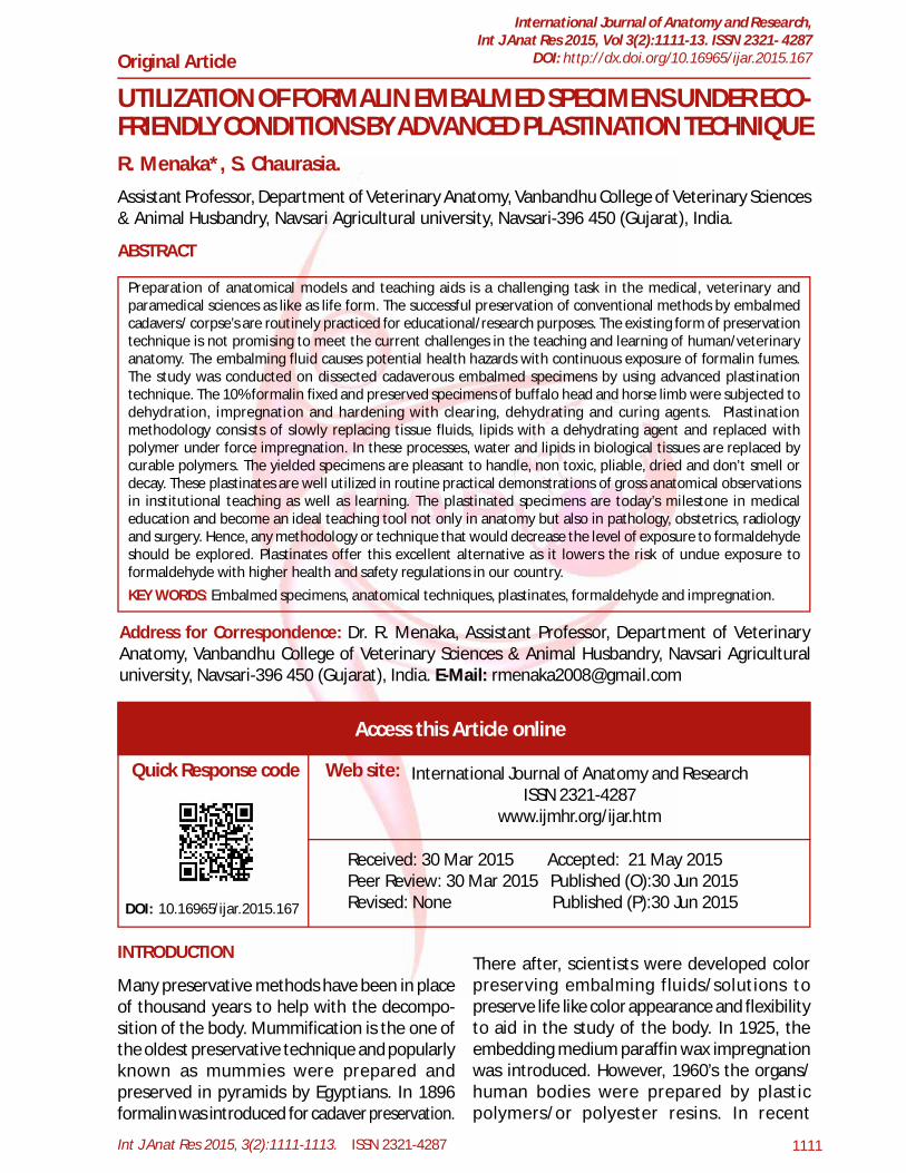
The study was conducted on the formalinpreserved cadaverous specimens. The specimenwas collected from the postmortem case fromthe department of Veterinary pathology, NavsariAgricultural University, Navsari, Gujarat. Thesespecimens were preserved in the 10% formalinand dissected out as per standard dissectionprocedure to understand the different layers ofmuscles, tendon and ligaments. The sagittalsection of buffalo head and longitudinal sectionof horse limb were taken instead of preservingyears together in the embalming tank after thedissection practices and demonstration in theundergraduate laboratory. It subsequently, 3-4changes had given at 4 days interval in acetonefor dehydration. The clearing and impregnationwas done with 1:1 ratio of chloroform andmelamyne solution for 4-5 days. Then, 2-3changes had been given in same ratio of solu-tion at 4 days interval in same mixture. Further,the limb specimen was subjected to the curingprocess by soaking in 9:1 ratio of melamyne andhardner for 4 days [3]. Finally they were shadowdried without exposing to direct sunlight.
RESULTS & DISCUSSIONIt was observed that the plastinated specimenwas more pleasant to touch and easy handlingfor demonstration of superficial and deeplayers of tendons, ligaments and muscles andbones (Fig. 1 to 3). There were many advantagesin palatinates over formalin preservedspecimens/ handling of wet specimens [4].However, the plastinated specimens are forteaching anatomy and pathology is well known[5 &6]. The formalin fixed specimens areeffectively utilized and preserved in eco-friendlyconditions.Fig. 1: Plastinates of horse limb (Longitudinal Section).
MATERIALS AND METHODS
Int J Anat Res 2015, 3(2):1111-1113. ISSN 2321-4287 1113
R. Menaka, S.Chaurasia. UTILIZATION OF FORMALIN EMBALMED SPECIMENS UNDER ECO-FRIENDLY CONDITIONS BY ADVANCEDPLASTINATION TECHNIQUE.
CONCLUSION
Conflicts of Interests: None
REFERENCES
Fig.2: Plastinates of buffalo head (Sagittal Section).
The plastination is an alternative method tomeet the current requirement of teaching tools/aids in the anatomical laboratory of medicalinstitutions across the country. Animal welfareorganizations and ethical committees raisedtheir curiosity to find out the new footstep inbiological tissue preservation. Plastination is agood anatomical technique which is increasinglygaining popularity for its own benefits in teach-ing and research of anatomy both medical andveterinary field. It might be promising to pre-serve the biological life on our planet especiallyendangered and significantly extinct species toshow to next generation.
1. Von Hagens, G (1985). Heidelberg plastinationfolder: Collection of all technical leaflets forplastination, English Edn. Anatomists InsHut 1,Universitat, Heidelberg.
2. Coleman R. Reducing the levels of formaldehydeexposure in gross anatomy laboratories.Anatomical Record. 1995;243:531-533.
3. R. Menaka., S. Chaurasia and N.H. Kelawala.Plastination of goat (kid) cadaver-A teaching model.Indian Journal of Veterinary Anatomy.2010;22(1):50-51.
4. Ramkrishna, V., Gadre, K.M., Pawar, A and Doolappa,A. Plastination –A viable atlternative of preservingthe biological specimens. Indian VeterinaryJournal. 2002;79:1158-59.
5. Von Hagens, G. Impregnation of soft biologicalspecimens with thermo setting resins andelastomers. Anat. Rec. 1979a;194:247-256.
6. Von Hagens, G. Emulsifying resins for plastination.Der Preparator. 1979b;25: 43-50.
How to cite this article:R. Menaka, S.Chaurasia. UTILIZATION OF FORMALIN EMBALMEDSPECIMENS UNDER ECO-FRIENDLY CONDITIONS BY ADVANCEDPLASTINATION TECHNIQUE. Int J Anat Res 2015;3(2):1111-1113.DOI: 10.16965/ijar.2015.167