intermediate filament accumulation can stabilize ... · network of actin, tubulin, and intermediate...
TRANSCRIPT
Intermediate filament accumulation can stabilizemicrotubules in Caenorhabditis elegans motor neuronsNaina Kurupa, Yunbo Lia, Alexandr Goncharova, and Yishi Jina,b,1
aNeurobiology Section, Division of Biological Sciences, University of California, San Diego, La Jolla, CA 92093; and bDepartment of Cellular and MolecularMedicine, University of California, San Diego, La Jolla, CA 92093
Edited by H. Robert Horvitz, Massachusetts Institute of Technology, Cambridge, MA, and approved February 11, 2018 (received for review December 21, 2017)
Neural circuits utilize a coordinated cellular machinery to form andeliminate synaptic connections, with the neuronal cytoskeletonplaying a prominent role. During larval development of Caenorhabditiselegans, synapses of motor neurons are stereotypically rewiredthrough a process facilitated by dynamicmicrotubules (MTs). Through agenetic suppressor screen on mutant animals that fail to rewire synap-ses, and in combination with live imaging and ultrastructural studies,we find that intermediate filaments (IFs) stabilize MTs to prevent syn-apse rewiring. Genetic ablation of IFs or pharmacological disruption ofIF networks restores MT growth and rescues synapse rewiring defectsin themutant animals, indicating that IF accumulation directly altersMTstability. Ourwork sheds light on the impact of IFs onMT dynamics andaxonal transport, which is relevant to themechanistic understanding ofseveral humanmotor neuron diseases characterized by IF accumulationin axonal swellings.
microtubules | intermediate filaments | synapses | C. elegans | neurons
Arobust cytoskeletal network is essential to many aspects ofneuronal function, including axon outgrowth, guidance, and
maintenance, as well as synapse formation and plasticity (1–3).Intermediate filaments (IFs) are one such cytoskeletal polymerand include different classes that are expressed in a variety oftissues. Neurofilaments (NFs) are a specific IF class expressed inneurons that provide structural support for the establishment ofaxon caliber in large-diameter myelinated axons (3, 4). Over-expression of NFs in some mouse models has linked NF accu-mulation to motor neuron death, with impairment of axonaltransport thought to be a precursor to neuron degeneration (5–7). Changes in NF levels have also been associated with neuro-psychiatric disorders such as bipolar disorder and schizophrenia,although whether NFs alter circuit activity remains unknown (8).The complexity in subunit composition and the variety of post-translational modifications associated with NFs suggest potentialroles for these filaments in neuronal functions outside axon-caliber maintenance (9). For example, several studies haveshown a requirement for microtubule (MT) motors in medi-ating IF motility and report a close association between IFs andMTs during MT assembly (10–12), supporting a mechanisticinteraction between the two cytoskeletal networks.In this study, we describe a role for IF assembly in promoting
MT stabilization in Caenorhabditis elegans, a genetically tractableanimal model extensively used to examine neuronal cytoskele-tal architecture (13, 14). A comprehensive comparison of theC. elegans connectome in larvae and adults revealed a strikingswitch in motor neuron connectivity during larval development,independent of axon outgrowth or retraction (15–17). We pre-viously found that an up-regulation in the number of dynamicMTs was required for synaptic vesicle transport during motorneuron synapse rewiring (18). Here, we show that IFs preventsynapse rewiring by hyperstabilizing neuronal MTs in mutantanimals with defective synapse rewiring. Our results reveal anunexpected regulatory role for IFs in neurons, furthering ourunderstanding of the complex relationship between various cyto-skeletal elements in vivo.
ResultsIdentification of IF Genes That Regulate Synapse Rewiring. At theend of larval stage 1 (L1), the dorsal D (DD)-type motor neuronsrewire their presynaptic connections from the ventral nerve cord(VNC) to the dorsal nerve cord (DNC), concurrent with thebirth of ventral D (VD)-type motor neurons, which then formsynapses along the VNC (19). We visualized DD-neuron pre-synaptic terminals using a GFP-tagged synaptobrevin (SNB-1::GFP) reporter (juIs137: Pflp-13 SNB-1::GFP). In L1 animals,discrete synaptic puncta were present along the ventral neurites(18), but in late larvae and adults, synaptic puncta were only seenalong the dorsal neurites (Fig. 1 A and B). We had previouslyshown that DD synapse remodeling depends on synergistic in-teractions between the MT cytoskeleton and the conservedMAPKKK DLK-1 using double-mutant animals with a missenseα-tubulin mutation [tba-1(gf)] and a loss of function in dlk-1(0)that were defective in DD synapse rewiring (18). Adult tba-1(gf)dlk-1(0) animals retained synaptic puncta along the VNC, withlittle to no synapses along the DNC (Fig. 1 A and B), resultingfrom a loss of dynamic MTs that impaired synaptic vesicletransport during rewiring (18).To identify regulators of MT dynamics during synapse rewir-
ing, we screened for mutants that suppressed synapse rewiringdefects in tba-1(gf) dlk-1(0) animals (18, 20). We identified twoindependent alleles (ju982 and ju963) of a C. elegans IF homo-log, ifp-1 (Fig. S1A). IFP-1 contains a conserved rod domain anda C-terminal lamin tail domain and shares 31% sequence iden-tity with human NF α-internexin (21, 22) (Fig. S1A). ju982caused a conserved leucine 363 to phenylalanine change in therod domain, while ju963 changed proline 443 to serine adjacent
Significance
A principal component of the nerve cell is its cytoskeleton—anetwork of actin, tubulin, and intermediate filament polymersthat maintains cell structure, directs cell growth, and transportof cargo. Here, we show that intermediate filaments can alterthe dynamics of microtubules to affect developmental synapseremodeling in Caenorhabditis elegans. Accumulation of in-termediate filaments is concurrent with hyperstablization ofmicrotubules, which in turn prevents synaptic vesicle transportduring rewiring. Our results identify a regulatory role for in-termediate filaments in neurons in vivo, providing insight intothe mechanisms behind neurological disorders such as ALS,where patients often present with accumulations of in-termediate filaments in dying nerve cells.
Author contributions: N.K. and Y.J. designed research; N.K., Y.L., and A.G. performedresearch; N.K., Y.L., A.G., and Y.J. analyzed data; and N.K. and Y.J. wrote the paper.
The authors declare no conflict of interest.
This article is a PNAS Direct Submission.
Published under the PNAS license.1To whom correspondence should be addressed. Email: [email protected].
This article contains supporting information online at www.pnas.org/lookup/suppl/doi:10.1073/pnas.1721930115/-/DCSupplemental.
Published online March 6, 2018.
3114–3119 | PNAS | March 20, 2018 | vol. 115 | no. 12 www.pnas.org/cgi/doi/10.1073/pnas.1721930115
Dow
nloa
ded
by g
uest
on
Apr
il 27
, 202
0

to the rod domain (Fig. S1A). Both alleles restored synapses tothe dorsal neurite in tba-1(gf) dlk-1(0). A null mutation, ifp-1(ok1609) [hereafter denoted ifp-1(0)], that removes most of therod domain and causes a premature stop showed stronger sup-pression of the synaptic defects of tba-1(gf) dlk-1(0) than eitherju982 or ju963, suggesting that they were hypomorphic alleles ofifp-1 (Fig. 1 A and B). Transgenic overexpression of wild-typeIFP-1 specifically in the DD neurons of tba-1(gf) dlk-1(0); ifp-1(ju982) and tba-1(gf) dlk-1(0); ifp-1(ju963) animals restored thesynapse rewiring defects seen in tba-1(gf) dlk-1(0) alone (Fig.1B). These results indicated that loss of function in ifp-1 sup-presses tba-1(gf) dlk-1(0) phenotypes, and that ifp-1 acts cellautonomously to regulate DD synapse rewiring. ifp-1 loss-of-function mutants alone displayed normal synapses, and over-expression of IFP-1 in wild-type animals also did not have aneffect on the number of synapses of DD neurons (Fig. 1 A andB), suggesting that the effect of ifp-1 on synapse formation wasdependent on tba-1(gf) dlk-1(0).We next characterized IF expression in wild-type and tba-1(gf)
dlk-1(0) animals. Using a transcriptional GFP reporter (driven
by 1 kb upstream of the transcription start site of ifp-1), we foundthat IFP-1 was strongly expressed in the intestine and pharynx, aspreviously reported (23) (Fig. S1B). Low levels of neuronal ex-pression might have been obscured by robust nonneuronal ex-pression. To test this, we used RNAi to reduce the GFP signal innonneuronal cells, since neurons in C. elegans are refractory tosystemic RNAi (24). In Pifp-1-GFP transgenic animals withdampened intestinal and pharyngeal GFP expression, we ob-served weak GFP expression in the VNC and some head neu-rons, which colocalized with a synaptic marker expressed in Dneurons (Fig. 1C and Fig. S1C). We then examined IFP-1 pro-tein expression by generating extrachromosomal transgenic ani-mals expressing GFP fused to the C terminus of genomic IFP-1.We verified that the fusion of GFP did not alter IFP-1 function,based on the rescue of behavioral suppression of tba-1(gf) dlk-1(0); ifp-1(0) animals by addition of the transgene (Fig. S1D). Inwild-type animals, IFP-1::GFP localized to filaments and punctain the head, pharynx, and intestine in adult animals (Fig. 1D).In contrast, expression of IFP-1(L363F)::GFP, mimicking ifp-1(ju982), showed a near complete loss of filamentous expression(Fig. S1E). In tba-1(gf) dlk-1(0) animals, we observed an increasein IFP-1::GFP filament intensity in the intestine, while the IFP-1::GFP puncta in the head appeared to have been reduced(Fig. S1F).
A
B*
*
*
*
WT ifp-1(0)
tba-1(gf) dlk-1(0); ifp-1(0)tba-1(gf) dlk-1(0)
C
+/+
ifp-1(
ok160
9)
DD-IFP-1 +/+
ifp-1(
ok160
9)
ifp-1(
ju982)
ifp-1(
ju963)
ifp-1(
ju982)
ifp-1(
ju963)
0
50
100ns *** ****** nsns
juIs137
tba-1(gf) dlk-1(0)
DD-IFP-1
ns
# of
syn
aptic
pun
cta
in
DN
C
Head Intestine
DHead VNC
Pifp-1-IFP-1::GFP
Pifp-1-GFP (+GFP RNAi)
A PD
V
Fig. 1. Loss of ifp-1 suppresses synapse remodeling defects in tba-1(gf) dlk-1(0). (A) Representative images of DD synapses along the VNC and DNC usingPflp-13-SNB-1::GFP (juIs137) in adult animals. Schematics of DD neuron (gray)with location of presynaptic vesicles (green) are shown Left of each fluorescentimage. White asterisks denote DD neuron cell bodies. (Scale bar: 10 μm.) (B)Quantification of synaptic puncta in the DNC of adult animals. Data aremean ± SEM; n > 10 animals per genotype. DD-IFP-1 denotes extrachromo-somal copies of IFP-1(cDNA) expressed in DD neurons under the flp-13 pro-moter. Statistics: one-way ANOVA followed by Tukey’s posttest; ***P < 0.001;ns, not significant. (C) GFP-RNAi treatment of worms expressing ifp-1 pro-moter-driven GFP (Pifp-1-GFP) revealed expression in head neurons and theVNC. (Scale bars: 50 μm.) (D) Expression pattern of IFP-1 protein in the headand intestine of WT worms using a C-terminal GFP-tagged IFP-1 expressedunder its own promoter. (Scale bars: 50 μm.) See also Fig. S1.
A B
Ctb
a-1(
gf) d
lk-1
(0)
tba-1(gf) dlk-1(0)
IFA-4::GFP
IFA-4::GFP (+GFP degron)
F
*
*
VNC
DN
CVN
CD
NC
tba-1(gf) dlk-1(0); ifa-4(0)
tba-1(gf) dlk-1(0); ifd-1(0)
+/+ ifc-1(0) ifd-1(0) ifd-2(0) ifa-4(0) ifp-1(0)0
20
40
60
80
100
ns nsns
***
***
***tba-1(gf) dlk-1(0); juIs137
# of
syn
aptic
pun
cta
in D
NC
E
D
Excretory canal
Rectum
IFA-4::GFP (L1)
sqt-1
LoxPLoxP
hs::Cre hygR
Self-Excising Cassette (SEC)
GFP
Heat Shockifa-4 gene
ifa-4 gfp (ju1576)
GFP
ifa-4 gene
GFPanti-GFP ZIF-1
IFA-4
D-neuron GFP degron
Cullin mediated degradation
nanobody
Fig. 2. ifa-4(0) also suppresses DD synapse remodeling defects in tba-1(gf)dlk-1(0). (A) Representative images of DD synapses in adult animals(juIs137). White asterisks denote DD neuron cell bodies. (Scale bar:10 μm.) (B) Quantification of synaptic puncta in the DNC of adult animals. Dataare mean ± SEM; n > 10 animals per genotype. Statistics: one-way ANOVAfollowed by Tukey’s posttest; ***P < 0.001; ns, not significant. (C) Schematic ofCRISPR-Cas9–mediated knockin of GFP at 3′ end of ifa-4 gene. After homolo-gous recombination of the repair plasmid into the genomic locus, the self-excising cassette is excised following heat shock at 37 °C for 1 h to drive theexpression of Cre recombinase. (D) Representative image of a wild-typeL1 animal expressing endogenous GFP-tagged IFA-4 in the excretory canal andrectum. (Scale bar: 10 μm.) (E) Schematic of GFP-degron system targeting IFA-4::GFP in D neurons for cullin-mediated degradation. (F) Representative images ofthe dorsal neurites of adult animals expressing Punc-25-mCherry::RAB-3. (Scalebar: 10 μm.) See also Fig. S2.
Kurup et al. PNAS | March 20, 2018 | vol. 115 | no. 12 | 3115
GEN
ETICS
Dow
nloa
ded
by g
uest
on
Apr
il 27
, 202
0
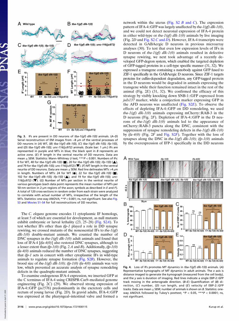
The C. elegans genome encodes 11 cytoplasmic IF homologs,at least 5 of which are essential for development, as null mutantsexhibit embryonic or larval lethality (23, 25–28) (Fig. S2A). Totest whether IFs other than ifp-1 played a role in DD synapserewiring, we crossed mutants of the nonessential IFs to tba-1(gf)dlk-1(0) double-mutant animals. We counted the number ofDNC synapses in tba-1(gf) dlk-1(0) adult animals and found thatloss of IFA-4 [ifa-4(0)] also restored DNC synapses, although toa lesser extent than ifp-1(0) (Fig. 2 A and B). Additionally, ifp-1(0)ifa-4(0) animals reduced the number of DNC synapses, suggestingthat ifp-1 acts in concert with other cytoplasmic IFs in wild-typeanimals to regulate synapse formation (Fig. S2B). However, thebrood size of tba-1(gf) dlk-1(0); ifp-1(0) ifa-4(0) animals was verylow, which prevented an accurate analysis of synapse remodelingdefects in the quadruple-mutant animals.To examine endogenous IFA-4 expression, we inserted GFP at
the C terminus of IFA-4 using CRISPR-Cas9–mediated genomeengineering (Fig. 2C) (29). We observed strong expression ofIFA-4::GFP (ju1576) predominantly in the excretory cells andrectum of young larvae (Fig. 2D). In gravid adults, IFA-4::GFPwas expressed at the pharyngeal–intestinal valve and formed a
network within the uterus (Fig. S2 B and C). The expressionpattern of IFA-4::GFP was largely unaffected by tba-1(gf) dlk-1(0),and we could not detect neuronal expression of IFA-4 proteinin either wild-type or tba-1(gf) dlk-1(0) animals by live imaging(Fig. 2D and Fig. S2 C and D). However, IFA-4 transcripts weredetected in GABAergic D neurons in previous microarrayanalyses (30). To test that even low expression levels of IFs inD neurons of tba-1(gf) dlk-1(0) animals resulted in defectivesynapse rewiring, we next took advantage of a recently de-veloped GFP-degron system, which enabled the targeted depletionof GFP-tagged proteins in a cell-type specific manner (31, 32). Weexpressed a transgene containing a nanobody against GFP fused toZIF-1 specifically in the GABAergic D neurons. Since ZIF-1 targetsproteins for cullin-dependent degradation, any GFP-tagged proteinin the D neurons would be degraded in animals expressing thetransgene while their function remained intact in the rest of theanimal (Fig. 2E) (31, 32). We confirmed the efficacy of thisstrategy by visibly knocking down SNB-1::GFP expressed fromjuIs137 marker, while a coinjection marker expressing GFP inthe AFD neurons was unaffected (Fig. S2E). To observe theeffects of depleting IFA-4::GFP on DD remodeling, we usedtba-1(gf) dlk-1(0) animals expressing mCherry::RAB-3 in theD neurons (Fig. 2F). Depletion of IFA-4::GFP in the D neu-rons of tba-1(gf) dlk-1(0) animals led to the appearance ofmCherry::RAB-3 puncta along the DNC, consistent with thesuppression of synapse remodeling defects in tba-1(gf) dlk-1(0)by ifa-4(0) (Fig. 2F and Fig. S2F). Together with the loss ofsynapses along the DNC in tba-1(gf) dlk-1(0); ifp-1(0) animalsby the overexpression of IFP-1 specifically in the DD neurons
G
F
BA
C
WT +/+ ifp-1(0) unc-116(ju972)0
2
4
6
8
10tba-1(gf) dlk-1(0)
***
IF L
engt
h (
m)
D
E
WT +/+ ifp-1(0)unc-116(ju972)0
2
4
6
8
10 tba-1(gf) dlk-1(0)M
T Le
ngth
(m
)
Wild type tba-1(gf) dlk-1(0)
tba-1(gf) dlk-1(0); ifp-1(0) tba-1(gf) dlk-1(0); unc-116(ju972)
WT +/+ ifp-1(0) unc-116(ju972)0
5
10
15 **
nsns
Av. M
T #/
sec
tion tba-1(gf) dlk-1(0)
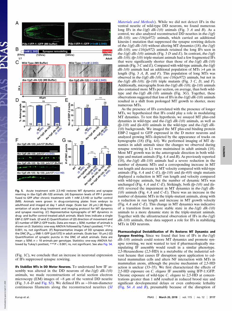
Fig. 3. IFs are present in DD neurons of tba-1(gf) dlk-1(0) animals. (A–D)Serial reconstruction of EM images from ∼8 μm of the ventral processes ofDD neurons in (A) WT, (B) tba-1(gf) dlk-1(0), (C) tba-1(gf) dlk-1(0); ifp-1(0),and (D) tba-1(gf) dlk-1(0); unc-116(ju972) animals. (Scale bar: 1 μm.) IFs arerepresented in purple and MTs in blue; the black spot in B represents anactive zone. (E) IF length in the ventral neurite of DD neurons. Data aremean ± SEM. Statistics: Mann–Whitney U test; ***P < 0.001. Numbers of IFs:0 for WT, 40 for tba-1(gf) dlk-1(0) (■), 20 for tba-1(gf) dlk-1(0); ifp-1(0) (▲),and 79 for tba-1(gf) dlk-1(0); unc-116(ju972) (▼). (F) MT length in the ventralneurite of DD neurons. Data are mean ± SEM. Red line delineates MTs >4 μmin length. Numbers of MTs: 24 for WT (●), 22 for tba-1(gf) dlk-1(0) (■),102 for tba-1(gf) dlk-1(0); ifp-1(0) (▲), and 19 for tba-1(gf) dlk-1(0); unc-116(ju972) (▼). (G) Number of MTs per section in the ventral neurite ofvarious genotypes (each data point represents the mean number of MTs per50-nm section in 2-μm regions of the axon; symbols as described in E and F).A total of 120 cross-sections in random order from each strain were analyzedto correlate with actual number of MTs, irrespective of the length of theMTs. Statistics: one-way ANOVA; **P < 0.001; ns, not significant. See also Fig.S3 and Movies S1–S4 for full reconstructions of DD neurites.
Wild Type
tba-1(gf) dlk-1(0) tba-1(gf) dlk-1(0); ifp-1(0)
ifp-1(0)
50 m
43.2
s
tba-1(gf) dlk-1(0); ifa-4(0)
ifa-4(0)
C
WT ifp-1(0) ifa-4(0) +/+ ifp-1(0) ifa-4(0)0
20
40
60
80
100
10 5 11 7
tba-1(gf) dlk-1(0)
5 8
% A
nter
ogra
de
WT ifp-1(0) ifa-4(0) +/+ ifp-1(0) ifa-4(0)0
10
20
30
40
50
ns
tba-1(gf) dlk-1(0)
***
***ns
No.
of E
BP-
2 tr
acks
B
A
D tba-1(gf) dlk-1(0)
WT ifp-1(0) ifa-4(0) +/+ ifp-1(0) ifa-4(0)0.0
0.2
0.4
0.6
0.8
1.0***
******ns
*** ***
Velo
city
(m
/s)
E
WT ifp-1(0) ifa-4(0) +/+ ifp-1(0) ifa-4(0)0
2
4
6
8
10
* ***
******
***
***
tba-1(gf) dlk-1(0)
Run
Len
gth
(m
)
Fig. 4. Loss of IFs promotes MT dynamics in tba-1(gf) dlk-1(0) animals. (A)Representative kymographs of MT dynamics in adult animals. The x axis isdistance imaged to generate the kymograph (measured from the cell body),and the y axis is duration of imaging. Red lines indicate a single EBP-2::GFPtrack moving in the anterograde direction. (B–E) Quantification of (B) di-rection, (C ) number, (D) run length, and (E ) velocity of EBP-2::GFPtracks. Data are mean ± SEM; number of animals is shown on B. Statistics: one-way ANOVA followed by Tukey’s posttest; *P < 0.05, ***P < 0.0001; ns,not significant.
3116 | www.pnas.org/cgi/doi/10.1073/pnas.1721930115 Kurup et al.
Dow
nloa
ded
by g
uest
on
Apr
il 27
, 202
0
(Fig. 1C), we conclude that an increase in neuronal expressionof IFs suppressed synapse rewiring.
IFs Stabilize MTs in DD Motor Neurons. To understand how IF as-sembly was altered in the DD neurons of tba-1(gf) dlk-1(0)animals, we made reconstructions of serial section electronmicroscopy (EM) images of ∼8 μm of the ventral DD neurite(Fig. 3 A–D and Fig. S3). We defined IFs as ∼10-nm-diametercontinuous filaments along the reconstructed neurites (SI
Materials and Methods). While we did not detect IFs in theventral neurite of wild-type DD neurons, we found numerouslong IFs in tba-1(gf) dlk-1(0) animals (Fig. 3 A and B). As acontrol, we also analyzed reconstructed DD neurites in tba-1(gf)dlk-1(0); unc-116(ju972) animals, which carried an additionalkinesin-1 mutation that suppressed the synapse rewiring defectsof tba-1(gf) dlk-1(0) without altering MT dynamics (18). tba-1(gf)dlk-1(0); unc-116(ju972) animals retained the long IFs seen intba-1(gf) dlk-1(0) animals (Fig. 3 D and E). In contrast, tba-1(gf)dlk-1(0); ifp-1(0) triple-mutant animals had a few fragmented IFsthat were significantly shorter than those of tba-1(gf) dlk-1(0)animals (Fig. 3 C and E). Compared with wild-type animals, tba-1(gf)dlk-1(0) animals had an additional population of MTs >4 μm inlength (Fig. 3 A, B, and F). This population of long MTs wasobserved in tba-1(gf) dlk-1(0); unc-116(ju972) animals, but not intba-1(gf) dlk-1(0); ifp-1(0) triple mutants (Fig. 3 C, D, and F).Additionally, micrographs from tba-1(gf) dlk-1(0); ifp-1(0) animalsalso contained more MTs per section, on average, than both wild-type and tba-1(gf) dlk-1(0) animals (Fig. 3G). Together, theseobservations suggested that loss of IFs in tba-1(gf) dlk-1(0) animalsresulted in a shift from prolonged MT growth to shorter, morenumerous MTs.As the presence of IFs correlated with the presence of longer
MTs, we hypothesized that IFs could play a role in modulatingMT dynamics. To test this hypothesis, we assayed MT plus-enddynamics in wild-type and tba-1(gf) dlk-1(0) animals, as well asifp-1(0) and ifa-4(0) animals in the wild-type and tba-1(gf) dlk-1(0) backgrounds. We imaged the MT plus-end binding proteinEBP-2 tagged to GFP expressed in the D motor neurons andanalyzed growing MTs depicted by the appearance of tracks onkymographs (18) (Fig. 4A). We performed imaging of MT dy-namics in adult animals since the changes we observed duringsynapse rewiring in L1 were maintained in adult animals (18).Most MT growth was in the anterograde direction in both wild-type and mutant animals (Fig. 4 A and B). As previously reported(18), tba-1(gf) dlk-1(0) animals had a severe reduction in thenumber of dynamic MTs and a corresponding increase in MTrun length and decrease in MT velocity compared with wild-typeanimals (Fig. 4 A and C–E). ifp-1(0) and ifa-4(0) single mutantsdisplayed a reduction in MT run length and velocity comparedwith wild-type animals, but the number of dynamic MTs wasunchanged (Fig. 4 A and C–E). Strikingly, both ifp-1(0) and ifa-4(0) reversed the impairment in MT dynamics in tba-1(gf) dlk-1(0) animals (Fig. 4 A and C–E). There was an increase in thenumber of dynamic MTs in the triple-mutant animals, along witha reduction in run length and increase in MT growth velocity(Fig. 4 A and C–E). This change in MT dynamics was indicativeof a transition from a more stable state in tba-1(gf) dlk-1(0)animals to a more dynamic state in the triple-mutant animals.Together with the ultrastructural observation of IFs in tba-1(gf)dlk-1(0) animals, these data support a role for IFs in stabilizingMTs in DD neurons.
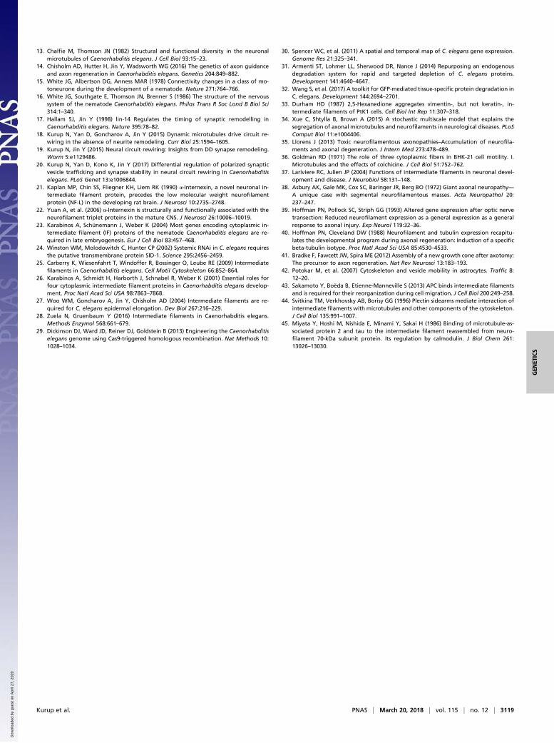
Pharmacological Destabilization of IFs Restores MT Dynamics andSynapse Rewiring. Since we found that loss of IFs in tba-1(gf)dlk-1(0) animals could restore MT dynamics and promote syn-apse rewiring, we next wanted to test if pharmacologically ma-nipulating IF assembly would result in a similar phenotype.2,5-Hexanedione (2,5-HD) is a metabolite of the industrial sol-vent hexane that causes IF disruption upon application to cul-tured mammalian cells and alters NF interaction with MTs inmammalian axons, although the precise mechanism of 2,5-HDaction is unclear (33–35). We first characterized the effects of2,5-HD exposure on C. elegans IF assembly using IFP-1::GFP.Chronic exposure of wild-type C. elegans to 2,5-HD at concen-trations greater than 1 mM resulted in reduced brood size andsignificant developmental delays or even embryonic lethality(Fig. S4 A and B), presumably because of the disruption of
A
m
24 s
40
tba-1(gf) dlk-1(0) (M9 Buffer) tba-1(gf) dlk-1(0) (+1mM 2,5HD)
Wild Type (M9 Buffer) Wild Type (+1mM 2,5HD)C
E
WT WT
tba-1(gf) d
lk-1(0
)
tba-1(gf) d
lk-1(0
)0
5
10
15
20 nsns
***
***
No.
of E
BP-
2 tr
acks
(2, 5H
D)
(2, 5H
D)
Pifp-1-IFP-1::GFP (M9 buffer)
B(0 hrs ) (56 hrs)
Synapse remodeling(12 hrs ) (34hrs )
L1 Adult
1mM 2, 5HD Imaging
D
tba-1(gf) dlk-1(0); juIs137 (Buffer)
tba-1(gf) dlk-1(0); ifp-1(0); juIs137 (Buffer)
tba-1(gf) dlk-1(0); ifp-1(0); juIs137 (+2,5 HD)
tba-1(gf) dlk-1(0); juIs137 (+2,5HD)
F G
WT WT
tba-1(gf) d
lk-1(0
)
tba-1(gf) d
lk-1(0
)
(2, 5H
D)0
20
40
60
80
100 ns
% A
nter
ogra
de
(2, 5H
D)15 14 1011
Pifp-1-IFP-1::GFP (+1mM 2,5HD)
WT WT
tba-1(gf)d
lk-1(0
)
tba-1(gf)d
lk-1(0
)
tba-1(gf) d
lk-1(0
); ifp-1(
0)
tba-1(gf) d
lk-1(0
); ifp-1(
0)0
50
100
150 ns
ns
***
# of
DN
C p
unct
a
(2, 5H
D)
(2, 5H
D)(2,
5HD)
Fig. 5. Acute treatment with 2,5-HD restores MT dynamics and synapserewiring in tba-1(gf) dlk-1(0) animals. (A) Expression levels of IFP-1 proteinfused to GFP after chronic treatment with 1 mM 2,5-HD or buffer control(M9). Animals were grown in drug-containing plates from embryo toadulthood and imaged at day 1 adult stage. (Scale bar: 20 μm.) (B) Repre-sentation of acute drug treatment and imaging protocol for MT dynamicsand synapse rewiring. (C) Representative kymographs of MT dynamics indrug- and buffer control-treated adult animals. Black lines indicate a singleEBP-2::GFP track. (D and E) Quantification of (D) direction of movement and(E) number of EBP-2::GFP tracks. Data are mean ± SEM; number of animals isshown on D. Statistics: one-way ANOVA followed by Tukey’s posttest; ***P <0.001; ns, not significant. (F) Representative images of DD synapses alongthe DNC [Pflp-13-SNB-1::GFP (juIs137)] in adult animals. (Scale bar: 10 μm.) (G)Quantification of synaptic puncta in the DNC of adult animals. Data aremean ± SEM; n > 10 animals per genotype. Statistics: one-way ANOVA fol-lowed by Tukey’s posttest; ***P < 0.001; ns, not significant. See also Fig. S4.
Kurup et al. PNAS | March 20, 2018 | vol. 115 | no. 12 | 3117
GEN
ETICS
Dow
nloa
ded
by g
uest
on
Apr
il 27
, 202
0

essential IF networks in the intestine and epidermis (26, 27).Animals grown on 1 mM 2,5-HD were superficially wild-type indevelopment and brood size (Fig. S4 A and B). Under thiscondition, 2,5-HD caused a strong reduction of IFP-1::GFP ex-pression in the head, but IFP-1::GFP in the intestine appeared toaggregate (Fig. 5A and Fig. S4B).We then tested whether this change in IF assembly could alter
MT dynamics, by treating wild-type and tba-1(gf) dlk-1(0) L1 ani-mals with 1 mM 2,5-HD, acutely for ∼9 h before the onset ofsynapse rewiring (Fig. 5B). We found that there was no change inthe number or direction of growth of dynamic MTs in wild-typeanimals after the acute 2,5-HD treatment, consistent with wild-typeDD neurons lacking detectable IFs (Fig. 5 C–E). Interestingly, intba-1(gf) dlk-1(0) animals, acute 2,5-HD treatment caused a sig-nificant increase in MT dynamics, while the overall direction of MTgrowth was unchanged (Fig. 5 C–E). We also monitored synapserewiring after acute treatment of L1 larvae with 1 mM 2,5-HD (Fig.5 F and G). Acute 2,5-HD treatment did not alter synapse rewiringin wild-type or tba-1(gf) dlk-1(0); ifp-1(0) animals (Fig. 5 F and G).However, there was a significant increase in the number of synapticpuncta successfully reaching the dorsal neurite of adult tba-1(gf)dlk-1(0) animals following acute 2,5-HD treatment, resulting in apartial rescue of the synapse rewiring defect (Fig. 5 F and G).Together, these results suggest that the presence of IFs was suffi-cient to shift the balance from dynamic to stable MTs to antagonizesynapse rewiring in C. elegans.
DiscussionActin, MTs, and IFs constitute the three major cytoskeletal el-ements in most eukaryotic cells, and their coordinated interac-tions have been the subject of intense study for decades. Theimportance of IF–MT interactions in maintaining the cellular IFnetwork was first demonstrated several years ago, when treat-ment of BHK-21 cells with colchicine to remove MTs resulted inIF aggregation as a juxtanuclear cap (36). Here, we focus on IF–MT interactions in neuronal development, using C. elegans as amodel. The synapse rewiring defects in tba-1(gf) dlk-1(0) doublemutants are a result of hyperstable MTs, and loss of function inIF genes ifp-1 and ifa-4 restored normal synapse rewiring in thisbackground. We further characterized IF–MT interactions inliving animals, revealing an underappreciated regulatory func-tion for neuronal IFs in modulating MT dynamics in vivo. Thefunctional significance of a bidirectional interaction between IFand MT networks is particularly relevant in neurodegenera-tive disease conditions like giant axonal neuropathy, ALS, andCharcot-Marie-Tooth disease, where toxic assemblies of IFs inneurons correlate with a fatal loss of motor and sensory function(37, 38). In wild-type DD motor neurons, absence of IFs doesnot alter circuit rewiring to a large extent, suggesting that regu-lation of MT dynamics by IFs depends on special conditions.Overexpression of IFP-1 also does not cause any defects in wild-type animals. These data suggest that IF protein expressionis tightly regulated in wild-type neurons in the absence ofadditional perturbations.Several observations are conducive to the idea that axonal MT
stability is directly linked to IF levels. IFs are down-regulated
following axonal injury in both the central and peripheral ner-vous systems (39, 40), which correlates with increased MT de-stabilization immediately after lesion in a variety of models (14,41). IF up-regulation is a hallmark of activated astrocytes, andboth IF and MT depolymerization can alter the directionalmotility of vesicles in rat astrocytes (42). Recently, vimentin IFshave also been shown to act as a template for MT stabilizationduring cell migration in wounded retinal epithelial cells (12).Together with our findings during C.elegans synapse rewiring,these results point to a universal role for IF–MT interactions inmodulating both IF and MT stability.The interactions between IF and MT networks have long been
thought to occur through a variety of cross-bridging moleculesthat contain both MT and IF binding domains. During astrocytemigration, the MT-dependent rearrangements of IFs require thetumor suppressor adenomatous polyposis coli (APC) as a cross-bridging molecule to mediate IF–MT interaction (43). Plectin(44) and MAP2/tau (45) have also been implicated as potentialIF–MT cross-bridging molecules, but mutant C. elegans tau [ptl-1(0)] (20) could not phenocopy IF subunit loss in tba-1(gf) dlk-1(0) animals. Homologs of APC (apr-1) and plectin (vab-10) areessential for C. elegans embryonic development; future experi-ments will be necessary to selectively eliminate these genes’function in neurons to elucidate their role in IF–MT interactions.IFs have also been thought to interact with MTs through MTmotors like kinesin and dynein (10); however, the kinesin anddynein mutations identified in our suppressor screen had noeffect on MT dynamics or composition in tba-1(gf) dlk-1(0) an-imals (Fig. 3 D and E) (18, 20). Another possible mechanism forIF regulation of MTs involves changes in IF posttranslationalmodifications like phosphorylation, which control IF interactionwith MT-associated proteins (9, 10). However, both ifp-1 and ifa-4 lack the KSP repeat motif that is typically phosphorylated inmammalian NFs (9, 10). Further studies are required to un-derstand how IFs and MTs interact during synapse development,and C.elegans provides a useful genetic model to dissect thepathways involved in mediating this interaction.
Materials and MethodsC. elegans strains were maintained at 20 to 22 °C on nematode growthmedium plates following standard practices. Information on alleles andgenotypes of strains and plasmids used for injection is summarized in TablesS1–S3. Additional details of imaging, EM image serial reconstruction, andstaging conditions for drug and RNAi treatment are provided in SI Materialsand Methods. Statistical analysis was performed using GraphPad Prism 5.Details of the statistical analyses performed for each graph are presented inthe corresponding figure legends.
ACKNOWLEDGMENTS. We thank A. D. Chisholm, K. McCulloch, N. H. Tang,C. Piggott, K. W. Kim, S. Park, and M. Andrusiak for comments on themanuscript; and other members of the Y.J. laboratory for useful discussionsduring the course of the study. We thank the Caenorhabditis GeneticsCenter, which is supported by the National Institutes of Health (NIH) (GrantP40 OD010440), and the Mitani laboratory (Tokyo Women’s Medical College)for strains used in this study. Y.J. was an Investigator and A.G. was a ResearchAssociate of the Howard Hughes Medical Institute (HHMI). N.K. was a recipientof the Latham & Watkins Graduate Fellowship. This work was supported bythe HHMI and by an NIH grant (NINDS R01 035546, to Y.J.).
1. Conde C, Cáceres A (2009) Microtubule assembly, organization and dynamics in axonsand dendrites. Nat Rev Neurosci 10:319–332.
2. Dillon C, Goda Y (2005) The actin cytoskeleton: Integrating form and function at thesynapse. Annu Rev Neurosci 28:25–55.
3. Yuan A, Rao MV, Veeranna, Nixon RA (2017) Neurofilaments and neurofilamentproteins in health and disease. Cold Spring Harb Perspect Biol 9:a018309.
4. Lee MK, Cleveland DW (1996) Neuronal intermediate filaments. Annu Rev Neurosci19:187–217.
5. Beaulieu JM, Nguyen MD, Julien JP (1999) Late onset of motor neurons in miceoverexpressing wild-type peripherin. J Cell Biol 147:531–544.
6. Lee MK, Marszalek JR, Cleveland DW (1994) A mutant neurofilament subunit causesmassive, selective motor neuron death: Implications for the pathogenesis of humanmotor neuron disease. Neuron 13:975–988.
7. Xu Z, Cork LC, Griffin JW, Cleveland DW (1993) Increased expression of neurofilamentsubunit NF-L produces morphological alterations that resemble the pathology ofhuman motor neuron disease. Cell 73:23–33.
8. Yuan A, Nixon RA (2016) Specialized roles of neurofilament proteins in synapses:Relevance to neuropsychiatric disorders. Brain Res Bull 126:334–346.
9. Snider NT, Omary MB (2014) Post-translational modifications of intermediate fila-ment proteins: Mechanisms and functions. Nat Rev Mol Cell Biol 15:163–177.
10. Chang L, Goldman RD (2004) Intermediate filaments mediate cytoskeletal crosstalk.Nat Rev Mol Cell Biol 5:601–613.
11. Bocquet A, et al. (2009) Neurofilaments bind tubulin and modulate its polymeriza-tion. J Neurosci 29:11043–11054.
12. Gan Z, et al. (2016) Vimentin intermediate filaments template microtubule networksto enhance persistence in cell polarity and directed migration. Cell Syst 3:252–263.e8.
3118 | www.pnas.org/cgi/doi/10.1073/pnas.1721930115 Kurup et al.
Dow
nloa
ded
by g
uest
on
Apr
il 27
, 202
0
13. Chalfie M, Thomson JN (1982) Structural and functional diversity in the neuronalmicrotubules of Caenorhabditis elegans. J Cell Biol 93:15–23.
14. Chisholm AD, Hutter H, Jin Y, Wadsworth WG (2016) The genetics of axon guidanceand axon regeneration in Caenorhabditis elegans. Genetics 204:849–882.
15. White JG, Albertson DG, Anness MAR (1978) Connectivity changes in a class of mo-toneurone during the development of a nematode. Nature 271:764–766.
16. White JG, Southgate E, Thomson JN, Brenner S (1986) The structure of the nervoussystem of the nematode Caenorhabditis elegans. Philos Trans R Soc Lond B Biol Sci314:1–340.
17. Hallam SJ, Jin Y (1998) lin-14 Regulates the timing of synaptic remodelling inCaenorhabditis elegans. Nature 395:78–82.
18. Kurup N, Yan D, Goncharov A, Jin Y (2015) Dynamic microtubules drive circuit re-wiring in the absence of neurite remodeling. Curr Biol 25:1594–1605.
19. Kurup N, Jin Y (2015) Neural circuit rewiring: Insights from DD synapse remodeling.Worm 5:e1129486.
20. Kurup N, Yan D, Kono K, Jin Y (2017) Differential regulation of polarized synapticvesicle trafficking and synapse stability in neural circuit rewiring in Caenorhabditiselegans. PLoS Genet 13:e1006844.
21. Kaplan MP, Chin SS, Fliegner KH, Liem RK (1990) α-Internexin, a novel neuronal in-termediate filament protein, precedes the low molecular weight neurofilamentprotein (NF-L) in the developing rat brain. J Neurosci 10:2735–2748.
22. Yuan A, et al. (2006) α-Internexin is structurally and functionally associated with theneurofilament triplet proteins in the mature CNS. J Neurosci 26:10006–10019.
23. Karabinos A, Schünemann J, Weber K (2004) Most genes encoding cytoplasmic in-termediate filament (IF) proteins of the nematode Caenorhabditis elegans are re-quired in late embryogenesis. Eur J Cell Biol 83:457–468.
24. Winston WM, Molodowitch C, Hunter CP (2002) Systemic RNAi in C. elegans requiresthe putative transmembrane protein SID-1. Science 295:2456–2459.
25. Carberry K, Wiesenfahrt T, Windoffer R, Bossinger O, Leube RE (2009) Intermediatefilaments in Caenorhabditis elegans. Cell Motil Cytoskeleton 66:852–864.
26. Karabinos A, Schmidt H, Harborth J, Schnabel R, Weber K (2001) Essential roles forfour cytoplasmic intermediate filament proteins in Caenorhabditis elegans develop-ment. Proc Natl Acad Sci USA 98:7863–7868.
27. Woo WM, Goncharov A, Jin Y, Chisholm AD (2004) Intermediate filaments are re-quired for C. elegans epidermal elongation. Dev Biol 267:216–229.
28. Zuela N, Gruenbaum Y (2016) Intermediate filaments in Caenorhabditis elegans.Methods Enzymol 568:661–679.
29. Dickinson DJ, Ward JD, Reiner DJ, Goldstein B (2013) Engineering the Caenorhabditiselegans genome using Cas9-triggered homologous recombination. Nat Methods 10:1028–1034.
30. Spencer WC, et al. (2011) A spatial and temporal map of C. elegans gene expression.Genome Res 21:325–341.
31. Armenti ST, Lohmer LL, Sherwood DR, Nance J (2014) Repurposing an endogenousdegradation system for rapid and targeted depletion of C. elegans proteins.Development 141:4640–4647.
32. Wang S, et al. (2017) A toolkit for GFP-mediated tissue-specific protein degradation inC. elegans. Development 144:2694–2701.
33. Durham HD (1987) 2,5-Hexanedione aggregates vimentin-, but not keratin-, in-termediate filaments of PtK1 cells. Cell Biol Int Rep 11:307–318.
34. Xue C, Shtylla B, Brown A (2015) A stochastic multiscale model that explains thesegregation of axonal microtubules and neurofilaments in neurological diseases. PLoSComput Biol 11:e1004406.
35. Llorens J (2013) Toxic neurofilamentous axonopathies–Accumulation of neurofila-ments and axonal degeneration. J Intern Med 273:478–489.
36. Goldman RD (1971) The role of three cytoplasmic fibers in BHK-21 cell motility. I.Microtubules and the effects of colchicine. J Cell Biol 51:752–762.
37. Lariviere RC, Julien JP (2004) Functions of intermediate filaments in neuronal devel-opment and disease. J Neurobiol 58:131–148.
38. Asbury AK, Gale MK, Cox SC, Baringer JR, Berg BO (1972) Giant axonal neuropathy—A unique case with segmental neurofilamentous masses. Acta Neuropathol 20:237–247.
39. Hoffman PN, Pollock SC, Striph GG (1993) Altered gene expression after optic nervetransection: Reduced neurofilament expression as a general expression as a generalresponse to axonal injury. Exp Neurol 119:32–36.
40. Hoffman PN, Cleveland DW (1988) Neurofilament and tubulin expression recapitu-lates the developmental program during axonal regeneration: Induction of a specificbeta-tubulin isotype. Proc Natl Acad Sci USA 85:4530–4533.
41. Bradke F, Fawcett JW, Spira ME (2012) Assembly of a new growth cone after axotomy:The precursor to axon regeneration. Nat Rev Neurosci 13:183–193.
42. Potokar M, et al. (2007) Cytoskeleton and vesicle mobility in astrocytes. Traffic 8:12–20.
43. Sakamoto Y, Boëda B, Etienne-Manneville S (2013) APC binds intermediate filamentsand is required for their reorganization during cell migration. J Cell Biol 200:249–258.
44. Svitkina TM, Verkhovsky AB, Borisy GG (1996) Plectin sidearms mediate interaction ofintermediate filaments with microtubules and other components of the cytoskeleton.J Cell Biol 135:991–1007.
45. Miyata Y, Hoshi M, Nishida E, Minami Y, Sakai H (1986) Binding of microtubule-as-sociated protein 2 and tau to the intermediate filament reassembled from neuro-filament 70-kDa subunit protein. Its regulation by calmodulin. J Biol Chem 261:13026–13030.
Kurup et al. PNAS | March 20, 2018 | vol. 115 | no. 12 | 3119
GEN
ETICS
Dow
nloa
ded
by g
uest
on
Apr
il 27
, 202
0