interim in vitro dose-response curve for the dicentric
TRANSCRIPT

Interim In vitro Dose-Response Curve for the Dicentric Biodosimeter Assay from a Philippine Radiotherapy Facility using a Linear Accelerator
Antonio Carlo D. De Guzman, MD,¹ Carmencita D. Padilla, MD, MAHPS,²Henri Cartier S. Co, MD,¹ Elrick T. Inocencio, MSc¹ and Edsel Allan G. Salonga, RMT²
1Section of Radiation Oncology, Department of Radiology, Philippine General Hospital, University of the Philippines Manila2Cytogenetics Laboratory, Institute of Human Genetics, National Institutes of Health, University of the Philippines Manila
ABSTRACT
Background. Accidental radiation exposure can occur anytime. Biodosimeters help in quantifying the absorbed dose of individuals who are not equipped with personal dosimeters during radiation exposure. The dicentric assay can quantify radiation damage by correlating radiation dose exposure with the frequency of dicentric chromosomes in the peripheral lymphocytes extracted from exposed individuals.
Objective. The study aims to present the interim results of the reference dose-response curve for a Philippine radiotherapy facility constructed using a 6MV linear accelerator (ClinacX, Varian).
Methods. Samples of peripheral blood from healthy volunteers were irradiated in a customized water phantom of doses 0.10 to 5.0 Gray using a linear accelerator. The irradiated samples were cultured and analyzed following the International Atomic Energy Agency Cytogenetic Dosimetry Protocol (2011) with modifications. Linear-quadratic model curve fiing and further statistical analysis were done using CABAS (Chromosome Aberration Calculation Software Version 2.0) and Dose Estimate (Version 5.2). Interim results of the samples were used to generate these curves.
Results. The dose-response curve generated from the preliminary results were comparable to published dose-response curves from international cytogenetic laboratories.
Conclusion. The generated dose-response calibration curve will be useful for medical triage of the public and radiologic staff accidentally exposed to radiation during medical procedures or in the event of nuclear accidents.
Key Words: biodosimeter, dicentric assay, radiation, cytogenetic dose-response curve
InTRoDuCTIon
Ionizing radiation exerts its effect on biological tissues through double-strand breaks. Radiation therapy, through ionizing radiation, also controls cancer growth by producing double-strand breaks and eventual mitotic death. The effects of radiation can be affected by cell repair, reassortment, reoxygenation, repopulation, and also radiosensitivity. Accumulation of such DNA breaks can produce chromosomal aberrations, one of which is the dicentric chromosome.¹
Biodosimetry is the analysis of chromosome aberrations to assess the absorbed dose of an individual. This is particularly useful in radiation protection and dose assess-ment. The International Atomic Energy Agency (IAEA) has supported the use of biodosimetry since 1978 and
Corresponding author: Antonio Carlo D. De Guzman, MDSection of Radiation OncologyDepartment of RadiologyPhilippine General HospitalUniversity of the Philippines ManilaTaft Avenue, Manila 1000, Philippines Email: [email protected]
VOL. 55 NO. 1 2021 ACTA MEDICA PHILIPPINA 117
ORIGINAL ARTICLE

also gives importance to its advancement. Biodosimetry is mainly used in radiation accidents resulting in mass exposure. It can be used for triaging exposed individuals as it gives an initial and rapid approximate of absorbed dose.²
Biodosimetry is of value in cases of accidental radiation exposures if dosimeters are not worn by exposed staff, or in cases of equivocal dosimetric measurement.² Its use is mainly to elucidate absorbed doses in cases where the details surrounding the accidental radiation exposure are unclear or no dose measurements are available. This is important in the assessment of stochastic effects such as carcinogenesis. A stochastic effect, by definition, is not dose-related; hence a radiation exposure of 1 Gray is no different from a 0.1 Gy exposure with regard to the risk of inducing carcinogenesis.¹ According to the International Commission on Radio-logical Protection (ICRP), for the general population exposed to low dose and low dose rate radiation, there is a 5% per sievert risk for carcinogenesis.¹ Investigation of absorbed doses is also important in high exposures such as during radiotherapy, wherein deterministic health effects are considered; wherein overexposure beyond an organ dose limit can result in toxicities.
Assays for biodosimetry include the dicentric assay (which is considered the gold standard), micronucleus assay, chromosome painting, premature chromosome condensation (PCC), and gamma H2AX focus test.³ Table 1 shows the different dosimetry protocols and their characteristics, as published by Kulka.³
The chromosomal aberrations observed in the lymphocytes of exposed individuals are interpreted in terms
of absorbed dose about a dose-response curve. Such dose-response curves are verifiable with a physical instrument like an ionization chamber. It is recommended that a dose-response curve be calibrated for each laboratory.²
Dicentric AssayDicentrics are unstable chromosome aberrations
and these are the main chromosomal aberration used for biodosimetry. It is defined as “an exchange between the centromeric pieces of two broken chromosomes which in its complete form is accompanied by a fragment composed of the acentric pieces of these chromosomes.” Multicentric configurations can also be formed after high doses.²
The dicentric chromosome assay was developed by Bender and Gooch in 1962 and has been the gold standard for biological dosimetry.4 Each institution should construct a dose-response curve that will likely represent the radiation type it frequently uses and from which the likelihood of a radiation accident is considered increased.5 The dose-response best fits the linear-quadratic model for low LET radiation, Y = αD + βD2, and linear for high LET radiations, Y = αD.5 By creating calibration curves unique to each laboratory, other factors such as dose rate, type, and energy of radiation, all of which influence alpha (α) and beta (β) can be taken into account.6
Significance and Impact on Existing PolicyThe main purpose of the essay is its use in radiation
protection of radiation workers, patients, and the general public. This is very important in the event of equivocal
Table 1. Comparison of Dicentric Assay (DIC), Micronucleus Assay (MN), Fluorescent Immunohistochemistry Translocation Assay (FISH), Premature Chromosome Condensation (PCC) and γH2AX staining assay, Electron Paramagnetic Resonance (EPR), and Optically Stimulated Luminescence (OSL). Reprinted with permission from the author. “RENEB – Running the European Network of Biological Dosimetry and Physical Retrospective Dosimetry” by Kulka et al., 2016. International Journal of Radiation Biology, 93:1, 2-14. Available online at: https://doi.org/10.1080/09553002.2016.1230239. Under Creative Commons Attribution-Non Commercial-No Derivatives License.³
Assay Sample type
Time frame for sample collectiona
Classification of Individuals: Green/Orange/Red (<1 Gy/1-2 Gy/>2 Gy)
Detection range (Gy) Robustnessc
Implication of individual sensitivityd
Stored material: Type and time range for further analysise
Time from sample receipt
to resultb
RENEB capacity (analyzed persons
per week)Dic blood days - months 52 hours ca. 1000 0.1-5 high yes fixed cells, slides: yearsMN blood days - months 75 hours ca. 400 0.2-5 medium yes slides: yearsFISH blood days - years 120 hours ca. 100 0.3-4 medium-high yes fixed cells: yearsPCC blood hours - months 2-8 hours ca. 50 0.1-20 high yes frozen lymphocytes,
fixed cells, slides: yearsγH2AX blood days 3 hours ca. 1800 0.2-5 low ? fixed cells, slides:
up to one yearEPR PEDf hours - years <1 hour ca. 770 >1 high no glass: yearsOSL PEDg hours - months <1 hour ca. 500 >0.1 high no resistors: weeks
aTime between irradiation and sample collection; bTime from arrival of a sample in the laboratory until the classification of a person, without time for transport/shipment; cRobustness: high: little influence of disturbing factors, medium: some influence of aging, smoking, other agents, low: large influence of other agents and factors; dConsidering the individual sensitivity of a person; eType of the stored material and time frame to perform further analysis; fPED, personal electronic device (glass touchscreen, e.g., smart phone); gPED, personal electronic device (resistors from circuit board, e.g., mobile or smart phone).
ACTA MEDICA PHILIPPINA VOL. 55 NO. 1 2021118
Dose-Response Curve for Dicentric Biodosimeter

measurements based on physical dosimeters and during radiation accidents. Since there is increasing use of ionizing radiation in medicine such as diagnostic radiology, interventional radiology, fluoroscopic procedures, and even nuclear medicine, the benefit of such radiation protection service for workers and patients cannot be overemphasized.¹
Studies like this also answer the call made by the IAEA to advance cytogenetic biodosimetry as well as establish a dose-response curve unique to one’s institution. Last 2007, Dr. Zhanat Carr of the World Health Organization (WHO) and IAEA called for the development of a global biodosimetry laboratory network for radiation emergencies. Such a global network will allow the sharing of protocols, criteria for quality assurance, comparative studies, and other vital operations.7
At our institutional level, this study can support policy modifications in the event of accidental exposure, equivocal dosimeter readings, or circumstances wherein measured readings do not correlate with perceived exposure. By simply providing a blood sample for the dicentric assay, the individual’s total absorbed dose can be correlated with the monitored doses from the physical biodosimeter, if worn by the individual. Hence, issues on accidents and equivocal readings can be addressed through quantitative data and further exposure can be minimized if warranted. Such monitoring is particularly important for radiation workers since ionizing radiation can manifest effects years after exposure, including carcinogenesis and infertility.
Cytogenetic assays are used in other countries to determine radiation exposure in vivo. The micronucleus assay, a type of cytogenetic assay and one of the most basic has also been studied as a reliable and standardized assay to determine radiation exposure.8 It has been used in monitoring cumulative radiation doses such as among industrial radiographers as reported by Shakeri, et al.9 The micronucleus assay has also been used to monitor hospital personnel, chemical, and agricultural workers for genomic damage due to chemical genotoxins.10 Chromosomal aberrations (including dicentrics), which are more specific for ionizing radiation than the micronucleus assay, are also used to monitor occupational exposures such as those of personnel working in angiocardiography laboratories.11 In the United States, bioassays are used in internal radiation dosimetry for cases who received an annual committed effective dose of 0.001 Sv or more from all occupational radionuclide intakes.12 In Bosnia, biodosimetry by chromosomal analysis is done for workers exposed to effective doses greater than 200mSv.13
Cytogenetic assays can also be used in radiotherapy for patient monitoring. LeFevre studied cytogenetics and its correlation with long term effects among patients who underwent radiotherapy.14
Aside from these, this research will also contribute to the limited body of data on the use of dicentric assay dose-response curves based on radiation produced by a
linear accelerator. The majority of previous dose-response curves were established using Cobalt teletherapy machines or gamma irradiators, both of which are no longer used as standards in current radiotherapy protocols. By using the linear accelerator, the dose-response curve produced in the institution’s laboratory is more accurate in estimating accidental dose exposures in the local setting.
oBJECTIVES
General Objective– establish a validated cytogenetic assay for biodosimetry
specific for the UP-Philippine General Hospital
Specific Objectives– construct the dose-response curve from the frequency
of dicentrics versus dose points of irradiation using dosimetric software
– validate the constructed dose-response curve
METHoDS
This research is an in vitro study of the dicentric assay for developing a dose-response curve based on the dicentric assay protocol from the International Atomic Energy Agency (IAEA). This study was made in cooperation with the University of the Philippines-Philippine General Hospital (UP-PGH) 2019 Resident’s Research Grant under the PGH Expanded Health Research Office (EHRO). The UP-Manila (UPM) National Institute of Health (NIH) Institute of Human Genetics and Department of Radiology, Philippine General Hospital has also supported the study.
Selection and Description of ParticipantsConvenience sampling was done by recruiting indivi-
duals known to the investigators or by word of mouth for volunteers for one month. Participants were recruited from employees of PGH and UPM. Recruitment posters were posted within the PGH and UPM campus as well as in UPM and PGH social media accounts. The primary investigator also contacted employee organizations within UPM and PGH to ask for assistance in recruitment. Each recruit was contacted by the primary investigator to explain the role of the volunteer, duration of participation, and risks involved; and also to screen the volunteer for eligibility. Participants were initially contacted by phone, and the study was explained personally thereafter.
Based on the IAEA Dicentric Analysis Protocol, subjects consisted of eight (8) healthy, voluntary individuals aged 19-50, with no known comorbidities, non-smokers, and did not receive radiation for the past six months.² They did not take antiparasitics, antibacterials, antibiotics, antineoplastics, or illegal drugs 25 days before sampling. Volunteers were not treated for tuberculosis prior and were not presumptive of pregnancy.
VOL. 55 NO. 1 2021 ACTA MEDICA PHILIPPINA 119
Dose-Response Curve for Dicentric Biodosimeter
Sample Preparation and Irradiation Once eligibility was assessed, history and physical
examination done, risks explained and informed consent taken, peripheral blood samples amounting to 10ml were extracted from each volunteer and stored in lithium heparinized tubes. Participation of the human participants ended after blood extraction. The samples were equally aliquoted into nine containers, and no excess blood samples were left unprocessed.
Irradiation of the samples was done using the UP-PGH Linear accelerator (CLinacX, Varian) at a dose rate of 3Gy/min.15 (Figure 1) The irradiation was carried out at 37°C in a water bath. Each sample was individually irradiated to the following doses: 0.10, 0.25, 0.50, 1.0, 2.0, 3.0, 4.0, and 5.0 Gray. The ninth sample was assigned as the negative control that received 0 Gray and was placed in a 37°C water bath. The samples were stored at 37°C for two hours then brought to the NIH-IHG laboratory in an ice bath below 20°C to prevent the progress of the cell cycle. During the transfer, the optimum temperature of 18-24°C was maintained. The samples were placed in a rigid, crushproof container and inside a secondary water-tight container with additional cushioning.
Dicentric Assay Protocol The dicentric assay protocol was based on the IAEA
guidelines as stated in the Cytogenetic Analysis for Radiation Dose Assessment Manual; while the lymphocyte culture and harvest was done using the optimized protocol of the Cytogenetics Laboratory of the University of the Philippines Manila - National Institutes of Health (UPM-NIH) Insitute of Human Genetics. Procedures hereafter were performed at the Cytogenetics Laboratory, Institute of Human Genetics, UPM-NIH.
One ml of irradiated, heparinized whole blood of the specified doses was aliquoted into labeled culture vessels, containing approximately 6.0ml of complete culture medium (RPMI/MEM with L-glutamine supplemented with fetal bovine serum, penicillin/streptomycin, or antibiotic/antimycotic and PHA). These were then incubated in the dark at 37℃ ± 0.5℃ for 72-96 hours. Before cell harvest, 100uL of thymidine was added to all tubes during the blocking step for cell cycle synchronization. These were then mixed thoroughly and returned into the 37℃ incubator. About 15 hours before harvest, 100uL 2-deoxycytidine was added to release from the cell block. The tubes were mixed thoroughly and re-incubated at 37℃. For the lymphocyte culture harvest, 50uL of colchicine was added to the culture
Figure 1. Customized water phantom for the irradiation of a microcontainer, containing a blood sample, at 3Gy/min using the ClinacX Linear Accelerator.
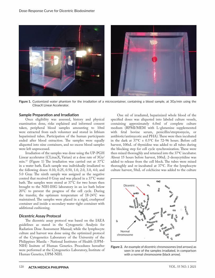
Figure 2. An example of dicentric chromosomes (red arrows) as seen in one of the samples irradiated, in comparison with a normal chromosome (black arrow).
Dicentric
Normal chromosome
ACTA MEDICA PHILIPPINA VOL. 55 NO. 1 2021120
Dose-Response Curve for Dicentric Biodosimeter

Health England Centre for Radiation, Chemical and Environmental Hazards (PHE CRCE), United Kingdom. Both chromosomal aberration analysis programs are endorsed by the IAEA for the construction of the dose-response calibration curves.
Ethical Considerations The research was conducted based on the principles
of good clinical practice. The protocol and consent were submitted and approved by the University of the Philippines - Manila Research Ethics Board (UPMREB). Subject privacy, confidentiality, and anonymity were maintained. Forms were anonymized and coded using identification numbers. Data linking documents will be kept only until the end of data analysis. A data protection plan compliant with the Data Privacy Act was put in place. Personal information and data retrieved from samples will remain confidential as discussed and written in the consent form. The staff of the Cytogenetics Laboratory followed the Data Security Plan of the Institute of Human Genetics Biorisk Management Manual Version 2.0 2015. The study adhered to the voluntary participation of subjects, and Informed consent for blood extraction, preparation of the dicentric assay, storage of chromosomal data/archived slides, and disclosure of results were secured before any data collection.
RESuLTS
Peripheral blood was donated by eight volunteers aged 25-39 (mean 33), without any vices (smoking, alcohol), comorbidities, previous treatment for tuberculosis, nor recent x-ray exposure. Donors were ensured to not have had prior exposure to radiotherapy and chemotherapy. Each donor was scored for approximately 300 - 400 cells.
After in vitro irradiation using the customized water phantom at 3Gy/min, the samples were sent for cytogenetic analysis and a total of 3,509 metaphase spreads were counted. As per the IAEA grading sheet, all chromosomal aberrations were counted. Preliminary data obtained from the samples are shown in Table 2. The study is still ongoing to count the remaining cells for a more robust dose-
to induce metaphase arrest and then mixed by inversion. The cell pellet was obtained through repeated centrifugation and was resuspended in 5ml 5% acetic acid to form a frothy head. The supernatant was aspirated and resuspended with 1ml of fixative dropwise. Fixation was done thrice, subsequently with repeated drying and resuspension. One to three drops of the cell suspension were placed at the center of a dried slide to prepare for slide analysis. Laboratory work and disposal of biologic materials followed the Institute of Human Genetics Biorisk Management Manual version 2.0 2015.
Slide Analysis and Data Recording The slides were coded to prevent bias. The whole slide
was scanned at low magnification (100-200x) for a quick scan of analyzable cells. Scanning was then done at higher magnification (1000x) (Figure 2). It is suggested that 500 cells or 100 dicentrics be scored to give a reasonably accurate dose. Only complete spreads were analyzed (i.e., 46 pairs). For this interim analysis, all of the 3,509 cells were analyzed.
Electronic systems were used to record information. In the laboratory, a score sheet was used based on the IAEA Handbook (2011) to facilitate dicentric scoring.² Data recording was done using a password-protected computer.
Analyzed microscopic slides will be kept under the optimal condition in the Institute of Human Genetics - Cytogenetics Laboratory indefinitely. These slides will serve as the reference data for the dose-response curve and may be used in further research regarding genotoxicity from radiation accidents. All the information gathered during the research and subsequent analysis will be guarded with strict confidentiality.
Statistical Analysis
The dose-response curves were constructed using CABAS Software version 2.0 and Dose Estimate version 5.2. To assess Poisson distribution, the dispersion index and normalized value of the index (u) were computed based on the IAEA Handbook.²
Dispersion index close to 1 and u values between ±1.96 indicates Poisson distribution. The above software was also used to compute for the goodness of fit and chi-square tests for homogeneity.
Correlation indices and goodness of fit were compared between the dose-response curves constructed. Validation of the dose-response curve and coefficients was done by comparing these to established dose-response curves from other cytogenetic laboratories.
The CABAS Software was used with permission from Dr. Andrzej Wojcik of the Centre for Radiation Protection Research, MBW Department, Stockholm University, Sweden. The Dose Estimate Software was also used with permission from Dr. Elizabeth Ainsbury of the Public
N–12(1–1/X)
u = (σ2/y-1)√dispersion index (σ2/y)
Table 2. Chromosomal Aberrations after acute irradiation as observed in human lymphocytes
Dose (Gy) Total Cells per Dose Dicentrics Ring Acentric0 499 2 0 00.1 300 4 0 00.25 600 0 1 00.5 634 12 7 121 558 24 2 212 301 15 8 93 504 303 77 4304 113 92 7 985 no growth
VOL. 55 NO. 1 2021 ACTA MEDICA PHILIPPINA 121
Dose-Response Curve for Dicentric Biodosimeter

response curve as advised by Dr. Kato of the Department of Environmental and Radiological Health Sciences, Colorado State University, USA (personal communication).
The frequency and distribution of the dicentrics are shown in Table 3. The dispersion index (σ/y) and U test were computed as per the IAEA EPR Biodosimetry Handbook.²
The sample irradiated at 0.25Gy did not yield any dicentric in the 600 cells; hence the dispersion index and U-test index were not computed. To be able to proceed with the statistical analysis, a value of 1 dicentric score (as background radiation) was substituted for dose 0.25Gy (with a score of 0) for the equation to be operable. This was by the IAEA consensus of background radiation of 0.5-1 dicentric per 1000 cells.²
Using the Chromosomal Aberrations Calculation Software (CABAS 2), the data was fitted using a linear quadratic model: y = C + αD + βD2 (Figure 3). The CABAS software computes the curve using maximum likelihood methods and resulted with the equation:
y = 0.0086 ± 0.0030) + (-0.0275 ± 0.0107)D + (0.0629 ± 0.0048)D2
For correlation, the curve (Figure 4) was also constructed using iteratively reweighted least square methods using the Dose Estimate Software version 5.0 that resulted in the equation:
Y = 0.0080 (± 0.0090) + (-0.0250 (± 0.0335)D + (0.0633 ± 0.0151)D2
Similar to the result of the dispersion index, there is overdispersion when computing for the two curves based
Table 3. Frequencies and Distributions of Dicentrics after acute irradiation as observed in human lymphocytesDose (Gray) Total Cells Dicentrics D0 D1 D2 D3 D4 D5 D6 Yield σ/y U test
0 499 2 497 2 0 0 0 0 0 0.004 0.998 -0.0450.1 300 4 296 4 0 0 0 0 0 0.013 0.99 -0.142
0.25 600 0 600 0 0 0 0 0 0 0 — —0.5 634 12 622 12 0 0 0 0 0 0.019 0.983 -0.323
1 558 24 536 21 0 1 0 0 0 0.043 1.21 3.572 301 15 289 9 3 0 0 0 0 0.05 1.35 4.53 504 303 284 154 50 15 1 0 0 0.601 1.07 1.074 113 92 50 41 18 2 1 1 0 0.814 1.06 0.488
Figure 4. The Dicentric Dose-Response Curve, based on observed dicentric frequencies, fitted using Dose Estimate Software version 5.2, with 95% confidence intervals.
Figure 3. The Dicentric Dose-Response Curve, based on observed dicentric frequencies, fitted using CABAS version 2.0.
ACTA MEDICA PHILIPPINA VOL. 55 NO. 1 2021122
Dose-Response Curve for Dicentric Biodosimeter

of cells should be counted for the low dose radiation points, especially for those irradiated to less than 1 Gray.²
Also, there is no growth for several samples irradiated at 5Gy. This is similar to the experiences of several cytogenetic studies at a high level of irradiation. The issue had been addressed by Pujol, et al. that one can increase the culture time of the lymphocytes to increase dicentric yield.17 Also, another measure is to score acentric and rings in addition to the dicentrics. Lastly, a technique known as Premature Chromosome Condensation (PCC) can be utilized at higher doses, to analyze heavily damaged cells blocked at the G2/M phase. The downside of the PCC is that dicentrics cannot be analyzed.17
Theoretically, the yield for dicentrics in samples irra-diated by x-rays follow a Poisson distribution. Our data shows that most of the dose points follow the Poisson distribution as indicated by a dispersion index approximating 1, and a U-test index of ± 1.96 as well as by the chi-square values. Overdispersion was observed in samples irradiated at 1 Gy and 2 Gy, as seen in Table 2. The adjustment was done using Estimate Dose Software to construct the dose-response calibration curve. Overdispersion can be attributed to the x-rays produce many tracks of radiation damage (i.e., quadratic) and this becomes more random at higher doses.18 Overdispersion can also occur at high radiation doses due to non-homogeneous irradiation of the sample.6
The majority of the cytogenetic assays use gamma and x-ray radiation, as these are the most common sources of radiation accidents.² The use of the linear accelerator as the reference and basis of calibration curves is surprisingly still new despite its increased use and availability in radiotherapy centers worldwide.
Based on a review of available literature, this study is among the few cytogenetic assays that used a linear accelerator to generate x-rays. It is the second study (after Bakkiam, et al.) to use a high dose rate of 3Gy/minute.15 Dose rate affects dose-response curves since the BD2 is
on a two-tailed chi-square test. This is likely attributable to the overdispersion at higher doses (1Gy and 2Gy), which is comparable to the reports of other cytogenetic laboratories. For the curve fitting, the r2 value of 0.9716 showed a good fit for the model as seen in Table 4.
DISCuSSIon
Biodosimeters function by measuring chromosome aberrations that reflect the radiation damage to the cell’s nucleus. Dose to soft tissues is a good approximation of the dose to the nucleus from photons and neutrons. The lymphocyte nucleus is very small compared to the secondary particles produced by photons and neutrons, hence the Bragg-Gray cavity theory is applicable in such cases.²
The dose determined based on cytogenetic studies using a dose-response curve is the average absorbed dose of the lymphocytes. Since blood circulates, this is the approximate averaged-out whole body dose received by an individual. The accuracy of biodosimetry is limited by non-uniform exposures, the intake of radionuclides, and delayed blood sampling.²
The half-life of dicentric chromosomes is reported to be 1.5 years.4 The usefulness of the dicentric assay is limited if the sampling is performed months or a few years after irradiation, although its sensitivity for whole-body irradiation can be as high as 0.1 Gy.16
The results showed that there is an increasing frequency of dicentrics with increasing doses, as seen in the yield per dose (Table 3). Based on the data from the Dose Estimate software, the correlation coefficient of the curve (r2) = 0.9716 means that there is a good relationship among the data points of the linear-quadratic dose model. Rings and acentric were also found to be dose-dependent. These were noted only for completeness as per the IAEA protocol. However, there were no dicentrics found in the 600 cells irradiated to 0.25Gy. This supports the recommendation that thousands
Table 4. Comparison of Linear and Quadratic Yield Coefficients of the CABAS and the Dose Estimate ProgramsC ± SE b ± SE a ± SE DF P-value r2
CABASDose estimate
0.0085± 0.00300.0080 ± 0.0090
0.0629 ± 0.00470.0633 ± 0.0151
-0.0275 ± 0.0107-0.0250 ± 0.0335
55
<.0001<.0001
—0.9716
Table 5. Dicentric Dose-Response Curves using Xrays produced by Linear AcceleratorsStudy Energy Dose Rate (Gy/min) Equation of Dose-Response Curve
DOSSOU (2000, France)20 18MV 0.16 Y = c + (0.052 ± 0.010)D + 0.037 ± (0.003)D2
LEMOS- PINTO (2015, Brazil)6 6MV 0.54Y = (0.001±0.007) + (0.013±0.007)D + (0.056±0.004)D2Y = (0.001±0.009) + (0.013±0.009)D + (0.056±0.006)D2
BAKKIAM (2015, India)15 6MV 3Y = (0.00037±0.0004) + (0.03667±0.0054)D + (0.05627±0.0025)D2Y = (0.00077±0.0005) + (0.03557±0.0057)D + (0.060870.0028)D2
LEE (2019, Korea)21 6MV 0.5 Y = (0.0011±0.0004) + (0.0119±0.0032)D +(0.0617±0.0019)D2
This study (2019, Philippines) 6MV 3Y = (0.0086±0.0030) + (-0.0275±0.0107)D + (0.0629±0.0048)D2
Y = (0.0080±0.0090) + (-0.0250±0.0335)D + (0.0633±0.01±51)D2
VOL. 55 NO. 1 2021 ACTA MEDICA PHILIPPINA 123
Dose-Response Curve for Dicentric Biodosimeter

dependent on it. A higher dose rate means a lower chance for DNA repair.19 When compared to cytogenetic dose-response curves using x-ray irradiation, the coefficients developed by this study’s curves are similar to published data (Table 5).
The calibration curve aims to determine an accurate dose from a set of analyzed cells and dicentric frequencies, ideally within 20% of the actual whole-body dose received by the individual. The standard error of ± 20% is sufficient to triage the individual for medical management. 22
Even though the dicentric assay is the gold standard for radiation dosimetry, its major disadvantages include being labor and time-intensive and being highly technical. A skilled cytogeneticist can take one whole day to analyze 200 cells.23 In this study, intra-comparison bias was minimized by having a senior cytogeneticist perform reviews.
Another factor that can affect the estimation of the dose is culture conditions. Authors have stated that changes in the laboratory culture protocol and handling of samples can affect metaphase chromosomes.24,25 Because of this factor, each cytogenetic laboratory is advised to construct their dose-response curves and coefficients.22
As of this time, the data of this report is still preliminary. To increase the robustness of the reference curves and to follow set international guidelines for the construction of such calibration curves, the study will continue to analyze approximately 2,400 cells per dose point or at least 100 dicentrics per dose point.
Although the calibration curve is based on preliminary data, it can be utilized to predict doses within 95% confidence intervals. To reduce the risk of overestimating doses, the IAEA also recommends predicting doses to 83% confidence intervals. Such changes can be made using the Estimate Dose software.²
ConCLuSIon
The curve coefficients generated by this study using CABAS (Chromosome Aberration Calculation Software Version 2.0) and Dose Estimate (Version 5.2) software are comparable to published data of dicentric dose-response curves of x-rays generated by linear accelerators. The curve also demonstrated goodness of fit (r²: 0.9716). This generated dose-response calibration curve will be useful for medical triage of the public and radiologic staff accidentally exposed to radiation during medical procedures or in the event of nuclear accidents. This analysis will be pursued to produce a more robust dose-response curve that meets international standards.
RecommendationsRecommendations after completion of the dose-response
curve include inter-laboratory comparison for quality assurance and dosimetric curve calibration for determining cut-offs for radiologic triage, such as for events needing rapid dose determination (ex. mass exposure to radiation).
AcknowledgmentsThe authors would like to thank the staff of the
Cytogenetics Laboratory, Institute of Human Genetics, National Institutes of Health - UP Manila. The authors would also like to acknowledge Dr. Andrzej Wojcik of the Centre for Radiation Protection Research, MBW Department, Stockholm University, Sweden for the use of the CABAS v2 Software, and Dr. Elizabeth Ainsbury of the Public Health England Centre for Radiation, Chemical and Environmental Hazards (PHE CRCE), the United Kingdom for the Dose Estimate Software. The authors would also like to thank Dr. Takamitsu Kato of the Department of Environmental & Radiological Health Sciences, Colorado State University, USA, and Dr. Mi-Ae Jang of the Department of Laboratory Medicine and Genetics, Soonchunhyang University Bucheon Hospital & Soonchunhyang University College of Medicine, Korea for their technical assistance on the protocol. The authors would also like to thank Dr. Ulrike Kulka for allowing the use of Table 1, as reprinted from her publication.
DisclaimerThe views expressed in this article are the authors’
own and do not reflect the views of the institution.
Statement of AuthorshipAll authors participated in data collection and analysis,
and approved the final version submitted.
Author DisclosureThe authors declare no conflicts of interest regarding
the content of this article.
Funding SourceThis study is supported by the 2019 Philippine
General Hospital, Expanded Health Research Grant. It is also partially supported by the Department of Radiology, Philippine General Hospital and the Institute of Human Genetics, National Institutes of Health, University of the Philippines - Manila.
REFEREnCES1. Hall EJ, Giaccia AJ. Radiobiology for the Radiologist. Philadelphia:
Lippincott Williams & Wilkins; 2011.2. International Atomic Energy Agency (IAEA). Cytogenetic Analysis
for Radiation Dose Assessment: A Manual. Technical Report Series 405. Vienna. 2011.
3. Kulka U, Abend M, Ainsbury E, Badie C, Barquinero JF, Barrios L, et al. RENEB – Running the European Network of biological dosimetry and physical retrospective dosimetry. Int J Radiat Biol. 2017 Jan; 93(1):2-14. doi: 10.1080/09553002.2016.1230239.
4. Wojcik A, Ostreicher U, Barrios L, Vral A, Terzoudi G, Ainsbury E, et al. The RENEB operational basis: complement of established biodosimetric assays. Int J Radiat Biol. 2017 Jan; 93(1):15-9. doi: 10.1080/09553002.2016.1235296.
5. International Atomic Energy Agency, no date. Lecture Module 4: Dicentric Assay. [Powerpoint presentation].
ACTA MEDICA PHILIPPINA VOL. 55 NO. 1 2021124
Dose-Response Curve for Dicentric Biodosimeter

16. Belyakov O. Review of the Established and New Biological Dosimetry Methods. International Atomic Energy Agency Presentation, Applied Radiation Biology and Radiotherapy Section, Division of Human Health. Presented at ASEANTOM, Bangkok, Thailand, March 8, 2017.
17. Pujol M, Barquinero J, Puig P, Puig R, Caballin MR, Barrios L. A new model of biodosimetry to integrate low and high doses. PLoS One. 2014 Dec; 9(12):e114137. doi:10.1371/journal.pone.0114137.
18. Vaurijoux A, Gruel G, Pouzoulet F, Grégoire E, Martin C, Roch-Lefèvre S, et al. Strategy for population triage based on dicentric analysis. Radiat Res. 2009 May; 171(5):541–8. doi: 10.1667/RR1664.1.
19. Perumal V, Sekaran TSG, Raavi V, Basheerudeen SAS, Kanagaraj K, Chowdhury AR, et al. Radiation signature on exposed cells: relevance in dose estimation. World J Radiol. 2015 Sep; 7(9): 266-78. doi: 10.4329/wjr.v7.i9.266.
20. Dossou J, M’kacher R, Bridier A, Girinsky T, Violot D, Legal JD, et al. Validation of Biological Dosimetry in patients conditioned with total body irradiation: conventional cytogenetics and in situ hybridization. Cancer Radiothér. 2000 Nov-Dec; 4(6):399-407. doi: 10.1016/s1278-3218(00)00013-5.
21. Lee Y, Jin YW, Wilkins RC, Jang S. Validation of the dicentric chromosome assay for radiation biological dosimetry in South Korea. J Radiat Res. 2019 Oct; 60(5):555–63. doi: 10.1093/jrr/rrz039.
22. Wilkins RC, Romm H, Kao T, Awa AA, Yoshida MA, Livingston GK, et al. Interlaboratory Comparison of the Dicentric Chromosome Assay for Radiation Biodosimetry in Mass Casualty Events. Radiat Res. 2008 May; 169(5):551-60. doi: 10.1667/RR1272.1.
23. Romm H, Oestreicher U, Kulka U. Cytogenetic damage analysed by the dicentric assay. Ann Ist Super Sanita. 2009. 45(3):251-9.
24. Stricklin D, Wilkinson D, Arvidsson E, Prud’homme-Lalonde L, Thorleifson E, Mullins D, et al. An initial limited biodosimetry inter-comparison exercise: FOI and DRDC Ottawa. Radiat Meas. 2007 July-Aug; 42(6-7):1133–7.
25. Roy L, Buard V, Delbos M, Durand V, Paillole N, Gregoire E, et al. 2004. International intercomparison for criticality dosimetry: the case of biological dosimetry. Radiat Prot Dosimetry. 2004; 110(1-4):471–6. doi: 10.1093/rpd/nch349.
6. Lemos-Pinto MMP, Cadena M, Santos N, Fernandes TS, Borges E, Amaral A. A Dose-response Curve for Biodosimetry from a 6 MV Electron Linear Accelerator. Braz J Med Biol Res. 2015 Oct; 48(10):908-14. doi: 10.1590/1414-431X20154470.
7. Blakely WF, Carr Z, Chu MCM, Dayal-Drager R, Fujimoto K, Hopmeir M, et al. WHO 1st Consultation on the Development of a Global Biodosimetry Laboratories Network for Radiation Emergencies (BioDoseNet). Radiat. Res. 2009 Jan; 171(1):127–39. doi: 10.1667/RR1549.1.
8. Vral A, Fenech M, Thierens H. The micronucleus assay as a biological dosimeter of in vivo ionising radiation exposure. Mutagenesis. 2011 Jan; 26(1):11–7. doi: 10.1093/mutage/geq078.
9. Shakeri M, Zakeri F, Changizi V, Rajabpour MR, Farshidpour MR. A cytogenetic biomonitoring of industrial radiographers occupationally exposed to low levels of ionizing radiation by using CBMN assay. Radiat Prot Dosimetry. 2017 Jun; 175(2):246-51. doi: 10.1093/rpd/ncw292.
10. Nersesyan A, Fenech M, Bolognesi C, Mišík M, Setayesh T, Wultsch G, et al. Use of The lymphocyte cytokinesis-block micronucleus assay in occupational biomonitoring of genome damage caused by in vivo exposure to chemical genotoxins: past, present and future. Mutat Res. 2016 Oct-Dec; 770(Pt A):1–11. doi: 10.1016/ j.mrrev.2016.05.003.
11. Zakeri F, Assaeii RG. Cytogenetic monitoring of personnel working in angiocardiography laboratories in Iran hospitals. Mutat Res. 2004 Aug; 562(1-2):1–9. doi: 10.1016/j.mrgentox.2004.04.005.
12. Electronic Code of Federal Regulations PART 835—OCCUPATIONAL RADIATION PROTECTION [Internet]. May 2018 [cited 2019 Oct]. Available from: https://www.ecfr.gov/cgi-bin/text-idx?SID=a069c156722da73e316046707c604ee2&c=true&node=pt10.4.835&rgn=div5# se10.4.835_1401
13. Dizdarević E. Regulation on the Medical Surveillance of Occupa-tionally Exposed Workers [Internet]. 2011 [cited 2019 Oct]. Available from: http://www.darns.gov.ba/en/EnglishDARNS/DownloadFile?fileName=Pravilnik_o_zdravstvenom_nadzoru_engleski.pdf
14. Roch-Lefevre S, Pouzoulet F, Giraudet AL, Voisin P, Vaurijoux A, Gruel G, et al. Cytogenetic assessment of heterogeneous radiation doses in cancer patients treated with fractionated radiotherapy. Br J Radiol. 2010 Sep; 83(993):759–66. doi: 10.1259/bjr/21022597
15. Bakkiam D, Bhavani M, Kumar A, Sonwani S, Venkatachalam P, Sivasubramanian K, et al. Dicentric assay: inter-laboratory comparison in Indian laboratories for routine and triage applications. Appl Radiat Isot. 2015 May; 99:77–85. doi: 10.1016/ j.apradiso.2015.02.015.
VOL. 55 NO. 1 2021 ACTA MEDICA PHILIPPINA 125
Dose-Response Curve for Dicentric Biodosimeter