interferons modulate expression of hormone receptors ... · sinase activity. after incubation, the...
TRANSCRIPT

Interferons Modulate the Expression of Hormone Receptorson the Surface of Murine Melanoma CellsKoichiro Kameyama,* Syuji Tanaka,* Yasuo Ishida,t and Vincent J. Hearing**Laboratory of Cell Biology, National Cancer Institute and *Laboratory of Immunology, National Institute ofAllergyand Infectious Diseases, National Institutes of Health, Bethesda, Maryland 20892
Abstract
The effects of IFN-a, IFN-j0, and IFN-'y on the differentiationof murine melanoma cells has been studied, in the presence andabsence of melanocyte-stimulating hormone (MSH); the cellswere highly responsive to treatment with MSH, which in-creased the rate of melanin production 25-fold and tyrosinaseactivity 6-fold within 4 d. Treatment of melanoma cells withIFN-a, IFN-,8, or IFN-'y alone had no stimulatory effect on
melanin production, but when the cells were cultured with IFNin the presence of MSH, pigment production was significantlyand synergistically increased relative to cells cultured withMSHonly. Flow cytometric analysis revealed that levels oftyrosinase in the cells were not affected by MSHor by IFN,which suggests that stimulation of melanogenic activity oc-
curred by activation of a preexisting cellular enzyme. Scatch-ard analyses showed that the number of MSHreceptors on
IFN-treated cells was significantly increased (- 2.5-fold) rela-tive to untreated cells (- 61,000/cell). These findings demon-strate that IFN stimulate differentiation (that is, pigmentation)of melanocytes by increasing the expression of surface MSHreceptors; this in turn suggests that such a mechanism may inpart be responsible for postinflammatory skin pigmentation,and provides an additional basis for action in the clinical re-
sponses of melanoma to IFN treatment.
Introduction
The phenomenon of postinflammatory skin pigmentation hasbeen observed after various kinds of inflammatory skin dis-ease, such as contact dermatitis and drug eruptions, althoughthe mechanism(s) involved have not yet been explained. Thesediseases have a common histologic feature, the presence ofinflammatory cells in the skin. Inflammatory cells are com-
posed of lymphocytes, monocytes, and neutrophils, all ofwhich produce IFN (1); in addition, lymphocytes infiltratingdysplastic nevi or melanoma lesions have been shown to pro-
duce IFN--y (2). IFN have been reported to promote differen-tiation of various kinds of cells including myoblasts (3), histio-cytic lymphoma cells (4), and promyelocytic leukemia cells(5). IFN have been reported in some studies to increase pig-ment production by melanoma cells (6-8), but in others, to
Dr. Kameyama's permanent address is the Department of Dermatol-ogy, Kitasato University School of Medicine, 1-15-1 Kitasato, Saga-mihara 228, Japan.
Address reprint requests to Dr. Vincent Hearing, Building 37,Room 1B22, National Institutes of Health.
Receivedfor publication 28 March 1988 and in revisedform 21 July1988.
reduce it (9-1 1). The specific effects of IFN on melanogenesisthus have not yet been conclusively determined. Such studiessuggest the possibility that IFN may elicit postinflammatoryskin pigmentation by stimulating differentiation, and thuspigmentation, of epidermal melanocytes. As a result, IFN havebeen used as potential antimelanoma agents in both animalmodels and clinical trials (12-14).
Melanocytes are highly suitable models for studies of dif-ferentiation because melanin is a specific, readily visible, andidentifiable product; the melanin pigment is produced by anenzyme termed tyrosinase (EC 1.14.18.1), which in turn canbe quantitated rather easily. There are also many physiologi-cally relevant agents that can induce differentiation in mam-malian melanocytes in vivo and in vitro, including ultravioletlight and melanocyte-stimulating hormone (MSH).' Ideally,studies of differentiation would use normal melanocytes, butbecause these cells are sparse in mammals, and are very diffi-cult if not impossible to culture in significant quantity, moststudies on melanogenesis have relied on the use of murinemelanomas, such as the B 16, S9 1, and Harding-Passey. Thosetumor lines were established > 30 yr ago, and thus there is apossibility that they have drifted significantly in phenotype,and no longer resemble the original pigmented tumors. Re-cently, the JB/MS melanoma was induced by carcinogens (15,16); this tumor has now been adapted to tissue culture, and is ahighly pigmented, relatively well-differentiated cell line, whichhas been proposed to serve as a more appropriate model formelanogenic and tumorigenic studies (15-18). In this report,JB/MS murine melanoma cells, as well as B16 and S91 cells,were used to investigate the effect of IFN on spontaneous andMSH-induced pigmentation. We have found that IFN-a,IFN-f3, and IFN-,y stimulate the expression of MSHreceptorson the surface of melanoma cells; this enhanced binding ca-pacity for MSHis followed by a significant synergistic stimula-tion of melanin production in the presence of MSH.
Methods
Materials. Natural murine IFN-a (1.4 X 10⁶ IU/mg) and IFN-ft (5.6X 10⁷ IU/mg) were purchased from Lee Biomolecular Research (SanDiego, CA); murine rIFN-,y (1.2 X 10⁷ IU/mg) was supplied by Gen-entech (19) (South San Francisco, CA). Purified synthetic a-MSH wasa gift from Dr. J. Pawelek (Yale University, NewHaven, CT), and wasalso purchased from Calbiochem-Behring Corp. (La Jolla, CA).
Cell culture. The recently derived, 7,12-dimethylbenzanthraceneinduced JB/MS melanoma (15, 16) was used in this study; the growthcharacteristics and culture methods for this cell line have been de-scribed in detail elsewhere (17, 18). In these experiments, JB/MS cells(at passage 6) were cultured a maximum of 2 mo; continual passagelonger than this resulted in significant phenotypic drift (unpublished
The Journal of Clinical Investigation, Inc.Volume 83, January 1989, 213-221
1. Abbreviations used in this paper: MSH, melanocyte-stimulatinghormone.
Stimulation of Melanin Production by Interferons 213

observations). Wehave also used B16 and S91 murine melanoma cellsin some experiments; these were derived as previously noted (18).DMEand other tissue culture supplies were purchased from GibcoLaboratories (Grand Island, NY); our routine culture medium con-sisted of DMEsupplemented with 10% heat-inactivated, mycoplasmaand virus-free FCS, 100 Ag/ml streptomycin, 100 U/ml penicillin, and0.25 Ag/mi amphotericin-B, nonessential amino acids, and glutamine.Unless otherwise noted, cells were harvested in exponential growthphase with trypsin/EDTA, and seeded at a concentration of 6 X 10⁵cells per 75-cm2 flask. The cells were maintained in a humidifiedincubator with 5%CO2 in air at 370C. 24 h later (day 0) the mediumwas removed and replaced with fresh medium containing IFN and/orMSHat the concentrations noted in the tables and figures; the appro-priate medium was changed daily thereafter. MSHwas routinely used(except as reported in Table IV) at 2 X l0-7 M; this concentration hasbeen shown to elicit maximal melanogenic response in murine mela-noma cells, as previously reported (20-22).
Determination of melanogenic activities. Melanogenic activitieswere measured by two radiometric assays as previously described (23,24); briefly, cells were harvested with trypsin/EDTA, and then solubi-lized at a concentration of I05 cells per 30 Ml with 1% NP-40 for I h at40C. The cell extracts were then centrifuged at 12,000 g for 10 min,and 30 Al of the soluble extract (i.e., 105 cell equivalents) were incu-bated with 10 Al labeled tyrosine (50 ,M as noted below) and 10 Ml ofDOPAcofactor (50 MM) at 370C in a 96-well microtiter plate. Foranalysis of melanin formation, L-[U-`4C]tyrosine (100 mCi/mmol, 25MCi/ml; NewEngland Nuclear, Boston, MA) was used; after the incu-bation, 20 ,l were removed to filter discs and washed three times with0.1 N HC1, two times with ethanol, and once with acetone. The discswere then dried and the radioactive, acid-insoluble melanin that hadbeen formed was measured in a liquid scintillation counter. The datareported in the tables used a 16-h incubation, but in separate experi-ments, we have determined that rates of melanin production are linearthroughout that time period. For analysis of tyrosinase activity, L-[3,5-3H]tyrosine (1 Ci/mmol, 250 MCi/ml; NewEngland Nuclear) wasused; after 1 h incubation, 20 Ml was removed and added to 980 Ml 0.1N HCl with activated charcoal and Celite 545 in 12 X 75 mmtubes;after mixing for 1 h at 23°C, the tubes were centrifuged for 1,300 g for10 min, and the [3H]water that had been formed as the result oftyrosine hydroxylation (which remains in the supernatant) was mea-sured in a scintillation counter. Enzyme assays were carried out at leastin triplicate and variability within the assays was always < ±15%;backgrounds were established by omitting either the enzyme sample orthe cofactor. In related experiments (not shown), we have determinedthat neither MSHnor IFN has any effect on either of the two radiomet-ric assays described above.
Flow cytometric analysis. Cells were harvested with 0.02% EDTAin HBSSand permeabilized with 70% ethanol for 30 min at 4°C; 106cells were reacted with 50 Ml tyrosinase-specific MAbTMH-l for 1 hat4°C; the specificity and reactivity of TMH-1 for mammalian tyrosi-nase has been previously reported (18, 25). After the incubation, thecells were washed twice in HBSSand then reacted with 40 Ml fluores-cein isothiocyanate-conjugated rabbit IgG anti-rat IgG (diluted 1:20in HBSS; Dako Corp., Santa Barbara, CA). The cells were then againwashed twice in HBSSand finally resuspended in HBSSand analyzedwith a FACSII (Becton Dickinson & Co., Mountain View, CA). Non-specific background staining was determined by omitting the first an-tibody and/or by staining of nonmelanocytic cells.
Iodination and purification of MSH. These techniques were per-formed using a modified procedure of Lambert and Lerner (26);briefly, 2.2 Mgof Iodogen (Pierce Chemical Co., Rockford, IL) in 20AMdichloromethane was plated on the bottom of 12 X 75 mmglass tubes,and then evaporated. Each iodination was carried out in one of thesetubes at 4°C, and the reaction mixture consisted of 5 mCi Na['l25](New England Nuclear) in 10 Ml 0.1 MNaOH, 100 Ml 0.1 Mboratebuffer, pH 8.2, and 1 1,l (6.5 nmol) of MSH. The reaction was termi-nated after 20 min by removing the reaction mixture from the tube andadding it to 0.9 ml borate-buffered saline containing 0.75 MDTTand
100 Mg/ml gentamycin; this solution was passed through a (22 X 0.45cm) Sephadex GlO column (Pharmacia Fine Chemicals, Piscataway,NJ) and eluted with DTT/gentamycin borate-buffered saline. The firstradioactive peak fractions were pooled and incubated at 37-C for 24 hto reduce oxidized methionine and maintain biological activity. Themixtures were then further purified by HPLC(model 344; BeckmanInstruments, Fullerton, CA) equipped with a 25 X 0.45 cm Altex C18reverse-phase column; the components of the mixture were elutedfrom the column at 1 ml/min using a linear gradient of increasingacetonitrile from 6 to 60% in 0.01 M trifluoroacetic acid, and theelution was monitored at 280 nm. The eluted material was collected in30-s fractions and monitored for radioactivity in a -y counter. Thepurified ['251]-MSH prepared in this manner had a specific activity of75-150 MCi/Mg.
Amino acid analysis. Amino acid analysis was performed as pre-viously described (27). Briefly, peptides were hydrolyzed with 6 NHCIcontaining 0.1% phenol for 24 h at 1 10C, after which the dried hydro-lysates were derivatized with phenylthiocarbamyl; these derivativeswere then analyzed using a Waters Picotag amino acid analysis system(Waters Associates, Milford, MA).
Biologic activity of iodinated MSH. 2 X 10⁵ JB/MS cells wereseeded into 25-cm2 flasks and 24 h later, the medium in each flask wasremoved and replaced with 5 ml fresh medium containing variousconcentrations of unlabeled MSHor '25I-MSH. 48 h later, the mediumin each flask was again changed, and 48 h later, the media were re-moved and the cells in the flasks were harvested with trypsin/EDTA.The cells were then solubilized as detailed above and assayed for tyro-sinase activity. After incubation, the aliquots were checked with a 'ycounter to measure [12511 contamination (slight levels were found inextracts of cells that had received the highest concentrations of labeledMSH). The samples were then processed as detailed above to measure[3H]water formation, and again checked for contaminating [1251](which was no longer detectable).
Receptor binding assay. Binding of the labeled MSHto surfacereceptors was measured by a modification of the method of Lambert etal. (28). Briefly, this involves harvesting the cells with EDTAas de-tailed above, and washing them twice with HBSS; the cells are thenresuspended in binding buffer, which consists of 0.5% BSA in DME,pH 6.8. The cells were centrifuged again, resuspended in fresh bindingbuffer, and cooled to 4°C. Known volumes of the suspension werethen mixed with binding buffer containing ['251]-MSH (to determinetotal binding of the ligand) or with [1251]-MSH plus a 1,000-fold excessof unlabeled MSH(to determine specific and nonspecific binding ofthe ligand). For all experiments, the incubation mixture had 1.5 X 10⁶cells in a final volume of 750 ,l in 12 X 75 mmsiliconized glass tubes;these were incubated for 2 h at 4°C with gentle shaking (unless other-wise noted). After determination of the total label in each tube in a ycounter, the cell-bound ['251]-MSH was separated from free ligand inthe following manner: 200-pl aliquots from each tube were layeredover discontinuous sucrose gradients (150 ,l 0.3 Msucrose overlaidwith 100 gl 0.15 M sucrose in 0.5 ml microcentrifuge tubes) andcentrifuged in a microfuge (model 5412; Brinkmann Instruments,Westbury, NY) for 10 s. After a further washing with 450 ,ul labelingbuffer, the ['251]-MSH associated with the cell pellets was determinedwith a y counter. In the analysis of the data, specific binding is definedas the amount of [ '251]-MSH bound to cells in the absence of unlabeledMSHcompared to the amount bound in the presence of a 1,000-foldexcess of unlabeled MSH.
Statistical analysis. The t test was used for statistical analyses.
Results
Effect of MSHand IFN-a, IFN-13, and IFN-,y on melanin pro-duction. Consistent with our earlier studies (17, 18), JB/MSmelanoma cells were highly responsive to MSHtreatment(Table I); they increased their melanogenic activity > 25-foldafter 4 d of treatment with the hormone. On the other hand,
214 K. Kameyama, S. Tanaka, S. Ishida, and V. J. Hearing

Table L Titration of IFN-a, IFN-f3, and IFN-,y Effectson MSH-induced Melanin Formation
Cell number (X 104/flask) Melanogenic activityAddition to
medium Without MSH With MSH Without MSH With MSH
pmol/J10 cells/J6 h
None 21±2 20±1 50±5 1,383±213IFN-a (0.1) 21±1 21±2 45±3 1,798±175IFN-a (1) 21±1 20±1 55±3 2,318±388*IFN-a (10) 19±1 19±2 53±5 2,573±350*IFN-a (100) 15±2* 15±1* 55±3 2,458±375*
IFN-# (0.1) 20±2 20±1 33+3* 1,503±113IFN-#t (1) 21±2 21±1 28±3* 2,225±175*IFN-# (10) 19±1 19±2 30±3* 2,448±200*IFN-# (100) 18±1 18±1 23±3* 1,780±138*IFN-y (0.1) 21±2 21±1 43±8 1,645±188IFN-y (1) 21±2 21±1 33+3* 1,783±75IFN-'y (10) 15±1* 15±1* 30±3* 1,208±38IFN-y (l00) 14±1* 14±1* 20±3* 310±75*
6 X i05 JB/MS cells were seeded in 75-cm2 flasks and 1 d later themedia were changed to include IFN (at IU/ml) as indicated andMSH(at 2 X I0` M) where noted; appropriate media were changeddaily. After 4 d, the cells were harvested, counted, solubilized, andmelanogenic activities were determined as detailed in Methods. Theresults are expressed as the means of picomoles [14C]tyrosine incor-porated into melanin in triplicate assays±SEM; * indicates a signifi-cant difference from the relevant control (on the top row) at P< 0.05 or better.
treatment with IFN-a, IFN-fl, or IFN--y alone did not signifi-cantly increase pigment production, but rather, IFN-a had nosignificant effect at any concentration tested, whereas IFN-f# orIFN-'y elicited dose-dependent decreases in melanogenic activ-ity. When the cells were cultured with IFN in the presence ofMSHhowever, melanin production was clearly synergisticallystimulated relative to cells treated with MSHalone (except atthe higher concentrations of IFN--y); the optimal concentra-tion for this stimulatory effect of MSHresponse for each IFNwas 1-10 IU/ml.
As can also be seen from the data in Table I, neither MSHnor any of the IFN (at concentrations of 1 or 0.1 IU/ml) hadany effect on cell growth during the 4-d time course of theseexperiments. However, IFN-a, IFN-f3, or IFN--y at 10 IU/mland above had slight antiproliferative effects on JB/MS cells;we thus routinely used IFN at 1 IU/ml as the optimal concen-tration to stimulate melanin production but to avoid effects onthe growth potential of the cells. In other experiments (notshown), IFN-y was shown to have similar effects on the prolif-eration of B16 cells, whereas S91 cells were resistant to IFN-induced growth inhibition at concentrations as high as 100IU/ml.
In a time course study, we examined the kinetics of theresponses to MSHand IFN (Table II). When the cells weretreated with IFN-a, IFN-,B, or IFN-,y alone, the melanogenicactivity at each time point was not significantly altered com-pared with the control. The relatively low levels of pigmentproduction by these cells at days 1 through 3 increased as theyapproached confluence and began spontaneous pigmentationat days 4 and 5. After subculture of the confluent cells at day 4,
melanogenic activity decreased as the cells began to prolifer-ate, and production of pigment slowly returned to basal levels.Note that the melanogenic activity of control cells, and theIFN treated cells, at day 1 was higher than at days 2 and 3; thiswas caused by residual melanogenic activity in the cell popula-tion used to initially seed the flasks.
The response of JB/MS cells to MSHwas rapid; a signifi-cant increase in demonstrable melanogenic activity occurredwithin 1 d, by which time it had doubled. The differencesbetween the melanogenic potentials of control cells and MSHtreated cells increased rapidly thereafter, was stimulated 6-foldby day 2, and 38-fold by day 3 (when the effect was mostpronounced). The levels of melanin production in MSH-treated cells were highest on day 5 and, surprisingly, decreasedthereafter until day 7, even though these cells were treated withMSHcontinuously.
The responses of JB/MS cells to treatment with MSHandIFN-a, IFN-fl, or IFN-y in this experiment showed that me-lanogenic activities were significantly increased over MSHalone by day 2, and that by day 4 were 15-60% higher (TableII). Perhaps more importantly, in cells treated with MSHandIFN the decreases in melanogenic activity after subculturing ofthe cells were significantly less pronounced, especially on day7, where the melanogenic potentials of MSHand IFN treatedcells were 7-8 times higher than those of MSHtreated cells.Virtually identical results were obtained in experiments testingthe effects of IFN--y and/or MSHon B16 and S91 cells (notshown), although the effects were somewhat less pronouncedwith S9 1 cells.
Flow cytometric analysis. To characterize the mechanismby which IFN stimulated melanogenic potential, we initiallydetermined whether treatment with IFN elicited increases inthe levels of tyrosinase produced by the cells. To do this, weused flow cytometric analysis, fixing and permeabilizing thecells, and then staining them with TMH-1, a MAbspecific for
Table IL Time Course of the Effects of MSHand IFN-a, IFN-f3,and IFN-,y on Melanin Production
Melanogenic activity
Without MSHtreatment With MSHtreatment
Day Cell number None IFN-a IFN-ft IFN--y None IFN-a IFN-# IFN-y
XltY6/flask pmoI/IJO cells/16 h
1 1.7±0.2 38 45 38 40 83 73 68 482 9.8±0.5 25 28 25 25 163 228 270 2183 17.8±1.3 25 28 33 28 945 1,503 1,030 1,0634 21.9±1.0 45 68 90 50 1,488 2,380 2,215 1,7135 0.6±0.1 190 238 170 193 1,570 2,305 1,985 2,0306 1.9±0.2 133 125 108 73 1,345 2,150 2,138 1,2587 5.2±0.4 43 25 18 30 143 1,128 1,008 1,1888 15.1±1.5 35 23 25 48 473 1,418 805 1,3009 18.7±1.0 45 50 45 35 1,273 2,580 2,343 1,880
The experimental protocol was as noted for Table I; flasks of cells were har-vested daily for determination of melanogenic activity, and remaining flaskswere subcultured at day 4. Again, the appropriate media were changed daily;MSHwas used at 2 X 10⁻⁷ Mand IFN at I IU/ml. The data are expressed asin Table I; for the sake of simplifying the table, standard errors are not re-ported but were < ± 15% in all cases. Cell numbers are expressed as the averageof the flasks on each day; there were no significant differences between the cellnumbers in controls, cells treated with MSH, or cells treated with IFN, at anytime point assayed.
Stimulation of Melanin Production by Interferons 215

Table III. Flow Cytometric Analysis of Tyrosinase Concentrationin Melanoma Cells
Mean fluorescence intensity
Treatment Without MSH 2 X 10⁻⁷ MMSH
None 482 480
IFN-a 485 458IFN-,B 475 460IFN-,y 463 472
The experimental protocol was as detailed in Table I; cells growingfor 4 d in media supplemented with IFN (at 1 IU/ml) as noted, wereharvested with EDTA, permeabilized with ethanol, and stained byindirect immunofluorescence with the tyrosinase specific MAbTMH-l as detailed in Methods. The data are expressed as mean fluo-rescence intensity determined by flow cytometry; background con-trols without the first antibody had a mean fluorescence intensity of320.
the predominant, enzymatically active form of tyrosinase (18,25). In Table III, data are shown on the intracellular tyrosinaseconcentrations of untreated, MSH-treated, and/or IFN-treatedJB/MS melanoma cells. The mean fluorescence intensity ofthe untreated JB/MS melanoma cells was 482 after 4 d inculture; cells treated with MSH, or with IFN-a, IFN-i3, orIFN-y had no significant difference in enzyme levels, even
1.00
- 0.80 _z Az
I 0.60cc
040tric
0.00-
LU
a-
~0.20
0 200 400 600 800CHANNELNUMBER
1.00
-juz 0.80zz B
0.60cc
0.40Z
uJ0.20-
0 200 400 600 800
CHANNELNUMBER
Figure 1. Flow cytometry of JB/MS cells labeled with anti-tyrosinaseMAb. JB/MS melanoma cells were harvested, fixed, permeabilized,and stained with TMH- I antibodies as detailed in Methods; in eachfigure, the curve on the left (---) is the negative control, the curve onthe right ( ) is the specific staining pattern. (A) Cells grown 4 d inDME. (B) Cells grown 4 d in DMEcontaining IFN-y (1 IU/ml) andMSH(2 X 10-' M).
when MSHand IFN were used at the same time. Fig. 1 showshistograms of two of these cell populations; there was no dif-ference in the shape and intensity of the specific histogramsbetween control cells (Fig. 1A) and MSH- and IFN-'y treatedcells (Fig. IB). These data demonstrate that the action of MSHand IFN is not at the level of stimulating synthesis of tyrosi-nase, which agrees with our previous observations for responseto MSHbased on metabolic labeling and immunoprecipita-tion analyses (18), and also with studies from other laborato-ries that used enzymatic criteria (20, 29).
Synthesis and purification of biologically active iodinatedMSH. Wenext examined the possibility that the stimulatoryeffects on melanin production elicited by IFN only in the pres-ence of MSHresulted from increased hormonal stimulationdue to modulation of the expression of surface MSHreceptors.To perform such studies, an MSHreceptor binding assay hadto be optimized. In HPLCanalysis of iodinated MSH(Fig. 2),two radioactive peaks eluted after the residual unlabeled MSH(peaks II and III). The radioactive peak (peak II) that elutedimmediately after the unlabeled MSHpeak (peak I) was con-firmed by amino acid analysis (not shown) to be monoiodin-ated MSH(peak II). The production of biologically active['2511]-MSH has been a point of contention in the literature forsome time (30, 31); it was suggested that the denaturation ofthe iodinated peptide resulted from the oxidation of methio-nine during the iodination procedure (32). The amino acidanalyses demonstrated that reduction with DTT immediatelyafter iodination preserves the methionine residue, and that theradioactive fraction eluting after the unlabeled MSHwas au-thentic ['25I]-MSH (peak II). The results in Table IV demon-strate that the biological activity of the iodinated MSHwaspreserved; the labeled MSHis equivalent to the original unla-beled MSH(at many different concentrations) in the ability tostimulate melanin production, that is, tyrosinase activity.
MSHbinding studies. In a time course study examiningthe rate of binding of labeled MSHto JB/MS cells (Fig. 3), 90%of maximal binding was observed within 1 h, and thus a 2 h
0.025 ..60
. 500.020 7M
E 40 >C <MO.015 M
Z ~~~~~~~~~~~~~~~~~~30~co~~ ~ ~~~~II4 0. Fcc 3'D~~~~~~~~~I m
'I I ~~3~ 20:
15 20TIME (min)
Figure 2. HPLCelution profile of MSHand iodinated MSH. MSHwas iodinated, reduced, and partially purified on a Sephadex G10column, as detailed in Methods. 100 ,l of the solution was resolvedon a C18 column using an acetonitrile gradient, and monitored byA280. 30-s fractions were collected and radioactivity was quantitatedin a y counter.
216 K. Kameyama, S. Tanaka, S. Ishida, and V. J. Hearing
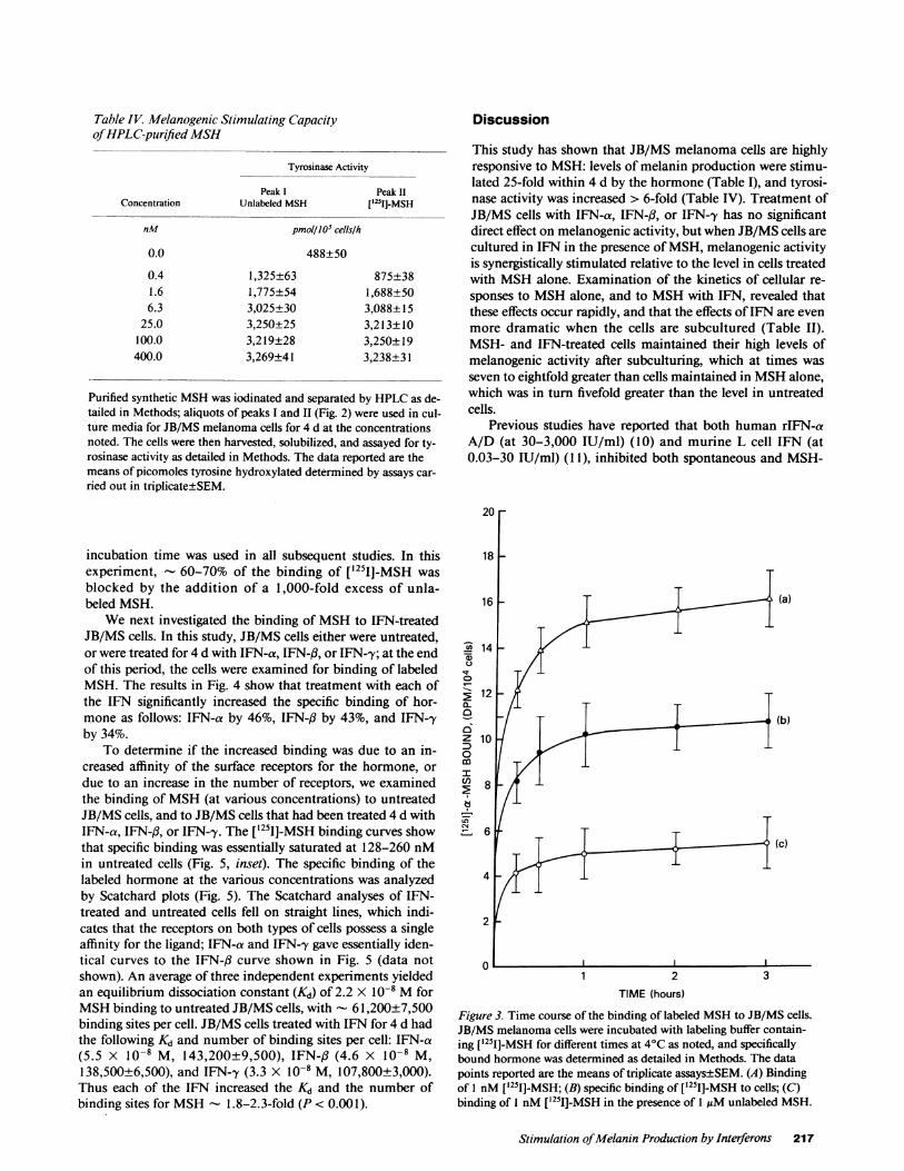
Table IV Melanogenic Stimulating Capacityof HPLC-purified MSH
Tyrosinase Activity
Peak I Peak IIConcentration Unlabeled MSH ['231]-MSH
nM pmol/JO' cells/h
0.0 488±50
0.4 1,325±63 875±381.6 1,775±54 1,688±506.3 3,025±30 3,088±15
25.0 3,250±25 3,213±10100.0 3,219±28 3,250±19400.0 3,269±41 3,238±31
Purified synthetic MSHwas iodinated and separated by HPLCas de-tailed in Methods; aliquots of peaks I and II (Fig. 2) were used in cul-ture media for JB/MS melanoma cells for 4 d at the concentrationsnoted. The cells were then harvested, solubilized, and assayed for ty-rosinase activity as detailed in Methods. The data reported are themeans of picomoles tyrosine hydroxylated determined by assays car-ried out in triplicate±SEM.
incubation time was used in all subsequent studies. In thisexperiment, - 60-70% of the binding of [1251J-MSH wasblocked by the addition of a 1,000-fold excess of unla-beled MSH.
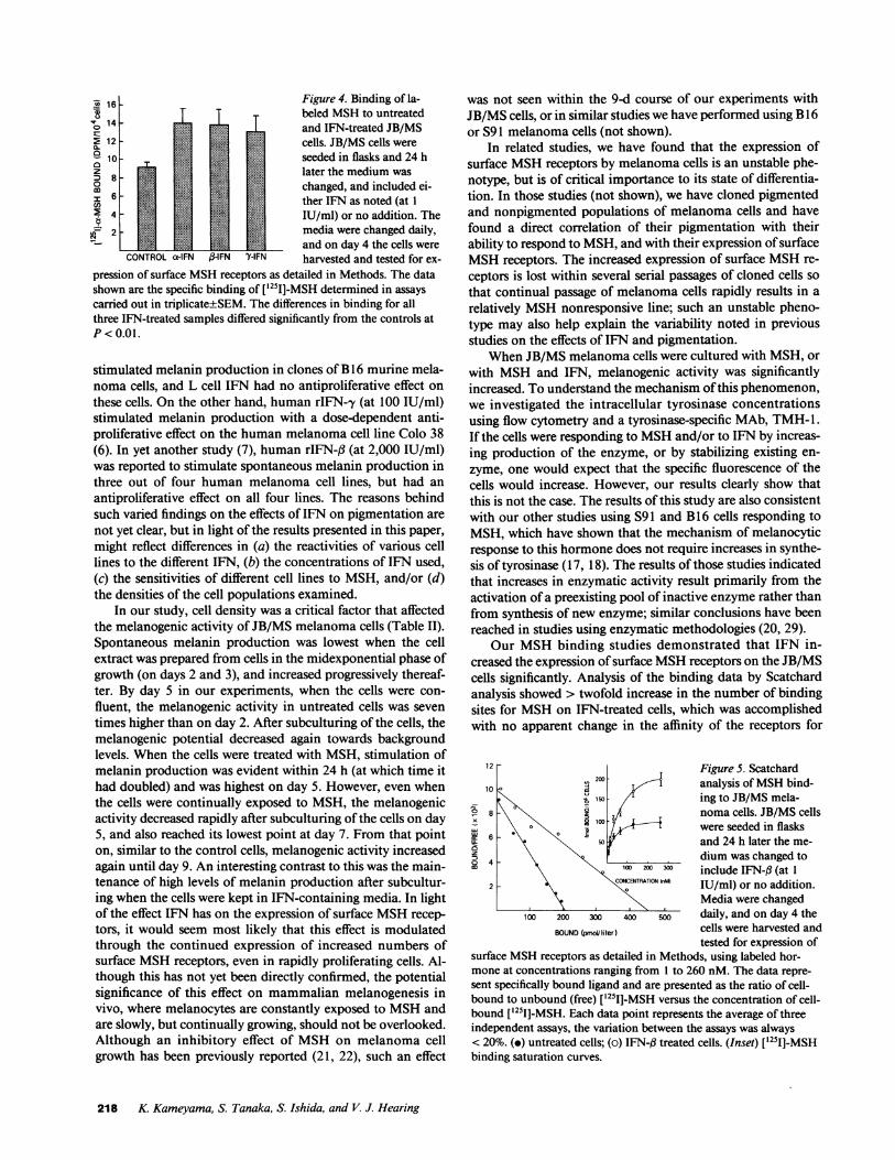
Wenext investigated the binding of MSHto IFN-treatedJB/MS cells. In this study, JB/MS cells either were untreated,or were treated for 4 d with IFN-a, IFN-f3, or IFN-y; at the endof this period, the cells were examined for binding of labeledMSH. The results in Fig. 4 show that treatment with each ofthe IFN significantly increased the specific binding of hor-mone as follows: IFN-a by 46%, IFN-fl by 43%, and IFN-'yby 34%.
To determine if the increased binding was due to an in-creased affinity of the surface receptors for the hormone, ordue to an increase in the number of receptors, we examinedthe binding of MSH(at various concentrations) to untreatedJB/MS cells, and to JB/MS cells that had been treated 4 d withIFN-a, IFN-fl, or IFN-,y. The ['251]-MSH binding curves showthat specific binding was essentially saturated at 128-260 nMin untreated cells (Fig. 5, inset). The specific binding of thelabeled hormone at the various concentrations was analyzedby Scatchard plots (Fig. 5). The Scatchard analyses of IFN-treated and untreated cells fell on straight lines, which indi-cates that the receptors on both types of cells possess a singleaffinity for the ligand; IFN-a and IFN-'y gave essentially iden-tical curves to the IFN-,B curve shown in Fig. 5 (data notshown). An average of three independent experiments yieldedan equilibrium dissociation constant (Kd) of 2.2 X 10⁻⁸ MforMSHbinding to untreated JB/MS cells, with - 61,200±7,500binding sites per cell. JB/MS cells treated with IFN for 4 d hadthe following Kd and number of binding sites per cell: IFN-a(5.5 X 10⁻⁸ M, 143,200±9,500), IFN-,B (4.6 X 10⁻⁸ M,138,500±6,500), and IFN-'y (3.3 X 10⁻⁸ M, 107,800±3,000).Thus each of the IFN increased the Kd and the number ofbinding sites for MSH - 1.8-2.3-fold (P < 0.001).
Discussion
This study has shown that JB/MS melanoma cells are highlyresponsive to MSH: levels of melanin production were stimu-lated 25-fold within 4 d by the hormone (Table I), and tyrosi-nase activity was increased > 6-fold (Table IV). Treatment ofJB/MS cells with IFN-a, IFN-fl, or IFN-y has no significantdirect effect on melanogenic activity, but when JB/MS cells arecultured in IFN in the presence of MSH, melanogenic activityis synergistically stimulated relative to the level in cells treatedwith MSHalone. Examination of the kinetics of cellular re-sponses to MSHalone, and to MSHwith IFN, revealed thatthese effects occur rapidly, and that the effects of IFN are evenmore dramatic when the cells are subcultured (Table II).MSH- and IFN-treated cells maintained their high levels ofmelanogenic activity after subculturing, which at times wasseven to eightfold greater than cells maintained in MSHalone,which was in turn fivefold greater than the level in untreatedcells.
Previous studies have reported that both human rIFN-aA/D (at 30-3,000 IU/ml) (10) and murine L cell IFN (at0.03-30 IU/ml) (1 1), inhibited both spontaneous and MSH-
20
18 -
16 - (a)
z14g|-_b
CD
12 -
0-
z 120
0
(C)
4
2
1 2 3TIME (hours)
Figure 3. Time course of the binding of labeled MSHto JB/MS cells.JB/MS melanoma cells were incubated with labeling buffer contain-ing ['251]-MSH for different times at 4°C as noted, and specificallybound hormone was determined as detailed in Methods. The datapoints reported are the means of triplicate assays±SEM. (A) Bindingof 1 nM ['251]-MSH; (B) specific binding of ['251]-MSH to cells; (C)binding of I nM ['251]-MSH in the presence of 1 uMunlabeled MSH.
Stimulation of Melanin Production by Interferons 217
16 Figure 4. Binding of la-beled MSHto untreatedand IFN-treated JB/MS
E 12 cells. JB/MS cells were10 . seeded in flasks and 24 h
D 8 later the medium wasco 6 - | j changed, and included ei-6'~~~~~~~~therIFN as noted (at 1
4 4 0 0 IU/ml) or no addition. The2 media were changed daily,
and on day 4 the cells wereCONTROLa-IFN j3-IFN 'YIFN harvested and tested for ex-
pression of surface MSHreceptors as detailed in Methods. The datashown are the specific binding of [ 51]-MSH determined in assayscarried out in triplicate+SEM. The differences in binding for allthree IFN-treated samples differed significantly from the controls atP< 0.01.
stimulated melanin production in clones of B16 murine mela-noma cells, and L cell IFN had no antiproliferative effect onthese cells. On the other hand, human rIFN-y (at 100 IU/ml)stimulated melanin production with a dose-dependent anti-proliferative effect on the human melanoma cell line Colo 38(6). In yet another study (7), human rIFN-,3 (at 2,000 IU/ml)was reported to stimulate spontaneous melanin production inthree out of four human melanoma cell lines, but had anantiproliferative effect on all four lines. The reasons behindsuch varied findings on the effects of IFN on pigmentation arenot yet clear, but in light of the results presented in this paper,might reflect differences in (a) the reactivities of various celllines to the different IFN, (b) the concentrations of IFN used,(c) the sensitivities of different cell lines to MSH, and/or (d)the densities of the cell populations examined.
In our study, cell density was a critical factor that affectedthe melanogenic activity of JB/MS melanoma cells (Table II).Spontaneous melanin production was lowest when the cellextract was prepared from cells in the midexponential phase ofgrowth (on days 2 and 3), and increased progressively thereaf-ter. By day 5 in our experiments, when the cells were con-fluent, the melanogenic activity in untreated cells was seventimes higher than on day 2. After subculturing of the cells, themelanogenic potential decreased again towards backgroundlevels. When the cells were treated with MSH, stimulation ofmelanin production was evident within 24 h (at which time ithad doubled) and was highest on day 5. However, even whenthe cells were continually exposed to MSH, the melanogenicactivity decreased rapidly after subculturing of the cells on day5, and also reached its lowest point at day 7. From that pointon, similar to the control cells, melanogenic activity increasedagain until day 9. An interesting contrast to this was the main-tenance of high levels of melanin production after subcultur-ing when the cells were kept in IFN-containing media. In lightof the effect IFN has on the expression of surface MSHrecep-tors, it would seem most likely that this effect is modulatedthrough the continued expression of increased numbers ofsurface MSHreceptors, even in rapidly proliferating cells. Al-though this has not yet been directly confirmed, the potentialsignificance of this effect on mammalian melanogenesis invivo, where melanocytes are constantly exposed to MSHandare slowly, but continually growing, should not be overlooked.Although an inhibitory effect of MSHon melanoma cellgrowth has been previously reported (21, 22), such an effect
was not seen within the 9-d course of our experiments withJB/MS cells, or in similar studies we have performed using B16or S91 melanoma cells (not shown).
In related studies, we have found that the expression ofsurface MSHreceptors by melanoma cells is an unstable phe-notype, but is of critical importance to its state of differentia-tion. In those studies (not shown), we have cloned pigmentedand nonpigmented populations of melanoma cells and havefound a direct correlation of their pigmentation with theirability to respond to MSH, and with their expression of surfaceMSHreceptors. The increased expression of surface MSHre-
ceptors is lost within several serial passages of cloned cells so
that continual passage of melanoma cells rapidly results in a
relatively MSHnonresponsive line; such an unstable pheno-type may also help explain the variability noted in previousstudies on the effects of IFN and pigmentation.
WhenJB/MS melanoma cells were cultured with MSH, or
with MSHand IFN, melanogenic activity was significantlyincreased. To understand the mechanism of this phenomenon,we investigated the intracellular tyrosinase concentrationsusing flow cytometry and a tyrosinase-specific MAb, TMH-1.If the cells were responding to MSHand/or to IFN by increas-ing production of the enzyme, or by stabilizing existing en-
zyme, one would expect that the specific fluorescence of thecells would increase. However, our results clearly show thatthis is not the case. The results of this study are also consistentwith our other studies using S91 and B16 cells responding to
MSH, which have shown that the mechanism of melanocyticresponse to this hormone does not require increases in synthe-sis of tyrosinase ( 17, 18). The results of those studies indicatedthat increases in enzymatic activity result primarily from theactivation of a preexisting pool of inactive enzyme rather thanfrom synthesis of new enzyme; similar conclusions have beenreached in studies using enzymatic methodologies (20, 29).
Our MSHbinding studies demonstrated that IFN in-creased the expression of surface MSHreceptors on the JB/MScells significantly. Analysis of the binding data by Scatchardanalysis showed > twofold increase in the number of bindingsites for MSHon IFN-treated cells, which was accomplishedwith no apparent change in the affinity of the receptors for
12 Figure 5. Scatchard
10 200 analysis of MSHbind-\ 150 ing to JB/MS mela-
E) 8 \ \ A} loo p noma cells. JB/MS cellsL \ \O :R / , were seeded in flasks
50 and 24 h later the me-4 dium was changed to
co X100 200 3 include IFN-# (at 12 E
COCENTRATION(nM) IU/ml) or no addition.Media were changed
100 200 30 00 0 daily, and on day 4 theBOUND(pmol/liter) cells were harvested and
tested for expression ofsurface MSHreceptors as detailed in Methods, using labeled hor-mone at concentrations ranging from 1 to 260 nM. The data repre-sent specifically bound ligand and are presented as the ratio of cell-bound to unbound (free) ['25I]-MSH versus the concentration of cell-bound ['251]-MSH. Each data point represents the average of threeindependent assays, the variation between the assays was always< 20%. (.) untreated cells; (o) IFN-fl treated cells. (Inset) [1251]-MSHbinding saturation curves.
218 K. Kameyama, S. Tanaka, S. Ishida, and V. J. Hearing
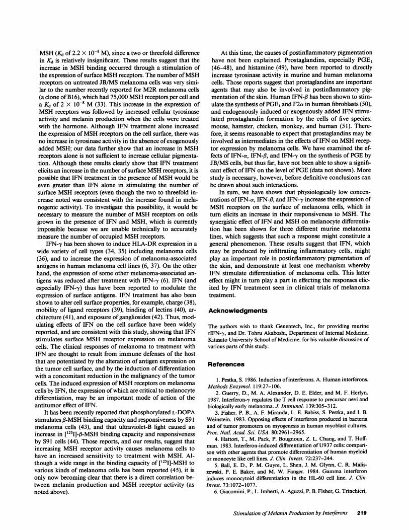
MSH(Kd of 2.2 X 10⁻⁸ M), since a two or threefold differencein Kd is relatively insignificant. These results suggest that theincrease in MSHbinding occurred through a stimulation ofthe expression of surface MSHreceptors. The number of MSHreceptors on untreated JB/MS melanoma cells was very simi-lar to the number recently reported for M2Rmelanoma cells(a clone of B16), which had 75,000 MSHreceptors per cell anda Kd of 2 X 10⁻⁸ M (33). This increase in the expression ofMSHreceptors was followed by increased, cellular tyrosinaseactivity and melanin production when the cells were treatedwith the hormone. Although IFN treatment alone increasedthe expression of MSHreceptors on the cell surface, there wasno increase in tyrosinase activity in the absence of exogenouslyadded MSH; our data further show that an increase in MSHreceptors alone is not sufficient to increase cellular pigmenta-tion. Although these results clearly show that IFN treatmentelicits an increase in the number of surface MSHreceptors, it ispossible that IFN treatment in the presence of MSHwould beeven greater than IFN alone in stimulating the number ofsurface MSHreceptors (even though the two to threefold in-crease noted was consistent with the increase found in mela-nogenic activity). To investigate this possibility, it would benecessary to measure the number of MSHreceptors on cellsgrown in the presence of IFN and MSH, which is currentlyimpossible because we are unable technically to accuratelymeasure the number of occupied MSHreceptors.
IFN-y has been shown to induce HLA-DR expression in awide variety of cell types (34, 35) including melanoma cells(36), and to increase the expression of melanoma-associatedantigens in human melanoma cell lines (6, 37). On the otherhand, the expression of some other melanoma-associated an-tigens was reduced after treatment with IFN-'y (6). IFN (andespecially IFN-'y) thus have been reported to modulate theexpression of surface antigens. IFN treatment has also beenshown to alter cell surface properties, for example, charge (38),mobility of ligand receptors (39), binding of lectins (40), ar-chitecture (41), and exposure of gangliosides (42). Thus, mod-ulating effects of IFN on the cell surface have been widelyreported, and are consistent with this study, showing that IFNstimulates surface MSHreceptor expression on melanomacells. The clinical responses of melanoma to treatment withIFN are thought to result from immune defenses of the hostthat are potentiated by the alteration of antigen expression onthe tumor cell surface, and by the induction of differentiationwith a concomitant reduction in the malignancy of the tumorcells. The induced expression of MSHreceptors on melanomacells by IFN, the expression of which are critical to melanocytedifferentiation, may be an important mode of action of theantitumor effect of IFN.
It has been recently reported that phosphorylated L-DOPAstimulates fl-MSH binding capacity and responsiveness by S9 1melanoma cells (43), and that ultraviolet-B light caused anincrease in ['251J-f3-MSH binding capacity and responsivenessby S91 cells (44). Those reports, and our results, suggest thatincreasing MSHreceptor activity causes melanoma cells tohave an increased sensitivity to treatment with MSH. Al-though a wide range in the binding capacity of ['25I]-MSH tovarious kinds of melanoma cells has been reported (45), it isonly now becoming clear that there is a direct correlation be-tween melanin production and MSHreceptor activity (asnoted above).
At this time, the causes of postinflammatory pigmentationhave not been explained. Prostaglandins, especially PGEI(46-48), and histamine (49), have been reported to directlyincrease tyrosinase activity in murine and human melanomacells. Those reports suggest that prostaglandins are importantagents that may also be involved in postinflammatory pig-mentation of the skin. Human IFN-(3 has been shown to stim-ulate the synthesis of PGEI and F2a in human fibroblasts (50),and endogenously induced or exogenously added IFN stimu-lated prostaglandin formation by the cells of five species:mouse, hamster, chicken, monkey, and human (51). There-fore, it seems reasonable to expect that prostaglandins may beinvolved as intermediates in the effects of IFN on MSHrecep-tor expression by melanoma cells. Wehave examined the ef-fects of IFN-a, IFN-f3, and IFN-'y on the synthesis of PGEbyJB/MS cells, but thus far, have not been able to show a signifi-cant effect of IFN on the level of PGE(data not shown). Morestudy is necessary, however, before definitive conclusions canbe drawn about such interactions.
In sum, we have shown that physiologically low concen-trations of IFN-a, IFN-fl, and IFN--y increase the expression ofMSHreceptors on the surface of melanoma cells, which inturn elicits an increase in their responsiveness to MSH. Thesynergistic effect of IFN and MSHon melanocyte differentia-tion has been shown for three different murine melanomalines, which suggests that such a response might constitute a
general phenomenon. These results suggest that IFN, whichmay be produced by infiltrating inflammatory cells, mightplay an important role in postinflammatory pigmentation ofthe skin, and demonstrate at least one mechanism wherebyIFN stimulate differentiation of melanoma cells. This lattereffect might in turn play a part in effecting the responses elic-ited by IFN treatment seen in clinical trials of melanomatreatment.
Acknowledgments
The authors wish to thank Genentech, Inc., for providing murinerIFN-y, and Dr. Tohru Akaboshi, Department of Internal Medicine,Kitasato University School of Medicine, for his valuable discussion ofvarious parts of this study.
References
1. Pestka, S. 1986. Induction of interferons. A. Humaninterferons.Methods Enzymol. 119:27-106.
2. Guerry, D., M. A. Alexander, D. E. Elder, and M. F. Herlyn.1987. Interferon-y regulates the T cell response to precursor nevi andbiologically early melanoma. J. Immunol. 139:305-312.
3. Fisher, P. B., A. F. Miranda, L. E. Babiss, S. Pestka, and I. B.Weinstein. 1983. Opposing effects of interferon produced in bacteriaand of tumor promoters on myogenesis in human myoblast cultures.Proc. Natl. Acad. Sci. USA. 80:2961-2965.
4. Hattori, T., M. Pack, P. Bougnoux, Z. L. Chang, and T. Hoff-man. 1983. Interferon-induced differentiation of U937 cells: compari-son with other agents that promote differentiation of human myeloidor monocyte like cell lines. J. Clin. Invest. 72:237-244.
5. Ball, E. D., P. M. Guyre, L. Shen, J. M. Glynn, C. R. Malis-zewski, P. E. Baker, and M. W. Fanger. 1984. Gammainterferoninduces monocytoid differentiation in the HL-60 cell line. J. Clin.Invest. 73:1072-1077.
6. Giacomini, P., L. Imberti, A. Aguzzi, P. B. Fisher, G. Trinchieri,
Stimulation of Melanin Production by Interferons 219

and S. Ferrone. 1985. Immunochemical analysis of the modulation ofmelanoma-associated antigens by DNArecombinant immune inter-feron. J. Immunol. 135:2887-2894.
7. Fisher, P. B., D. R. Prignoli, H. Hermo, I. B. Weinstein, and S.Pestka. 1985. Effects of combined treatment with interferon and me-zerein on melanogenesis and growth in human melanoma cells. J.Interferon Res. 5:11-22.
8. Blalock, J. E., and C. Harp. 1981. Interferon and adrencortico-tropic hormone induction of steroidogenesis, melanogenesis and anti-viral activity. Arch. Virol. 67:45-49.
9. Fisher, P. B., and S. Grant. 1985. Effects of interferon on differ-entiation of normal and tumor cells. Pharmacol. Ther. 27:143-166.
10. Fisher, P. B., H. Hermo, D. R. Prignoli, I. B. Weinstein, and S.Pestka. 1984. Hybrid recombinant human leucocyte interferon in-hibits differentiation in murine B- 16 melanoma cells. Biochem.Biophys. Res. Commun. 119:108-115.
11. Fisher, P. B., R. A. Mufson, and I. B. Weinstein. 1981. Inter-feron inhibits melanogenesis in B- 16 mouse melanoma cells. Biochem.Biophys. Res. Commun. 100:823-830.
12. Foon, K. A., S. A. Sherwin, P. G. Abrams, H. C. Stevenson, P.Holmes, A. E. Maluish, R. K. Oldham, and R. B. Herberman. 1985. Aphase I trial of recombinant gammainterferon in patients with cancer.Cancer Immunol. Immunother. 20:193-197.
13. Creagan, E. T., D. L. Ahmann, S. Frytak, H. J. Long, M. N.Chang, and L. M. Itri. 1986. Phase II trials of recombinant leukocyte Ainterferon in disseminated malignant melanoma: results in 96 patients.Cancer Treatment Rep. 70:619-624.
14. McLeod, G. R. C., D. B. Thomson, and P. Hersey. 1987.Recombinant interferon alfa-2a in advanced malignant melanoma. Aphase I-II study in combination with DTIC. Int. J. Cancer.l(Suppl):31-35.
15. Berkelhammer, J., R. W. Oxenhandler, R. R. Hook, Jr., andJ. M. Hennessy. 1982. Development of a new melanoma model inC57BL/6 mice. Cancer Res. 42:3157-3163.
16. Berkelhammer, J., T. N. Luethans, R. R. Hook, Jr., and R. W.Oxenhandler. 1986. Phenotypic instability of mouse melanomas afterpropagation in vivo and in vitro. Cancer Res. 46:2923-2928.
17. Hearing, V. J., G. B. Cannon, W. D. Vieira, M. Jimenez, K.Kameyama, and L. W. Law. 1988. JB/MS murine melanoma: a newmodel for studies on the modulation of differentiation, and of tumori-genic and metastatic potential. Int. J. Cancer. 41:275-282.
18. Jimenez, M., K. Kameyama, Y. Tomita, and V. J. Hearing.1988. Mammalian tyrosinase: biosynthesis, processing and modula-tion by melanocyte stimulating hormone. Proc. Natl. Acad. Sci. USA.85:3830-3834.
19. Gray, P. W., and D. V. Goeddel. 1983. Cloning and expressionof murine immune interferon cDNA. Proc. Nati. Acad. Sci. USA.80:5842-5846.
20. Fuller, B. B., J. B. Lunsford, and D. S. Iman. 1987. a-Melano-cyte-stimulating hormone regulation of tyrosinase in Cloudman S-91mouse melanoma cell cultures. J. Bio. Chem. 262:4024-4033.
21. Halaban, R., and A. B. Lerner. 1977. The dual effect of mela-nocyte-stimulating hormone (MSH) on the growth of cultured mousemelanoma cells. Exp. Cell Res. 108:111-117.
22. Abdel-Malek, Z. A., M. E. Hadley, M. D. Bregman, F. L.Meyskens, and V. J. Hruby. 1986. Actions of melanotropins on mousemelanoma cell growth in vitro. J. Nati. Cancer Inst. 76:857-863.
23. Hearing, V. J., and T. M. Ekel. 1976. Mammalian tyrosinase; acomparison of tyrosine hydroxylation and melanin formation. Bio-chem. J. 157:549-557.
24. Hearing, V. J. 1987. Mammalian monophenol monooxygenase(tyrosinase): purification, properties, and reactions catalyzed. MethodsEnzymol. 142:154-163.
25. Tomita, Y., P. M. Montague, and V. J. Hearing. 1985. Anti-T4-tyrosinase monoclonal antibodies-specific markers for melano-cytes. J. Invest. Dermatol. 85:426-430.
26. Lambert, D. T., and A. B. Lerner. 1983. Optimization of a
melanotropin-receptor binding assay by reversed-phase high-perfor-mance liquid chromatography. J. Chromatogr. 266:567-576.
27. Bidlingmeyer, B. A., S. A. Cohen, and T. L. Tarvin. 1984.Rapid analysis of amino acids using pre-column derivatization. J.Chromatogr. 336:93-104.
28. Lambert, D. T., P. E. Whitcombe, G. E. Moellmann, and A. B.Lerner. 1985. Basic characterization of the receptor for MSHonCloudman S91 melanoma cells. In Pigment Cell 1985. J. Bagnara,S. N. Klaus, E. Paul, and M. Schartl, editors. University of TokyoPress, Tokyo. 165-174.
29. Wong, G., and J. M. Pawelek. 1975. Melanocyte-stimulatinghormone promotes activation of pre-existing tyrosinase molecules inCloudman S91 melanoma cells. Nature (Lond.). 255:644-646.
30. Lambert, D. T., and J. M. Varga. 1981. lodination of melano-tropin: production of biologically active 1251I-a-MSH. In Pigment Cell1981. M. Seiji, editor. University of Tokyo Press, Tokyo. 347-353.
31. Lambert, D. T., C. Stachelek, J. M. Varga, and A. B. Lerner.1982. Iodination of fl-melanotropin. J. Biol. Chem. 257:8211-8215.
32. Heward, C. B., Y. C. S. Yang, T. K. Sawyer, M. D. Bregman,B. B. Fuller, V. J. Hruby, and M. E. Hadley. 1979. lodination asso-ciated inactivation of fl-melanocyte stimulating hormone. Biochem.Biophys. Res. Commun. 88:266-273.
33. Gerst, J. E., J. Sole, J. P. Mather, and Y. Salomon. 1986.Regulation of adenylate cyclase by fl-melanotropin in the M2Rmela-noma cell line. Mol. Cell Endocrinol. 46:137-147.
34. Kelley, V. E., W. Fiers, and T. B. Strom. 1983. Cloned humaninterferon-y, but not interferon-f, or -a, induces expression of HLA-DRdeterminants by fetal monocytes and myeloid leukemic cell lines.J. Immunol. 132:240-245.
35. Kameyama, K., T. Tone, H. Eto, S. Takezaki, T. Kanzaki, andS. Nishiyama. 1987. Recombinant gamma interferon induces HLA-DRexpression on squamous cell carcinoma, trichilemmoma, adeno-carcinoma cell lines, and cultured human keratinocytes. Arch. Der-matol. Res. 279:161-166.
36. Basham, T. Y., and T. C. Merigan. 1983. Recombinant y-in-terferon induces HLA-DR synthesis and expression. J. Immunol.130:1492-1494.
37. Kameyama, K., S. Takezaki, T. Kanzaki, and S. Nishiyama.1986. HLA-DR and melanoma-associated antigen (p97) expressionduring the cell cycle in human melanoma cell lines, and the effects ofrecombinant gamma-interferon: two-color flow cytometric analysis. J.Invest Dermatol. 87:313-318.
38. Knight, E., Jr., and B. D. Korant. 1977. A cell surface alterationin mouse L cells induced by interferon. Biochem. Biophys. Res. Com-mun. 74:707-713.
39. Matsuyama, M. 1979. Action of interferon on cell membraneof mouse lymphocytes: inhibition of ligand-induced redistribution ofsurface receptors. Exp. Cell Res. 124:253-259.
40. Huet, C., I. Gresser, M. T. Mandu, and P. Lindahl. 1974.Increased binding of concanavalin A to interferon-treated murine leu-kemia L12 10 cells. Proc. Soc. Exp. Biol. Med. 147:52-57.
41. Chang, E. H., F. T. Jay, and R. M. Friedman. 1978. Physical,morphological, and biochemical alterations in the membrane of AKRmouse cells after interferon treatment. Proc. Natl. Acad. Sci. USA.75:1859-1863.
42. Grollman, E. F., G. Lee, S. Romas, P. S. Lazo, H. R. Kaback,R. M. Friedman, and L. D. Kohn. 1978. Relationships of the structureand function of the interferon receptor to hormone receptors andestablishment of the antiviral state. Cancer Res. 38:4172-4185.
43. McLane, J., M. Osber, and J. M. Pawelek. 1987. Phosphory-lated isomers of L-DOPA stimulate MSHbinding capacity and respon-siveness to MSHin cultured melanoma cells. Biochem. Biophys. Res.Commun. 145:719-725.
44. Bologna, J., M. Murray, and J. M. Pawelek. 1987. Evidencethat UVB-induced melanogenesis is mediated through MSHreceptors.J. Invest. Dermatol. 88:478A.
45. Libert, A., G. Ghanem, R. Arnould, A. Verammen-Grandjean,
220 K. Kameyama, S. Tanaka, S. Ishida, and V. J. Hearing

and F. Lejeune. 1985. Binding of the a-melanocyte-stimulating hor-mone to human melanoma cells in culture. In Pigment Cell 1985. J.Bagnara, S. N. Klaus, E. Paul, and M. Schartl, editors. University ofTokyo Press, Tokyo. 175-181.
46. Kreiner, P. W., C. J. Gold, J. J. Keirns, W. A. Brock, and M. U.Bitensky. 1973. Hormonal control of melanocytes, MSH-sensitiveadenyl cyclase in the Cloudman melanoma. Yale J. Biol. Med.46:583-591.
47. Fuller, B. B., and J. Lebowitz. 1980. Decay of hormone respon-siveness in mouse melanoma cells in culture as a function of celldensity. J. Cell Physiol. 103:279-287.
48. Abdel-Malek, Z. A., V. B. Swope, N. Amornisiripanitch, andJ. J. Nordlund. 1987. In vitro modulation of proliferation and melani-
zation of S91 melanoma cells by prostaglandins. Cancer Res.47:3141-3146.
49. Bernd, A., P. Altmeyer, G. Schafer, W. Ch. Marsch, and H.Holzmann. 1986. Bleomycin enhances the tyrosinase activity ofhuman malignant melanoma cells in culture. Pharmacol. Res. Com-mun. 18:1075-1091.
50. Fuse, A., I. Mahmud, and T. Kuwata. 1982. Mechanism ofstimulation by human interferon of prostaglandin synthesis in humancell lines. Cancer Res. 42:3209-3214.
51. Fitzpatrick, F. A., and D. A. Stringfellow. 1980. Virus andinterferon effects on cellular prostaglandin biosynthesis. J. Immunol.125:431-437.
Stimulation of Melanin Production by Interferons 221