interaction of human β-defensin 2 (hbd2) with glycosaminoglycans
TRANSCRIPT

pubs.acs.org/Biochemistry Published on Web 11/09/2010 r 2010 American Chemical Society
10486 Biochemistry 2010, 49, 10486–10495
DOI: 10.1021/bi1011749
Interaction of Human β-Defensin 2 (HBD2) with Glycosaminoglycans†
Emily S. Seo,‡ B€arbel S. Blaum,‡ Thomas Vargues,‡ Martin De Cecco,‡ Jon A. Deakin,§ Malcolm Lyon,§
Perdita E. Barran,‡ Dominic J. Campopiano,‡ and Du�san Uhrın*,‡
‡EastChem, School of Chemistry, The University of Edinburgh, King’s Buildings, West Mains Road,Edinburgh EH9 3JJ, U.K., and §Glyco-Oncology Group, School of Cancer and Imaging Sciences,
The University of Manchester, Paterson Institute for Cancer Research, Wilmslow Road, Manchester M20 4BX, U.K.
Received July 24, 2010; Revised Manuscript Received November 2, 2010
ABSTRACT: Human β-defensin 2 (HBD2) is a member of the defensin family of antimicrobial peptides thatplays important roles in the innate and adaptive immune system of both vertebrates and invertebrates. Inaddition to their direct bactericidal action, defensins are also involved in chemotaxis and Toll-like receptoractivation. In analogy to chemokine/glycosaminoglycan (GAG) interactions, GAG-defensin complexes arelikely to play an important role in chemotaxis and in presenting defensins to their receptors. Using a gelmobility shift assay, we found that HBD2 bound to a range of GAGs including heparin/heparan sulfate (HS),dermatan sulfate (DS), and chondroitin sulfate.We usedNMR spectroscopy of 15N-labeledHBD2 tomap thebinding sites for twoGAGmodel compounds, a heparin/HS pentasaccharide (fondaparinux sodium; FX) andenzymatically preparedDS hexasaccharide (DSdp6). We identified a number of basic amino acids that form acommon ligand binding site, which indicated that these interactions are predominantly electrostatic. Thedissociation constant of the [DSdp6-HBD2] complex was determined byNMR spectroscopy to be 5( 5 μM.Binding of FX could not be quantified because of slow exchange on the NMR chemical shift time scale. FXwas found to induce HBD2 dimerization as evidenced by the analysis of diffusion coefficients, 15N relaxation,and nESI-MSmeasurements. The formation of FX-bridged HBD2 dimers exhibited features of a cooperativebinding mechanism. In contrast, the complex with DSdp6 was found to be mostly monomeric.
Defensins are small (2-4 kDa) cationic, cysteine-rich, anti-microbial peptides (AMPs)1 that play a crucial role in host defenseagainst pathogens (1-5). They are an integral part of the innateand adaptive immune system not only in mammals but also inbirds, fish, amphibians, insects, and plants. In humans, they areinvolved in fighting pulmonary inflammation (6), urinary tract (7),and gastrointestinal (8) tract infections, as well as acne (9), irritablebowel syndrome (10), and bacterial bone infection (11).
Defensins are divided into three classes (R, β, and θ) accordingto the spacing between their cysteine residues and topology of theirdisulfide bonds. β-Defensins are characterized by the disulfideconnectivities: Cys1-Cys5, Cys2-Cys4, and Cys3-Cys6. Themost studied peptides of the human β subclass include humanβ-defensins 1, 2, and 3 (HBD1-3). Their structures have been
solved (12-16) by X-ray crystallography and/or NMR spectros-copy andwere all found to be similar with three β-strands ( β1-β3)arranged in an antiparallel fashion and an N-terminal R-helix.Whereas HBD1 (13) and HBD2 (13, 15) were found to be mono-meric in solution, HBD3 was found to be dimeric (13). However,X-ray structures of HBD2 showed evidence of higher orderoligomerization (14).
The structural principle underlying the antimicrobial propertiesof defensins is their amphipathic design with spatially separatedclusters of hydrophobic and polar residues (17). However, somebacteria utilize the propensity of defensins to bind negativelycharged molecules as a defense mechanism. Proteinases secretedby several human pathogens degrade dermatan sulfate- (DS-)containing proteoglycans, thereby releasing the negatively chargedglycosaminoglycan (GAG) chains (18). Addition ofDS to bacteriaincubated with human neutrophil R-defensin-1 blocked thebactericidal activity of this defensin. Binding ofAMPs to freeGAGstherefore represents a possible virulence mechanism (18, 19).Numerous AMPs have been shown to bind heparin andDS (20).A qualitative correlation between the antimicrobial activity ofseveral β-defensins and related peptides and their gas-phasebinding to a heparin-derived disaccharide was observed (21). Inaddition to their antimicrobial action, β-defensins have also beenshown to stimulate the adaptive immune system (22). HBD1-4all display chemotactic properties by recruiting immature dendriticcells, memory T cells, and/or mast cells (23-25).
The structure and function of defensins overlap with those ofchemokines, a superfamily of some 50 (8-12 kDa) proteins,which are involved in leukocyte trafficking and activation (26).This similarity is best demonstrated by a comparison of HBD2
†This work was supported by the Engineering and Physical ScienceResearch Council, U.K. (EPSRC, Platform Grant EP/C541561/1), andthe University of Edinburgh.*Corresponding author. Tel: (44)131 650 7061. Fax: (44)131 650 7155.
E-mail: [email protected]: AMP, antimicrobial peptide; HBD, human β-defensin;
GAG, glycosaminoglycan; HS, heparan sulfate; DS, dermatan sulfate; CS,chondroitin sulfate; MIP-3R, macrophage inflammatory protein 3 alpha;MIP-1R, macrophage inflammatory protein 1 alpha; IL-8, interleukin-8;CCR6, chemokine receptor 6;RANTES, regulateduponactivation, normalT cell expressed and secreted; MCP-1, monocyte chemotactic protein-1;FGF1, acidic fibroblast growth factor; FX, fondaparinux sodium; DSdp6,dermatan sulfate hexasaccharide; CSdp12, chondroitin sulfate dodecasac-charide;GMSA,gelmobility shift assay;AMAC,2-aminoacridone;EDTA,ethylenediaminetetraacetic acid; HSQC, heteronuclear single-quantumcoherence; HISQC, heteronuclear in-phase single-quantum coherence;WATERGATE, water suppression through gradient tailored excitation;CCSD, combined chemical shift difference; DOSY, diffusion-orderedspectroscopy; LED, longitudinal encode-decode; nESI, nanoelectrosprayionization.

Article Biochemistry, Vol. 49, No. 49, 2010 10487
with the chemokineMIP-3R (CCL20). In structural termsHBD2can be considered as a simplified formofCCL20with a truncatedN-terminus and lacking the C-terminal helix (27), yet retainingthe antimicrobial and chemotactic properties of the chemokine;both species activate the CCR6 receptor (24, 28). In addition,a study of 30 chemokines has found 17 to be antimicrobial andattributed their activity to the occurrence of a large, topological,positively charged electrostatic patch on their surfaces (29). Suchstructural features are also responsible for the binding of chemokinesto GAGs. It has been demonstrated that GAG/chemokineinteractions assist in establishing the concentration gradientsrequired for chemotaxis (30). Beside their role in chemotaxis,the binding of chemokines to cell surface GAGs increases thejuxtamembrane concentration of chemokines, increasing thelikelihood of receptor activation (30, 31). In addition, chemokineshave a propensity to form biologically relevant dimers: mono-meric mutants of MCP-1 and RANTES lost the ability to recruitcells in vivo, although their ability to bind heparin and activatetheir receptor was maintained (30). The exact role of dimerformation in receptor activation remains controversial (32) but itwas shown that GAGs stimulate oligomerization of chemokines(31, 33), including formation of heterodimers (34).
The variety of roles that the GAG-chemokine, and presum-ably the GAG-defensin, interactions play in biological systemswarrants a thorough investigation of such complexes, preferablyat atomic resolution. However, because of precipitation and/ordifficulties to crystallize [GAG-chemokine(defensins)] com-plexes, only two X-ray structures of GAG disaccharides boundto chemokines (i.e.,RANTES (35) andCXCL12 (35,36)) havebeenreported to date; there is noX-ray structure of a [GAG-defensin]complex.
For the identification of amino acids involved in GAGbinding,biophysical and biochemical studies of mutant proteins (37) andtitration experiments monitored by NMR spectroscopy (38) arecommonly carried out. The advantage of the latter approach isthat wild-type proteins can be used and that it provides informa-tion about all residues at once. To our knowledge, there are noreports characterizing the GAG-binding sites of defensins.
In our study, we have focused on the interaction ofHBD2, a 41amino acid peptide, with two types of GAGs: heparin/heparinsulfate (HS) and DS. Mature HS contains variably sulfateddomains, which are interrupted by domains essentially lackingsulfation (39); the sulfated domains typically bind proteins (40).Consequently, heparin, which shares an identical carbohydrateskeleton with HS but displays a higher and more even distribu-tion of sulfates, is often used as a model compound in studiesof GAG-protein interactions. In this work we mainly used asynthetic pentasaccharide, fondaparinux sodium (FX, Figure 1a)as a highly sulfated HS mimetic. FX represents structures foundin HS and heparin and is the most important heparin epitope inthe HS-antithrombin III interaction (41). It has eight sulfategroups including one at the 3-O-position of its central glucos-amine. The secondGAG included in our studies isDS,whichwaschosen because of its occurrence in skin, a possible site of injuryand subsequent microbial infection, thus possessing a high prob-ability of interaction with HBD2 (42). Overall, FX and DS differin their constitutive monosaccharides, position of glycosidiclinkages, and the level/position of sulfation and represent a goodstarting point for the investigation of [GAG-HBD2] complexes.When compared to FX, theDS hexasaccharide (DSdp6, Figure 1b)used in this work represents a less sulfatedGAGoligosaccharide,containing only three sulfate groups (uniformly one per constituent
disaccharide), which should affect the strength of the electrostaticinteractions.
In this study we have elucidated the binding sites for HS andDSonHBD2 and examined the oligomeric state of [GAG-HBD2]complexes using a combination of NMR spectroscopy, compu-tational analysis, and mass spectrometry. We have revealedcommon HBD2 residues involved in binding both ligands butalso observed GAG-specific traits that result in differences in thenature of the complexes formed.
MATERIALS AND METHODS
Materials. 15N-Labeled HBD2 was prepared as describedpreviously (25, 43). FX was a gift from GlaxoSmithKline, andheparin, DS, and chondroitin sulfate (CS) were purchased fromSigma Aldrich. Heparin, DS, and CS oligosaccharides were pre-pared by enzymatic digestion of the corresponding GAGs andpurified as described previously (44).GelMobility Shift Assay (GMSA).Gel mobility shift assays
were carried out as described previously (44). Briefly,GAGoligo-saccharides were labeled at their reducing end with fluorescent2-aminoacridone (AMAC). LabeledGAGoligosaccharides (1 μg)were combined with equimolar amounts ofHBD2 and incubatedat room temperature for 30 min in phosphate-buffered salinecontaining 25% (v/v) glycerol. Samples were applied to the wellsof a 1% (w/v) agarose gel in 10 mM Tris-HCl/1 mM EDTA,pH 6.4. Electrophoresis was carried out at 120 V for 10 min with40mMTris-acetate/1mMEDTA, pH8.0, as the electrophoresisbuffer. Immediately after electrophoresis the migration of fluo-rescent oligosaccharides was monitored under UV on a UVItecgel analysis system coupled to a UVIphoto photographic imager(UVItec Ltd., Cambridge, U.K.).NMR Spectroscopy. All NMR spectra were recorded on
BrukerAVANCE instruments equippedwith cryoprobes operatingat 600 or 800 MHz at 25 or 10 �C. For the 2D 1H-15N HSQCexperiment, 8-16 scans were acquired for each increment usinga spectral width of 17 and 14 ppm for F1 (15N) and F2 (1H),respectively.Thedata setswere collectedusing2048and128complexpoints in F2 and F1, respectively. WATERGATE water suppres-sion (45) was used. 15N-Labeled HBD2 (45 μg, 0.010 μmol) wasdissolved in 20 mM deuterated sodium acetate buffer (420 μL)and D2O (22 μL) to give a final concentration of 24 μM and apHof 4.7. The solutionwas transferred to a Shigemi tubewithoutthe top insert. For titration of FX into HBD2, the followingmolar ratios ofGAGtopeptidewere used: 0.1, 0.2, 0.3, 0.4, 0.5, 1,2, 4, 8, and 16 to 1. Titrations of DSdp6 into HBD2 were carried
FIGURE 1: GAG oligosaccharides: (a) fondaparinux sodium, FX,and (b) DS hexasaccharide (DSdp6).
10488 Biochemistry, Vol. 49, No. 49, 2010 Seo et al.
out with the following ratios: 0, 0.1, 0.3, 0.5, 1, 1.5, 2, and 4 to 1.The pH was monitored for every titration step to ensure that itwas maintained. Combined chemical shift differences (CCSD) ofNH resonances were calculated using the equation:
CCSD ¼ffiffiffiffiffiffiffiffiffiffiffiffiffiffiffiffiffiffiffiffiffiffiffiffiffiffiffiffiffiffiffiffiffiffiffiffiffiffiffiffiffiffiffiðΔδHNÞ2 þðΔδN=5Þ2
qð1Þ
where ΔδHN andΔδN are the proton and nitrogen chemical shiftchanges upon ligand binding, respectively. NMR data were pro-cessed using Azara (Wayne Boucher and the Department of Bio-chemistry, University of Cambridge, http://www.ccpn.ac.uk/azara/)and analyzed using CcpNmrAnalysis (46). The assignment of theHBD2 NH cross-peaks originally done at 25 �C (43) was trans-ferred to assign the NH peaks at 10 �C by a series of spectracollected at progressively lower temperatures. The dissociationconstant,Kd, of the [DSdp6-HBD2] complex was determined byfitting the chemical shift changes to the equation (47):
Δδobs ¼ Δδmax=2Po½ðPo þLo þKdÞ- sqrtððPoþLoþKdÞ2 - 4PoLoÞ� ð2Þ
where Δδobs is the observed chemical shift change, Δδmax is themaximumchemical shift change at saturation,Po is the total peptideconcentration, and Lo is the total concentration of ligand added.Heteronuclear in-phase single-quantum coherence (HISQC) experi-ments were carried out as described previously (48). A spectralwidth of 3 ppm in F1 was sampled over 200 ms in the presenceof 2H decoupling. The 15N offset was set to 32.5 ppm. The totalacquisition time per experiment was 45 min.
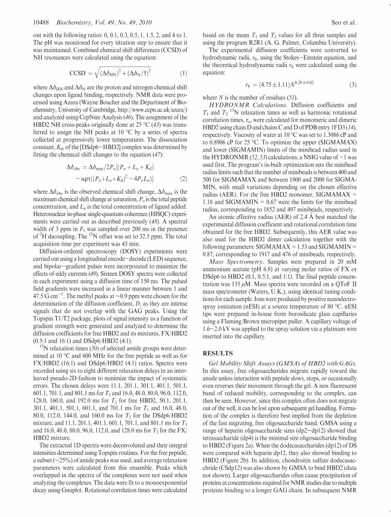
Diffusion-ordered spectroscopy (DOSY) experiments werecarried out using a longitudinal encode-decode (LED) sequence,and bipolar-gradient pulses were incorporated to minimize theeffects of eddy currents (49). Sixteen DOSY spectra were collectedin each experiment using a diffusion time of 150 ms. The pulsedfield gradients were increased in a linear manner between 1 and47.5 G cm-1. The methyl peaks at∼0.9 ppm were chosen for thedetermination of the diffusion coefficient, D, as they are intensesignals that do not overlap with the GAG peaks. Using theTopspin T1/T2 package, plots of signal intensity as a function ofgradient strength were generated and analyzed to determine thediffusion coefficients for free HBD2 and its mixtures, FX/HBD2(0.5:1 and 16:1) and DSdp6/HBD2 (4:1).
15N relaxation times (50) of selected amide groups were deter-mined at 10 �C and 600 MHz for the free peptide as well as forFX/HBD2 (16:1) and DSdp6/HBD2 (4:1) ratios. Spectra wererecorded using six to eight different relaxation delays in an inter-leaved pseudo-2D fashion to minimize the impact of systematicerrors. The chosen delays were 11.1, 201.1, 301.1, 401.1, 501.1,601.1, 701.1, and 801.1 ms forT1 and 16.0, 48.0, 80.0, 96.0, 112.0,128.0, 160.0, and 192.0 ms for T2 for free HBD2, 50.1, 201.1,301.1, 401.1, 501.1, 601.1, and 701.1 ms for T1 and 16.0, 48.0,80.0, 112.0, 144.0, and 160.0 ms for T2 for the DSdp6/HBD2mixture, and 11.1, 201.1, 401.1, 601.1, 701.1, and 801.1 ms for T1
and 16.0, 48.0, 80.0, 96.0, 112.0, and 128.0 ms for T2 for the FX/HBD2 mixture.
The extracted 1D spectra were deconvoluted and their integralintensities determined using Topspin routines. For the free peptide,a subset (∼25%) of amide peakswas used, and average relaxationparameters were calculated from this ensemble. Peaks whichoverlapped in the spectra of the complexes were not used whenanalyzing the complexes. The data were fit to a monoexponentialdecay using Gnuplot. Rotational correlation times were calculated
based on the mean T1 and T2 values for all three samples andusing the program R2R1 (A. G. Palmer, Columbia University).
The experimental diffusion coefficients were converted tohydrodynamic radii, rh, using the Stokes-Einstein equation, andthe theoretical hydrodynamic radii rh were calculated using theequation:
rh ¼ ð4:75( 1:11ÞN0:29( 0:02 ð3Þwhere N is the number of residues (51).HYDRONMR Calculations. Diffusion coefficients and
T1 and T215N relaxation times as well as harmonic rotational
correlation times, τc, were calculated for monomeric and dimericHBD2using chainDand chainsCandDofPDBentry 1FD3 (14),respectively. Viscosity of water at 10 �C was set to 1.3086 cP andto 0.8906 cP for 25 �C. To optimize the upper (SIGMAMAX)and lower (SIGMAMIN) limits of the minibead radius used intheHYDRONMR (52, 53) calculations, aNSIG value of-1 wasused first. The program’s in-built optimization sets the minibeadradius limits such that the number ofminibeads is between 400 and500 for SIGMAMAX and between 1800 and 2000 for SIGMA-MIN, with small variations depending on the chosen effectiveradius (AER). For the free HBD2 monomer, SIGMAMAX=1.18 and SIGMAMIN= 0.67 were the limits for the minibeadradius, corresponding to 1852 and 497 minibeads, respectively.
An atomic effective radius (AER) of 2.4 A best matched theexperimental diffusion coefficient and rotational correlation timeobtained for the free HBD2. Subsequently, this AER value wasalso used for the HBD2 dimer calculation together with thefollowing parameters: SIGMAMAX=1.53 and SIGMAMIN=0.87, corresponding to 1917 and 476 of minibeads, respectively.Mass Spectrometry. Samples were prepared in 20 mM
ammonium acetate (pH 6.8) at varying molar ratios of FX orDSdp6 to HBD2 (0:1, 0.5:1, and 1:1). The final peptide concen-tration was 135 μM. Mass spectra were recorded on a QToF IImass spectrometer (Waters, U.K.), using identical tuning condi-tions for each sample. Ionswere produced by positive nanoelectro-spray ionization (nESI) at a source temperature of 80 �C. nESItips were prepared in-house from borosilicate glass capillariesusing a Flaming/Brown micropipet puller. A capillary voltage of1.6-2.0 kV was applied to the spray solution via a platinum wireinserted into the capillary.
RESULTS
Gel Mobility Shift Assays (GMSA) of HBD2 with GAGs.In this assay, free oligosaccharides migrate rapidly toward theanode unless interaction with peptide slows, stops, or occasionallyeven reverses their movement through the gel. A new fluorescentband of reduced mobility, corresponding to the complex, canthen be seen. However, since this complex often does not migrateout of the well, it can be lost upon subsequent gel handling. Forma-tion of the complex is therefore best implied from the depletionof the fast migrating, free oligosaccharide band. GMSA using arange of heparin oligosaccharide sizes (dp2-dp12) showed thattetrasaccharide (dp4) is the minimal size oligosaccharide bindingto HBD2 (Figure 2a). When the dodecasaccharides (dp12) of DSwere compared with heparin dp12, they also showed binding toHBD2 (Figure 2b). In addition, chondroitin sulfate dodecasac-chride (CSdp12) was also shown by GMSA to bind HBD2 (datanot shown). Larger oligosaccharides often cause precipitation ofproteins at concentrations required forNMRstudies due tomultipleproteins binding to a longer GAG chain. In subsequent NMR

Article Biochemistry, Vol. 49, No. 49, 2010 10489
andMS interaction studies the smaller ligands FX pentasaccharideand DSdp6 hexasaccharide were therefore used.NMR-Monitored Titration of FX into 15N-LabeledHBD2.
The 1H-15N HSQC spectrum of free 15N-HBD2 assigned pre-viously (43) was used tomonitor chemical shift changes of the pep-tide amide resonances upon binding to FX (Figure 3). Preliminarytitration experiments using 800 and 600MHzNMR spectrometersand varying temperature showed signs of chemical exchangebroadening, as peaks sharpened at lower temperatures and alower magnetic field strength. The optimal experimental condi-tions for following the largest number of resonances during thetitrations were at a field strength of 600 MHz and a temperatureof 10 �C. All subsequent experiments were therefore performedusing these optimized conditions.
Even at these optimal conditions addition of only a 0.1 molequiv of FX had a dramatic effect on the appearance of the HBD21H-15NHSQC spectrum,with themajority of signals disappearingor weakening substantially (Supporting Information Figure S1).A subset of signals could be followed during subsequent titration
points; up to the addition of 0.5mol equiv ofFX, their behaviorwascharacteristic of a slow tomedium exchange on the NMR chemicalshift time scale. Chemical shift changes were accompanied by peakbroadening in the 1H NMR spectra of the complex (SupportingInformation Figure S2). This trend was reversed at FX/HBD2molar ratios >0.5:1, when the observable NH cross-peaks startedto move backward toward their original positions (circled peaks inFigure 3). Previously missing cross-peaks started to reappear, someat new positions, and at 4:1 FX/HBD2, all of the HBD2 NHresonances were again observed in the 1H-15N HSQC spectrum(Supporting InformationFigureS1).Very small changes in thepeakpositions between 4:1 and 8:1 molar ratios were detected, andpractically no changes were registered between the 8:1 and 16:1 FX/HBD2 titration points, indicating saturation of the binding site. Atthese high ligand concentrations, signals in the 1H NMR spectrumsharpened, although they never reached the line widths of the freeHBD2.
The changes observed in the 1H-15NHSQC spectrumofHBD2during the titration with FX (Figure 3) point to the existence of atleast two binding events. The CCSD were therefore calculated(eq 1) for two titration points: 0.5:1 and 4:1 ofFX/HBD2 (Figure 4).The interpretation of the first event is complicated by the fact thatonly a subset of NH resonances was observed at this point and willtherefore be addressed in the Discussion. Nevertheless, it becameclear that CCSDs of the observable NH cross-peaks up to 0.5:1FX/HBD2molar ratio did not directly correlate to the FX bindingsite. The binding site was identified by analyzing the 4:1 FX/HBD2spectrum, which showed aCCSDof>0.2 ppm forNH resonancesof five consecutive residues, 22RRYKQ26, and also of residues K39and K40. The former sequence matches the BBXB GAG-bindingmotif (B-basic residue) that has been identified in chemokines (37).The two C-terminal lysines, although distant in sequence, are closeto the GAG-binding loop as highlighted in Figure 5 using theHBD2monomer (14). NH signals of another two pairs of residues,F19-C20 and I14-C15, also highlighted in Figure 5, showedCCSD> 0.1 ppm. The former pair directly precedes the BBXBbinding site, whereas the latter one is part of the dimer interfaceseen in the crystal structure of HBD2 (14).NMR-Monitored Titration of DSdp6 into 15N-Labeled
HBD2. The titration experiments were repeated using DSdp6.
FIGURE 2: Gel mobility shift assays of GAG oligosaccharides andHBD2ona1%agarose gel (1μgofGAGandequimolar concentrationof HBD2). Minus (-) and plus (þ) signs indicate absence and pres-ence of HBD2, respectively. (a) Size dependency of the bindingof AMAC-labeled heparin oligosaccharides (disaccharide, dp2, tododecasaccharide, dp12). (b) Binding of heparin and DS dodeca-saccharides.
FIGURE 3: 600MHz2D1H-15NHSQCspectrumof freeHBD2super-imposed with spectra from FX titrations at 10 �C. FX/HBD2 molarratios of 0 (black), 0.1 (red), 0.2 (beige), 0.3 (yellow), 0.4 (green),0.5 (blue), 1 (violet), 2 (pink), and 4 (turquoise):1. Circles highlight thecross-peaks that change thedirectionofmovement after 0.5:1FX/HBD2ratio.
FIGURE 4: Histogram of combined chemical shift differences (CCSD)between free and bound HBD2 calculated according to eq 1 forFX-HBD2 (filled rectangles) andDSdp6-HBD2 (open rectangles)using 4:1 GAG:HBD2 ratios. Dashed lines indicate the cutoff forresidues highlighted in Figure 5. Secondary structure elements areindicated. The inset shows CCSD obtained at the 0.5:1 FX:HBD2titration point.

10490 Biochemistry, Vol. 49, No. 49, 2010 Seo et al.
For this ligand, we observed fast binding on the NMR time scalethroughout the titration (Figure 6). Continuous chemical shiftchanges occurred with increasing ligand concentrations, and allNH cross-peaks were observable for every titration point. Therewas no evidence for two distinct binding events as seen in the FXtitration (compare the signals circled in the spectra of Figures 3and 6), and 1H NMR spectra acquired during the DSdp6 titration(Supporting Information Figure S3) showed smaller line broad-ening compared to the spectra of free HBD2. CCSD calculated atthe 4:1 DS/HBD2 ratio (Figure 4) again showed major chemicalshift changes for theNH signals of residues 22RRYKQ26, K39, andK40, although the observed changeswere smaller than those seen inthe FX titration. Saturation of the binding site was practicallyreached at a 4:1DS/HBD2 ratio, at which almost no further chemi-cal shift change was observed for K40, the residue which experi-enced the largest chemical shift changes in both titrations.Assuming1:1 binding the dissociation constant, Kd, for the [DSdp6-HBD2]complex was determined using eq 2. TheKd values ranged from 1.3to 17.9 μMfor the different residues in theHBD2 sequence, with anaverage of 5 ( 5 μM (Supporting Information Table S1), corre-sponding to 92%occupancyof the binding site at 4:1DSdp6/HBD2ratio. Because of the multiple binding events and slow exchangeobserved in the FX titration, it was not possible to determine thedissociation constant for its interaction with HBD2.HISQC Spectra of FX and DS Complexes of HBD2.
Heteronuclear in-phase single-quantum coherence (HISQC) (48)experiments, optimized for the detection of NH3
þ side chainresonances of lysine residues, were collected for free HBD2 and
HBD2 in complex with FX (1:16) or DSdp6 (1:4) (Figure 7.) Thespectrum of free HBD2 contains three distinct signals and twopeaks significantly broadened by water exchange, adding up tothe five lysine side chains in theHBD2 sequence. On the contrary,five and four lysine side chains were prominent in the HISQCspectra of the [FX-HBD2] and [DSdp6-HBD2] complexes,respectively, indicating increased protection of exchangeableNH3
þ protons upon ligand binding. The individual lysine sidechain resonances cannot be assigned without a 13C,15N-labeledsample of HBD2. Nevertheless, from the chemical shift changesobserved in theHISQC spectra it is evident that (i) the side chainsof some lysine residues are involved in complex formation withGAGs and (ii) these amine groups are affected by binding of FXand DSdp6 in a similar manner.Rotational Correlation Times of HBD2 and Its Complexes
with GAGs. The differences between the response of HBD2 toFX and DSdp6, as evidenced by the 1H-15N HSQC spectra,could possibly be explained by different oligomerization states ofthe peptide induced by the presence of the two different GAGs.We have therefore conducted further NMR-based investigationsto obtain diffusion coefficients and rotational correlation times ofthe complexes.
Rotational correlation times reflect the size of peptides andproteins and can be obtained via analysis of 15N relaxation data (54).Because of the low concentration ofHBD2 (24 μM), the 15NT1 andT2 relaxation timeswere determined from1Drelaxation experiments
FIGURE 5: Delineation of the FX binding site in HBD2 on a surface map of the crystal structure of monomer HBD2 (PDB entry 1FD3).The following color coding is used: Residues that show CCSD>0.2 ppm for the 1:4 FX/HBD2 (Figure 4) are highlighted in blue. Residues withCCSD>0.1 ppmare shown in turquoise (extendedGAGbinding site) or red (β1 strand; peptide-peptide interface). ResidueswithCCSD>0.1 ppmin the 0.5:1 FX/HBD2 mixture are highlighted in orange. Proline 41 is shown in marine blue.
FIGURE 6: 600 MHz 2D 1H-15N HSQC spectrum of HBD2 super-imposed with DSdp6 titrations at 10 �C. DSdp6/HBD2 molar ratiosof 0 (black), 0.1 (red), 0.3 (lightbrown), 0.5 (yellow), 1 (green), 1.5 (blue),2 (violet), and 4 (pink) to 1. Circles highlight those cross-peaks thatchange the direction of movement in the FX titration but are notaffected in the DS titration.
FIGURE 7: HISQC spectra of (a) free HBD2, (b) FX/HBD2 (16:1),and (c) HBD2/DSdp6 (4:1). The NH3
þ lysine side chain peaks arecircled in the spectra to distinguish them from NH2D
þ signals thatare marked by an asterisk. The identity of the latter signals, whichshow a constant deuterium isotope shift indicated for the top signalby a full arrow in (b), was confirmed by acquiring spectra withoutdeuterium decoupling. The dashed lines emphasize similarities inchemical shifts between the two complexes.

Article Biochemistry, Vol. 49, No. 49, 2010 10491
using a minimum set of nine resolved NH 1H resonances of the freeHBD2 and GAG/HBD2 mixtures (FX/HBD2, 16:1; DSdp6/HBD2, 4:1). Residue-specific relaxation times of the free peptidecan be found in Supporting Information Figure S4. Small standardvariations of the average T1 and T2 values, σT1,2
(Table 1), suggestthatHBD2 is a compactly foldedpeptide,which is in agreementwiththe published X-ray and NMR structures (14-16). Residues C37,C38, and K39, located at the C-terminus of HBD2, did not exhibitelevatedmobility despite the high crystallographicB factors reportedpreviously for the C-terminus (14). The relaxation times reported inTable 1 represent meanT1 andT2 values. The rotational correlationtime, τc, of free HBD2 at 10 �C calculated based on the T1/T2 ratiowas 4.0 ( 0.2 ns. For comparison, ubiquitin, a protein which hasalmost twice asmany residues asHBD2,has τc=4.6ns at 25 �C(55).Ubiquitin, a monomeric globular protein, has a diameter of∼30 A,while HBD2 is a disk-shaped protein with dimensions 28 � 26 �15 A. Given the temperature difference between the two measure-ments and similarities in the largest dimension between the twoproteins, we can conclude HBD2 is a monomeric protein.
The trends in the experimental values of 15N relaxation times(i.e., increasing T1 and decreasing T2 relaxation times) suggestthat the molecular size is increasing in the order of HBD2 <[DSdp6-HBD2] complex<[FX-HBD2] complex. These trendsare reflected in the calculated rotational correlation times. Forboth complexes, the GAG concentrations used for the relaxationmeasurements were sufficient to saturate the GAG-binding site.Nevertheless, these relaxation times may represent weightedaverages of different species where such exist and, additionally,may be biased by contributions from chemical exchange to theapparent relaxation times.We have therefore further investigatedthe GAG-induced oligomerization of HBD2 by measuring diffu-sion coefficients using diffusion-ordered spectroscopy (DOSY).Diffusion Coefficients of HBD2 and Its Complexes with
GAGs.Measuring the diffusion coefficients viaDOSY is a usefultool in determining the oligomeric state of proteins and peptidesat low concentrations (56, 57). Oligomeric properties of HBD2and HBD3 have been previously investigated by DOSY experi-ments at 25 �C (13). Our DOSY experiments on HBD2 andHBD2 in complexwithFX (1:16 and 0.5:1) andDSdp6 (1:4) wereperformed at 10 �C, in line with the optimized conditions used forthe titration experiments, but were repeated at 25 �C for directcomparison with the literature data. The obtained diffusioncoefficients are reported in Table 2. The experimental diffusioncoefficients decreased steadily from HBD2 through its complexwith DSdp6 and then with FX, at both temperatures, confirmingthe trend revealed by the rotational correlation times. Diffusioncoefficients obtained for the 0.5:1 FX/HBD2 ratio were practi-cally identical to those obtained at the excess of the FX.Theoretical Hydrodynamic Parameters ofHBD2 and Its
Complexes withGAGs.The experimental diffusion coefficientsof HBD2 and its complexes determined at 25 �C were, in the
first instance, used to calculate the hydrodynamic radii using theStokes-Einstein equation. The values obtained were 13.1, 14.7,and 15.9 A for free HBD2, DSdp6/HBD2 (4:1), and FX/HBD2(16:1), respectively. The theoretical values calculated using eq 3for theHBD2monomer and dimer are 13.9 and 17.0 A. For com-parison, literature values (13) for HBD2 monomer and HBD3dimer were 14.7 and 18.3 A, respectively. Our values suggest thatHBD2 is monomeric and the [DSdp6-HBD2] complex existsmostly asmonomerwhile the [FX-HBD2] complex existsmostlyas a dimer.
To interpret our data more rigorously, we calculated theoreticalcorrelation times and diffusion coefficients using the programHYDRONMR (52, 53). Monomeric and dimeric X-ray struc-tures ofHBD2 (14) were used to approximate the [GAG-HBD2]complexes. The absolute values of τc and D calculated byHYDRONMR depend on the chosen effective radius (AER),which reflects the size of the hydration layer. The optimal AERvalue has been shown to vary for different proteins (52). Theexperimental diffusion coefficient and correlation time obtainedfor free monomeric HBD2 were therefore used to optimize theAER value for our calculations. Values between 2.3 and 2.5 Ayielded the best match between the experimental and calculatedparameters for the HBD2 monomer (Table 2). A value of 2.4 Awas used to calculate τc and D for the HBD2 dimer (14), as anapproximation to a possible dimer in the presence of FX orDSdp6 (Table 2). The experimental and theoretical data arepresented in Figure 8 in the form of a plot of τc vsD. Our resultssuggests that both [GAG-HBD2] complexes havebiophysical char-acteristics of species larger than free HBD2, but with [FX-HBD2]being closer to the HBD2 dimer and [DSdp6-HBD2] beingcloser to the HBD2 monomer.nESI Mass Spectra of HBD2 and Its Complexes with
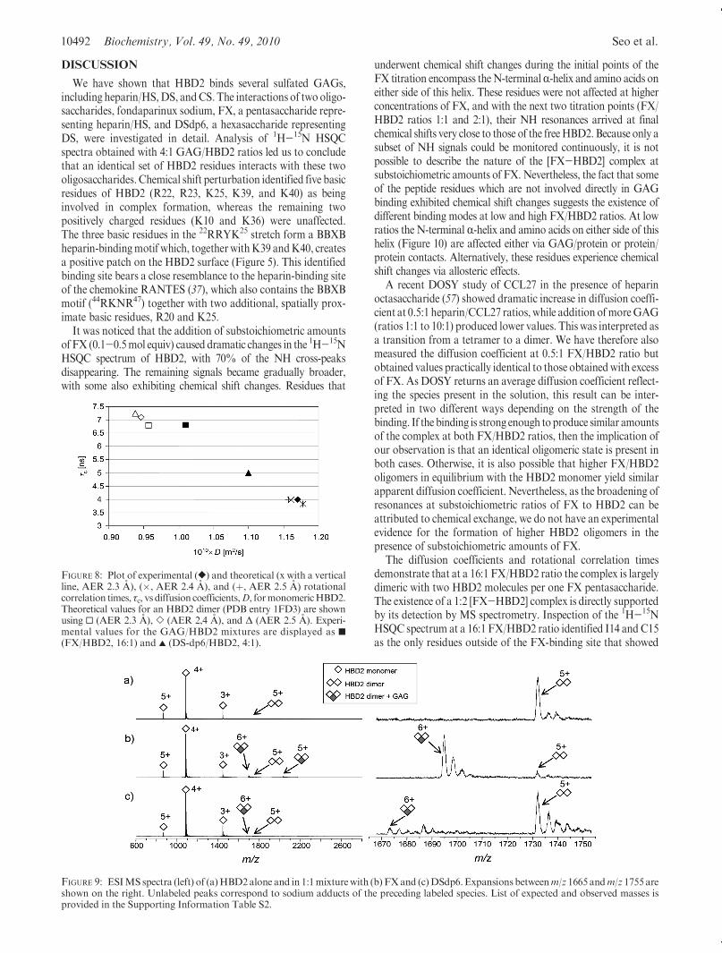
GAGs. Mass spectrometry was employed to further investigatethe oligomeric states of free HBD2 and its complexes with FXand DSdp6. Under our MS experimental conditions the HBD2monomer in the 4þ charge statewas seen as themost intense peakin the MS spectra (Figure 9). A very small amount of dimericHBD2 in the 5þ state was also observed. Addition of FX in a 1:1molar ratio resulted in the appearance of a small amount of a 1:2[FX-HBD2] complex as manifested by the 6þ and 5þ specieswith corresponding m/z ratios. No monomeric 1:1 [FX-HBD2]complex was observed. For DSdp6, a very small peak corre-sponding to a 1:2 [DSdp6-HBD2] complex was observed,and again, no 1:1 [DSdp6-HBD2] complex was detected. Whensprayed from a solution with a ratio of FX/HBD2 of 0.5:1, nohigher order oligomers were detected, though an increasedamount of the 1:2 [FX-HBD2] complex was observed (datanot shown).
Table 1: 15NNMRRelaxationData for HBD2 andGAG/HBD2Mixtures
at 10 �C
sample (no. of cross-peaks used) T1;a σT1
b (ms) T2;a σT2
b(ms) τcorrc (ns)
free HBD2 (12) 428 ( 15; 10 163 ( 6; 18 4.0( 0.2
DSdp6/HBD2, 4:1 (10) 466 ( 20; 27 131 ( 5; 8 5.0( 0.2
FX/HBD2, 16:1 (9) 560 ( 21; 32 103 ( 4; 11 6.8( 0.2
aGiven with fitting errors. bStandard variations of the average values.cCalculated rotational correlation times.
Table 2: Experimental and Theoretical Diffusion Coefficients and Rotational
Correlation Times of HBD2 and [GAG-HBD2] Complexesa
sample
1010Dexp
at 10 �C(at 25 �C) (m2/s)
1010Dcalc
at 10 �C(at 25 �C) (m2/s)
τc (exptl)at 10 �C(ns)
τc (calcd)at 10 �C(ns)
HBD2 monomer 1.17 (1.87) 1.16 (1.80) 4.0( 0.2 4.0
HBD2 dimer 0.95 (1.46) 6.8
DSdp6/HBD2 (4:1) 1.10 (1.67) 5.0( 0.2
FX/HBD2 (16:1) 1.01 (1.54) 6.8( 0.2
FX/HBD2 (0.5:1) 0.99 (1.51)
aCalculated values were determined using HYDRONMR and mono-meric or dimeric X-ray structures of HBD2 (14).
10492 Biochemistry, Vol. 49, No. 49, 2010 Seo et al.
DISCUSSION
We have shown that HBD2 binds several sulfated GAGs,including heparin/HS,DS, andCS. The interactions of two oligo-saccharides, fondaparinux sodium, FX, a pentasaccharide repre-senting heparin/HS, and DSdp6, a hexasaccharide representingDS, were investigated in detail. Analysis of 1H-15N HSQCspectra obtained with 4:1 GAG/HBD2 ratios led us to concludethat an identical set of HBD2 residues interacts with these twooligosaccharides. Chemical shift perturbation identified five basicresidues of HBD2 (R22, R23, K25, K39, and K40) as beinginvolved in complex formation, whereas the remaining twopositively charged residues (K10 and K36) were unaffected.The three basic residues in the 22RRYK25 stretch form a BBXBheparin-bindingmotif which, togetherwithK39 andK40, createsa positive patch on the HBD2 surface (Figure 5). This identifiedbinding site bears a close resemblance to the heparin-binding siteof the chemokine RANTES (37), which also contains the BBXBmotif (44RKNR47) together with two additional, spatially prox-imate basic residues, R20 and K25.
It was noticed that the addition of substoichiometric amountsof FX (0.1-0.5mol equiv) caused dramatic changes in the 1H-15NHSQC spectrum of HBD2, with 70% of the NH cross-peaksdisappearing. The remaining signals became gradually broader,with some also exhibiting chemical shift changes. Residues that
underwent chemical shift changes during the initial points of theFX titration encompass theN-terminalR-helix and amino acids oneither side of this helix. These residues were not affected at higherconcentrations of FX, and with the next two titration points (FX/HBD2 ratios 1:1 and 2:1), their NH resonances arrived at finalchemical shifts very close to those of the freeHBD2.Because only asubset of NH signals could be monitored continuously, it is notpossible to describe the nature of the [FX-HBD2] complex atsubstoichiometric amounts of FX. Nevertheless, the fact that someof the peptide residues which are not involved directly in GAGbinding exhibited chemical shift changes suggests the existence ofdifferent binding modes at low and high FX/HBD2 ratios. At lowratios the N-terminal R-helix and amino acids on either side of thishelix (Figure 10) are affected either via GAG/protein or protein/protein contacts. Alternatively, these residues experience chemicalshift changes via allosteric effects.
A recent DOSY study of CCL27 in the presence of heparinoctasaccharide (57) showed dramatic increase in diffusion coeffi-cient at 0.5:1 heparin/CCL27 ratios, while addition ofmoreGAG(ratios 1:1 to 10:1) produced lower values. This was interpreted asa transition from a tetramer to a dimer. We have therefore alsomeasured the diffusion coefficient at 0.5:1 FX/HBD2 ratio butobtained values practically identical to those obtainedwith excessof FX. As DOSY returns an average diffusion coefficient reflect-ing the species present in the solution, this result can be inter-preted in two different ways depending on the strength of thebinding. If the binding is strong enough toproduce similar amountsof the complex at both FX/HBD2 ratios, then the implication ofour observation is that an identical oligomeric state is present inboth cases. Otherwise, it is also possible that higher FX/HBD2oligomers in equilibrium with the HBD2 monomer yield similarapparent diffusion coefficient. Nevertheless, as the broadening ofresonances at substoichiometric ratios of FX to HBD2 can beattributed to chemical exchange, we do not have an experimentalevidence for the formation of higher HBD2 oligomers in thepresence of substoichiometric amounts of FX.
The diffusion coefficients and rotational correlation timesdemonstrate that at a 16:1 FX/HBD2 ratio the complex is largelydimeric with two HBD2 molecules per one FX pentasaccharide.The existence of a 1:2 [FX-HBD2] complex is directly supportedby its detection by MS spectrometry. Inspection of the 1H-15NHSQC spectrum at a 16:1 FX/HBD2 ratio identified I14 and C15as the only residues outside of the FX-binding site that showed
FIGURE 8: Plot of experimental ([) and theoretical (x with a verticalline, AER 2.3 A), (�, AER 2.4 A), and (þ, AER 2.5 A) rotationalcorrelation times, τc, vs diffusion coefficients,D, formonomericHBD2.Theoretical values for an HBD2 dimer (PDB entry 1FD3) are shownusing 0 (AER 2.3 A), ] (AER 2,4 A), and Δ (AER 2.5 A). Experi-mental values for the GAG/HBD2 mixtures are displayed as 9(FX/HBD2, 16:1) and 2 (DS-dp6/HBD2, 4:1).
FIGURE 9: ESIMS spectra (left) of (a)HBD2alone and in 1:1mixturewith (b) FXand (c)DSdp6.Expansions betweenm/z 1665 andm/z 1755 areshown on the right. Unlabeled peaks correspond to sodium adducts of the preceding labeled species. List of expected and observed masses isprovided in the Supporting Information Table S2.
Article Biochemistry, Vol. 49, No. 49, 2010 10493
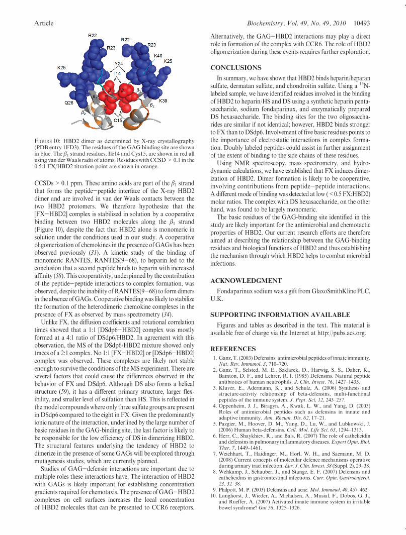
CCSDs>0.1 ppm. These amino acids are part of the β1 strandthat forms the peptide-peptide interface of the X-ray HBD2dimer and are involved in van der Waals contacts between thetwo HBD2 protomers. We therefore hypothesize that the[FX-HBD2] complex is stabilized in solution by a cooperativebinding between two HBD2 molecules along the β1 strand(Figure 10), despite the fact that HBD2 alone is monomeric insolution under the conditions used in our study. A cooperativeoligomerization of chemokines in the presence ofGAGs has beenobserved previously (31). A kinetic study of the binding ofmonomeric RANTES, RANTES(9-68), to heparin led to theconclusion that a second peptide binds to heparin with increasedaffinity (58). This cooperativity, underpinned by the contributionof the peptide-peptide interactions to complex formation, wasobserved, despite the inability ofRANTES(9-68) to formdimersin the absence ofGAGs. Cooperative bindingwas likely to stabilizethe formation of the heterodimeric chemokine complexes in thepresence of FX as observed by mass spectrometry (34).
Unlike FX, the diffusion coefficients and rotational correlationtimes showed that a 1:1 [DSdp6-HBD2] complex was mostlyformed at a 4:1 ratio of DSdp6/HBD2. In agreement with thisobservation, the MS of the DSdp6/HBD2 mixture showed onlytraces of a 2:1 complex. No 1:1 [FX-HBD2] or [DSdp6-HBD2]complex was observed. These complexes are likely not stableenough to survive the conditions of theMS experiment. There areseveral factors that could cause the differences observed in thebehavior of FX and DSdp6. Although DS also forms a helicalstructure (59), it has a different primary structure, larger flex-ibility, and smaller level of sulfation than HS. This is reflected inthemodel compoundswhere only three sulfate groups are presentin DSdp6 compared to the eight in FX. Given the predominantlyionic nature of the interaction, underlined by the large number ofbasic residues in the GAG-binding site, the last factor is likely tobe responsible for the low efficiency of DS in dimerizing HBD2.The structural features underlying the tendency of HBD2 todimerize in the presence of some GAGs will be explored throughmutagenesis studies, which are currently planned.
Studies of GAG-defensin interactions are important due tomultiple roles these interactions have. The interaction of HBD2with GAGs is likely important for establishing concentrationgradients required for chemotaxis. The presence ofGAG-HBD2complexes on cell surfaces increases the local concentrationof HBD2 molecules that can be presented to CCR6 receptors.
Alternatively, the GAG-HBD2 interactions may play a directrole in formation of the complex with CCR6. The role of HBD2oligomerization during these events requires further exploration.
CONCLUSIONS
In summary, we have shown that HBD2 binds heparin/heparansulfate, dermatan sulfate, and chondroitin sulfate. Using a 15N-labeled sample, we have identified residues involved in the bindingof HBD2 to heparin/HS andDS using a synthetic heparin penta-saccharide, sodium fondaparinux, and enzymatically preparedDS hexasaccharide. The binding sites for the two oligosaccha-rides are similar if not identical; however, HBD2 binds strongertoFX than toDSdp6. Involvement of five basic residues points tothe importance of electrostatic interactions in complex forma-tion. Doubly labeled peptides could assist in further assignmentof the extent of binding to the side chains of these residues.
Using NMR spectroscopy, mass spectrometry, and hydro-dynamic calculations, we have established that FX induces dimer-ization of HBD2. Dimer formation is likely to be cooperative,involving contributions from peptide-peptide interactions.A different mode of binding was detected at low (<0.5 FX:HBD2)molar ratios. The complex with DS hexasaccharide, on the otherhand, was found to be largely monomeric.
The basic residues of the GAG-binding site identified in thisstudy are likely important for the antimicrobial and chemotacticproperties of HBD2. Our current research efforts are thereforeaimed at describing the relationship between the GAG-bindingresidues and biological functions of HBD2 and thus establishingthe mechanism through which HBD2 helps to combat microbialinfections.
ACKNOWLEDGMENT
Fondaparinux sodiumwas a gift fromGlaxoSmithKline PLC,U.K.
SUPPORTING INFORMATION AVAILABLE
Figures and tables as described in the text. This material isavailable free of charge via the Internet at http://pubs.acs.org.
REFERENCES
1. Ganz, T. (2003)Defensins: antimicrobial peptides of innate immunity.Nat. Rev. Immunol. 3, 710–720.
2. Ganz, T., Selsted, M. E., Szklarek, D., Harwig, S. S., Daher, K.,Bainton, D. F., and Lehrer, R. I. (1985) Defensins. Natural peptideantibiotics of human neutrophils. J. Clin. Invest. 76, 1427–1435.
3. Kluver, E., Adermann, K., and Schulz, A. (2006) Synthesis andstructure-activity relationship of beta-defensins, multi-functionalpeptides of the immune system. J. Pept. Sci. 12, 243–257.
4. Oppenheim, J. J., Biragyn, A., Kwak, L. W., and Yang, D. (2003)Roles of antimicrobial peptides such as defensins in innate andadaptive immunity. Ann. Rheum. Dis. 62, 17–21.
5. Pazgier, M., Hoover, D. M., Yang, D., Lu, W., and Lubkowski, J.(2006) Human beta-defensins. Cell. Mol. Life Sci. 63, 1294–1313.
6. Herr, C., Shaykhiev, R., and Bals, R. (2007) The role of cathelicidinand defensins in pulmonary inflammatory diseases.Expert Opin. Biol.Ther. 7, 1449–1461.
7. Weichhart, T., Haidinger, M., Horl, W. H., and Saemann, M. D.(2008) Current concepts of molecular defence mechanisms operativeduring urinary tract infection.Eur. J. Clin. Invest. 38 (Suppl. 2), 29–38.
8. Wehkamp, J., Schauber, J., and Stange, E. F. (2007) Defensins andcathelicidins in gastrointestinal infections. Curr. Opin. Gastroenterol.23, 32–38.
9. Philpott, M. P. (2003) Defensins and acne. Mol. Immunol. 40, 457–462.10. Langhorst, J., Wieder, A., Michalsen, A., Musial, F., Dobos, G. J.,
and Rueffer, A. (2007) Activated innate immune system in irritablebowel syndrome? Gut 56, 1325–1326.
FIGURE 10: HBD2 dimer as determined by X-ray crystallography(PDB entry 1FD3). The residues of the GAG binding site are shownin blue. The β1 strand residues, Ile14 and Cys15, are shown in red allusing van derWaals radii of atoms.ResidueswithCCSD>0.1 in the0.5:1 FX/HBD2 titration point are shown in orange.
10494 Biochemistry, Vol. 49, No. 49, 2010 Seo et al.
11. Varoga, D., Tohidnezhad, M., Paulsen, F., Wruck, C. J., Brandenburg,L., Mentlein, R., Lippross, S., Hassenpflug, J., Besch, L., Muller, M.,Jurgens, C., Seekamp, A., Schmitt, L., and Pufe, T. (2008) The role ofhuman beta-defensin-2 in bone. J. Anat. 213, 749–757.
12. Hoover, D. M., Chertov, O., and Lubkowski, J. (2001) The structureof human beta-defensin-1: new insights into structural properties ofbeta-defensins. J. Biol. Chem. 276, 39021–39026.
13. Schibli, D. J., Hunter, H. N., Aseyev, V., Starner, T. D., Wiencek,J. M., McCray, P. B., Jr., Tack, B. F., and Vogel, H. J. (2002) Thesolution structures of the human beta-defensins lead to a betterunderstanding of the potent bactericidal activity of HBD3 againstStaphylococcus aureus. J. Biol. Chem. 277, 8279–8289.
14. Hoover, D. M., Rajashankar, K. R., Blumenthal, R., Puri, A.,Oppenheim, J. J., Chertov,O., andLubkowski, J. (2000) The structureof human beta-defensin-2 shows evidence of higher order oligomer-ization. J. Biol. Chem. 275, 32911–32918.
15. Sawai, M. V., Jia, H. P., Liu, L., Aseyev, V., Wiencek, J.M.,McCray,P. B., Jr., Ganz, T., Kearney,W.R., and Tack, B. F. (2001) TheNMRstructure of human beta-defensin-2 reveals a novel alpha-helicalsegment. Biochemistry 40, 3810–3816.
16. Bauer, F., Schweimer, K., Kluver, E., Conejo-Garcia, J. R., Forssmann,W. G., Rosch, P., Adermann, K., and Sticht, H. (2001) Structuredetermination of human and murine beta-defensins reveals structuralconservation in the absence of significant sequence similarity. ProteinSci. 10, 2470–2479.
17. Zasloff, M. (2002) Antimicrobial peptides of multicellular organisms.Nature 415, 389–395.
18. Schmidtchen, A., Frick, I. M., and Bjorck, L. (2001) Dermatansulphate is released by proteinases of common pathogenic bacteriaand inactivates antibacterial alpha-defensin. Mol. Microbiol. 39,708–713.
19. Kuschert, G. S. V., Coulin, F., Power, C. A., Proudfoot, A. E. I.,Hubbard, R. E., Hoogewerf, A. J., and Wells, T. N. C. (1999) Glyco-saminoglycans interact selectively with chemokines and modulatereceptor binding and cellular responses. Biochemistry 38, 12959–12968.
20. Andersson, E., Rydengard, V., Sonesson, A., Morgelin, M., Bjorck,L., and Schmidtchen, A. (2004) Antimicrobial activities of heparin-binding peptides. Eur. J. Biochem. 271, 1219–1226.
21. McCullough, B. J., Kalapothakis, J.M., Chin,W., Taylor, K., Clarke,D. J., Eastwood, H., Campopiano, D., MacMillan, D., Dorin, J., andBarran, P. E. (2010) Binding a heparin derived disaccharide todefensin inspired peptides: insights to antimicrobial inhibition fromgas-phase measurements. Phys. Chem. Chem. Phys. 12, 3589–3596.
22. Yang, D., Chertov, O., Bykovskaia, S. N., Chen, Q., Buffo, M. J.,Shogan, J., Anderson, M., Schroder, J. M., Wang, J. M., Howard,O.M., and Oppenheim, J. J. (1999) Beta-defensins: linking innate andadaptive immunity through dendritic and T cell CCR6. Science 286,525–528.
23. Chen, X., Niyonsaba, F., Ushio, H., Hara, M., Yokoi, H., Matsumoto,K., Saito, H., Nagaoka, I., Ikeda, S., Okumura, K., and Ogawa, H.(2007) Antimicrobial peptides human beta-defensin (hBD)-3 and hBD-4activate mast cells and increase skin vascular permeability. Eur. J.Immunol. 37, 434–444.
24. Hoover, D.M., Boulegue, C., Yang,D., Oppenheim, J. J., Tucker, K.,Lu, W., and Lubkowski, J. (2002) The structure of human macro-phage inflammatory protein-3alpha /CCL20. Linking antimicrobialand CC chemokine receptor-6-binding activities with human beta-defensins. J. Biol. Chem. 277, 37647–37654.
25. Vargues, T., Morrison, G. J., Seo, E. S., Clarke, D. J., Fielder, H. L.,Bennani, J., Pathania, U., Kilanowski, F., Dorin, J. R., Govan,J. R. W., Mackay, C. L., Uhrin, D., and Campopiano, D. J. (2009)Efficient production of human beta-defensin 2 (HBD2) in Escherichiacoli. Protein Pept. Lett. 16, 668–676.
26. Yang, D., Biragyn, A., Kwak, L. W., and Oppenheim, J. J. (2002)Mammalian defensins in immunity: more than just microbicidal.Trends Immunol. 23, 291–296.
27. Perez-Canadillas, J. M., Zaballos, A., Gutierrez, J., Varona, R.,Roncal, F., Albar, J. P., Marquez, G., and Bruix, M. (2001) NMRsolution structure of murine CCL20/MIP-3 alpha, a chemokine thatspecifically chemoattracts immature dendritic cells and lymphocytesthrough its highly specific interaction with the beta-chemokine receptorCCR6. J. Biol. Chem. 276, 28372–28379.
28. Yang, D., Liu, Z. H., Tewary, P., Chen, Q., de la Rosa, G., andOppenheim, J. J. (2007) Defensin participation in innate and adaptiveimmunity. Curr. Pharm. Des. 13, 3131–3139.
29. Yang, D., Chen, Q., Hoover, D. M., Staley, P., Tucker, K. D.,Lubkowski, J., andOppenheim, J. J. (2003)Many chemokines includ-ing CCL20/MIP-3 alpha display antimicrobial activity. J. LeukocyteBiol. 74, 448–455.
30. Proudfoot, A. E. I., Handel, T. M., Johnson, Z., Lau, E. K., LiWang,P., Clark-Lewis, I., Borlat, F., Wells, T. N. C., and Kosco-Vilbois,M. H. (2003) Glycosaminoglycan binding and oligomerization areessential for the in vivo activity of certain chemokines. Proc. Natl.Acad. Sci. U.S.A. 100, 1885–1890.
31. Hoogewerf, A. J., Kuschert, G. S. V., Proudfoot, A. E. I., Borlat, F.,ClarkLewis, I., Power, C.A., andWells, T.N.C. (1997)Glycosamino-glycansmediate cell surfaceoligomerizationof chemokines.Biochemistry36, 13570–13578.
32. Springael, J. Y., Urizar, E., and Parmentier, M. (2005) Dimerizationof chemokine receptors and its functional consequences. CytokineGrowth Factor Rev. 16, 611–623.
33. Lau, E. K., Paavola, C. D., Johnson, Z., Gaudry, J. P., Geretti, E.,Borlat, F., Kungl, A. J., Proudfoot, A. E., and Handel, T. M. (2004)Identification of the glycosaminoglycan binding site of the CCchemokine, MCP-1 - Implications for structure and function in vivo.J. Biol. Chem. 279, 22294–22305.
34. Crown, S. E., Yu, Y. H., Sweeney, M. D., Leary, J. A., and Handel,T.M. (2006)Heterodimerization ofCCR2 chemokines and regulationby glycosaminoglycan binding. J. Biol. Chem. 281, 25438–25446.
35. Shaw, J. P., Johnson, Z., Borlat, F., Zwahlen, C., Kungl, A., Roulin,K., Harrenga, A., Wells, T. N. C., and Proudfoot, A. E. I. (2004) TheX-ray structure of RANTES: heparin-derived disaccharides allowsthe rational design of chemokine inhibitors. Structure 12, 2081–2093.
36. Murphy, J. W., Cho, Y., Sachpatzidis, A., Fan, C. P., Hodsdon,M. E., andLolis, E. (2007) Structural and functional basis of CXCL12(strornal cell-derived factor-1 alpha) binding to heparin. J. Biol.Chem. 282, 10018–10027.
37. Proudfoot, A. E. I., Fritchley, S., Borlat, F., Shaw, J. P., Vilbois, F.,Zwahlen,C.,Trkola,A.,Marchant,D.,Clapham,P.R., andWells,T.N.C.(2001) The BBXB motif of RANTES is the principal site for heparinbinding and controls receptor selectivity. J. Biol. Chem. 276, 10620–10626.
38. McCornack, M. A., Cassidy, C. K., and LiWang, P. J. (2003) Thebinding surface and affinity of monomeric and dimeric chemokinemacrophage inflammatory protein 1 beta for various glycosamino-glycan disaccharides. J. Biol. Chem. 278, 1946–1956.
39. Murphy, K. J., Merry, C. L. R., Lyon, M., Thompson, J. E., Roberts,I. S., andGallagher, J. T. (2004)Anewmodel for the domain structureof heparan sulfate based on the novel specificity of K5 lyase. J. Biol.Chem. 279, 27239–27245.
40. Imberty, A., Lortat-Jacob, H., and Perez, S. (2007) Structural view ofglycosaminoglycan-protein interactions. Carbohydr. Res. 342, 430–439.
41. Hricovini, M., and Torri, G. (1995) Dynamics in aqueous solutions ofthe pentasaccharide corresponding to the binding site of heparin forantithrombin III studied by NMR relaxation measurements.Carbohydr.Res. 268, 159–175.
42. Philpott, M. P. (2003) Defensins and acne. Mol. Immunol. 40, 457–462.43. Seo, E. S., Vargues, T., Clarke, D. J., Uhrin, D., and Campopiano,
D. J. (2009) Preparation of isotopically labelled recombinant beta-defensin for NMR studies. Protein Expression Purif. 65, 179–184.
44. Lyon,M.,Deakin, J. A., Lietha,D., Gherardi, E., andGallagher, J. T.(2004) The interactions of hepatocyte growth factor/scatter factor andits NK1 and NK2 variants with glycosaminoglycans using a modifiedgel mobility shift assay. Elucidation of theminimal size of binding andactivatory oligosaccharides. J. Biol. Chem. 279, 43560–43567.
45. Piotto, M., Saudek, V., and Sklenar, V. (1992) Gradient-tailoredexcitation for single-quantum NMR spectroscopy of aqueous solu-tions. J. Biomol. NMR 2, 661–665.
46. Vranken, W. F., Boucher, W., Stevens, T. J., Fogh, R. H., Pajon, A.,Llinas, M., Ulrich, E. L., Markley, J. L., Ionides, J., and Laue, E. D.(2005) The CCPN data model for NMR spectroscopy: developmentof a software pipeline. Proteins 59, 687–696.
47. Masterson, L. R., Mascioni, A., Traaseth, N. J., Taylor, S. S., andVeglia, G. (2008) Allosteric cooperativity in protein kinase A. Proc.Natl. Acad. Sci. U.S.A. 105, 506–511.
48. Iwahara, J., Jung, Y. S., andClore, G.M. (2007)HeteronuclearNMRspectroscopy for lysine NH(3) groups in proteins: unique effect ofwater exchange on (15)N transverse relaxation. J. Am. Chem. Soc.129, 2971–2980.
49. Wu, D., Chen, A., and Johnson, C. S., Jr. (1995) An improveddiffusion-ordered spectroscopy bipolar-gradient pulses. J. Magn.Reson., Ser. A 115, 260–264.
50. Kay, L. E.,Nicholson, L.K.,Delaglio, F., Bax,A., andTorchia,D.A.(1992) Pulse sequences for removal of the effects of cross-correlationbetween dipolar and chemical shift anisotropy relaxation mechanismon the measurement of heteronuclear T1 and T2 values in proteins.J. Magn. Reson. 97, 359–375.
51. Wilkins, D.K., Grimshaw, S. B., Receveur, V., Dobson, C.M., Jones,J. A., and Smith, L. J. (1999) Hydrodynamic radii of native and

Article Biochemistry, Vol. 49, No. 49, 2010 10495
denatured proteins measured by pulse field gradient NMR tech-niques. Biochemistry 38, 16424–16431.
52. de la Torre, J. G., Huertas,M. L., andCarrasco, B. (2000) Calculationof hydrodynamic properties of globular proteins from their atomic-level structure. Biophys. J. 78, 719–730.
53. de la Torre, J. G., Huertas, M. L., and Carrasco, B. (2000)HYDRONMR: prediction of NMR relaxation of globular proteinsfrom atomic-level structures and hydrodynamic calculations. J. Magn.Reson. 147, 138–146.
54. Palmer, A. G. (2001) NMR probes of molecular dynamics: overviewand comparison with other techniques. Annu. Rev. Biophys. Biomol.Struct. 30, 129–155.
55. Lee, A. L., and Wand, A. J. (1999) Assessing potential bias in thedetermination of rotational correlation times of proteins by NMRrelaxation. J. Biomol. NMR 13, 101–112.
56. Johnson, C. S., Jr (1999) Diffusion ordered nuclear magnetic reso-nance spectroscopy: principles and applications. Prog. Nucl. Magn.Reson. Spectrosc. 34, 203–256.
57. Jansma, A. L., Kirkpatrick, J. P., Hsu, A. R., Handel, T. M., andNietlispach, D. (2010) NMR analysis of the structure, dynamics, andunique oligomerization properties of the chemokine CCL27. J. Biol.Chem. 285, 14424–14437.
58. Vives, R. R., Sadir, R., Imberty, A., Rencurosi, A., and Lortat-Jacob,H. (2002) A kinetics and modeling study of RANTES(9-68) bindingto heparin reveals a mechanism of cooperative oligomerization.Biochemistry 41, 14779–14789.
59. Silipo, A., Zhang, Z. Q., Canada, F. J., Molinaro, A., Linhardt, R. J.,and Jimenez-Barbero, J. (2008) Conformational analysis of a dermatansulfate-derived tetrasaccharide by NMR, molecular modeling, andresidual dipolar couplings. ChemBioChem 9, 240–252.