interaction of glua1 ampa receptor with synapse-associated ... · cracking the code of chikungunya...
TRANSCRIPT

Interaction of GluA1 AMPA Receptor with Synapse-Associated Protein 97
LOTTA VON OSSOWSKI
dissertationes scholae doctoralis ad sanitatem investigandam universitatis helsinkiensis 11/2017
11/2017
Helsinki 2017 ISSN 2342-3161 ISBN 978-951-51-2887-4
LOTTA
VO
N O
SSOW
SKI In
teraction of G
luA1 A
MP
A R
eceptor with Syn
apse-Associated P
rotein 97
Recent Publications in this Series
81/2016 Finny S. VargheseCracking the Code of Chikungunya Virus: Inhibitors as Tools to Explore Alphavirus Biology82/2016 Vera ShirokovaTranscription Factor Foxi3 in Hair Follicle Development and Homeostasis83/2016 Daria BulanovaNovel Genetic Determinants of Breast Cancer Progression84/2016 Hugh ChapmanThe hERG1 (KV11.1) Potassium Channel: Its Modulation and the Functional Characterisation of Genetic Variants85/2016 Katja RostiExpression and Characterization of Neuronal Membrane Receptor Proteins86/2016 Irepan Salvador-MartínezEstimating Complexity and Adaptation in the Embryo: A Statistical Developmental Biology Approach87/2016 Vigneshwari SubramanianField-based Proteochemometric Models Derived from 3D Protein Structures: A Novel Approach to Visualize Affinity and Selectivity Features88/2016 Anita LampinenSignalling and Expression of the Ang-Tie Pathway in Tumor Vasculature1/2017 Essi HavulaTranscriptional Control of Dietary Sugar Metabolism and Homeostasis by Mondo-Mlx Transcription Factors2/2017 Satu MassinenSpecific Reading Disorder: Cellular and Neurodevelopmental Functions of Susceptibility Genes3/2017 Margarita AndreevskayaEcological Fitness and Interspecies Interactions of Food-Spoilage-Associated Lactic Acid Bacteria: Insights from the Genome Analyses and Transcriptome Profiles4/2017 Mikko SiuralaImproving Adenovirus-Based Immunotherapies for Treatment of Solid Tumors5/2017 Inken KörberMicroglial Dysfunction in Cstb-/- Mice, a Model for the Neurodegenerative Disorder Progressive Myoclonus Epilepsy of Unverricht-Lundborg Type, EPM16/2017 Shrikanth KulashekharThe Role of Cortical Oscillations in the Estimation and Maintenance of Sensory and Duration Information in Working Memory7/2017 Xu YanNew Insight into Mechanisms of Transcellular Propagation of Tau and α-Synuclein in Neurodegenerative Diseases8/2017 Noora BergAccumulation of Disadvantage from Adolescence to Midlife. A 26-Year Follow-Up Study of 16-Year Old Adolescents9/2017 Asta HautamäkiGenetic and Structural Variations Associated with the Activity of Exudative Age-Related Macular Degeneration Lesion10/2017 Veera PohjolainenHealth-Related Quality of Life and Cost-Utility in Bulimia Nervosa and Anorexia Nervosa in Women
DIVISION OF BIOCHEMISTRY AND BIOTECHNOLOGYDEPARTMENT OF BIOSCIENCESFACULTY OF BIOLOGICAL AND ENVIRONMENTAL SCIENCESDOCTORAL PROGRAMME IN INTEGRATIVE LIFE SCIENCEUNIVERSITY OF HELSINKI

Interaction of GluA1 AMPA receptor with
Synapse-Associated Protein 97
Lotta von Ossowski
Division of Biochemistry and Biotechnology Department of Biosciences
Faculty of Biological and Environmental Sciences University of Helsinki
and
Doctoral School in Health Sciences
Doctoral Program in Integrative Life Science
Academic Dissertation
To be presented for public examination with the permission of the Faculty of Biological and Environmental Sciences of the University of Helsinki in lecture hall 2 (Raisio-sali),
Metsätieteiden talo (enter through Viikki A-building, Latokartanonkaari 9) on February 3rd 2017 at 12 noon.
Helsinki 2017

2
Supervisor Professor Kari Keinänen PhD Department of Biosciences Faculty of Biological and Environmental Sciences University of Helsinki Pre-Examiners Docent Tuomo Glumoff PhD Faculty of Biochemistry and Molecular medicine University of Oulu Professor Kalle Saksela MD, PhD Department of Virology Faculty of Medicine University of Helsinki
Opponent Docent Ulla Petäjä-Repo PhD Institute of Biomedicine Faculty of Medicine University of Oulu Custos Professor Kari Keinänen PhD Department of Biosciences Faculty of Biological and Environmental Sciences University of Helsinki Dissertationes Scholae Doctoralis Ad Sanitatem Investigandam Universitatis Helsinkiensis ISSN 2342-3161 (print) ISSN 2342-317X (online) ISBN 978-951-51-2887-4 (paperback) ISBN 978-951-51-2888-1 (PDF, http://ethesis.helsinki.fi/) Hansaprint, Helsinki 2017

3
TABLE OF CONTENTS LIST OF ORIGINAL PUBLICATIONS 5 ABBREVIATIONS 6 ABSTRACT 7 1. REVIEW OF THE LITERATURE 9 1.1 The synapse 9 1.2 Glutamate receptors 10 1.2.1 AMPA receptors and their structure 12 1.2.1.1 The N-terminal domain 12 1.2.1.2 The ligand-binding domain 13 1.2.1.3 The transmembrane pore-forming region 15 1.2.1.4 The intracellular C-terminal domain 15 1.2.2 AMPA receptor regulation through C-terminal post-translational modifications and protein interactions 16 1.2.2.1 Phosphorylation of GluA1 CTD 16
1.2.2.2 Ubiquitination of GluA1 CTD 18 1.2.2.3 GluA1 palmitoylation 18 1.2.2.4 S-nitrosylation at glutamatergic synapses 19 1.2.2.5 GluA1 C-terminal protein interactions 20 1.2.2.5.1 PDZ domain mediated GluA1 C-terminal interactions 21 1.2.2.5.2 Non-PDZ domain mediated GluA1 C-terminal interactions 21
1.3 SAP97 21 1.3.1. SAP97 - PDZ binding partner of GluA1 and a member of the
MAGUK disks large (DLG) subfamily 21 1.3.2. Modular structure of SAP97 and other DLG MAGUKs 22 1.3.2.1. Alternative splicing of DLGs 23 1.3.2.2. The PDZ domains 24 1.3.2.3. The SH3 and GUK domains 26 1.3.2.4 Supramodules 27 1.3.3 SAP97-GluA1 interaction 30 1.3.4 SAP97 and GluA1 -role in neuronal development and synaptic function 30
1.3.4.1. SAP97 and GluA1 in synaptic plasticity 31 1.3.4.2. SAP97 in GluA1 trafficking 31 1.3.4.3. SAP97 in synaptogenesis 32 1.3.4.4. SAP97 and GluA1 in dendrite branching 33
2. AIMS OF THE STUDY 35 3. MATERIALS AND METHODS 36 4. RESULTS AND DISCUSSION 39 4.1 Biochemical and structural studies with GluA1 C-terminus and SAP97 PDZ domains (I and II) 39 4.1.1 Selectivity of GluA1-PDZ binding (II) 39

4
4.1.2 Assessment of the oligomeric state of purified PDZ2 domains (I) 41 4.1.3 Peptide binding studies on GluA1-SAP97 interaction (I) 42 4.1.4 NMR studies on GluA1-SAP97 interaction (I) 43 4.1.5 A two-step binding model for the SAP97-GluA1 interaction (I) 43
4.1.6 X-ray studies on SAP97PDZ2 (II) 44 4.1.6.1 Comparison of SAP97PDZ2 crystal to closely related PDZ structures (II) 45
4.1.6.2 The ligand bound structure of SAP97PDZ2 (II) 45 4.1.6.3 Conformational changes in SAP97PDZ2 upon peptide binding (II) 46 4.2 Functional role of C-terminal C893 of GluA1 (III) 47 4.2.1 The contribution of C893 on GluA1-SAP97 interaction (III) 47 4.2.2 The contribution of C893 on GluA1 protein expression (III) 47 4.3 GluA1 as target for S-nitrosylation (III) 48 4.4 Association of SAP97 and GluA1 with nNOS (III) 48 5. CONCLUDING REMARKS 50 6. ACKNOWLEDGEMENTS 52 7. REFERENCES 53 APPENDIX: Original publications I-III

5
LIST OF ORIGINAL PUBLICATIONS This thesis is based on the following articles, which are referred to by their Roman numerals (I-III) in the text. In addition, some unpublished data is included. I. von Ossowski, L., Tossavainen, H., von Ossowski, I., Cai, C., Aitio, O., Fredriksson,
K., Permi, P., Annila, A., and Keinänen, K. (2006) Peptide binding and NMR analysis of the interaction between SAP97 PDZ2 and GluR-A: Potential involvement of a disulfide bond. Biochemistry 45, 5567-5575.
II. von Ossowski, I., Oksanen, E., von Ossowski, L., Cai, C., Sundberg, M., Goldman,
A., and Keinänen, K. (2006) Crystal structure of the second PDZ domain of SAP97 in complex with a GluR-A C-terminal peptide. FEBS J. 273, 5219-5229.
III. von Ossowski, L., Li, L.-L., Möykkynen, T., Coleman, S. K., Courtney, M. J., and
Keinänen, K. (2016) Cysteine 893 is a target of regulatory thiol modifications of GluA1 AMPA receptors. Submitted
Articles I and II are reprinted with the permission of the publishers.

6
ABBREVIATIONS AKAP A-kinase anchoring protein AMPA α-amino-3-hydroxy-5-methyl-4-isoxazole propionate CaMKII Ca2+/calmodulin-dependent protein kinase II CASK Ca2+/calmodulin-dependent protein kinase cGMP cyclic guanosine monophosphate CNS central nervous system CTD C-terminal domain CysNO nitrosocysteine DLG Disks large eNOS endothelial nitric oxide synthase GSNO nitrosoglutathione HEK human embryonic kidney iGluR ionotropic glutamate receptor iNOS inducible nitric oxide synthase kDa kilodalton LBD ligand-binding domain LTD long-term depression LTP long-term potentiation MAGUK membrane-associated guanylate kinase (homolog) mGluR metabotropic glutamate receptor MIS multi-innervated spine NADP+ nicotinamide adenine dinucleotide phosphate (oxidized form) NADPH nicotinamide adenine dinucleotide phosphate (reduced form) NMDA N-methyl-D-aspartate nNOS neuronal nitric oxide synthase NO nitric oxide NTD N-terminal domain PDZ Postsynaptic density 95/Disks large/Zona Occludens-1 PKA protein kinase A; cyclic-AMP-dependent protein kinase PKC protein kinase C PSD postsynaptic density SAP synapse-associated protein SAP97 synapse-associated protein 97; hDlg; DLG1 sGC soluble guanylate cyclase TARP transmembrane AMPA receptor regulatory protein TMD transmembrane domain wt wild-type Å Ångström (10-10 m)

7
ABSTRACT Alpha-amino-3-hydroxy-5-methyl-4-isoxazole propionate (AMPA) receptors are glutamate-gated cation channels and mediators of fast excitatory neurotransmission in the mammalian central nervous system. Trafficking and functional regulation of AMPA receptors GluA1-4 is carried out through numerous intracellular protein interactions and post-translational modifications. The aim of this thesis work was to study the selective interaction between AMPA receptor subunit GluA1 and synapse-associated protein 97 (SAP97), a protein scaffold belonging to the protein family of membrane associated guanylate kinase homologs. The interaction between SAP97 and GluA1 has been implicated in AMPA receptor trafficking, neuronal development and synaptic plasticity, while disturbances in normal levels of both GluA1 and SAP97 have been linked to neuropathologies such as Alzheimer’s disease and schizophrenia. In the present study, a combination of biochemical and structural work was employed to gain detailed information on the selective interaction of GluA1 with SAP97 identifying molecular determinants involved in and regulating the interaction. X-ray crystallization screens of the second PDZ domain of SAP97 (SAP97PDZ2) yielded well-diffracting crystals both for the apo and ligand bound form. The solved crystal structure of the SAP97PDZ2-GluA1 peptide complex conformed to a conventional class I PDZ interaction with hydrogen bonds forming between the carboxylate group of the ultimate C-terminal residue of the peptide and main chain nitrogens in the carboxylate binding loop of the PDZ domain, and a hydrogen bond between the antepenultimate residue of the peptide and a conserved histidine in the αB helix lining the peptide binding groove. Beside these typical PDZ interactions, as a novel finding we observed contacts within the PDZ domain reorganizing upon peptide binding leading to a slight opening of the peptide binding groove facilitating better accommodation of the ligand. In vitro binding analysis of isolated PDZ domains and short GluA1 peptides showed that, in addition to the prototypic PDZ interaction, a C-terminal cysteine, C893 located upstream from the short PDZ binding motif on GluA1 participated in the interaction through a disulfide bond formed with cysteine C378 of SAP97 under in vitro conditions. Streptavidin pull-down experiments with full-length molecules expressed in cultured cells showed that the C893S mutation leads to a substantial reduction in binding of GluA1 to SAP97, confirming the involvement of C893 in the regulation of the interaction in live cells. Reactive cysteines, like C893, can in addition to disulfide bonds participate in other thiol modifications. In our work we constructed several deletion and cysteine replacement mutants of GluA1 and tested their sensitivity to S-nitrosylating agents nitrosoglutathione and nitrosocysteine. Out of the three C-terminal cysteine residues in GluA1, we identified C893 as the sole cysteine residue sensitive to a post-translational modification by NO. Furthermore, we found evidence of a physical link between GluA1 and the NO generating neuronal enzyme nitric oxide synthase nNOS via SAP97. The results of the present study provide, for the first time, detailed structural information on the interaction between GluA1 AMPA receptor and SAP97. In addition to a canonical PDZ

8
interaction, the association with SAP97 involves a reactive cysteine residue, C893, in GluA1 C-terminal tail, a potential regulatory target for nitric oxide and oxidative conditions.
9
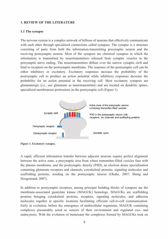
1. REVIEW OF THE LITERATURE 1.1 The synapse The nervous system is a complex network of billions of neurons that effectively communicate with each other through specialized connections called synapses. The synapse is a structure consisting of parts from both the information-transmitting presynaptic neuron and the receiving postsynaptic neuron. Most of the synapses are chemical synapses in which the information is transmitted by neurotransmitters released from synaptic vesicles in the presynaptic nerve ending. The neurotransmitters diffuse over the narrow synaptic cleft and bind to receptors on the postsynaptic membrane. The response of the postsynaptic cell can be either inhibitory or excitatory. Excitatory responses increase the probability of the postsynaptic cell to produce an action potential while inhibitory responses decrease the probability for an action potential in the receiving cell. Most excitatory synapses are glutamatergic (i.e., use glutamate as neurotransmitter) and are located on dendritic spines, specialized membranous protrusions on the postsynaptic cell (Figure 1).
Figure 1. Excitatory synapse. A rapid, efficient information transfer between adjacent neurons require perfect alignment between the active zone, a presynaptic area from where transmitter-filled vesicles fuse with the plasma membrane, and the postsynaptic density (PSD), an electron dense specialization containing glutamate receptors and channels, cytoskeletal proteins, signaling molecules and scaffolding proteins, residing on the postsynaptic neuron (Okabe, 2007; Sheng and Hoogenraad, 2007). In addition to postsynaptic receptors, among principal building blocks of synapses are the membrane-associated guanylate kinase (MAGUK) homologs. MAGUKs are scaffolding proteins bringing cytoskeletal proteins, receptors, signaling molecules, and adhesion molecules together at specific locations facilitating efficient cell-to-cell communication. Early in evolution, before the emergence of multicellular organisms, MAGUK containing complexes presumably acted as sensors of their environment and regulated exo- and endocytosis. With the evolution of metazoans the complexes formed by MAGUKs took on

10
more specialized functions such as the cell-to-cell contacts of neuronal synapses (de Mendoza et al., 2010; Zheng et al., 2011). This literature review will focus on synapse-associated protein 97 (SAP97), a MAGUK protein with several important roles in the development and plasticity of synapses, as well as its major interactor and an important mediator of excitatory neurotransmission in the central nervous system (CNS), the alpha-amino-3-hydroxy-5-methyl-4-isoxazole propionate (AMPA) glutamate receptor GluA1. 1.2. Glutamate receptors Glutamate is the principal excitatory neurotransmitter in the mammalian CNS (Robinson and Coyle, 1987). The physiological effects of glutamate are mediated by various types of glutamate receptors expressed predominantly in the brain. Glutamate receptors are divided into two categories depending on how they convey their effect on the target cell upon ligand binding. Metabotropic glutamate receptors (mGluR) are G-protein coupled proteins that act through second messengers in the target cells conferring slow responses (see for review Conn and Pin, 1997), while ionotropic glutamate receptors (iGluR) are responsible for fast-acting neurotransmission exerting their effects via cations flowing through the ion channel pore once the ligand has bound. The iGluRs can be further divided into subclasses based on structural homology and their pharmacological preference for the non-native agonists AMPA, N-methyl-D-aspartate (NMDA) and kainate. In addition to AMPA-, NMDA- and kainate receptors, there is a fourth subclass, the δ-receptor, which shares structural homology, but does not yet have any identified agonist (Traynelis et al., 2010). Each subclass consists of several subunits (Figure 2). The nomenclature of iGluRs recommended by the International Union of Pharmacology Committee on Receptor Nomenclature and Drug Classification (NC-IUPHAR) is presented in Table 1 (Palmer et al., 2005; Collingridge et al., 2009; Traynelis et al., 2010). The three major ionotropic glutamate receptor subclasses all have distinct roles in the CNS: AMPA receptors are the primary mediators of fast synaptic transmission (Jonas and Sakmann, 1992), NMDA receptors are involved in the induction of synaptic plasticity, and kainate receptors, are involved in modulation of neuronal function in less-well characterized ways (Traynelis et al., 2010). Most AMPA and NMDA receptors reside in the postsynaptic membrane, whereas kainate receptors localize also to presynaptic sites, where they regulate both inhibitory and excitatory transmitter release (Lerma, 2006; Pinheiro and Mulle; 2008; Jane et al., 2009; Traynelis et al., 2010; Contractor et al., 2011). In the following chapters, structural features of AMPA receptors will be discussed, but all iGluR subclasses show similarity at the level of amino acid sequence, domain organization, tetrameric assembly and three-dimensional structure.

11
Figure 2. Ionotropic glutamate receptor classification. Table 1. Harmonized nomenclature for ionotropic glutamate receptors recommended by NC-IUPHAR.

12
1.2.1. AMPA receptors and their structure Functional AMPA receptors are tetrameric integral membrane proteins composed of four subunits GluA1, GluA2, GluA3 or GluA4 forming either homomeric (consisting of identical subunits) or heteromeric (consisting of two or more subunit types) ion channels. Each of the approximately 900 amino acids long subunits have a modular structure consisting of an extracellular N-terminal domain (NTD), an extracellular ligand-binding domain (LBD), a transmembrane domain (TMD) and an intracellular C-terminal domain (CTD) (Figure 3). While the NTD, LBD and TMD are highly conserved amongst the different subunits, the CTDs are more varied and thus likely contributing to subunit-dependent functional differences. X-ray structures of near full-length AMPA receptor tetramers comprising the NTD, LBD and TMD have been obtained, representing different activity states, for both homomeric (Sobolevsky et al., 2009; Dürr et al., 2014; Chen et al., 2014) and heteromeric (Herguedas et al., 2016) AMPA receptors. The structural domains of AMPA receptors are discussed in the next chapters.
Figure 3. AMPA receptor topology and structure. A schematic representation of an AMPA receptor subunit shown in the middle. Functional AMPA receptors are either homomeric, comprised of four identical subunits (shown on the left side), or heteromeric comprised of four differing subunits (shown on the right side). The homomeric GluA2 receptor (ProteinDataBank [pdb] coordinate 4UQJ) and heteromeric GluA2/3 receptor (pdb coordinates 5FWY [top view], 5IDE [side view]) are drawn with PyMol. 1.2.1.1. The N-terminal domain AMPA receptors contain a large, approximately 400 residue long, extracellular domain homologous to the LBD of mGluRs and bacterial amino acid binding proteins (O'Hara et al., 1993). The NTDs of AMPA receptors have no known ligands and the function of the NTD is largely unknown. NTDs are not obligatory receptor modules, since functional homomeric AMPA receptors form even in the absence of the NTD (Pasternack et al., 2002). Isolated NTDs form dimers in solution and have been suggested to play a role in the initial steps of subtype-specific receptor assembly (Kuusinen et al., 1999; Leuschner and Hoch 1999; Ayalon and Stern-Bach 2001; Ayalon et al., 2005; Matsuda et al., 2005; Gan et al., 2015). Crystal structures show that the NTD dimer is comprised of two clamshell-like structures,

13
each composed of an upper lobe and a lower lobe separated by a cleft. Residues in both the lower and upper lobe participate in the initial NTD dimer assembly, whereafter the dimer pairs form a tetramer through interactions between the lower lobes (Clayton et al., 2009; Jin et al., 2009; Yao et al., 2011; Kumar and Mayer; 2013). Although the NTDs of AMPA receptors have no known ligands, the dimeric NTDs are positioned with their clefts facing outwards to opposite directions, suggestive of a role as functional ligand-binding units (Krieger et al., 2015; Herguedas et al., 2016). While the homomeric GluA2 receptors adopt an elongated structure resembling the letter Y (Sobolevsky et al., 2009), the heteromeric GluA2/3 receptor adopts a compressed structure similar to the structure of the obligatory homomeric NMDA receptor GluAN1/N2B (Karakas and Furukawa, 2014; Herguedas et al., 2016). The differences derive from the NTD assembly. In heteromeric GluA2/3 and GluA2/4 the NTDs from the four subunits alternate around a central axis to create, when viewed from the top, a circular (O-shaped) structure differing from the loose zigzagging (N-shaped) arrangement seen in GluA2 homomers. The NTDs in heteromeric AMPA receptors are situated closer to the LBDs than the NTDs of homomeric receptors, thus giving the receptor a more compact structure. Simulations suggest that the receptor can alternate between the “O” and “N” states, which thus represent two allosteric conformations. Crystal structures of heteromeric GluA2/3 and GluA2/4 also indicate that structures in the lower lobe of NTDs may act as key determinants for subunit-selective receptor heteromerization (Herguedas et al., 2016). 1.2.1.2 The ligand-binding domain Two approximately 150 residues long extracellular segments S1 and S2 separated by transmembrane helices 1-3 (M1-M3) (Stern-Bach et al., 1994) form the LBD of AMPA receptors. Like the NTDs, the LBDs form a clamshell-like structure with two distinct lobes. The agonist binds in a crevice between the lobes and induces a closure of the clamshell (Armstrong et al., 1998, 2000). The LBDs form a back-to-back dimer through contacts mediated by the upper lobes (Sobolevsky et al., 2009; Kumar and Mayer, 2013). The lower lobes of the dimer are in contact with the transmembrane segments M1 and M3, which flank the channel pore, and when the agonist binds, the resulting closure of the binding site cleft leads to an upward and lateral movement of the lower lobes, which pulls the receptor channel open. The resulting structure (binding site closed, channel open) is very unstable and is rearranged in a millisecond timescale into an more stable, desensitized state (binding site closed, channel closed) by disruption of the dimer interface formed by LBD upper lobes (Armstrong et al., 2000; Traynelis et al., 2010; Meyerson et al., 2014) (Figure 4). The primary structure of LBD is affected by alternative splicing and RNA editing. In the LBD, a part of the upper lobe that forms the dimer interface includes a 38 amino acid long segment which as a result of alternative splicing occurs in two alternative forms, the “flip” and “flop” splice variants, which differ in 9 to 11 amino acids depending on AMPA receptor subunit (Sommer et al., 1990). Flip and flop isoforms exhibit different channel kinetics with flip splice variants generally desensitizing four to five times slower than flop variants (Mosbacher et al., 1994; Koike et al., 2000). They also show different sensitivities to allosteric modulators such as cyclothiazide (Partin et al., 1994; Kessler et al., 2000), 4-[2-

14
(phenylsulfonylamino)ethylthio]-2,6-difluoro-phenoxyacetamide (PEPA) (Sekiguchi et al., 1997; Sekiguchi et al., 1998), zinc (Shen and Yang, 1999), and lithium (aKarkanias and Papke, 1999; bKarkanias and Papke, 1999), that fine-tune the kinetics of the receptor channel. Moreover, differences in the early trafficking of receptors have been reported with flip and flop isoforms. Homomeric GluA1 and GluA4 flop isoforms accumulate in the endoplasmic reticulum (ER) possibly through interactions with luminal ER proteins, while flip isoforms are efficiently transported to the cell surface (Coleman et al., 2006). The ER retention can be rescued with stargazin, a transmembrane AMPA receptor regulatory protein (TARP), or through co-expression with flip isoforms (Chen et al., 2000; Coleman et al., 2006). In addition to the alternative splicing, an amino acid residue at a so-called R/G-site, located immediately before the flip/flop region is targeted by RNA editing. The primary RNA transcripts of GluA2, GluA3 and GluA4 genes are edited so that the codon AGA, for the amino acid arginine (R), is - to varying extent - converted by adenosine deaminase to IGA (I standing for inosine which has base-pairing properties of G), encoding the amino acid glycine (G) in the translated protein. The resulting editing isoforms (R and G) have different kinetic properties. Edited (G) receptors, containing glycine, show quicker resensitization (i.e. recovery from desensitization) than the unedited (R) receptors, while the editing has different effects on the onset of desensitization rates depending on subunit type (Lomeli et al., 1994; Krampfl et al., 2002).
Figure 4. Schematic representation showing conformational changes in AMPA receptor LBD and TMD upon channel activation. The LBD consisting of an upper and lower lobe form a clamshell with a ligand-binding cleft between the two lobes. The cylinder represents transmembrane segment M3, which move upon agonist binding to open the ion channel (inactive in resting state [channel closed], active with ligand bound [channel open], desensitized, i.e. inactive with ligand bound [channel closed]). The NTDs and CTDs are omitted from the figure for clarity.

15
1.2.1.3 The transmembrane pore-forming region The transmembrane domain (TMD) is composed of four membrane-associated segments, M1-M4, each composed of largely hydrophobic residues. Three of these (M1, M3, and M4) are transmembrane helices, whereas the M2 segment forms a re-entrant pore (P) loop. The TMD anchors the receptor to the plasma membrane and forms the ion channel of the receptor with a four-fold rotational symmetry (Sobolevsky et al., 2009; Herguedas et al., 2016). In the crystal structure, the ion channel is shaped like a pyramid with a cut off top. The broad base is facing towards the cytoplasm and the top is extracellular. Agonist binding to the LBD and closure of the clamshell leads to movement in three regions linking LBD to TMD, linkers S1-M1, M3-S2 and S2-M4. This movement pulls apart the M3 helices that line the outer cavity of the pore leading to channel opening, i.e. the channel is activated (Sobolevsky et al., 2009). AMPA receptor channels are permeable to monovalent cations and, in the absence of GluA2 subunit, to Ca2+. M2 (pore loop) lines the inner cavity of the ion channel pore and determines the Ca2+ permeability of the receptor through RNA editing affecting a single residue in the so-called Q/R-site in the M2 encoding part of the GluA2 subunit. While the genomic DNA of the GluA2 subunit encodes the amino acid glutamine (Q), the majority of GluA2 transcripts undergo RNA editing by adenosine deaminase to yield functional GluA2 receptors containing an arginine (R), at position 6071. Due to the size and charge of the arginine side chain, the receptor channel shows low conductance and permeability to Ca2+ (Sommer et al., 1991; Cha et al., 1994; Seeburg et al., 1998; Seeburg and Hartner, 2003 Burnashev et al., 1992; Burnashev et al., 1995; Swanson et al., 1997). It has been estimated that the majority (~80%) of hippocampal synaptic AMPA receptors are heteromeric GluA2-containing receptors, which are calcium-impermeable, while roughly 8% of the total pool of hippocampal AMPA receptors are calcium-permeable homomeric GluA1 receptors (Wenthold et al., 1996; Lu et al., 2009). 1.2.1.4 The intracellular C-terminal domain Compared to the prominent sequence homology amongst the extracellular and transmembrane domains of the AMPA receptors, the cytosolic C-terminal tails of different AMPA receptor subunits vary in terms of amino acid sequence and length. The AMPA receptor CTDs are of two types: short (50 residues) and long (68-80 residues) depending on subunit type (Traynelis et al., 2010). GluA1 subunits have a long tail (80-81 residues in mammalian species) and GluA3 a short tail. Adding to the diversity of the CTDs is the ability of GluA2 and GluA4 to undergo alternative splicing to produce both long- and short-tailed isoforms. The majority of GluA2 exists in the short form, while GluA4 is predominantly of the long form (Gallo et al., 1992; Köhler et al., 1994). The low degree of sequence homology amongst the CTDs of different AMPA receptor subtypes is reflected in differences in post-translational modifications and protein interactions influencing the intracellular trafficking and regulation of the receptor (Figure 5).
1 The amino acid numbering for AMPA receptor subunits, throughout this thesis, is for the total protein including the signal peptide (initiating methionine is numbered 1).

16
Importantly, all crystal structures of AMPA receptors have been obtained from deletion mutants, which lack the CTD. Thus, there is very little structural information for the CTDs of AMPA receptors. Primary sequence analysis algorithms predict that the CTDs of AMPA receptors are intrinsically disordered and thus unlikely to adopt a stable fold. Generally, an unfolded nature is common for cytosolic CTDs of mammalian membrane proteins and in combination with short conserved sequence motifs they are optimal for probing and successfully connecting with multiple scaffolding protein partners (Minezaki et al., 2007).
Figure 5. C-terminal AMPA receptor sequences from rat (Gene bank accession number following subunit name) showing post-translational modifications (● palmitoylation; ○ ubiquitinylation; + phosphorylation; * S-nitrosylation). Numbers to the right indicate the length of the amino acid sequence of the receptor (Traynelis et al., 2010 Na et al., 2012; Wagner et al., 2012; Selvakumar et al., 2013). 1.2.2 AMPA receptor regulation through C-terminal post-translational modifications and protein interactions The intracellular C-terminal tails of AMPA receptors contain several sites for reversible post-translational modifications, such as phosphorylation, ubiquitination, S-palmitoylation and S-nitrosylation, as well as sites for protein interactions playing critical roles in receptor trafficking, localization, and function. In the following chapters, the focus will be on post-translational modifications of the cytosolic C-terminus of AMPA receptor subunit GluA1 specifically. Similar post-translational modifications have been described for the other subunits, and contribute to their specific functional regulation. 1.2.2.1 Phosphorylation of GluA1 CTD Phosphorylation is the addition of a phosphoryl group (PO3
2-) to threonine, serine and tyrosine residues on target proteins. In rare cases histidine, lysine and arginine residues can also serve as sites for phosphorylation (Ciesla et al, 2011). The phosphoryl group changes the hydrophobicity and electric charge of the protein, as well as the size of its side chain, which can result in functional changes due to structural alterations in the protein or due to newly gained or lost protein interactions. In glutamate receptors phosphorylation of CTD has been

17
reported to regulate intracellular trafficking and channel properties (Wang et al., 2005; Lussier et al., 2015). In neuronal cultures or slice preparations, the C-terminus of GluA1 is phosphorylated at multiple sites by protein kinase C (PKC), cyclic-AMP-dependent protein kinase (PKA) and Ca2+/calmodulin dependent protein kinase II (CaMKII) in an activity-dependent manner. Phosphorylation of the GluA1 C-tail modulates the gating properties of the receptor channel. Phosphorylation at serine residue 849 (S849) by CaMKII increases the single channel conductance, whereas phosphorylation of S863 by PKA increases the mean open probability of homomeric GluA1 receptors (Roche et al., 1996; Barria et al., 1997; Mammen et al., 1997; Derkach et al., 1999). The enhanced conductance of homomeric GluA1 receptors by S849 phosphorylation can be overhauled by coexpression with the GluA2 subunit (Oh and Derkach, 2005). Yet again, the increased effect of S849 phosphorylation can be restored in GluA1/GluA2 heteromeric receptors when co-expressed with TARPs (Kristensen et al., 2011), adding a layer of complexity to the regulation of AMPA receptors through phosphorylation. In addition to the modulatory role on channel properties, phosphorylation of C-terminal residues in GluA1 is also regulating the receptor trafficking. Phosphorylation of S863 has been shown to drive GluA1 to extrasynaptic sites for subsequent delivery to synapses during long-term potentiation (LTP), the long-lasting change on synaptic strength in response to synaptic activity, a form of synaptic plasticity essential for learning and memory (Lee et al., 2003; Oh et al., 2006). For LTP expression, a concomitant phosphorylation of S863, S849 and S836 is likely needed. Phosphorylation of S849 and S863 lowers the threshold for LTP induction (Lee et al., 2003; Lee et al., 2010; Makino 2011), while phosphorylation of S836 drives the synaptic incorporation of GluA1 and is critical for the LTP expression (Boehm 2006). How phosphorylation of GluA1 can influence trafficking and synaptic plasticity is somewhat unclear. One plausible explanation is, that phosphorylation can modulate GluA1 protein interactions involved in AMPA receptor trafficking. An example of this is the phosphorylation of GluA1 CTD residues S834 and S836 by PKC, which enhances the interaction between GluA1 and actin-binding protein 4.1N, leading to an increased exocytosis of AMPA receptors at extrasynaptic sites and subsequent insertion in synapses at LTP induction (Lin et al., 2009). Long-term depression (LTD), the activity-dependent removal of AMPA receptors from synaptic sites, on the other hand, depends solely on dephosphorylation of S863, but not of S849 (Lee et al, 1998; Kameyama et al, 1998; Lee et al., 2003; Lee et al., 2010). Moreover, a decrease in the GluA1 phosphorylation on threonine 858 (T858), after NMDA receptor induced activity, has been implicated in LTD as well (Lee et al., 2007; Delgado et al., 2007).

18
1.2.2.2 Ubiquitination of GluA1 CTD Many cellular processes such as protein turnover, apoptosis, cell-cycle progression, transcriptional regulation, trafficking and receptor down-regulation by endocytosis are regulated by a small 76 amino acids long ubiquitously expressed protein, ubiquitin. Ubiquitination (also known as ubiquitylation) is a reversible post-translational modification where ubiquitin is covalently linked to lysine, cysteine, threonine or serine residues or to the N-terminal amino group of target proteins catalyzed by ubiquitin ligases and reverted by deubiquitinating proteases (deubiquitinases). Ubiquitination generally steers proteins to degradation by proteasomes (Breitschopf et al., 1998; Hershko and Ciehanover, 1998; Cadwell and Coscoy, 2005; Wang et al., 2007). Ubiquitination is also an important molecular mechanism regulating the synaptic function. All mammalian AMPA receptors are subject to ubiquitination on specific C-terminal lysine residues in an activity- and Ca2+-dependent manner. The process requires the activity of L-type voltage gated Ca2+ channels and CaMKII. Receptor activation leads to endocytosis and subsequent lysosomal accumulation and degradation of ubiquitinated AMPA receptors. It seems however, that AMPA receptor activity-induced receptor endocytosis is not due to ubiquitination per see, as ubiquitination of the receptor only occurs after internalization. Analysis of C-terminal lysine mutants pinpoints one ubiquitination site in GluA1, the lysine 886 (K886) (Widagdo et al., 2015). 1.2.2.3 GluA1 palmitoylation Glutamate receptors, as well as several other proteins in glutamatergic synapses are regulated through S-palmitoylation, the reversible and covalent addition of palmitic acid (palmitate), a 16-carbon saturated fatty acid, to cysteine residues of target proteins. The cysteines sensitive for palmitoylation, by palmitoyl acyl transferases, are surrounded by basic and hydrophobic residues and located in close vicinity of the cytosolic inner face of the hydrophobic cell membrane. Palmitoylation increases the hydrophobicity of proteins thus generally facilitating membrane insertion. In addition to stabilizing proteins at the plasma membrane, palmitoylation regulates the intracellular trafficking of proteins. AMPA receptor subunits are palmitoylated at two conserved cysteine residues located in close vicinity of the cytosolic face of the membrane, one located on the C-terminal tail and the other on the intracellular loop between M2 and M3. It has been reported that the cyclic action of palmitoylation and depalmitoylation (by palmitoyl thioesterases) of AMPA receptors, or their interacting proteins, facilitates dynamic control of receptor trafficking. Palmitoylation on the intracellular loop between M2 and M3, close to the channel pore, of GluA1 and GluA2 leads to an accumulation of the receptor in the Golgi and a reduction of receptor surface expression (Hayashi et al., 2005; Yang et al., 2009; Han et al., 2015). Palmitoylation of the membrane proximate cysteine in the GluA1 C-terminal tail also decreases the cell surface trafficking of the receptor by destroying the interaction to protein 4.1N having a pivotal role in stabilizing the receptors at the cell surface (Shen et al., 2000; Coleman et al., 2003; Hayashi et al., 2005). Of note, AMPA- (and NMDA-) receptor activity induced by glutamate rapidly depalmitoylates the AMPA receptors promoting surface trafficking (Hayashi et al., 2005).

19
1.2.2.4 S-nitrosylation at glutamatergic synapses Nitric oxide (NO) is an unstable and reactive gaseous signaling molecule with important roles in immune defense, regulation of vascular function and neuronal plasticity. NO exerts its biological effects through at least two distinct molecular mechanisms. Firstly, NO binding activates soluble guanylyl cyclase (sGC) leading to the formation of a second messenger, cyclic guanosine monophosphate (cGMP), involved in downstream signaling cascades. Secondly, through a covalent reversible and non-enzymatic post-translational modification, called S-nitrosylation, NO is attached to thiol (-SH) moieties of cysteine residues generating S-nitrosylated proteins (S-nitrosoproteins [-SNO]) with modified functions (Broillet, 1999). The term S-nitrosylation was coined in 1992 (Stamler et al., 1992; Nakamura and Lipton, 2011) to this process involving NO, in analogy with phosphorylation and the phosphoryl group PO3
2-. NO is produced from arginine and molecular oxygen by the enzyme nitric oxide synthase (NOS) through a two-step reaction (Figure 6). There are three mammalian isoforms of NOS, the neuronal nitric oxide synthase (nNOS; NOS1), the inducible nitric oxide synthase (iNOS; NOS2) and the endothelial nitric oxide synthase (eNOS; NOS3). The nNOS and eNOS are constitutively expressed in cells and their activity is regulated by Ca2+ via Ca2+-binding protein calmodulin (CaM). At basal concentrations, nNOS and eNOS are inactive, but in response to an elevated intracellular Ca2+ concentration Ca2+/CaM bind to nNOS and eNOS activating the enzyme resulting in NO synthesis. iNOS binds CaM permanently at physiological Ca2+ concentrations and is typically induced during inflammation to produce NO as an immune defense mechanism. As a free radical with an unpaired electron, NO can easily be converted to harmful nitrites and nitrates when synthesized in excess (Knowles and Moncada, 1994; Stuehr, 1999; Bradley and Steinert, 2016).
Figure 6. Reaction step for NO production. NOS catalyzes the production of NO and L-citrulline from L-arginine and oxygen with electrons donated from NADPH. As a by-product two molecules of water are formed. Chemical structures were obtained from the ChemSpider database (www.chemspider.com), with oxygen and hydroxyl groups in red and amine groups in blue.

20
S-nitrosylation of protein thiols was demonstrated for the first time in the early nineties (Stamler et al., 1992) and in 2001, Jaffrey and co-workers introduced a method, the so-called biotin-switch assay, to selectively biotinylate nitrosylation sites in proteins. By swopping the labile NO to a stable biotin molecule, they were able to detect several proteins endogenously S-nitrosylated by nNOS in brain tissue by a simple affinity precipitation utilizing streptavidin-sepharose. Since then an ever-growing number of proteins have been shown to be subject to S-nitrosylation. In the brain, NO signaling has been reported to regulate several important molecules in glutamatergic synapses through reversible cysteine S-nitrosylation, such as NMDA receptors (Choi et al., 2000; Choi and Lipton, 2000), kainate receptors (Zhang et al., 2012) and stargazing (Selvakumar et al., 2009). The effects of NO on glutamate receptors can be mediated indirectly via S-nitrosylation of interacting proteins or directly through S-nitrosylation of receptors themselves, influencing their trafficking, signaling, localization and function and by doing so, modulating normal physiological processes such as neuronal survival and synaptic plasticity. NO signaling regulates also AMPA receptors, both via cGMP-pathway and S-nitrosylation (Serulle et al., 2007; Vielma et al., 2014; Selvakumar et al., 2009; Selvakumar et al., 2013; Selvakumar et al., 2014). Recently, it was reported that C-terminal cysteines in GluA1 are nitrosylation targets regulating receptor conductance and endocytosis. S-nitrosylation of GluA1 CTD residue C893 was reported to augment S849 phosphorylation and facilitate an increase in the conductance of AMPA receptors, and to promote endocytosis by increasing the interaction between GluA1 and AP2 (Selvakumar et al., 2013), a protein participating in clathrin-mediated endocytosis (Pearse et al., 2000). 1.2.2.5 GluA1 C-terminal protein interactions In addition to several post-translational modification sites, the C-terminus of GluA1 contains binding sites for interacting proteins regulating cellular trafficking and/or surface expression of GluA1 containing receptors with minor differences in their spatio-temporal association to GluA1 (Figure 7).
Figure 7. C-terminal sequence of GluA1. Post-translational modifications (● palmitoylation; ○ ubiquitinylation; + phosphorylation; * S-nitrosylation) and protein interactions indicated.

21
1.2.2.5.1 PDZ domain mediated GluA1 C-terminal interactions The C-terminus of GluA1 harbors a C-terminal class I PDZ binding motif (see chapter 1.3.2.2) that has been reported to bind to PDZ domain containing proteins SAP97 (Leonard et al., 1998), mLin-10 (Stricker and Huganir, 2003), Shank (Uchino et al., 2006) and sorting nexin 27 (Hussain et al., 2014; Loo et al., 2014). The interaction between SAP97 and GluA1, as well as its role in neuronal function will be discussed in detail in chapter 1.3. Shank3 is a PDZ domain containing scaffolding protein expressed in the spines of developing neurons where it was reported to associate with the surface expressed pool of GluA1 receptors. In addition to Shank3, two other members of the Shank family (Shank1 and Shank2) were reported to bind GluA1 CTD (Uchino et al., 2006). Sorting nexin 27 has been reported to bind, via its PDZ domain to GluA1 and promote receptor re-cycling to the cell surface (Hussain et al., 2014; Loo et al., 2014). Unlike Shank3 and sorting nexin 27, mLin-10 associates not only with GluA1, but also with GluA2 that lacks a class I PDZ binding motif (but has a class II motif instead). Consistent with an unconventional nature of this PDZ interaction, a deletion of the 15 last C-terminal residues in GluA1 only reduces but does not completely abolish the interaction. The interaction with mLin-10 is suggested to regulate GluA1 trafficking as a PDZ point mutation enhances the surface expression of AMPA receptors (Stricker and Huganir, 2003). 1.2.2.5.2 Non-PDZ domain mediated GluA1 C-terminal interactions In addition to the GluA1 interacting protein 4.1., discussed earlier in the context of C-terminal post-translational modifications, other non-PDZ domain mediated C-terminal GluA1 interacting proteins include the reversion-induced LIM protein (RIL) (Schulz et al., 2004), the cGMP-dependent protein kinase II (cGKII) (Serulle et al., 2007) and the α/β-Hydrolase domain-containing 6 (ABHD6) (Wei et al., 2016). RIL is enriched at excitatory synapses where it plays a role in the endosomal re-cycling. Through its LIM domain RIL binds to GluA1 and via its PDZ domain it binds to actin and is able to promote surface expression of internalized GluA1 receptors (Schultz et al., 2004). cGKII, is another protein that has been reported to promote surface expression of GluA1 receptors. GluA1 binds to cGKII in vicinity of the catalytic site of the kinase and the binding increases when cGKII is activated by cGMP. When activated, cGKII phosphorylates GluA1 at site S863 thus enhancing surface expression of GluA1 (Serulle et al., 2007). The binding of GluA1 to ABHD6 on the other hand reduces the surface expression of receptors and excitatory postsynaptic responses (Wei et al., 2016). 1.3 SAP97 1.3.1 SAP97 - PDZ binding partner of GluA1 and a member of the MAGUK disks large (DLG) subfamily SAP97, also known as Disks large homolog 1 (DLG1), is a cytosolic protein with a calculated molecular mass of 97 kDa which is ubiquitously expressed not only in brain, but also throughout the body, often at sites for cell-to-cell contact (Müller et al., 1995). SAP97 belongs to the MAGUK family of scaffolding proteins that facilitate efficient cell-to-cell communication by clustering cytoskeletal proteins, receptors, signaling molecules, and

22
adhesion molecules at specific locations in the cell. The MAGUKs are divided into 10 subfamilies according to sequence comparisons. These subfamilies are: discs large (DLG), DLG5, zona occludens (ZO), calcium/calmodulin-dependent protein kinase (CASK), membrane protein palmitoylated 1 (MPP1), MPP2-7, MPP5, caspase recruitment domain containing MAGUK protein (CARMA), calcium channel β subunit (CACNB), and MAGUK with an inverted repeat (MAGI) (de Mendoza et al., 2010; Zheng et al., 2011; Oliva et al., 2012). SAP97 (Lue et al., 1994; Müller et al., 1995) belongs to the DLG subfamily of MAGUKs together with three other members, the postsynaptic density protein 93 (PSD-93; DLG2) (Kim et al., 1996), synapse-associated protein 102 (SAP102; DLG3) (Müller et al., 1996) and postsynaptic density protein 95 (PSD-95; DLG4) (Cho et al., 1992; Kistner et al., 1993). The physiological importance of SAP97 is demonstrated by the phenotype of null mutant mice for the Dlg1 gene. Mice devoid of SAP97 express defects in several organs and die shortly after birth due to severe developmental abnormalities in the cardiovascular system obstructing normal respiration (Iizuka-Kogo et al., 2015). Moreover, the loss of DLG, the sole DLG MAGUK member in Drosophila results in an uncontrolled cell proliferation and lethality in Drosophila larvae (Woods and Bryant, 1989; Woods and Bryant, 1991). A conditional knock-out of SAP97 in motor neurons leads to a reduction in the dendritic branching and overall dendritic tree size as well as reduced dendrite length (Zhou et al., 2008). While SAP97 is omnipresent, being expressed in the brain and in most other organs and tissues, the other members of the DLG subfamily of MAGUKs localize predominantly to the CNS (Cho et al., 1992; Kistner et al., 1993; Müller et al., 1995; Müller et al., 1996; Kim et al., 1996; Aoki et al., 2001). The spatio-temporal expression pattern of SAP97 and the other DLGs in the brain are partly overlapping. DLG MAGUK immunoreactivity can be found in a variety of brain regions such as the cerebellum, hippocampus and cerebral cortex (Cho et al., 1992; Müller et al., 1995; Kim et al., 1996; Müller et al., 1996; Sans et al., 2000). PSD-95 and PSD-93 expression is low in embryonic and early postnatal rat brain tissue and increases in juvenile (from postnatal day 15 onwards) and adult tissue (Cho et al., 1992; Kistner et al., 1993; Hsueh and Sheng, 1999; Sans et al., 2000), while SAP97 expression is at its highest in juvenile rat brain tissue with slowly decreasing expression in adult tissue (Müller et al., 1995). The temporal expression of SAP102 differs from the other members of the DLG subfamily, with an earlier onset of expression and a rapid decline before adulthood. High levels of SAP102 can be detected in rat cerebral cortex and hippocampus already at postnatal days 1 and 2, with a clearly detectable decline in expression after postnatal day 30 (Müller et al., 1996; Sans et al., 2000). 1.3.2 Modular structure of SAP97 and other DLG MAGUKs The DLGs contain several protein interaction domains: three PDZ domains, owing their name to three proteins in where the domain was originally discovered (the postsynaptic density protein PSD-95, the tumor suppressor protein disks large 1 of Drosophila [Dlg] and the tight junction protein Zona Occludens-1 [ZO-1]), one Src Homology 3 domain (SH3 domain) and one catalytically inactive guanylate kinase-like domain (GUK domain) (Mayer et al., 1988; Berger et al., 1989; Woods and Bryant, 1989; Woods and Bryant, 1991; Cho et al., 1992;

23
Gaidarov et al., 1993; Willott et al.,1993; Zschocke et al., 1993; Kistner et al., 1995; Stahl et al., 1988). 1.3.2.1 Alternative splicing of DLGs All members of the DLG subfamily of MAGUKs are subject to alternative splicing producing several structural variants (Figure 8). The N-terminus of SAP97, PSD-93 and PSD-95 is composed either of an approximately 50-60 amino acids long L27 heterodimerization domain (“β-isoform”), a protein interaction domain initially found in Caenorhabditis elegans proteins Lin-2 and Lin-7, or a 10 residues long sequence containing two palmitoylated cysteines (“α-isoform”) (Doerks et al., 2000; aChetkovich et al., 2002; Schlüter et al., 2006). The majority of SAP97 in the CNS is of the β-isoform while PSD-95 in the brain is predominantly of the palmitoylated α-isoform, as only ~ 10% of PSD-95 exists in the β-isoform (aChetkovich et al, 2002; Schlüter et al., 2006). The α- and β-isoforms bestow proteins with distinct characteristics. The L27 domain containing β-isoforms are able to form homo- and heteromers with other L27 domain containing proteins and thus increase their capacity of clustering membrane channels (and other proteins bound to the DLGs) at specific locations in the cell and forming multiprotein complexes to facilitate efficient signaling (Marfatia et al., 2000; Lee et al., 2002; Nakagawa et al., 2004). Although all DLGs can be found both presynaptically and postsynaptically, along the plasma membrane or within the cytoplasm, PSD-95 is most typically found postsynaptically, where roughly half of the protein is anchored firmly in the PSD through the two palmitoylated N-terminal cysteines while the other half is non-synaptic (Al-Hallaq et al., 2001; Aoki et al., 2001). SAP97 localizes both to presynaptic and to postsynaptic sites. On the postsynaptic neuron, SAP97 is present in the PSD but mostly found at perisynaptic sites surrounding the PSD (Müller et al., 1995; DeGiorgis et al., 2006; Waites et al., 2009). PSD-93 and SAP102 are also typically localized to the post-synaptic site, where PSD-93 shows a cellular distribution similar to PSD-95 in the PSD (likely due to the two palmitoylated N-terminal cysteines present in PSD-93 but lacking in SAP102), while SAP102 shows a broader distribution within the spine (Al-Hallaq et al., 2001; DeGiorgis et al., 2006; Zheng et al., 2010). The subcellular localization within the postsynaptic cell, into synaptic and non-synaptic compartments, suggests dual roles for the DLGs in the synapses: in the PSD, the DLGs maintain receptors at the synapse, while the non-synaptic DLGs may bind a reserve pool of receptors that are transported to the plasma membrane by the DLGs when needed. Beside the far N-terminal splicing producing the α- and β-isoforms, the SAP97 polypeptide has internal regions affected by alternative splicing. Between the N-terminus and the first PDZ domain, two different short insertions may be added after residue 161 to produce I1A and I1B isoforms. The N-terminal segment preceding the first PDZ domain is involved in intra- and intermolecular association with SH3 domains (Lue et al., 1994; Wu et al., 2000; McLaughlin et al., 2002; Cai et al., 2006). A similar alternatively spliced N-terminal region has been reported for SAP102 (Müller et al., 1996). The third alternatively spliced region in SAP97, situated between the SH3 and GUK domains, called the Hook region, contains four alternatively spliced insertions named I2, I3, I4 and I5 (Lue et al., 1994; Mori et al, 1998; McLaughlin et al., 2002). Alternative splicing of the Hook region is documented for PSD-93

24
and SAP102 as well (Kim et al., 1996; Müller et al., 1996). The I3 insertion is unique to SAP97. SAP97 containing the insertion I3 localizes to the plasma membrane and sites of cell-to-cell contact. The I3 insertion is involved in binding to 4.1N protein, an interaction critical for cell surface and synaptic targeting of SAP97 and its associated AMPA receptors (Lue et al., 1994; McLaughlin et al., 2002; Rumbaugh et al, 2003). The I2 insertion on the other hand, gives SAP97 a more diffuse expression pattern in the soma and in the dendrites, and a proposed nuclear-targeted expression (McLaughlin et al., 2002; Rumbaugh et al, 2003; Roberts et al., 2007). Functional effects of I4 and I5 insertions are less well characterized.
Figure 8. Modular structure of DLGs. Illustration of the domain organization and splice variants of SAP97. All DLG MAGUKs share the same overall architecture as they consist of three PDZ domains, an SH3 domain and a GUK domain. In addition SAP97, PSD-95 and PSD-93 contain either an N-terminal L27 domain or two cysteines that can be post-translationally modified. The alternative splice forms I1a/I1b and I3 are unique for SAP97 (Adapted from Fourie et al., 2014). 1.3.2.2 The PDZ domains The PDZ domains are ~90 amino acids long globular protein interaction domains often coexisting with other modular protein interaction domains within the same protein (Cho et al., 1992; Willott et al., 1993; Woods and Bryant, 1993). Each protein interaction domain in the scaffold protein have their own interacting partners, and in this way PDZ containing scaffold proteins can cluster large multiprotein complexes at defined compartments in the cell, thus facilitating efficient signal transduction (Sheng & Sala, 2001; Nourry et al., 2003; Kim and Sheng, 2004). The PDZ domain is one of the most abundant protein interaction domain and over 260 non-redundant PDZ domains have been reported to be encoded by the human proteome (Luck et al., 2012). It can be found in a variety of species ranging from bacteria and yeast to mammalian species (Ponting, 1997). Typically, PDZ domains bind to a short C-terminal binding motif on their target proteins and can be classified into three major types based on the consensus sequence of the last four C-terminal amino acids of their ligands. PDZ domains of SAP97 and the other members of the DLG subfamily recognize class I PDZ binding motifs: -S/T-X-Ф (S/T standing for a serine or threonine, X standing for any amino acid, and Ф standing for a hydrophobic C-terminal amino acid). Based on structural studies, the canonical PDZ domain contains five to six β-strands (βA-βF) and two α-helices (αA and αB). The C-terminal peptide ligands bind to a

25
groove between the βB strand and the αB helix. The ligand peptide assumes an extended form antiparallel to the βB strand with main chain contacts with the βB strand. A loop, the so-called carboxylate binding loop, between βA and βB strands containing a conserved repeat of amino acids, -G-L-G-F-, docks the carboxylate group of the ultimate C-terminal amino acid (at position P0) of the ligand via hydrogen bonds to main chain amides and forms a hydrophobic pocket accommodating the aliphatic side chain of the C-terminal residue. In addition, an arginine (or lysine) residue in the carboxylate binding loop, preceding the –G-L-G-F- repeat interacts with the carboxylate via a water molecule. The requirement for a S/T residue at the third to last position P-2 of the PDZ ligand is explained by a hydrogen bond which forms between the hydroxyl oxygen of S/T residues and a conserved histidine residue at the N-terminal end of the αB helix (the αB1 position) in class I PDZ domains (Doyle et al., 1996) (Figure 9). In addition to class I PDZ binding motifs and domains, there are class II and class III PDZ domains which contain different amino acids at position αB1 than histidine with preferences for ligands with hydrophobic amino acids (class II PDZ binding motif; -X-Ф-X-Ф) or negatively charged amino acids (class III PDZ binding motif; -X-D/E-X-V) at the antepenultimate P-2 position (Sheng and Sala, 2001).
Figure 9. Canonical class I PDZ interaction (PSD-95PDZ3 and CRIPT peptide). (A) The PDZ3 structure of PSD-95 in complex with a CRIPT peptide showing a typical PDZ fold comprising two α-helices (αA, αB) and six β-strands (βA-βF). The peptide (green) is situated in a groove between the βB strand and the αB helix. (B) Canonical class I PDZ interaction in the peptide binding groove through contacts made by the ultimate and penanteultimate residue of the peptide (the last four amino acid residues of the peptide [green stick] is shown in the peptide binding groove) to the carboxylate binding loop (-G-L-G-F-) and the αB helix. For clarity only hydrogen bonds between the ultimate C-terminal amino acid valine at position P0 and main chain amides of conserved residues in the carboxylate binding loop and the hydrogen bond between threonine in position P-2 and the conserved histidine at the N-terminal end of the αB helix are shown.
26
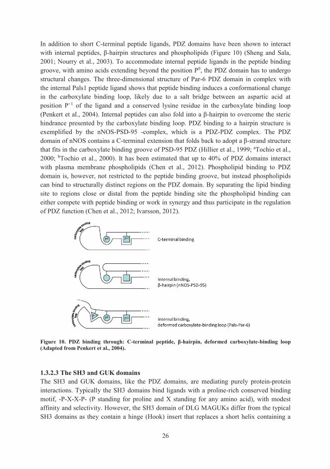
In addition to short C-terminal peptide ligands, PDZ domains have been shown to interact with internal peptides, β-hairpin structures and phospholipids (Figure 10) (Sheng and Sala, 2001; Nourry et al., 2003). To accommodate internal peptide ligands in the peptide binding groove, with amino acids extending beyond the position P0, the PDZ domain has to undergo structural changes. The three-dimensional structure of Par-6 PDZ domain in complex with the internal Pals1 peptide ligand shows that peptide binding induces a conformational change in the carboxylate binding loop, likely due to a salt bridge between an aspartic acid at position P+1 of the ligand and a conserved lysine residue in the carboxylate binding loop (Penkert et al., 2004). Internal peptides can also fold into a β-hairpin to overcome the steric hindrance presented by the carboxylate binding loop. PDZ binding to a hairpin structure is exemplified by the nNOS-PSD-95 -complex, which is a PDZ-PDZ complex. The PDZ domain of nNOS contains a C-terminal extension that folds back to adopt a β-strand structure that fits in the carboxylate binding groove of PSD-95 PDZ (Hillier et al., 1999; aTochio et al., 2000; bTochio et al., 2000). It has been estimated that up to 40% of PDZ domains interact with plasma membrane phospholipids (Chen et al., 2012). Phospholipid binding to PDZ domain is, however, not restricted to the peptide binding groove, but instead phospholipids can bind to structurally distinct regions on the PDZ domain. By separating the lipid binding site to regions close or distal from the peptide binding site the phospholipid binding can either compete with peptide binding or work in synergy and thus participate in the regulation of PDZ function (Chen et al., 2012; Ivarsson, 2012).
Figure 10. PDZ binding through: C-terminal peptide, β-hairpin, deformed carboxylate-binding loop (Adapted from Penkert et al., 2004). 1.3.2.3 The SH3 and GUK domains The SH3 and GUK domains, like the PDZ domains, are mediating purely protein-protein interactions. Typically the SH3 domains bind ligands with a proline-rich conserved binding motif, -P-X-X-P- (P standing for proline and X standing for any amino acid), with modest affinity and selectivity. However, the SH3 domain of DLG MAGUKs differ from the typical SH3 domains as they contain a hinge (Hook) insert that replaces a short helix containing a

27
conserved tyrosine crucial for canonical peptide binding, which is present in other SH3 domains. In addition, the SH3 domain of DLG MAGUKs is structurally discontinuous as the most C-terminal β-strand (βF) in DLG MAGUK SH3 domains is situated only after the GUK domain (McGee et al., 2001). With the exception of PSD-95 SH3 domain reported to bind to GluK5 kainate receptor CTD (Garcia et al., 1998), no specific binding partners have been identified for the SH3 domains of SAP97 and the other DLG MAGUKs. Instead, the SH3 domains of DLGs participate in intra- and intermolecular homo- and heterotypic interactions with the GUK domain and with proline-rich N-terminal sequences in DLGs. These SH3 domain interactions are atypical, as no proline-rich binding motifs are present in the GUK or NTD regions (McGee and Bredt, 1999; McLaughlin et al., 2002). Although named guanylate kinase homology domain, the GUK domain is catalytically inactive due to lack of critical amino acid residues needed for guanylate kinase catalyzed ATP-dependent phosphorylation of GMP to GDP (Lue et al., 1994; Kistner et al., 1995), and is believed to mediate specific protein interactions instead. The intramolecular interactions between SH3 and GUK domains or SH3 and N-terminal sequences can regulate the binding between GUK and other proteins. In SAP97, the intramolecular interaction between SH3 Hook region and GUK hinders the interaction between GUK and the GUK associated protein (GKAP). However, an intramolecular interaction between an N-terminal sequence (situated between the L27 and PDZ1 domains) and SH3 in SAP97 alters the SAP97 conformation facilitating GKAP binding (Wu et al., 2000). The crystal structure of SH3-GUK of SAP97 in complex with a mitotic spindle regulatory protein LGN peptide showed that the GUK domain mediated binding to target proteins involves recognition of a phosphorylated serine/threonine residue. Within all members of the DLG MAGUKs the residues forming the phospho-serine/threonine binding pocket are absolutely conserved, and therefore, the GMP-binding site of GUK domains have likely evolved to bind phosphorylated ligands (Zhu et al, 2011). 1.3.2.4 Supramodules Although the binding specificity of PDZ domains is often determined by recognition of only a few residues at the C-terminus of their target proteins by the ligand-binding groove of the PDZ domain, for some PDZ domains structures and sequences beyond their canonical PDZ fold contribute to specificity, affinity and regulation of the interaction with their targets. These so-called extended PDZ domains include individual PDZ domains with short extensions in their N- or C-terminus, multiple PDZs in tandem arrays (homotypic supramodules) or PDZ domains acting in concert with other protein interaction domains such as SH3 or/and GUK (heterotypic supramodules) (Feng and Zhang, 2009; Ye and Zhang, 2013). PSD-95, the prototypical member of DLG subfamily of MAGUKs, can form extended PDZ domains through a C-terminal extension (PDZ3- helix αC) as well as homotypic (PDZ1-PDZ2) and heterotypic supramodules (PDZ3-SH3-GUK). The first canonical PDZ interaction resolved by X-ray crystallography was the PSD-95 PDZ3 in complex with a C-terminal peptide from cysteine-rich interactor of PDZ3 (CRIPT) (Doyle et al., 1996). PSD-95 PDZ3 contains a C-terminal extension folding to an α-helix bending towards the peptide binding groove (Figure 11). This additional element, the so-called helix αC (or helix α3), was not

28
discussed in the original paper by Doyle and co-workers, but subsequent work has shown that the αC helix participates in the ligand-PDZ interaction by making contact up to the eight residue on the ligand extending from the canonical peptide binding groove. A deletion of the αC helix does not change the overall structure of PSD-95 PDZ3, but the affinity towards C-terminal peptides is substantially reduced (Petit et al., 2009; Chi et al, 2012).
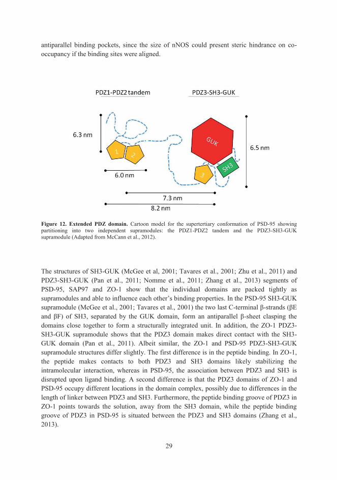
Figure 11. Extended PDZ domain. PSD-95PDZ3 with a C-terminal extension. The figure is drawn with PyMol (pdb coordinates 1IE9). All DLG MAGUKs have been reported to partition into homotypic PDZ1-PDZ2 supramodules and heterotypic PDZ3-SH3-GUK supramodules in solution (Figure 12) (McCann et al., 2012; Zhang et al., 2013). Studies on the PSD-95 PDZ1-PDZ2 tandem, connected by a short eight residues long conserved linker, have demonstrated that peptide binding has a strong effect on domain orientation (Wang et al., 2009) and that the tandem is capable of both antiparallel and parallel alignment of the individual binding pockets (Figure 13) (Long et al., 2003; Sainlos et al., 2011; McCann et al., 2011). The orientations of the ligand binding sites have implications for the PSD-95 ligand selection. PSD-95 binds multiple ligands, such as the short C-terminal tails of several membrane proteins as well as sizable cytosolic proteins such as nNOS. Short C-terminal peptide sequences by membrane proteins are able to co-occupy parallel binding pockets in the PSD-95, which is how PSD-95 is suggested to cluster membrane channels and receptors (Long et al., 2003). Co-occupancy of PDZ1-PDZ2 by a C-terminal tail of a membrane protein and nNOS is most likely through
29
antiparallel binding pockets, since the size of nNOS could present steric hindrance on co-occupancy if the binding sites were aligned.
Figure 12. Extended PDZ domain. Cartoon model for the supertertiary conformation of PSD-95 showing partitioning into two independent supramodules: the PDZ1-PDZ2 tandem and the PDZ3-SH3-GUK supramodule (Adapted from McCann et al., 2012). The structures of SH3-GUK (McGee et al, 2001; Tavares et al., 2001; Zhu et al., 2011) and PDZ3-SH3-GUK (Pan et al., 2011; Nomme et al., 2011; Zhang et al., 2013) segments of PSD-95, SAP97 and ZO-1 show that the individual domains are packed tightly as supramodules and able to influence each other’s binding properties. In the PSD-95 SH3-GUK supramodule (McGee et al., 2001; Tavares et al., 2001) the two last C-terminal β-strands (βE and βF) of SH3, separated by the GUK domain, form an antiparallel β-sheet clasping the domains close together to form a structurally integrated unit. In addition, the ZO-1 PDZ3-SH3-GUK supramodule shows that the PDZ3 domain makes direct contact with the SH3-GUK domain (Pan et al., 2011). Albeit similar, the ZO-1 and PSD-95 PDZ3-SH3-GUK supramodule structures differ slightly. The first difference is in the peptide binding. In ZO-1, the peptide makes contacts to both PDZ3 and SH3 domains likely stabilizing the intramolecular interaction, whereas in PSD-95, the association between PDZ3 and SH3 is disrupted upon ligand binding. A second difference is that the PDZ3 domains of ZO-1 and PSD-95 occupy different locations in the domain complex, possibly due to differences in the length of linker between PDZ3 and SH3. Furthermore, the peptide binding groove of PDZ3 in ZO-1 points towards the solution, away from the SH3 domain, while the peptide binding groove of PDZ3 in PSD-95 is situated between the PDZ3 and SH3 domains (Zhang et al., 2013).

30
Figure 13. Possible orientations of PDZ1-PDZ2 tandem influencing their function. A parallel arrangement of the peptide binding grooves enables the MAGUK proteins to cluster receptors by binding to their short C-terminal tails at the cell membrane and enhances their affinity to receptors through multivalency. Through antiparallel arrangement on the other hand, the MAGUK molecule is able to interact simultaneously with C-terminal tails of membrane bound proteins and sizable cytosolic proteins in a rather limited space offered in the PSD of a synapse (Modified from Long et al., 2003; McCann et al., 2011). 1.3.3 SAP97 – GluA1 interaction SAP97 is the only member of the DLG subfamily of MAGUKs capable of binding the AMPA-type glutamate receptor GluA1 through a direct interaction. In principle, the interaction is a canonical class I PDZ interaction between the second PDZ domain of SAP97 and the short PDZ binding motif (-A-T-G-L) residing in the ultimate C-terminus of GluA1 (Leonard et al., 1998). The threonine at position P-2 and leucine at position P0 of GluA1 are important for the interaction, as mutations to alanine in these residues abolish the interaction between GluA1 and SAP97 (Cai et al., 2002). In addition to the short PDZ binding motif, a tripeptide sequence (-S-S-G-) located 9-11 residues upstream from the C-terminus of GluA1 has been found to contribute to the interaction (Cai et al., 2002). 1.3.4 SAP97 and GluA1 –role in neuronal development and synaptic function In the brain, SAP97 has several roles in the development of glutamatergic synapses. Although SAP97 is not believed to participate in the initial steps of synaptogenesis its expression in brain, and that of the other members of the DLG subfamily, quickly picks up and peaks at around postnatal day 15 in rats (Cho et al., 1992; Kistner et al., 1993; Müller et al., 1995; Müller et al., 1996; Hsueh and Sheng, 1999; Sans et al., 2000). This time coincides with the period when developing pups begin to hear and open their eyes for the first time and when the newly established synapses are molded. A number of studies indicate that SAP97 together with PSD-95 and PSD-93 play an important role in the trafficking of glutamate receptors and signaling molecules to these newly formed synapses (Chen et al., 2000; El-Husseini et al., 2000; Sans et al., 2001; Rumbaugh et al., 2003; Ehrlich and Malinow, 2004; Elias et al., 2006; Howard et al., 2010; Zheng and Keifer, 2014). Overexpression of SAP97, the only member of the DLG family capable of direct interaction with both AMPA and NMDA receptors (Leonard et al., 1998; Bassand et al., 1999; Cai et al., 2002), increases the amount of synaptic GluA1 and the frequency of miniature excitatory postsynaptic currents (Rumbaugh et al., 2003; Nakagawa et al., 2004).

31
1.3.4.1 SAP97 and GluA1 in synaptic plasticity The synaptic connections are continuously remodeled, strengthened or rendered weaker, in response to neuronal activity in a process called synaptic plasticity. Repetitive synaptic activity triggers NMDA receptor activation and a subsequent rise in the intracellular Ca2+-level, critical for the induction of long-term potentiation (LTP) or long-term depression (LTD), long lasting changes in the synaptic efficacy due to insertion or removal of AMPA receptors, respectively, at synaptic sites (Lüscher and Malenka, 2012; Herring and Nicoll, 2016). The insertion of calcium-permeable GluA1 containing AMPA receptors to the synapse is essential for LTP expression and implicated in learning and memory formation (Shi et al., 1999; Hayashi et al., 2000; Shi et al., 2001; Lee et al., 2003; Lynch, 2004; Plant et al., 2006). As discussed in chapter 1.3.2.1, SAP97 and PSD-95 exists as two alternatively spliced isoforms containing either a shorter palmitoylated N-terminus (α-isoform) or an N-terminal L27 domain (β-isoform). The unique N-terminal sequences confer the isoforms with different subcellular destinations in the cell. The vast majority of SAP97 in the brain is of the β-isoform and localize to perisynaptic regions outside the PSD while PSD-95 predominantly exists in the α-isoform localizing to the PSD. The α- and β-isoforms also influence the AMPA receptor mediated synaptic strength differently. The α-isoform regulates the synaptic insertion of AMPA receptors during basal activity, in the absence of NMDA receptor induced activity, whereas β-isoforms participate in activity-induced potentiation of the synaptic strength through CaMKII regulated insertion of AMPA receptors at the synapse (Schlüter et al., 2006). During basal activity palmitoylation of PSD-95 N-terminal cysteines in the α-isoform leads to membrane insertion of the protein and concomitant surface expression of AMPA receptors. An increase in the synaptic activity and the rise in intracellular Ca2+ levels through NMDA receptor channels on the other hand activate Ca2+/calmodulin (CaM) that bind to the N-terminus of PSD-95 preventing its palmitoylation and leading to decreased surface expression of AMPA receptors through PSD-95 (Topinka and Bredt, 1998; El-Husseini et al., 2002; Noritake et al., 2009; Zhang et al., 2014). As PSD-95 does not bind AMPA receptors directly the effect of PSD-95 on AMPA receptor surface expression is likely mediated through SAP97 or TARPs able of binding both PSD-95 and AMPA receptors simultaneously (Leonard et al., 1998; Cai et al., 2006; bChetkovich et al., 2002; Schnell et al., 2002). Elevated intracellular Ca2+ also activates CaMKII, which has been shown to phosphorylate a serine residue (S39) in the N-terminal L27 domain of SAP97 driving SAP97 to spines (Mauceri et al., 2004). This modification might explain how SAP97, which predominantly exists in the β-isoform containing the L27 domain and lacking C-terminal cysteines, can potentiate the synaptic strength during NMDA receptor induced activity by inserting GluA1 containing AMPA receptors at the synapse. 1.3.4.2 SAP97 in GluA1 trafficking To exert their function GluA1 receptors need to reach their destination at the synapse. Through a series of regulated steps each likely involving interacting proteins (and post-translational modifications), the newly formed receptors are transported from the ER via Golgi complex to the vicinity of the plasma membrane at spines waiting to be inserted in the perisynaptic membrane in response to synaptic activity (Sans et al., 2001; Rumbaugh et al.,

32
2003; Nakagawa et al., 2004; Waites et al., 2009). SAP97 is reported to bind GluA1 early in the biosynthetic pathway and participate in the forward trafficking of GluA1 subunit containing receptors to the dendritic spines (Sans et al., 2001). Both the NMDA receptor activity-induced insertion of GluA1 containing AMPA receptors into the synapse, as well as basal AMPA receptor re-cycling, involve an actin-based motor protein myosin VI interacting with GluA1 receptors via SAP97. It has been shown, in hippocampal neurons, that inhibiting the interaction between the N-terminus of SAP97 containing the L27 domain and the cargo domain of myosin VI causes a notable decrease in the number of synapses and a reduction in the number of surface exposed synaptic GluA1 AMPA receptors. Moreover, inhibiting the SAP97-myosin VI interaction also negatively influences the increase in miniature excitatory postsynaptic currents generally associated with recruitment of AMPA receptors to the cell surface (Nash et al., 2010). Transport of GluA1 containing AMPA receptors within the cell could also occur along microtubules mediated by motor proteins from the kinesin superfamily, via a SAP97 interaction. SAP97 in the brain can interact directly with kinesin KIF1Bα through PDZ interactions (Mok et al., 2002). GluA1 interacts via SAP97 also with A-kinase anchoring protein (AKAP79/150), a scaffolding protein for signaling proteins PKA, PKC and calcineurin (also called Ca2+/calmodulin dependent protein phosphatase 2B, PP2B). In response to LTP induction, AKAP79/150 is palmitoylated at two N-terminal cysteine residues and recruited to dendritic spines with a subsequent increase in surface expressed GluA1 receptors (Keith et al., 2012). Once at the synaptic surface, the activity of the GluA1 receptors can be modulated by phosphorylation/ dephosphorylation through signaling proteins bound to AKAP79/150 to regulate receptor activity and re-cycling. 1.3.4.3 SAP97 in synaptogenesis The importance of SAP97 (and PSD-95) in the maturation of synapses has been demonstrated, by overexpressing SAP97 (or PSD-95) in mammalian synapses (El-Husseini et al., 2000; Nikonenko et al., 2008; Poglia et al., 2011). Overexpression of SAP97 in rat hippocampal pyramidal neurons increases the spine volume by threefold compared to the control spines. The size of PSDs also significantly increases within the spines, on an average, by 7.5-fold. In addition, overexpression of SAP97 promotes the formation of multi-innervated spines (MIS) that is, single spines with synaptic contacts to multiple axons. Moreover, SAP97 also induces the formation of multiple excitatory synapses directly on dendritic shafts. Similar changes can be observed in hippocampal pyramidal neurons, which overexpress PSD-95 albeit in a lesser degree. Intriguingly, both the formation of MIS and dendritic shaft synapses is blocked by the NOS inhibitor L-NG-nitro arginine methyl ester (L-NAME). Additionally, SAP97 overexpression results in recruitment of nNOS (and PSD-95) to synapses suggesting the morphological changes on the synapses are mediated by NO signaling via SAP97 and PSD-95. The ability of SAP97 to influence spine morphology through NO signaling has been implied to occur through a ternary complex were SAP97 binds nNOS via PSD-95 (Nikonenko et al., 2008; Poglia et al., 2011). However, SAP97 is able to interact with nNOS also directly (Chang et al., 2011 and this study, III). One

33
distinguishing feature seen only when overexpressing SAP97, but not PSD-95, is the enwrapping and engulfing of the presynaptic terminals (Poglia et al., 2011). A possible explanation for this is the difference in the subcellular localization of SAP97 and PSD-95. While PSD-95 localizes evenly in the spine, clearly present in the PSD as well as in cytoplasmic compartments, SAP97 concentrates perisynaptically, at the edges of PSD (DeGiorgis et al., 2006; Waites et al., 2009). The preferential localization of SAP97 along the edges of the PSD, rather than within the PSD like PSD-95, could also explain the larger PSDs observed during SAP97 overexpression as compared to PSD-95 overexpression (Nikonenko et al., 2008; Poglia et al., 2011). SAP97 can also affect the development of the presynaptic neuron. Unlike the other DLGs, which predominantly have a postsynaptic, dendritic localization, SAP97 is also present in axons. It has been demonstrated in developing neurites of mice that SAP97, through a PDZ interaction with the somatostatin receptor subtype 1, participates in the axonal growth cone formation (Cai et al., 2008). The effects of SAP97 on the presynaptic neuron is not only restricted to presynaptically expressed SAP97. Postsynaptic overexpression of SAP97 has been reported to increase the size of the active zone and accumulation of presynaptic proteins through an unknown retrograde signaling event. This presynaptic effect is critically dependent on the PDZ domains, and to a lesser extent, the L27 and GUK domains of SAP97. More specifically, the effect of SAP97 overexpression on presynaptic growth requires simultaneous binding of yet unidentified ligand(s) to the first and second PDZ domains of SAP97. This presynaptic growth promoting effect can also be demonstrated by overexpressing PSD-95 or SAP102, although the effect is significantly more modest than with SAP97 (Regalado et al., 2006). The difference may be related to the unique features of SAP97: the N-terminal insertions between the L27 domain and the first PDZ domain (Lue et al., 1994; McLaughlin et al., 2002) and the I3 insertion in the Hook region (Lue et al., 1994). Especially the I3 insertion might be relevant, since it binds protein 4.1 granting multimolecular complexes of SAP97 access to postsynaptic surface from where it may promote presynaptic growth more effectively than the other DLGs (Rumbaugh et al., 2003). 1.3.4.4 SAP97 and GluA1 in dendrite branching In the light of research results from several research groups highlighting the importance of SAP97 in GluA1 trafficking it was rather surprising when Zhou and co-workers reported in 2008 that SAP97 had no discernible influence on GluA1 trafficking and cell surface association. Instead, their experiments showed that GluA1, through its C-terminal PDZ motif, is essential for bringing SAP97 to the plasma membrane, where SAP97 is able to translate the activity of GluA1 receptors into dendrite growth. The interaction is critically dependent on the second PDZ domain of SAP97, as a mutation disrupting the PDZ binding site of PDZ2 abolishes the ability of GluA1 to chaperone SAP97 to the cell surface. For the growth promoting effect to take place at the plasma membrane GluA1 and SAP97 no longer need to be physically linked through a PDZ interaction. How SAP97 is able to control GluA1-dependent dendrite growth is suggested to take place through a locally produced and diffusible messenger that signals from activated GluA1 receptors to SAP97 (Zhou et al., 2008). Subsequent work on the growth promoting effects of SAP97 on dendrites following
34
GluA1 receptor activation have revealed that ligands to PDZ1, PDZ2, I3 and especially PDZ3, when localized at the plasma membrane, contribute to the dendrite growth (Zhang et al., 2015). The growth promoting effect of SAP97 and GluA1 on dendrites is synergistic, and is expressed at several levels including dendritic branching, size of the dendritic arbor, and dendritic length (Zhou et al., 2008; Zhang et al, 2015). SAP97 alone at the plasma membrane, in the absence of GluA1, is not able to promote dendrite growth, and it has been suggested that calcium influx through calcium permeable GluA1 channels drives the dendritic growth (Jeong et al., 2006; Zhang et al., 2015).

35
2. AIMS OF THE STUDY The aim of the study was to gain information on the structural basis and regulation of the interaction between GluA1 and SAP97. This interaction has been implicated in AMPA receptor cellular trafficking, synaptic plasticity and neuronal development, and in various neuropathologies, and thus the results will contribute to our understanding on the molecular basis of these important processes. The specific aims of the study were:
1. to gain detailed structural information on the selective interaction of GluA1 with SAP97 identifying important determinants involved in the regulation of the interaction (Publications I and II)
2. to study the role of C-terminal GluA1 residue C893 (Publications I, II and III) 3. to study the sensitivity of GluA1 for post-translational modification by NO and to
explore the possibility that GluA1 is physically linked to nitric oxide synthase via SAP97 (Publication III)
36
3. MATERIALS AND METHODS With the exception of size exclusion chromatography and glutathione S-transferase (GST) pull-down studies, the materials and methods used in this study have been described in detail in the original publications (I-III). Therefore, only a list of those methods is provided here. Method Original publication _____________________________________________________________________ Biotin-switch assay (BSA) III Buffer exchange I Bacterial protein expression I, II Bacterial protein expression –isotope labeled I Chemical shift assignment I* Crystal structure determination II* Immunoprecipitation/ Streptavidin-precipitation II, III Mammalian cell culture II, III Mass spectrometry analysis I Microplate binding assay II NMR model building I* NMR spectroscopy I* Peptide binding assay I Plasmid construction I, II, III Preparation of CysNO III Protein crystallization II* Protein purification I, II SDS-PAGE I, II, III Transfection II, III Western blotting I, II, III _____________________________________________________________________ * Methods not utilized by the author of this thesis Size-exclusion chromatography (I) Purified wt SAP97PDZ2 and SAP97PDZ2 C378G were analyzed by size-exclusion chromatography. For the purpose, a Superose 12 HR 10/30 gel filtration column (GE Healthcare Life Sciences) was equilibrated in 10 mM Bis-Tris buffer, pH 6.5. Standard proteins (Blue Dextran 2000 and HMW gel filtration calibration kit [GE Healthcare Life Sciences], aprotinin [Sigma]), wt SAP97PDZ2 in the presence or absence of 10-15 mM dithiothreitol (DTT) and SAP97PDZ2 C378G in absence of DTT, were run in an ÄKTA Fast protein liquid chromatography (FPLC) system (GE Healthcare Life Sciences) operating at 4 °C. Samples and standard proteins were diluted to 1-10 mg/ml in the eluent (10 mM Bis-Tris buffer, pH 6.5). Elution was monitored by UV absorbance at 280 nm and peak fractions were analyzed on non-reducing SDS-PAGE gels.

37
Generation of DNA constructs for GST pull-down studies (unpublished) SAP97PDZ1-3/PSD-95PDZ1-3 and PSD-95PDZ1-3/SAP97PDZ1-3 chimeras were generated through overlap extension PCR (OE-PCR) first amplifying separately the N-terminus (comprising the entire PDZ1 sequence and the N-terminal part of PDZ2) and the C-terminus (comprising the C-terminal part of PDZ2 and the entire PDZ3 sequence) of each MAGUK using full-length PSD-95 and SAP97 plasmid constructs as templates. Appropriate primers were used to introduce complementary sequences to the PDZ2 region of the SAP97 and PSD-95 PCR products, which in the subsequent PCR reactions were mixed, annealed and used as templates to produce chimeric SAP97PDZ1-3/PSD-95PDZ1-3 and PSD-95PDZ1-3/SAP97PDZ1-3 PCR products. The OE-PCR primers for the PDZ2 region, which were used to produce chimeras SAP97PDZ1-
3/PSD-95PDZ1-3 and PSD-95PDZ1-3/SAP97PDZ1-3 contained a silent mutation introducing an XhoI site, for analytical reasons, not present in the wt SAP97 and PSD-95 PDZ1-3 segments. For expression of His-tagged proteins the chimeric PCR products were cloned into a T7 promoter-containing pTFT74HIPMC NcoI-XbaI-treated vector as previously described in the construction of bacterial expression vectors encoding wt PDZ1-3 domain segments of SAP97 and PSD-95 (Cai et al., 2002). The mutations C378G and G219C were introduced to SAP97PDZ1-3/PSD-95PDZ1-3 and PSD-95PDZ1-3/SAP97PDZ1-3 chimeras respectively through PCR by amplifying the N-terminus (comprising the entire PDZ1 sequence and the N-terminal part of PDZ2) with 3’ primers containing the desired point mutation (C378G or G219C) as well as a silent mutation generating the restriction site for XhoI. Full-length PSD-95 and SAP97 plasmid constructs were used as templates. The resulting PCR products were cloned into NcoI-XhoI-treated SAP97PDZ1-3/PSD-95PDZ1-3 and PSD-95PDZ1-3/SAP97PDZ1-3 chimeras described above producing SAP97PDZ1-3 C378G/PSD-95PDZ1-3 and PSD-95PDZ1-3 G219C /SAP97PDZ1-3. The generation of a GST fusion protein of wt GluA1 CTD has been described previously (Cai et al., 2002). The correctness of the chimeric constructs were verified by analytical restriction digestions and sequencing. Bacterial expression of proteins for GST pull-down assay (unpublished) Recombinant proteins used in GST pull-down assays were produced in Escherichia coli (E. coli) strain BL21 (GST-tagged GluA1 CTD) or BL21(DE3)pLysS (His-tagged proteins) as follows: cultures from single bacterial colonies grown overnight in 2 ml of LB medium (containing 100 μg/ml ampicillin) were diluted 1:80 with fore mentioned medium and the growth was continued vigorously shaking at 37 °C for approximately 3 h until the culture reached an absorbance between 0.4-0.6 when measured at wavelength 600 nm. Thereafter, isopropyl-D-thio-β-galactopyranoside was added to a final concentration of 0.5 mM to induce protein expression for 3 h while the culture was incubated under continuous shaking at 37 °C. Induced cells were pelleted, suspended in TNE-buffer (containing 50 mM Tris-HCl pH 7.5, 120 mM NaCl, 1.0 mM EDTA, 1% (w/v) Triton X-100, 1 mM phenylmethylsulfonyl fluoride (PMSF), 5-10 μg/ml aprotinin and 5-10 μg/ml leupeptin) and sonicated on ice with a tip sonicator. Insoluble cell debris was removed by centrifugation (13500 g for 10 min at 4 °C) and the supernatant containing recombinant fusion protein was used in GST pull-down assays.

38
GST pull-down assay (unpublished) Bacterial lysates containing expressed protein of PDZ1-3 domain segments of SAP97, PSD-95 or chimeras SAP97/PSD-95, PSD-95/SAP97, SAP97 C378G/PSD-95 and PSD-95 G219C/SAP97 were incubated with GST-tagged GluA1 CTD attached to glutathione-sepharose beads overnight at 4 °C (with or without 10 mM DTT). The glutathione-sepharose beads were pelleted at 850 g for 1 min and washed three times with TNE-buffer (with or without 10 mM DTT) and once with PBS (with or without 10 mM DTT). The bound proteins were eluted in SDS sample buffer and analyzed by immunoblotting. Specific binding of His-tagged proteins to GST-tagged GluA1 CTD was assessed with an anti-His primary antibody and an alkaline phosphatase conjugated secondary antibody. The blots were developed in a colorimetric reaction using 5-bromo-4-chloro-3-indolyl phosphate and nitroblue tetrazolium as substrate.

39
4. RESULTS AND DISCUSSION 4.1 Biochemical and structural studies with GluA1 C-terminus and SAP97 PDZ domains (I and II) Within the DLG subfamily of MAGUKs SAP97 is the only member reported to bind AMPA receptor subunit GluA1 directly (Leonard et al., 1998; Cai et al., 2002; Zhou et al., 2008). This highly selective binding of GluA1 to SAP97, but not to the closely homologous PSD-93, PSD-95 or SAP102, is surprising considering the extent of sequence homology and the functional redundancy observed in several loss-of-function experiments showing that the DLGs are able to compensate for each other (Elias et al., 2006; Howard et al., 2010; Sun and Turrigiano, 2011). 4.1.1 Selectivity of GluA1-PDZ binding (II, unpublished) Earlier studies on the GluA1-SAP97 interaction had shown that the interaction between GluA1 and SAP97 involves the second PDZ domain of SAP97 (SAP97PDZ2) and is critically dependent on the C-terminal class I PDZ binding motif (-A-T-G-L) in GluA1 (I, Figure 1b) (Cai et al., 2002). Other pulldown studies, however, proclaimed the GluA1-SAP97 interaction to involve the first PDZ1 domain of SAP97 (SAP97PDZ1) instead (Mehta et al., 2001; Gardoni et al., 2003). To resolve this discrepancy, we performed a complementary experiment by coating microtiter plate wells with His-tagged SAP97PDZ1, SAP97PDZ2 and SAP97PDZ3 domains expressed in an E. coli BL21(DE3)pLysS strain and purified by immobilized metal chelation affinity chromatography (IMAC). The binding of GST-tagged C-terminal GluA1 wt or PDZ binding motif mutant (L907A) 11-mer peptides expressed in E. coli strain BL21 and purified by GST-sepharose was determined by utilizing anti-GST-horseradish peroxidase as a marker. The results confirmed that both SAP97PDZ1 and SAP97PDZ2 (but not SAP97PDZ3) are able to bind GluA1 through a classical PDZ interaction, although the strongest binding was observed between SAP97PDZ2 and GluA1 (II, Figure 2). This is not surprising, since it is known that PDZ1 and PDZ2 domains often share binding partners (Lim et al., 2002). Importantly, GluA1 point mutant L907A showed strongly reduced binding to both PDZ1 and PDZ2 domains (II, Figure 2), consistent with type I PDZ interaction. Mutations to the C-terminal -A-T-G-L PDZ binding motif of GluA1 had shown that the threonine at position P-2 and leucine at position P0, when mutated to alanine destroyed the interaction to SAP97, while mutations to alanine and glycine in positions P-3 and P-1 had no effect on the interaction (Cai et al., 2002). Residues in positions P-2 and P0 make contacts with SAP97PDZ2 residues in the carboxylate binding loop (-G-L-G-F-) and to the first residue in the αB helix (histidine) which are conserved in all class I PDZ domains (Doyle et al., 1996) (I, Figure 2b; II, Figure 3). Thus, other residues differing between SAP97 and the other members of the DLG subfamily had to account for the selectivity of the interaction. To pinpoint the critical residues in SAP97PDZ2 contributing to the highly selective interaction to GluA1, His-tagged PDZ1-3 chimeras comprising parts from SAP97 and PSD-95 were constructed. We chose to work with PSD-95 since the PDZ2 domains of SAP97 and PSD-95

40
differ only in nine amino acid residues (Figure 14). The PDZ1 and PDZ3 domains were kept intact and the crossover point between SAP97 and PSD-95 was within the PDZ2 domain leaving three of the differences to the N-terminal side and six to the C-terminal side of the chimeras. SAP97PDZ1-3 and PSD-95PDZ1-3 wt and chimeric variants were expressed in E. coli strain BL21(DE3)pLysS and their binding to recombinant GST-tagged GluA1 CTD expressed in BL21 was assessed in a GST pull-down assay directly from crude cell lysates. The PSD-95PDZ1-3 wt and PSD-95PDZ1-3/SAP97PDZ1-3 chimera repeatedly failed to bind to GluA1 CTD while SAP97PDZ1-3 wt and SAP97PDZ1-3/PSD-95PDZ1-3 chimera bound avidly to GluA1 CTD (Figure 15a), indicating that the ability to bind to GluA1 is conferred by the three SAP97-specific residues present in the PDZ2 segment N-terminal from the crossover point. The three differences between SAP97 and PSD-95, include two which conserve the nature of the side chain (L371 and K364 in SAP97 and I212 and R205 in PSD-95, respectively), and one nonconservative difference, cysteine (C378) in SAP97 and glycine (G219) in PSD-95. A GST pull-down assay performed under reducing conditions with GST-tagged GluA1 CTD, wt SAP97PDZ1-3 and wt PSD-95PDZ1-3 suggested that the interaction between GluA1 and SAP97 is stabilized by a disulfide bond, since the presence of reducing agent DTT throughout the GST pull-down assay was able to abolish the interaction between SAP97 and GluA1 almost completely (Figure 15b). The involvement of SAP97 cysteine C378 in the interaction with GluA1, suggested by the result of the GST pull-down under reducing conditions, was further studied by introducing cysteine-to-glycine (or glycine-to-cysteine) point mutations to the existing SAP97PDZ1-3/PSD-95PDZ1-3 and PSD-95PDZ1-
3/SAP97PDZ1-3 chimeras. The new chimeras (SAP97PDZ1-3 C378G /PSD-95PDZ1-3 and PSD-95PDZ1-3 G219C /SAP97PDZ1-3) were used in GST pull-down assays along with wt SAP97PDZ1-
3 and PSD-95PDZ1-3. While the SAP97PDZ1-3/PSD-95PDZ1-3 chimera (containing three SAP97 PDZ2 specific residues C378, L371 and K364) had bound avidly to GluA1 (Figure 15a), the SAP97PDZ1-3 C378G /PSD-95PDZ1-3 chimera (containing two SAP97 PDZ2 specific residues L371 and K364) did not. The equivalent point mutation (G219C) to the PSD-95PDZ1-
3/SAP97PDZ1-3 chimera in turn, resulted in a protein with the ability to bind GluA1 indicating that C378 in SAP97 plays a dominant role in the interaction with GluA1 (Figure 15c). To study the interaction between GluA1 and SAP97, and the role of cysteines therein, in greater detail we pursued peptide binding assays, NMR spectroscopy and X-ray crystallography with purified SAP97PDZ2 domains and C-terminal GluA1 13- or 18-mer peptides (I, Figure 1c).
Figure 14. PDZ2 (SAP97 and PSD-95) alignment showing amino acid residue differences in blue letters. Residues in the peptide binding pocket marked with red letters (Chen et al., 2008). Conserved residues in the carboxylate-binding loop and the N-terminal histidine in αB helix, making contact with the ultimate and antepenultimate amino acid residues of the ligand are marked yellow. The crossover point for the chimeric SAP97PDZ1-3 and PSD-95PDZ1-3 constructs is marked with a dotted line.
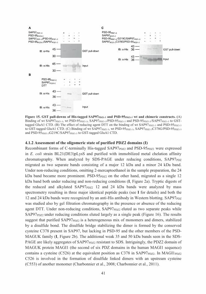
41
Figure 15. GST pull-downs of His-tagged SAP97PDZ1-3 and PSD-95PDZ1-3 wt and chimeric constructs. (A) Binding of wt SAP97PDZ1-3, wt PSD-95PDZ1-3, SAP97PDZ1-3/PSD-95PDZ1-3 and PSD-95PDZ1-3/SAP97PDZ1-3 to GST-tagged GluA1 CTD. (B) The effect of reducing agent DTT on the binding of wt SAP97PDZ1-3 and PSD-95PDZ1-3 to GST-tagged GluA1 CTD. (C) Binding of wt SAP97PDZ1-3, wt PSD-95PDZ1-3, SAP97PDZ1-3C378G/PSD-95PDZ1-3 and PSD-95PDZ1-3G219C/SAP97PDZ1-3 to GST-tagged GluA1 CTD. 4.1.2 Assessment of the oligomeric state of purified PDZ2 domains (I) Recombinant forms of C-terminally His-tagged SAP97PDZ2 and PSD-95PDZ2 were expressed in E. coli strain BL21(DE3)pLysS and purified with immobilized metal chelation affinity chromatography. When analyzed by SDS-PAGE under reducing conditions, SAP97PDZ migrated as two separate bands consisting of a major 12 kDa and a minor 24 kDa band. Under non-reducing conditions, omitting 2-mercaptoethanol in the sample preparation, the 24 kDa band became more prominent. PSD-95PDZ2 on the other hand, migrated as a single 12 kDa band both under reducing and non-reducing conditions (I, Figure 2a). Tryptic digests of the reduced and alkylated SAP97PDZ2 12 and 24 kDa bands were analyzed by mass spectrometry resulting in three major identical peptide peaks (see I for details) and both the 12 and 24 kDa bands were recognized by an anti-His antibody in Western blotting. SAP97PDZ was studied also by gel filtration chromatography in the presence or absence of the reducing agent DTT. Under non-reducing conditions, SAP97PDZ2 eluted as two separate peaks while SAP97PDZ2 under reducing conditions eluted largely as a single peak (Figure 16). The results suggest that purified SAP97PDZ2 is a heterogeneous mix of monomers and dimers, stabilized by a disulfide bond. The disulfide bridge stabilizing the dimer is formed by the conserved cysteine C378 present in SAP97, but lacking in PSD-95 and the other members of the PSD-MAGUK family (I, Figure 2b). The additional weak 35 and 50 kDa bands seen in the SDS-PAGE are likely aggregates of SAP97PDZ2 resistant to SDS. Intriguingly, the PDZ2 domain of MAGUK protein MAGI1 (the second of six PDZ domains in the human MAGI1 sequence) contains a cysteine (C526) at the equivalent position as C378 in SAP97PDZ2. In MAGI1PDZ2
C526 is involved in the formation of disulfide linked dimers with an upstream cysteine (C553) of another monomer (Charbonnier et al., 2008; Charbonnier et al., 2011).

42
Figure 16. Size exclusion chromatography of SAP97PDZ2 in the presence and absence of DTT. In the absence of the reducing agent DTT SAP97PDZ2 elutes as two separate, dimer (elution volume 15.1 ml) and monomer (elution volume 15.9 ml), peaks. Addition of DTT to the sample increases the size of the monomer peak. 4.1.3. Peptide binding studies on GluA1-SAP97 interaction (I) To characterize the GluA1-SAP97 interaction, a peptide binding assay with biotinylated 18-mer C-terminal GluA1 peptides (Bio-GluA118) and SAP97PDZ2 was performed. 96-well microtiter plates were coated with SAP97PDZ2 or with a SAP97NTD control protein and the binding of Bio-GluA118 was assessed with streptavidin-alkaline phosphatase conjugate and colorigenic substrate. A biotinylated 14-mer C-terminal peptide (Bio-GluN2A14) from the GluN2A subunit of the NMDA receptor, which is known to bind to the first and second PDZ domains of PSD-MAGUKs (Niethammer et al., 1996; Cai et al., 2002), was used as a positive control. The affinities assessed through the peptide binding assay were 5.98 ± 1.20 μM for Bio-GluA1 and 0.40 ± 0.05 μM for Bio-GluN2A (I, Figure 3a). In the presence of unlabeled GluA118 the binding between Bio-GluA118 and SAP97PDZ2 was strongly reduced, whereas unlabeled GluN2A14 had little effect on the Bio-GluA118 and SAP97PDZ2 binding. Similarly, the binding between Bio-GluN2A and SAP97PDZ2 was strongly inhibited in the presence of unlabeled GluN2A, while GluA118 reduced the binding only slightly. Moreover, DTT strongly reduced the Bio-GluA118 -SAP97PDZ2 binding, while the effect was only minute on the Bio-GluN2A -SAP97PDZ2 interaction (I, Figure 3b). Furthermore, the binding could also be inhibited by pre-treating SAP97PDZ2 with N-ethylmaleimide, a reagent that alkylates free thiol groups found in cysteines irreversibly (I, Figure 3c). The results of the peptide binding assays suggested, that besides the rather low-affinity binding mediated by the PDZ binding motif in GluA1 and SAP97PDZ2, a covalent disulfide bond formed between C893 in GluA1 and C378 in SAP97. To confirm the results of the peptide binding assay, of a disulfide complex between the GluA1 peptide and SAP97PDZ2, we employed an electrophoretic approach. Equal amounts of Bio-GluA118 and SAP97PDZ2 or Bio-GluA118 and SAP97PDZ2 C378S point mutant were mixed and analyzed by SDS-PAGE run under reduced and nonreduced conditions. Blotting with alkaline-phosphatase conjugated streptavidin revealed a covalent SDS-resistant complex between Bio-GluA118 and SAP97PDZ2 under nonreducing conditions, which could not be detected with the SAP97PDZ2 C378S point mutant (I, Figure 3d).

43
4.1.4 NMR studies on the GluA1-SAP97 interaction (I) The purified SAP97PDZ2 had a strong tendency to form dimers in solution in particular upon prolonged storage at 4 °C and to prevent SAP97PDZ2 dimer formation NMR spectroscopy (with 13C/15N-enriched protein) was performed under reducing conditions (10 mM DTT). Upon peptide titration, chemical shift changes (indicative of ligand binding) were observed mainly in the peptide binding groove of SAP97PDZ2 and no differences were observed in SAP97PDZ2 chemical shifts when titrated with either 13-mer or 18-mer GluA1 peptides (I, Figure 4a). Of note, none of the nine residues that differ between SAP97PDZ2 and PSD-95PDZ2 resides in the peptide binding groove. Of the nine residues differing between SAP97PDZ2 and PSD-95PDZ2 (Figure 14), significant chemical shift changes were observed at three residues of SAP97PDZ2 which were all situated outside the peptide binding groove: L371 (corresponding to I212 in PSD-95PDZ2), T383 (M224) and T389 (A230). T383 is preceding the αB helix and situated next to the conserved H384 in the αB helix participating in all known class I PDZ interactions. The T389 resides on the αB helix, whereas L371 is situated on the βD strand on the surface, more than 8 Å away from the peptide. The equivalent chemical shift changes in T383 (M224) and T389 (A230) upon peptide binding are observed in the PSD-95PDZ2-nNOS complex as well (Wang et al, 2000). In the case of L371, the chemical shift change is probably due to structural rearrangement of the PDZ domain upon peptide binding, since the distance does not allow for a direct interaction with the peptide. In complementary experiments, SAP97PDZ2 (or PSD-95PDZ2) was titrated in a 13-mer GluA1 peptide solution. Chemical shift changes were observed solely at the C-terminal part of the peptide (-A-T-G-L) with no significant differences between SAP97PDZ2 and PSD-95PDZ2. Thus, under reducing conditions, the C-terminal class I PDZ binding motif (-A-T-G-L) in GluA1 accounts solely for the binding, and the binding shows no selectivity between SAP97 and PSD-95. These NMR studies indicate that under reducing conditions, GluA1 peptides bind equally to PDZ2 domains of SAP97 and PSD-95, suggesting that the selective (and apparently high-affinity) interaction seen between GluA1 and SAP97 in binding experiments with full-length molecules, could not derive simply through a canonical class I PDZ interaction involving only the last four GluA1 residues. As our earlier GST-pulldown experiments with chimeric PDZ domains and the plate binding assays with Bio-GluA118 peptides and SAP97PDZ2 suggested that the binding was strongly influenced by redox conditions, NMR experiments were also performed under non-reducing conditions. Titrations with GluA118 under non-reducing conditions resulted in a chemical shift in the 13Cβ atom of C378 from ~28 ppm to ~41 ppm indicating a change from reduced to oxidized form, i.e. demonstrating the formation of a disulfide-bridged complex. The formation of a covalent GluA1-SAP97 complex was further confirmed by mass spectrometry, which indicated that all SAP97PDZ2 protein was covalently bound to GluA118.
4.1.5 A two-step binding model for the GluA1-SAP97 interaction (I) An increasing number of studies suggest that specificity and affinity of PDZ interactions are determined not only by the rather limited contacts between the extreme C-terminal PDZ binding motif and the canonical peptide binding groove, but that additional structures located

44
outside the region defining a classical PDZ interaction will add to the affinity and specificity of the interaction. This contribution of the sequence context can involve either extensions immediately preceding or following the PDZ binding motif or the PDZ domain, or alternatively protein interaction domains and regions situated further away from the region can strengthen the canonical PDZ interaction (Wang et al., 2010; Luck et al., 2012). Based on our peptide binding studies and NMR data a structural model of the SAP97PDZ2-GluA118 interaction was built showing, at least in theory, that there were no structural constraints for simultaneous occupation of the peptide binding groove of PDZ2 by GluA1 C-terminus and formation of a disulfide between C893 in GluA1 and C378 in SAP97 (I, Figure 5b). Such dual occupancy would present additional specificity to the (apparently) low affinity interaction between the peptide binding groove of PDZ2 of SAP97 (and PSD-95) and the ultimate C-terminus of GluA1. However, judged from the chemical shifts of SAP97PDZ2 in covalent complex with GluA118 only a small fraction of the C-terminus of covalently bound peptide is occupying the peptide binding groove at any given time. To reconcile these observations, we suggest a two-step binding mechanism for the interaction: an initial low affinity interaction involving the canonical peptide binding groove of the PDZ2 domain and the ultimate C-terminal PDZ binding motif in GluA1 would position the interacting molecules in a favorable way to facilitate, in the second step, disulfide bridge formation between the interacting proteins (I, Figure 7a). Once the covalent bond is formed, the binding would become independent of the canonical PDZ interaction. Interestingly, the peptide titration experiments indicated that the upstream tripeptide –S-S-G- sequence in GluA1, found earlier to participate in the GluA1-SAP97 binding (Cai et al., 2002), was not involved in the chemical shift changes in SAP97PDZ2 induced by the peptide. It is thus likely that the –S-S-G- sequence does not participate directly in the interaction with SAP97PDZ2, but may rather introduce structural flexibility to the C-terminus of GluA1 needed to establish stable binding via the proposed two-step mechanism. In order to analyze the potential physiological relevance of SAP97-GluA1 complexes stabilized by a disulfide bond, we studied whether the electrophoretic size of native SAP97 and GluA1 is affected by non-reducing conditions. SDS-PAGE run under non-reducing conditions of GluA1 and SAP97 in detergent extracts of rat brain produced high-molecular weight complexes, including a 250-kDa species seen in both SAP97 and GluA1 immunoblots, providing indirect support for the proposed model of a disulfide-linked GluA1-SAP97 complex forming in native tissue (I, Supporting Figure 1). 4.1.6 X-ray studies on SAP97PDZ2 (II) To study the structural details of the GluA1-SAP97 interaction further, we pursued X-ray crystallography and set up crystallization screens with SAP97PDZ2 and 18-mer GluA1 C-terminal peptides. Due to the tendency of SAP97PDZ2 to form disulfide-linked dimers through C378 (see above, 4.1.2), we constructed variants of SAP97PDZ2 in which the C378 was replaced by either glycine (C378G) (the corresponding amino acid in the homologous PSD-95PDZ2 and devoid of side chain) or serine (C378S) (differs from cysteine in one single atom, -SH in cysteine, and -OH in serine). Unlike the wild-type PDZ2, the C378 mutants had no

45
tendency to dimerize. The variants were expressed, purified and included in the crystal screens. Well-diffracting crystals were obtained both for free (SAP97PDZ2 C378G [2.44 Å resolution] and SAP97PDZ2 C378S [1.8 Å resolution]) and ligand bound SAP97PDZ2 (wt SAP97PDZ2 + GluA118 [2.35 Å resolution] and SAP97PDZ2 C378G + GluA118 [2.2 Å resolution]). The crystal structures obtained for SAP97PDZ2 were at the time the first SAP97 PDZ2 structures to be solved both in its liganded and apo form. The crystal structure of SAP97PDZ2 was similar to other previously solved PDZ structures, containing 2 α-helices and 6 β-strands folding into a typical compact globular protein interacting domain (Doyle et al., 1996). The glycine and serine residues replacing the C378 in the SAP97PDZ2 variants reside on the outer surface and the side chain of serine points outwards into the solution (II, Figure 4 and 5). 4.1.6.1 Comparison of SAP97PDZ2 crystal to closely related PDZ structures (II) SAP97PDZ2 C378S and closely related SAP102PDZ2 and PSD-95PDZ2 in their apo forms were superposed to compare their structures. The backbone structures show high similarity with minor differences only in two loop areas extending from either side of the βB strand that forms one side of the peptide binding groove: βA- βB and βB- βC (II, Figure 5). In SAP97PDZ2 C378S, the βA- βB loop is closer to the αB helix, which forms the other side of the peptide binding groove, than in PSD-95 or SAP102. In addition, the βB- βC loop is bent closer to the C-terminus in PSD-95PDZ2, while it points outwards to the solution in SAP97PDZ2 C378S and SAP102PDZ2. Differences observed in the loop areas of both βA- βB and βB- βC have been shown to add specificity and affinity to the canonical PDZ interaction involving the last four ligand residues and the peptide binding groove (Chi et al., 2009; Luck et al., 2012). 4.1.6.2 The ligand bound structure of SAP97PDZ2 (II) Well diffracting crystals in complex with a GluA118 peptide ligand were attained for both SAP97PDZ2 wt and the C378G variant. In both wild-type and C378G complexes, only the last four C-terminal residues (-A-T-G-L) of the ligand are seen in the crystal structure. The peptide is situated in the peptide binding groove between the βB strand and αB helix of the PDZ and antiparallel with the βB strand. The ultimate C-terminal residue L907, at position P0, docks the peptide in the peptide binding groove through hydrogen bonds between its carboxylate group and main chain amide nitrogens of L329, G330 and F331 of the carboxylate binding loop. An additional hydrogen bond is seen between the side chain of T905 at position P-2 of GluA1 and the conserved H384 at the N-terminal end of the αB-helix in SAP97PDZ2 (II, Figure 6). The interactions observed did thus not diverge beyond the canonical class I PDZ interaction seen in other known PDZ structures, and were consistent with the effects of point mutations reported earlier (Cai et al., 2002). The results from previous studies suggested that in addition to the four residues long C-terminal PDZ binding motif, -A-T-G-L, additional residues, -S-S-G- (at positions P-10 to P-8) (Cai et al., 2002) and the cysteine C893 at position P-14 (I) contribute to the interaction. However, in the crystal structure obtained with the 18-mer GluA1 ligand there was no electron density marking peptide binding to the PDZ domain beyond the last four residues and thus did not provide us with an answer to the high specificity and affinity interaction observed with intact molecules.

46
Regarding the SSG tripeptide, our NMR experiments (I) argued against its direct participation in binding and the crystallographic findings support this notion. The reasons for the notable absence of interactions beyond the very C-terminus from the crystal structure are currently unclear. It is possible that complexes, which include the inferred additional interactions, do not crystallize readily or are simply not formed under the conditions found optimal for the crystallization. Longer C-terminal peptides or even the entire CTD could result in more illuminating complex structures. However, the C-terminus of GluA1 like C-terminal tails of other membrane proteins are predicted to be intrinsically disordered (Minezaki et al., 2007; Xue et al., 2009) and disordered proteins do not arrange into a stable fold but rather sample alternative conformations, which makes them hard to crystallize. A structurally disordered sequence in the C-terminal tail in close vicinity of a PDZ binding motif allows conformational freedom to search and connect with interacting partners (Magidovich et al., 2006). Thus the -S-S-G- sequence, containing disorder promoting amino acids serine and glycine (Williams et al., 2001; Romero et al., 2001), would not be required for the direct interaction between GluA1 and SAP97, but necessary in providing a flexible fold allowing perfect alignment of short sequences and residues involved in the high specificity and affinity binding. Moreover, our attempts to produce a longer C-terminal GluA1 domain suitable for crystallization screens have been unsuccessful due to the modest expression levels and poor stability of the purified recombinant GluA1 CTD. 4.1.6.3 Conformational changes in SAP97PDZ2 upon peptide binding (II) While the wt SAP97PDZ2 crystals never diffracted without ligand, well-diffracting crystals were obtained from the C378G and C378S variants. By comparing the structures of ligand bound and free SAP97PDZ2, we discovered conformational changes taking place upon ligand binding. When a ligand is bound, the βA-βB loop (carboxylate binding loop) and αB-βF loop which are closest to the C-terminal end of the peptide move outwards away from each other. In the unliganded structure K324 preceding the -G-L-G-F- repeat in the βA-βB loop makes contact with D396 in the αB-βF loop. However, in the ligand-bound structure K324 moves to form hydrogen bonds to main chain and side chain oxygens in T394 and main chain oxygen in S395 (II, Figure 7). Such conformational change to better accommodate the peptide in the peptide binding groove had not been described previously for a PDZ-peptide interaction. Subsequent work by Chi and co-workers in 2009 with SAP97PDZ2 and a peptide of human papillomavirus type 18 protein E6 (HPV-18 E6) reported a similar conformational change and suggested that the interaction is an allosteric two-step mechanism, with an initial complex forming through a fast an unspecific association, which then undergoes a conformational change to lock the complex via an induced fit mechanism. The sequential mechanism allows the PDZ domain to screen a vast number of ligands and “lock” those with highest affinity and it was suggested that all peptides bind SAP97PDZ2 via the induced fit mechanism (Chi et al., 2009). However, such a mechanism is unlikely to have a strong contribution to the affinity and strict SAP97-selectivity of the GluA1 interaction, as the crystal structure of the complex does not reveal any direct interactions additional those involving the last four residues.

47
4.2 Functional role of C-terminal C893 of GluA1 (III) Previous work with synthetic GluA1 C-terminal peptides (I, II) identified C893 as a reactive residue capable of forming an intermolecular disulfide with C378 of SAP97PDZ2. To study the functional role of C893 in a more physiological context we in the next set of experiments chose to work with full-length GluA1 and SAP97 molecules. 4.2.1 The contribution of C893 on GluA1-SAP97 interaction (III) To examine the contribution of C893 on SAP97 interaction with full-length molecules, GluA1 wt and the point mutant GluA1 C893S were co-expressed together with a recombinant biotin-tagged SAP97 (Bio-SAP97) in transiently transfected human embryonic kidney cells (HEK 293(T) cells). The cells were lysed and SAP97-GluA1 complexes were harvested through a streptavidin-sepharose pulldown assay and analyzed by western blotting. The results show that while GluA1 wt and GluA1 C893S both bind to SAP97, the mutation strongly reduces the binding (III, Figure 2c, d). This result with full-length molecules is consistent with the observations seen with short GluA1 peptides and individual PDZ2 domains (I), that C893 is an important residue contributing, however not absolutely necessary, to the interaction with SAP97. Therefore, it is possible that a transient disulfide bond will form between GluA1 and SAP97 under suitable conditions, thereby increasing the amount of the complex that can be detected at any given time. However, the ability of GluA1 C893S to form a complex with SAP97 that is stable enough to be isolated by streptavidin pulldown clearly indicates that disulfide- or C893- independent mechanisms are sufficient for the interaction to take place. 4.2.2 The contribution of C893 on GluA1 protein expression (III) We noticed that a mutation to C893 also has an effect on the expression levels of receptor molecules in HEK 293 cells. Full-length GluA1 wt or GluA1 C893S were expressed either alone or co-expressed with SAP97, whereafter the GluA1 protein levels were quantified from western blot signals. The expression levels of GluA1 C893S were consistently two to three times higher than the GluA1 wt expression levels, and unaffected by co-expressed SAP97. The expression levels of GluA1 wt on the other hand experienced a roughly twofold increase when co-expressed with SAP97 (III, Figure 2a, b), in itself an indication of an interaction between the molecules. The results suggest that the loss of thiol function (i.e. cysteine to serine mutation) stabilizes the receptor in the absence of GluA1 ligand. On the other hand, the increased expression level of the wt receptor when co-expressed with SAP97 indicates that SAP97 is able to regulate GluA1 stability through an interaction involving C893. It is possible that GluA1 CTD residue C893 plays a supportive role in receptor turnover, and an interaction with SAP97 would shield the C893 in GluA1 thus making it inaccessible for posttranslational cysteine modifications or competing protein interactions which would lead to receptor degradation.

48
4.3 GluA1 as target for S-nitrosylation (III) Our earlier study with GluA1 18-mer peptides (I) revealed that C893 contains a reactive thiol which is able to form a disulfide with C378 in SAP97PDZ2 and is modified by alkylation. Thus, we wondered if the reactive C893 could also be a target for reversible post-translational thiol modifications. As many of the proteins, residing in the glutamatergic synapse have been shown to be sensitive to S-nitrosylation the effect of NO on GluA1 cysteines, and C893 specifically, was studied in particular. Several GluA1 N- and C-terminal deletion mutants, as well as several short C-terminal domains of GluA1 with single C-terminal cysteine point mutations (targeting the three CTD cysteines separately) were made and their susceptibility to NO were assessed through the biotin switch assay (Jaffrey and Snyder, 2001). After subjecting the samples to S-nitrosylation, in the first step of the assay the free thiols insensitive to NO are alkylated whereafter, the NO-modified thiols, i.e. nitrosothiols, are reduced to free thiols by ascorbate. In the last steps of the assay the thiol groups, marking originally S-nitrosylated cysteines, are biotinylated. Biotinylated proteins are then harvested with streptavidin-sepharose and analyzed on western blots. A signal on the western blot is an indication of the existence of S-nitrosylated protein in the sample. Initially we determined the receptiveness of full-length GluA1 receptors to cell-permeable NO donor, S-nitrosocysteine (CysNO) in live HEK293 cells using the biotin switch assay. A positive signal was indeed observed using the assay and the result was confirmed by treating HEK293 cell lysates expressing full-length GluA1 or N- and C-terminal deletion mutants thereof with S-nitrosoglutathione (GSNO) another commonly used but cell-impermeant NO donor. Positive signals were observed not only for the full-length molecule, but also for the N- and C- terminal deletion mutants suggesting that several cysteines in different parts of the GluA1 receptor are sensitive to S-nitrosylation (III, Figure 4a, b, c). As our primary interest was in the three C-terminal cysteines of GluA1 including C893, we investigated the susceptibility of short C-terminal domains of wt and cysteine-to-serine point mutants, C829S, C843S and C893S to NO. Of the three C-terminal cysteines, only C893 was found to be sensitive for S-nitrosylation (III, Figure 4d), confirming the reactive nature of C893 and suggesting that GluA1 protein interactions might be redox-regulated by thiol modifications to C893. 4.4 Association of SAP97 and GluA1 with nNOS (III) Intrigued by the finding that cysteine residues in GluA1 are indeed S-nitrosylated we wondered if GluA1 could associate with neuronal nitric oxide synthase (nNOS) analogous to the NMDA receptors. The NMDA receptors are functionally regulated by S-nitrosylation and physically linked to nNOS via PSD-95. The first and second PDZ domains of PSD-95 bind to the C-terminus of NMDA receptors through a classical type I PDZ interaction (Lim et al., 2002). PSD-95 also interacts with nNOS through an unconventional PDZ-PDZ interaction (Brenman et al., 1996; Christopherson et al., 1999). NMDA receptor activity leads to an

49
elevated intracellular Ca2+ concentration and activation of nNOS via Ca2+/calmodulin. This leads to generation of NO and subsequent S-nitrosylation of cysteine residues in nearby proteins, including the extracellular portion of the NMDA receptor regulating the conductance through the receptor channel, especially during hypoxia, thus presenting a neuroprotective role for NO through S-nitrosylation (Lei et al., 1992; Choi et al, 2000; Takahashi et al., 2007). Furthermore, PSD-95 is also sensitive to S-nitrosylation. The C-terminus of the major isoform of PSD-95 contains two conserved N-terminal cysteines C3 and C5 that when palmitoylated stabilize membrane association of PSD-95 and interacting proteins. Ca2+ influx through NMDA receptor channels and the subsequent NO production results in S-nitrosylation of the C-terminal cysteines in PSD-95 counteracting the stabilizing effect of palmitoylation on PSD-95 membrane association. In this way, PSD-95 is able to regulate the signal transduction by reciprocal S-nitrosylation and palmitoylation of its N-terminal cysteines (Ho et al., 2011). Recognizing several similarities between NMDA and GluA1 receptor types such as calcium-permeability, NO sensitivity, and the association with a MAGUK protein, we sought to find out if SAP97 could bridge GluA1 to nNOS similar to PSD-95 in the ternary NMDA-PSD-95-nNOS- complex. For this purpose, we first studied the interaction of recombinant biotin-tagged SAP97 and PSD-95 with nNOS in a streptavidin-pulldown assay. Binary complexes were observed between nNOS and the positive control, PSD-95 (nNOS-PSD-95), but also between nNOS and SAP97 (nNOS-SAP97) (III, Figure 5a, b). The nNOS-SAP97 binary complex was also detected in immunoprecipitations of native brain tissue from postnatal day 7 rat (III, Figure 5c), supporting its physiological relevance. Encouraged by the findings we examined the formation of a potential ternary complex GluA1-SAP97-nNOS. For this purpose we performed streptavidin-pulldowns from HEK293T cell lysates triply transfected with biotin-tagged nNOS, GluA1 and SAP97. GluA1 indeed co-precipitated with nNOS suggesting a ternary complex bridged by SAP97 in analogy with the NMDA-PSD-95 –nNOS- complex, as no direct interaction was observed between nNOS and GluA1 (III, Figure 5d). Interestingly, in the absence of GluA1 we observed that the association between SAP97 and nNOS decreased, suggesting that GluA1 may promote SAP97-nNOS binding, possibly by inducing a conformational change in SAP97 upon binding to increase its binding affinity to nNOS. It has been shown, that a PSD-95 PDZ1-PDZ2 tandem construct, 90 % identical to the corresponding SAP97 segment, adopts different conformations depending on whether it is free or occupied by ligands. The ligand-free PSD-95 PDZ tandem is conformationally restrained, with parallel arrangement of peptide binding grooves. Upon ligand-binding, the flexibility of the short linker between PDZ1 and PDZ2 increases to allow for antiparallel ligand binding and increased ligand binding affinity (Figure 13) (Wang et al., 2009). As both the PDZ1 and PDZ2 domains of SAP97 have been implicated in interactions with GluA1 (II) and nNOS (Chang et al., 2011), a similar rearrangement of the SAP97 PDZ1-PDZ2 tandem is also likely to take place upon simultaneous binding of GluA1 and nNOS.

50
5. CONCLUDING REMARKS Since 1998, when the interaction between SAP97 and GluA1 was initially discovered by Leonard and co-workers, we have been presented with numerous studies indicating important roles for SAP97 in AMPA receptor trafficking, targeting and function as well as in neuronal development (Sans et al., 2001; Rumbaugh et al., 2003; Dell’Acqua et al., 2006; Schlüter et al., 2006; Zhou et al., 2008; Waites et al., 2009; Howard et al., 2010; Poglia et al., 2011; Zhang et al., 2015). The specific aims of this study was to gain insights on the structural basis and regulation of the specific interaction between SAP97 and GluA1, and for that purpose we pursued several biochemical (I, II, III) and structural studies (I, II). In our search for structural determinants accounting for the specific highly selective interaction between GluA1 and SAP97, it soon became evident that additional elements beyond the canonical type I PDZ interaction, i.e. the last four C-terminal residues in GluA1 and the PDZ binding groove of the second PDZ domain in SAP97, must be involved. In addition to the low-affinity binding observed for the short peptide sequence (-A-T-G-L) in the peptide groove we identified C893 upstream the PDZ binding motif in the intracellular cytosolic tail of GluA1 and C378 situated on the surface of the second PDZ domain of SAP97 outside the peptide binding groove as reactive cysteines participating in the interaction through a covalent disulfide bond (I). There is accumulating evidence of the importance of extensions to the classical PDZ interaction in determining protein specificity and affinity. These extensions include upstream ligand residues beyond the PDZ binding motif and regions outside the peptide binding groove of the PDZ domain (Ye and Zhang, 2013; Wang et al., 2009). Intracellular disulfide formation is considered relatively rare due to the reducing potential of the eukaryotic cytosol and traditionally protein oxidation has been regarded as an event linked almost solely to oxidative stress and pathological conditions. However, there is increasing evidence supporting the formation of cytosolic intra- and intermolecular disulfides and their redox regulation under normal physiological conditions, and cytosolic disulfide stabilized interactions have been reported for PDZ scaffolds as well (Kimple et al., 2001; van den Berk et al., 2005; Mishra et al., 2007; Liu et al., 2011). The crystal structure obtained of SAP97PDZ2 in complex with a GluA1 18-mer peptide shows a typical class I PDZ interaction similar to the structures of many other PDZ-peptide complexes. Disappointingly, only the last four residues of the GluA1 peptide are visible in the structure, including the hydrogen bonds between the C-terminal peptide residue L907 (position P0) and the residues L329, G330, and F331 in the conserved carboxylate binding loop, as well as a hydrogen bond between the peptide residue T905 (position P-2) and the conserved H384 in the αB helix (II). As there was no discernible electron density for bound peptide beyond the last four residues of peptide the crystal structure did not provide an explanation for the contribution of upstream elements (C893, -S-S-G- sequence) to SAP97 binding (I). The plausible reason for the lack of electron density for the C-terminal residues extending beyond the PDZ binding motif is that the C-terminus of GluA1 is predicted to be

51
disordered (Minezaki et al., 2007; Xue et al., 2009) and thus do not arrange into a stable fold which makes it hard to crystallize. A novel observation from the crystals in their liganded and apo form was the structural changes taking place in two loop areas, βA-βB loop and αB-βF, upon peptide binding. Peptide binding leads to a slight opening of the binding groove to better facilitate peptide binding in the cavity (II). In addition to its role in a direct interaction with SAP97 (I, III) the thiol group of GluA1 C893 was shown to be a target for reversible post-translational S-nitrosylation (III). Moreover, we discovered that GluA1 is physically linked to nNOS via SAP97 in vitro (III). The thiol modifications, i.e. the intermolecular disulfide and the post-translational S-nitrosylation, are not mutually exclusive, rather one can envision that the thiol modifications can both take place and be transiently regulated depending on the cellular context. At the level of ER-Golgi, where the initial interaction between GluA1 and SAP97 has been reported to take place (Sans et al., 2001) the interaction with SAP97 can offer stability to the newly formed glutamate receptor through an intermolecular interaction involving C893. At the plasma membrane on the other hand the situation is different. Homomeric GluA1 receptors are, when activated through agonist binding, calcium-permeable. SAP97 binds to both GluA1 and nNOS thus linking receptor activity directly to NO formation at the plasma membrane. Subsequent S-nitrosylation of C893 has been reported to lead to receptor endocytosis (Selvakumar et al., 2013), which might serve a protective role in neuronal cells. From our work it is not clear whether GluA1 and SAP97 can form disulfide linked complexes at the plasma membrane, which is the site where S-nitrosylation of GluA1 C893 is reported to take place. Further work with endogenous molecules is clearly warranted and will clarify the functional and physiological role of the reactive C893 in GluA1. Nevertheless, our work have identified C893 of GluA1 and C378 of SAP97 as reactive cysteines participating both in a direct interaction between the proteins and due to their reactive nature serving as targets for S-nitrosylation and possibly other post-translational thiol modifications such as palmitoylation, ubiquitination, S-glutathionylation to mention just a few. It is plausible that thiol modifications like S-nitrosylation, disulfide formation and others can regulate reactive cysteines (Hess and Stamler, 2012) although proteomic analysis of cysteine modifications of proteins suggest only modest overlapping of redox-dependent cysteine modifications (Gould et al., 2013).

52
6. ACKNOWLEDGEMENTS This study was carried out in the Division of Biochemistry and Biotechnology within the Faculty of Biological and Environmental Sciences at the University of Helsinki, and under the supervision of Professor Kari Keinänen. Funding for this work was made possible through financial support of the National Doctoral Program in Informational and Structural Biology, the University of Helsinki, the Magnus Ehrnrooth Foundation, and Medicinska Understödsföreningen Liv och Hälsa r.f. I wish to express my deepest gratitude to my supervisor, Professor Kari Keinänen. His enthusiastic attitude to science and his ability for generating ideas have always impressed me. Moreover, his optimism and encouraging attitude has helped carry this research project over those difficult periods when the outcome seemed doubtful. Here, he had trusted me to manage my project, and as well given me the freedom to do research on my own. This allowed me to develop into an independent scientific researcher, and for that I am grateful. In addition, I warmly thank all the past and present members of the glutamate receptor group for making the working atmosphere both stimulating and enjoyable. Special thanks go to my former colleague Annukka, who helped me to settle into the lab during those first few early months. As well, both Professor Kalle Saksela and Docent Tuomo Glumoff are gratefully acknowledged for their careful review of the thesis manuscript. To all my co-authors, a sincere thank you, as without your contribution and fruitful co-operation this work would not have been possible. Thank you to all the lovely people at the Division of Biochemistry and Biotechnology, you will be fondly remembered for the friendly coffee room discussions. To Professor Emeritus Carl Gahmberg, I offer my genuine appreciation for your encouragement and interest in my work over the years. My heartfelt thank you goes to my family, parents, and sisters. My many thanks for being there, as your love and support means the world to me. Ingemar, how can I ever thank you enough? You have carried me through the toughest times and without your continuous support and belief in me, I feel I could not have reached the finishing line with this thesis. Sofia, mammas söta gulle, my blissful thanks for coming into my life − you are my everlasting sunshine.

53
7. REFERENCES
Al-Hallaq, R. A., Yasuda, R. P., and Wolfe, B. B. (2001) Enrichment of N-methyl-D-aspartate NR1 splice variants and synaptic proteins in rat postsynaptic densities. J. Neurochem. 77, 110-119.
Aoki, C., Miko, I., Oviedo, H., Mikeladze-Dvali, T., Alexandre, L., Sweeney, N., and Bredt, D. S. (2001) Electron microscopic immunocytochemical detection of PSD-95, PSD-93, SAP-102, and SAP-97 at postsynaptic, presynaptic, and nonsynaptic sites of adult and neonatal rat visual cortex. Synapse. 40, 239-257.
Armstrong, N., and Gouaux, E. (2000) Mechanisms for activation and antagonism of an AMPA-sensitive glutamate receptor: Crystal structures of the GluR2 ligand binding core. Neuron. 28, 165-181.
Armstrong, N., Sun, Y., Chen, G., and Gouaux, E. (1998) Structure of a glutamate-receptor ligand-binding core in complex with kainate. Nature. 395, 913-917.
Ayalon, G., Segev, E., Elgavish, S., and Stern-Bach, Y. (2005) Two regions in the N-terminal domain of ionotropic glutamate receptor 3 form the subunit oligomerization interfaces that control subtype-specific receptor assembly. J. Biol. Chem. 280, 15053-15060.
Ayalon, G., and Stern-Bach, Y. (2001) Functional assembly of AMPA and kainate receptors is mediated by several discrete protein-protein interactions. Neuron. 31, 103-113.
Barria, A., Derkach, V., and Soderling, T. (1997) Identification of the Ca2+/Calmodulin-dependent protein kinase II regulatory phosphorylation site in the α-amino-3-hydroxyl-5-methyl-4-isoxazole-propionate-type glutamate receptor. Journal of Biological Chemistry. 272, 32727-32730.
Bassand, P., Bernard, A., Rafiki, A., Gayet, D., and Khrestchatisky, M. (1999) Differential interaction of the tSXV motifs of the NR1 and NR2A NMDA receptor subunits with PSD-95 and SAP97. Eur. J. Neurosci. 11, 2031-2043.
Berger, A., Schiltz, E., and Schulz, G. E. (1989) Guanylate kinase from saccharomyces cerevisiae. isolation and characterization, crystallization and preliminary X-ray analysis, amino acid sequence and comparison with adenylate kinases. Eur. J. Biochem. 184, 433-443.
Boehm, J., Kang, M. G., Johnson, R. C., Esteban, J., Huganir, R. L., and Malinow, R. (2006) Synaptic incorporation of AMPA receptors during LTP is controlled by a PKC phosphorylation site on GluR1. Neuron. 51, 213-225.
Bradley, S. A., and Steinert, J. R. (2016) Nitric oxide-mediated posttranslational modifications: Impacts at the synapse. Oxid Med. Cell. Longev. 2016, 5681036.
Breitschopf, K., Bengal, E., Ziv, T., Admon, A., and Ciechanover, A. (1998) A novel site for ubiquitination: The N-terminal residue, and not internal lysines of MyoD, is essential for conjugation and degradation of the protein. EMBO J. 17, 5964-5973.

54
Brenman, J. E., Chao, D. S., Gee, S. H., McGee, A. W., Craven, S. E., Santillano, D. R., Wu, Z., Huang, F., Xia, H., Peters, M. F., Froehner, S. C., and Bredt, D. S. (1996) Interaction of nitric oxide synthase with the postsynaptic density protein PSD-95 and α1-syntrophin mediated by PDZ domains. Cell. 84, 757-767.
Broillet, M. C. (1999) S-nitrosylation of proteins. Cell Mol. Life Sci. 55, 1036-1042.
Burnashev, N., Monyer, H., Seeburg, P. H., and Sakmann, B. (1992) Divalent ion permeability of AMPA receptor channels is dominated by the edited form of a single subunit. Neuron. 8, 189-198.
Burnashev, N., Zhou, Z., Neher, E., and Sakmann, B. (1995) Fractional calcium currents through recombinant GluR channels of the NMDA, AMPA and kainate receptor subtypes. J. Physiol. 485 ( Pt 2), 403-418.
Cadwell, K., and Coscoy, L. (2005) Ubiquitination on nonlysine residues by a viral E3 ubiquitin ligase. Science. 309, 127-130.
Cai, C., Coleman, S. K., Niemi, K., and Keinanen, K. (2002) Selective binding of synapse-associated protein 97 to GluR-A alpha-amino-5-hydroxy-3-methyl-4-isoxazole propionate receptor subunit is determined by a novel sequence motif. J. Biol. Chem. 277, 31484-31490.
Cai, C., Li, H., Kangasniemi, A., Pihlajamaa, T., Von Ossowski, L., Kerkela, K., Schulz, S., Rivera, C., and Keinanen, K. (2008) Somatostatin receptor subtype 1 is a PDZ ligand for synapse-associated protein 97 and a potential regulator of growth cone dynamics. Neuroscience. 157, 833-843.
Cai, C., Li, H., Rivera, C., and Keinanen, K. (2006) Interaction between SAP97 and PSD-95, two maguk proteins involved in synaptic trafficking of AMPA receptors. J. Biol. Chem. 281, 4267-4273.
Cha, J. H., Kinsman, S. L., and Johnston, M. V. (1994) RNA editing of a human glutamate receptor subunit. Brain Res. Mol. Brain Res. 22, 323-328.
Chang, B. H., Gujral, T. S., Karp, E. S., BuKhalid, R., Grantcharova, V. P., and MacBeath, G. (2011) A systematic family-wide investigation reveals that ~30% of mammalian PDZ domains engage in PDZ-PDZ interactions. Chem. Biol. 18, 1143-1152.
Charbonnier, S., Nomine, Y., Ramirez, J., Luck, K., Chapelle, A., Stote, R. H., Trave, G., Kieffer, B., and Atkinson, R. A. (2011) The structural and dynamic response of MAGI-1 PDZ1 with noncanonical domain boundaries to the binding of human papillomavirus E6. J. Mol. Biol. 406, 745-763.
Charbonnier, S., Stier, G., Orfanoudakis, G., Kieffer, B., Atkinson, R. A., and Trave, G. (2008) Defining the minimal interacting regions of the tight junction protein MAGI-1 and HPV16 E6 oncoprotein for solution structure studies. Protein Expr. Purif. 60, 64-73.
Chen, J. R., Chang, B. H., Allen, J. E., Stiffler, M. A., and MacBeath, G. (2008) Predicting PDZ domain-peptide interactions from primary sequences. Nat. Biotechnol. 26, 1041-1045.

55
Chen, L., Chetkovich, D. M., Petralia, R. S., Sweeney, N. T., Kawasaki, Y., Wenthold, R. J., Bredt, D. S., and Nicoll, R. A. (2000) Stargazin regulates synaptic targeting of AMPA receptors by two distinct mechanisms. Nature. 408, 936-943.
Chen, L., Durr, K. L., and Gouaux, E. (2014) X-ray structures of AMPA receptor-cone snail toxin complexes illuminate activation mechanism. Science. 345, 1021-1026.
Chen, Y., Sheng, R., Kallberg, M., Silkov, A., Tun, M. P., Bhardwaj, N., Kurilova, S., Hall, R. A., Honig, B., Lu, H., and Cho, W. (2012) Genome-wide functional annotation of dual-specificity protein- and lipid-binding modules that regulate protein interactions. Mol. Cell. 46, 226-237.
aChetkovich, D. M., Bunn, R. C., Kuo, S. H., Kawasaki, Y., Kohwi, M., and Bredt, D. S. (2002) Postsynaptic targeting of alternative postsynaptic density-95 isoforms by distinct mechanisms. J. Neurosci. 22, 6415-6425.
bChetkovich, D. M., Chen, L., Stocker, T. J., Nicoll, R. A., and Bredt, D. S. (2002) Phosphorylation of the postsynaptic density-95 (PSD-95)/discs large/zona occludens-1 binding site of stargazin regulates binding to PSD-95 and synaptic targeting of AMPA receptors. J. Neurosci. 22, 5791-5796.
31. Chi, C. N., Bach, A., Engstrom, A., Wang, H., Stromgaard, K., Gianni, S., and Jemth, P. (2009) A sequential binding mechanism in a PDZ domain. Biochemistry. 48, 7089-7097.
Chi, C. N., Haq, S. R., Rinaldo, S., Dogan, J., Cutruzzola, F., Engstrom, A., Gianni, S., Lundstrom, P., and Jemth, P. (2012) Interactions outside the boundaries of the canonical binding groove of a PDZ domain influence ligand binding. Biochemistry. 51, 8971-8979.
Cho, K. O., Hunt, C. A., and Kennedy, M. B. (1992) The rat brain postsynaptic density fraction contains a homolog of the drosophila discs-large tumor suppressor protein. Neuron. 9, 929-942.
Choi, Y. B., and Lipton, S. A. (2000) Redox modulation of the NMDA receptor. Cell Mol. Life Sci. 57, 1535-1541.
Choi, Y. B., Tenneti, L., Le, D. A., Ortiz, J., Bai, G., Chen, H. S., and Lipton, S. A. (2000) Molecular basis of NMDA receptor-coupled ion channel modulation by S-nitrosylation. Nat. Neurosci. 3, 15-21.
Christopherson, K. S., Hillier, B. J., Lim, W. A., and Bredt, D. S. (1999) PSD-95 assembles a ternary complex with theN-methyl-d-aspartic acid receptor and a bivalent neuronal NO synthase PDZ domain. Journal of Biological Chemistry. 274, 27467-27473.
Ciesla, J., Fraczyk, T., and Rode, W. (2011) Phosphorylation of basic amino acid residues in proteins: Important but easily missed. Acta Biochim. Pol. 58, 137-148.
Coleman, S. K., Cai, C., Mottershead, D. G., Haapalahti, J. P., and Keinanen, K. (2003) Surface expression of GluR-D AMPA receptor is dependent on an interaction between its C-terminal domain and a 4.1 protein. J. Neurosci. 23, 798-806.
56
Coleman, S. K., Moykkynen, T., Cai, C., von Ossowski, L., Kuismanen, E., Korpi, E. R., and Keinanen, K. (2006) Isoform-specific early trafficking of AMPA receptor flip and flop variants. J. Neurosci. 26, 11220-11229.
Collingridge, G. L., Olsen, R., Peters, J. A., and Spedding, M. (2009) Ligand gated ion channels. Neuropharmacology. 56, 1.
Conn, P. J., and Pin, J. P. (1997) Pharmacology and functions of metabotropic glutamate receptors. Annu. Rev. Pharmacol. Toxicol. 37, 205-237.
Contractor, A., Mulle, C., and Swanson, G. T. (2011) Kainate receptors coming of age: Milestones of two decades of research. Trends Neurosci. 34, 154-163.
de Mendoza, A., Suga, H., and Ruiz-Trillo, I. (2010) Evolution of the MAGUK protein gene family in premetazoan lineages. BMC Evol. Biol. 10, 93-2148-10-93.
DeGiorgis, J. A., Galbraith, J. A., Dosemeci, A., Chen, X., and Reese, T. S. (2006) Distribution of the scaffolding proteins PSD-95, PSD-93, and SAP97 in isolated PSDs. Brain Cell. Biol. 35, 239-250.
Delgado, J. Y., Coba, M., Anderson, C. N., Thompson, K. R., Gray, E. E., Heusner, C. L., Martin, K. C., Grant, S. G., and O'Dell, T. J. (2007) NMDA receptor activation dephosphorylates AMPA receptor glutamate receptor 1 subunits at threonine 840. J. Neurosci. 27, 13210-13221.
Dell'Acqua, M. L., Smith, K. E., Gorski, J. A., Horne, E. A., Gibson, E. S., and Gomez, L. L. (2006) Regulation of neuronal PKA signaling through AKAP targeting dynamics. Eur. J. Cell Biol. 85, 627-633.
Derkach, V., Barria, A., and Soderling, T. R. (1999) Ca2+/calmodulin-kinase II enhances channel conductance of alpha-amino-3-hydroxy-5-methyl-4-isoxazolepropionate type glutamate receptors. Proc. Natl. Acad. Sci. U. S. A. 96, 3269-3274.
Doerks, T., Bork, P., Kamberov, E., Makarova, O., Muecke, S., and Margolis, B. (2000) L27, a novel heterodimerization domain in receptor targeting proteins lin-2 and lin-7. Trends Biochem. Sci. 25, 317-318.
Doyle, D. A., Lee, A., Lewis, J., Kim, E., Sheng, M., and MacKinnon, R. (1996) Crystal structures of a complexed and peptide-free membrane protein-binding domain: Molecular basis of peptide recognition by PDZ. Cell. 85, 1067-1076.
Durr, K. L., Chen, L., Stein, R. A., De Zorzi, R., Folea, I. M., Walz, T., Mchaourab, H. S., and Gouaux, E. (2014) Structure and dynamics of AMPA receptor GluA2 in resting, pre-open, and desensitized states. Cell. 158, 778-792.
Ehrlich, I., and Malinow, R. (2004) Postsynaptic density 95 controls AMPA receptor incorporation during long-term potentiation and experience-driven synaptic plasticity. J. Neurosci. 24, 916-927.

57
El-Husseini, A. E., Schnell, E., Chetkovich, D. M., Nicoll, R. A., and Bredt, D. S. (2000) PSD-95 involvement in maturation of excitatory synapses. Science. 290, 1364-1368.
El-Husseini, A., Schnell, E., Dakoji, S., Sweeney, N., Zhou, Q., Prange, O., Gauthier-Campbell, C., Aguilera-Moreno, A., Nicoll, R. A., and Bredt, D. S. (2002) Synaptic strength regulated by palmitate cycling on PSD-95. Cell. 108, 849-863.
Elias, G. M., Funke, L., Stein, V., Grant, S. G., Bredt, D. S., and Nicoll, R. A. (2006) Synapse-specific and developmentally regulated targeting of AMPA receptors by a family of MAGUK scaffolding proteins. Neuron. 52, 307-320.
Feng, W., and Zhang, M. (2009) Organization and dynamics of PDZ-domain-related supramodules in the postsynaptic density. Nature Reviews Neuroscience. 10, 87-99.
Fourie, C., Li, D., and Montgomery, J. M. (2014) The anchoring protein SAP97 influences the trafficking and localisation of multiple membrane channels. Biochim. Biophys. Acta. 1838, 589-594.
Gaidarov, I. O., Suslov, O. N., and Abdulaev, N. G. (1993) Enzymes of the cyclic GMP metabolism in bovine retina. FEBS Lett. 335, 81-84.
Gallo, V., Upson, L. M., Hayes, W. P., Vyklicky, L.,Jr, Winters, C. A., and Buonanno, A. (1992) Molecular cloning and development analysis of a new glutamate receptor subunit isoform in cerebellum. J. Neurosci. 12, 1010-1023.
Gan, Q., Salussolia, C. L., and Wollmuth, L. P. (2015) Assembly of AMPA receptors: Mechanisms and regulation. J. Physiol. 593, 39-48.
Garcia, E. P., Mehta, S., Blair, L. A., Wells, D. G., Shang, J., Fukushima, T., Fallon, J. R., Garner, C. C., and Marshall, J. (1998) SAP90 binds and clusters kainate receptors causing incomplete desensitization. Neuron. 21, 727-739.
Gardoni, F., Mauceri, D., Fiorentini, C., Bellone, C., Missale, C., Cattabeni, F., and Di Luca, M. (2003) CaMKII-dependent phosphorylation regulates SAP97/NR2A interaction. J. Biol. Chem. 278, 44745-44752.
Gould, N., Doulias, P. T., Tenopoulou, M., Raju, K., and Ischiropoulos, H. (2013) Regulation of protein function and signaling by reversible cysteine S-nitrosylation. J. Biol. Chem. 288, 26473-26479.
Han, J., Wu, P., Wang, F., and Chen, J. (2015) S-palmitoylation regulates AMPA receptors trafficking and function: A novel insight into synaptic regulation and therapeutics. Acta Pharm. Sin. B. 5, 1-7.
Hayashi, T., Rumbaugh, G., and Huganir, R. L. (2005) Differential regulation of AMPA receptor subunit trafficking by palmitoylation of two distinct sites. Neuron. 47, 709-723.
Hayashi, Y., Shi, S. H., Esteban, J. A., Piccini, A., Poncer, J. C., and Malinow, R. (2000) Driving AMPA receptors into synapses by LTP and CaMKII: Requirement for GluR1 and PDZ domain interaction. Science. 287, 2262-2267.

58
Herguedas, B., Garcia-Nafria, J., Cais, O., Fernandez-Leiro, R., Krieger, J., Ho, H., and Greger, I. H. (2016) Structure and organization of heteromeric AMPA-type glutamate receptors. Science. 352.
Herring, B. E., and Nicoll, R. A. (2016) Long-term potentiation: From CaMKII to AMPA receptor trafficking. Annu. Rev. Physiol. 78, 351-365.
Hershko, A., and Ciechanover, A. (1998) The ubiquitin system. Annu. Rev. Biochem. 67, 425-479.
Hess, D. T., and Stamler, J. S. (2012) Regulation by S-nitrosylation of protein post-translational modification. J. Biol. Chem. 287, 4411-4418.
Hillier, B. J., Christopherson, K. S., Prehoda, K. E., Bredt, D. S., and Lim, W. A. (1999) Unexpected modes of PDZ domain scaffolding revealed by structure of nNOS-syntrophin complex. Science. 284, 812-815.
Ho, G. P., Selvakumar, B., Mukai, J., Hester, L. D., Wang, Y., Gogos, J. A., and Snyder, S. H. (2011) S-nitrosylation and S-palmitoylation reciprocally regulate synaptic targeting of PSD-95. Neuron. 71, 131-141.
Howard, M. A. (2010) The role of SAP97 in synaptic glutamate receptor dynamics. 107, 3805-3810.
Hsueh, Y. P., and Sheng, M. (1999) Regulated expression and subcellular localization of syndecan heparan sulfate proteoglycans and the syndecan-binding protein CASK/LIN-2 during rat brain development. J. Neurosci. 19, 7415-7425.
Hussain, N. K., Diering, G. H., Sole, J., Anggono, V., and Huganir, R. L. (2014) Sorting nexin 27 regulates basal and activity-dependent trafficking of AMPARs. Proc. Natl. Acad. Sci. U. S. A. 111, 11840-11845.
Iizuka-Kogo, A., Senda, T., Akiyama, T., Shimomura, A., Nomura, R., Hasegawa, Y., Yamamura, K., Kogo, H., Sawai, N., and Matsuzaki, T. (2015) Requirement of DLG1 for cardiovascular development and tissue elongation during cochlear, enteric, and skeletal development: Possible role in convergent extension. PLoS One. 10, e0123965.
Ivarsson, Y. (2012) Plasticity of PDZ domains in ligand recognition and signaling. FEBS Lett. 586, 2638-2647.
Jaffrey, S. R., and Snyder, S. H. (2001) The biotin switch method for the detection of S-nitrosylated proteins. Sci. STKE. 2001, pl1.
Jane, D. E., Lodge, D., and Collingridge, G. L. (2009) Kainate receptors: Pharmacology, function and therapeutic potential. Neuropharmacology. 56, 90-113.
Jeong, G. B., Werner, M., Gazula, V. R., Itoh, T., Roberts, M., David, S., Pfister, B., Cohen, A., Neve, R. L., Hollmann, M., and Kalb, R. (2006) Bi-directional control of motor neuron dendrite remodeling by the calcium permeability of AMPA receptors. Mol. Cell. Neurosci. 32, 299-314.

59
Jin, R. (2009) Crystal structure and association behaviour of the GluR2 amino-terminal domain. EMBO J. 28, 1812.
Jonas, P., and Sakmann, B. (1992) Glutamate receptor channels in isolated patches from CA1 and CA3 pyramidal cells of rat hippocampal slices. J. Physiol. 455, 143-171.
Kameyama, K., Lee, H., Bear, M. F., and Huganir, R. L. (1998) Involvement of a postsynaptic protein kinase A substrate in the expression of homosynaptic long-term depression. Neuron. 21, 1163-1175.
Karakas, E., and Furukawa, H. (2014) Crystal structure of a heterotetrameric NMDA receptor ion channel. Science. 344, 992-997.
aKarkanias, N. B., and Papke, R. L. (1999) Lithium modulates desensitization of the glutamate receptor subtype gluR3 in xenopus oocytes. Neurosci. Lett. 277, 153-156.
bKarkanias, N. B., and Papke, R. L. (1999) Subtype-specific effects of lithium on glutamate receptor function. J. Neurophysiol. 81, 1506-1512.
Keith, D. J., Sanderson, J. L., Gibson, E. S., Woolfrey, K. M., Robertson, H. R., Olszewski, K., Kang, R., El-Husseini, A., and Dell'acqua, M. L. (2012) Palmitoylation of A-kinase anchoring protein 79/150 regulates dendritic endosomal targeting and synaptic plasticity mechanisms. J. Neurosci. 32, 7119-7136.
Kessler, M., Rogers, G., and Arai, A. (2000) The norbornenyl moiety of cyclothiazide determines the preference for flip-flop variants of AMPA receptor subunits. Neurosci. Lett. 287, 161-165.
Kim, E., Cho, K. O., Rothschild, A., and Sheng, M. (1996) Heteromultimerization and NMDA receptor-clustering activity of chapsyn-110, a member of the PSD-95 family of proteins. Neuron. 17, 103-113.
Kim, E., and Sheng, M. (2004) PDZ domain proteins of synapses. Nat. Rev. Neurosci. 5, 771-781.
Kimple, M. E., Siderovski, D. P., and Sondek, J. (2001) Functional relevance of the disulfide-linked complex of the N-terminal PDZ domain of InaD with NorpA. EMBO J. 20, 4414-4422.
Kistner, U., Garner, C. C., and Linial, M. (1995) Nucleotide binding by the synapse associated protein SAP90. FEBS Lett. 359, 159-163.
Kistner, U., Wenzel, B. M., Veh, R. W., Cases-Langhoff, C., Garner, A. M., Appeltauer, U., Voss, B., Gundelfinger, E. D., and Garner, C. C. (1993) SAP90, a rat presynaptic protein related to the product of the drosophila tumor suppressor gene dlg-A. J. Biol. Chem. 268, 4580-4583.
Knowles, R. G., and Moncada, S. (1994) Nitric oxide synthases in mammals. Biochem. J. 298 ( Pt 2), 249-258.

60
Kohler, M., Kornau, H. C., and Seeburg, P. H. (1994) The organization of the gene for the functionally dominant alpha-amino-3-hydroxy-5-methylisoxazole-4-propionic acid receptor subunit GluR-B. J. Biol. Chem. 269, 17367-17370.
Koike, M., Tsukada, S., Tsuzuki, K., Kijima, H., and Ozawa, S. (2000) Regulation of kinetic properties of GluR2 AMPA receptor channels by alternative splicing. J. Neurosci. 20, 2166-2174.
Krampfl, K., Schlesinger, F., Zorner, A., Kappler, M., Dengler, R., and Bufler, J. (2002) Control of kinetic properties of GluR2 flop AMPA-type channels: Impact of R/G nuclear editing. Eur. J. Neurosci. 15, 51-62.
Krieger, J., Bahar, I., and Greger, I. (2015) Structure, dynamics, and allosteric potential of ionotropic glutamate receptor N-terminal domains. Biophys. J. 109, 1136-1148.
Kristensen, A. S., Jenkins, M. A., Banke, T. G., Schousboe, A., Makino, Y., Johnson, R. C., Huganir, R., and Traynelis, S. F. (2011) Mechanism of Ca2+/calmodulin-dependent kinase II regulation of AMPA receptor gating. Nat. Neurosci. 14, 727-735.
Kumar, J., and Mayer, M. L. (2013) Functional insights from glutamate receptor ion channel structures. Annu. Rev. Physiol. 75, 313-337.
Kuusinen, A., Abele, R., Madden, D. R., and Keinanen, K. (1999) Oligomerization and ligand-binding properties of the ectodomain of the alpha-amino-3-hydroxy-5-methyl-4-isoxazole propionic acid receptor subunit GluRD. J. Biol. Chem. 274, 28937-28943.
Lee, H. K., Kameyama, K., Huganir, R. L., and Bear, M. F. (1998) NMDA induces long-term synaptic depression and dephosphorylation of the GluR1 subunit of AMPA receptors in hippocampus. Neuron. 21, 1151-1162.
Lee, H., Takamiya, K., Han, J., Man, H., Kim, C., Rumbaugh, G., Yu, S., Ding, L., He, C., Petralia, R. S., Wenthold, R. J., Gallagher, M., and Huganir, R. L. (2003) Phosphorylation of the AMPA receptor GluR1 subunit is required for synaptic plasticity and retention of spatial memory. Cell. 112, 631-643.
Lee, H., Takamiya, K., He, K., Song, L., and Huganir, R. L. (2010) Specific roles of AMPA receptor subunit GluR1 (GluA1) phosphorylation sites in regulating synaptic plasticity in the CA1 region of hippocampus. J. Neurophysiol. 103, 479-489.
Lee, H., Takamiya, K., Kameyama, K., He, K., Yu, S., Rossetti, L., Wilen, D., and Huganir, R. L. (2007) Identification and characterization of a novel phosphorylation site on the GluR1 subunit of AMPA receptors. Molecular and Cellular Neuroscience. 36, 86-94.
Lee, S., Fan, S., Makarova, O., Straight, S., and Margolis, B. (2002) A novel and conserved protein-protein interaction domain of mammalian lin-2/CASK binds and recruits SAP97 to the lateral surface of epithelia. Mol. Cell. Biol. 22, 1778-1791.
Lei, S. Z., Pan, Z. H., Aggarwal, S. K., Chen, H. S., Hartman, J., Sucher, N. J., and Lipton, S. A. (1992) Effect of nitric oxide production on the redox modulatory site of the NMDA receptor-channel complex. Neuron. 8, 1087-1099.

61
Leonard, A. S., Davare, M. A., Horne, M. C., Garner, C. C., and Hell, J. W. (1998) SAP97 is associated with the alpha-amino-3-hydroxy-5-methylisoxazole-4-propionic acid receptor GluR1 subunit. J. Biol. Chem. 273, 19518-19524.
Lerma, J. (2006) Kainate receptor physiology. Current Opinion in Pharmacology. 6, 89-97.
Leuschner, W. D., and Hoch, W. (1999) Subtype-specific assembly of alpha-amino-3-hydroxy-5-methyl-4-isoxazole propionic acid receptor subunits is mediated by their n-terminal domains. J. Biol. Chem. 274, 16907-16916.
Lim, I. A., Hall, D. D., and Hell, J. W. (2002) Selectivity and promiscuity of the first and second PDZ domains of PSD-95 and synapse-associated protein 102. J. Biol. Chem. 277, 21697-21711.
111. Lin, D. T., Makino, Y., Sharma, K., Hayashi, T., Neve, R., Takamiya, K., and Huganir, R. L. (2009) Regulation of AMPA receptor extrasynaptic insertion by 4.1N, phosphorylation and palmitoylation. Nat. Neurosci. 12, 879-887.
Liu, W., Wen, W., Wei, Z., Yu, J., Ye, F., Liu, C. H., Hardie, R. C., and Zhang, M. (2011) The INAD scaffold is a dynamic, redox-regulated modulator of signaling in the drosophila eye. Cell. 145, 1088-1101.
Lomeli, H., Mosbacher, J., Melcher, T., Hoger, T., Geiger, J. R., Kuner, T., Monyer, H., Higuchi, M., Bach, A., and Seeburg, P. H. (1994) Control of kinetic properties of AMPA receptor channels by nuclear RNA editing. Science. 266, 1709-1713.
Long, J. F., Tochio, H., Wang, P., Fan, J. S., Sala, C., Niethammer, M., Sheng, M., and Zhang, M. (2003) Supramodular structure and synergistic target binding of the N-terminal tandem PDZ domains of PSD-95. J. Mol. Biol. 327, 203-214.
Loo, L. S., Tang, N., Al-Haddawi, M., Dawe, G. S., and Hong, W. (2014) A role for sorting nexin 27 in AMPA receptor trafficking. Nat. Commun. 5, 3176.
Lu, W., Shi, Y., Jackson, A. C., Bjorgan, K., During, M. J., Sprengel, R., Seeburg, P. H., and Nicoll, R. A. (2009) Subunit composition of synaptic AMPA receptors revealed by a single-cell genetic approach. Neuron. 62, 254-268.
Luck, K., Charbonnier, S., and Trave, G. (2012) The emerging contribution of sequence context to the specificity of protein interactions mediated by PDZ domains. FEBS Lett. 586, 2648-2661.
Lue, R. A., Marfatia, S. M., Branton, D., and Chishti, A. H. (1994) Cloning and characterization of hdlg: The human homologue of the drosophila discs large tumor suppressor binds to protein 4.1. Proc. Natl. Acad. Sci. U. S. A. 91, 9818-9822.
Luscher, C., and Malenka, R. C. (2012) NMDA receptor-dependent long-term potentiation and long-term depression (LTP/LTD). Cold Spring Harb Perspect. Biol. 4, 10.1101/cshperspect.a005710.

62
Lussier, M. P., Sanz-Clemente, A., and Roche, K. W. (2015) Dynamic regulation of N-methyl-d-aspartate (NMDA) and alpha-amino-3-hydroxy-5-methyl-4-isoxazolepropionic acid (AMPA) receptors by posttranslational modifications. J. Biol. Chem. 290, 28596-28603.
Lynch, M. A. (2004) Long-term potentiation and memory. Physiol. Rev. 84, 87-136.
Magidovich, E., Fleishman, S. J., and Yifrach, O. (2006) Intrinsically disordered C-terminal segments of voltage-activated potassium channels: A possible fishing rod-like mechanism for channel binding to scaffold proteins. Bioinformatics. 22, 1546-1550.
Makino, Y., Johnson, R. C., Yu, Y., Takamiya, K., and Huganir, R. L. (2011) Enhanced synaptic plasticity in mice with phosphomimetic mutation of the GluA1 AMPA receptor. Proceedings of the National Academy of Sciences. 108, 8450-8455.
Mammen, A. L., Kameyama, K., Roche, K. W., and Huganir, R. L. (1997) Phosphorylation of the α-amino-3-hydroxy-5-methylisoxazole4-propionic acid receptor GluR1 subunit by calcium/ calmodulin-dependent kinase II. Journal of Biological Chemistry. 272, 32528-32533.
Marfatia, S. M., Byron, O., Campbell, G., Liu, S. C., and Chishti, A. H. (2000) Human homologue of the drosophila discs large tumor suppressor protein forms an oligomer in solution. identification of the self-association site. J. Biol. Chem. 275, 13759-13770.
Matsuda, S., Kamiya, Y., and Yuzaki, M. (2005) Roles of the N-terminal domain on the function and quaternary structure of the ionotropic glutamate receptor. J. Biol. Chem. 280, 20021-20029.
Mauceri, D., Cattabeni, F., Di Luca, M., and Gardoni, F. (2004) Calcium/calmodulin-dependent protein kinase II phosphorylation drives synapse-associated protein 97 into spines. J. Biol. Chem. 279, 23813-23821.
Mayer, B. J., Hamaguchi, M., and Hanafusa, H. (1988) A novel viral oncogene with structural similarity to phospholipase C. Nature. 332, 272-275.
McCann, J. J., Zheng, L., Chiantia, S., and Bowen, M. E. (2011) Domain orientation in the N-terminal PDZ tandem from PSD-95 is maintained in the full-length protein. Structure. 19, 810-820.
McCann, J. J., Zheng, L., Rohrbeck, D., Felekyan, S., Kuhnemuth, R., Sutton, R. B., Seidel, C. A., and Bowen, M. E. (2012) Supertertiary structure of the synaptic MAGuK scaffold proteins is conserved. Proc. Natl. Acad. Sci. U. S. A. 109, 15775-15780.
McGee, A. W., and Bredt, D. S. (1999) Identification of an intramolecular interaction between the SH3 and guanylate kinase domains of PSD-95. J. Biol. Chem. 274, 17431-17436.
McGee, A. W., Dakoji, S. R., Olsen, O., Bredt, D. S., Lim, W. A., and Prehoda, K. E. (2001) Structure of the SH3-guanylate kinase module from PSD-95 suggests a mechanism for regulated assembly of MAGUK scaffolding proteins. Mol. Cell. 8, 1291-1301.
63
McLaughlin, M., Hale, R., Ellston, D., Gaudet, S., Lue, R. A., and Viel, A. (2002) The distribution and function of alternatively spliced insertions in hDlg. J. Biol. Chem. 277, 6406-6412.
Mehta, S., Wu, H., Garner, C. C., and Marshall, J. (2001) Molecular mechanisms regulating the differential association of kainate receptor subunits with SAP90/PSD-95 and SAP97. J. Biol. Chem. 276, 16092-16099.
Meyerson, J. R., Kumar, J., Chittori, S., Rao, P., Pierson, J., Bartesaghi, A., Mayer, M. L., and Subramaniam, S. (2014) Structural mechanism of glutamate receptor activation and desensitization. Nature. 514, 328-334.
Minezaki, Y., Homma, K., and Nishikawa, K. (2007) Intrinsically disordered regions of human plasma membrane proteins preferentially occur in the cytoplasmic segment. J. Mol. Biol. 368, 902-913.
Mishra, P., Socolich, M., Wall, M. A., Graves, J., Wang, Z., and Ranganathan, R. (2007) Dynamic scaffolding in a G protein-coupled signaling system. Cell. 131, 80-92.
Mok, H., Shin, H., Kim, S., Lee, J. R., Yoon, J., and Kim, E. (2002) Association of the kinesin superfamily motor protein KIF1Balpha with postsynaptic density-95 (PSD-95), synapse-associated protein-97, and synaptic scaffolding molecule PSD-95/discs large/zona occludens-1 proteins. J. Neurosci. 22, 5253-5258.
Mori, K., Iwao, K., Miyoshi, Y., Nakagawara, A., Kofu, K., Akiyama, T., Arita, N., Hayakawa, T., and Nakamura, Y. (1998) Identification of brain-specific splicing variants of the hDLG1 gene and altered splicing in neuroblastoma cell lines. J. Hum. Genet. 43, 123-127.
Mosbacher, J., Schoepfer, R., Monyer, H., Burnashev, N., Seeburg, P. H., and Ruppersberg, J. P. (1994) A molecular determinant for submillisecond desensitization in glutamate receptors. Science. 266, 1059-1062.
Muller, B. M., Kistner, U., Kindler, S., Chung, W. J., Kuhlendahl, S., Fenster, S. D., Lau, L. F., Veh, R. W., Huganir, R. L., Gundelfinger, E. D., and Garner, C. C. (1996) SAP102, a novel postsynaptic protein that interacts with NMDA receptor complexes in vivo. Neuron. 17, 255-265.
Muller, B. M., Kistner, U., Veh, R. W., Cases-Langhoff, C., Becker, B., Gundelfinger, E. D., and Garner, C. C. (1995) Molecular characterization and spatial distribution of SAP97, a novel presynaptic protein homologous to SAP90 and the drosophila discs-large tumor suppressor protein. J. Neurosci. 15, 2354-2366.
Na, C. H., Jones, D. R., Yang, Y., Wang, X., Xu, Y., and Peng, J. (2012) Synaptic protein ubiquitination in rat brain revealed by antibody-based ubiquitome analysis. J. Proteome Res. 11, 4722-4732.
Nakagawa, T., Futai, K., Lashuel, H. A., Lo, I., Okamoto, K., Walz, T., Hayashi, Y., and Sheng, M. (2004) Quaternary structure, protein dynamics, and synaptic function of SAP97 controlled by L27 domain interactions. Neuron. 44, 453-467.

64
Nakamura, T., and Lipton, S. A. (2011) Redox modulation by S-nitrosylation contributes to protein misfolding, mitochondrial dynamics, and neuronal synaptic damage in neurodegenerative diseases. Cell Death Differ. 18, 1478-1486.
Nash, J. E., Appleby, V. J., Correa, S. A., Wu, H., Fitzjohn, S. M., Garner, C. C., Collingridge, G. L., and Molnar, E. (2010) Disruption of the interaction between myosin VI and SAP97 is associated with a reduction in the number of AMPARs at hippocampal synapses. J. Neurochem. 112, 677-690.
Niethammer, M., Kim, E., and Sheng, M. (1996) Interaction between the C terminus of NMDA receptor subunits and multiple members of the PSD-95 family of membrane-associated guanylate kinases. J. Neurosci. 16, 2157-2163.
Nikonenko, I., Boda, B., Steen, S., Knott, G., Welker, E., and Muller, D. (2008) PSD-95 promotes synaptogenesis and multiinnervated spine formation through nitric oxide signaling. J. Cell Biol. 183, 1115-1127.
Nomme, J., Fanning, A. S., Caffrey, M., Lye, M. F., Anderson, J. M., and Lavie, A. (2011) The src homology 3 domain is required for junctional adhesion molecule binding to the third PDZ domain of the scaffolding protein ZO-1. Journal of Biological Chemistry. 286, 43352-43360.
Noritake, J., Fukata, Y., Iwanaga, T., Hosomi, N., Tsutsumi, R., Matsuda, N., Tani, H., Iwanari, H., Mochizuki, Y., Kodama, T., Matsuura, Y., Bredt, D. S., Hamakubo, T., and Fukata, M. (2009) Mobile DHHC palmitoylating enzyme mediates activity-sensitive synaptic targeting of PSD-95. The Journal of Cell Biology. 186, 147-160.
Nourry, C., Grant, S. G., and Borg, J. P. (2003) PDZ domain proteins: Plug and play! Sci. STKE. 2003, RE7.
Oh, M. C., and Derkach, V. A. (2005) Dominant role of the GluR2 subunit in regulation of AMPA receptors by CaMKII. Nat. Neurosci. 8, 853-854.
Oh, M. C., Derkach, V. A., Guire, E. S., and Soderling, T. R. (2006) Extrasynaptic membrane trafficking regulated by GluR1 serine 845 phosphorylation primes AMPA receptors for long-term potentiation. J. Biol. Chem. 281, 752-758.
O'Hara, P. J., Sheppard, P. O., Thogersen, H., Venezia, D., Haldeman, B. A., McGrane, V., Houamed, K. M., Thomsen, C., Gilbert, T. L., and Mulvihill, E. R. (1993) The ligand-binding domain in metabotropic glutamate receptors is related to bacterial periplasmic binding proteins. Neuron. 11, 41-52.
Okabe, S. (2007) Molecular anatomy of the postsynaptic density. Mol. Cell. Neurosci. 34, 503-518.
Oliva, C., Escobedo, P., Astorga, C., Molina, C., and Sierralta, J. (2012) Role of the MAGUK protein family in synapse formation and function. Dev. Neurobiol. 72, 57-72.
65
Palmer, C. L., Cotton, L., and Henley, J. M. (2005) The molecular pharmacology and cell biology of alpha-amino-3-hydroxy-5-methyl-4-isoxazolepropionic acid receptors. Pharmacol. Rev. 57, 253-277.
Pan, L., Chen, J., Yu, J., Yu, H., and Zhang, M. (2011) The structure of the PDZ3-SH3-GuK tandem of ZO-1 protein suggests a supramodular organization of the membrane-associated guanylate kinase (MAGUK) family scaffold protein core. J. Biol. Chem. 286, 40069-40074.
Partin, K. M., Patneau, D. K., and Mayer, M. L. (1994) Cyclothiazide differentially modulates desensitization of alpha-amino-3-hydroxy-5-methyl-4-isoxazolepropionic acid receptor splice variants. Mol. Pharmacol. 46, 129-138.
Pearse, B. M., Smith, C. J., and Owen, D. J. (2000) Clathrin coat construction in endocytosis. Curr. Opin. Struct. Biol. 10, 220-228.
Penkert, R. R., DiVittorio, H. M., and Prehoda, K. E. (2004) Internal recognition through PDZ domain plasticity in the par-6-Pals1 complex. Nat. Struct. Mol. Biol. 11, 1122-1127.
Petit, C. M., Zhang, J., Sapienza, P. J., Fuentes, E. J., and Lee, A. L. (2009) Hidden dynamic allostery in a PDZ domain. Proc. Natl. Acad. Sci. U. S. A. 106, 18249-18254.
Pinheiro, P. S., and Mulle, C. (2008) Presynaptic glutamate receptors: Physiological functions and mechanisms of action. Nat. Rev. Neurosci. 9, 423-436.
Plant, K., Pelkey, K. A., Bortolotto, Z. A., Morita, D., Terashima, A., McBain, C. J., Collingridge, G. L., and Isaac, J. T. (2006) Transient incorporation of native GluR2-lacking AMPA receptors during hippocampal long-term potentiation. Nat. Neurosci. 9, 602-604.
Poglia, L., Muller, D., and Nikonenko, I. (2011) Ultrastructural modifications of spine and synapse morphology by SAP97. Hippocampus. 21, 990-998.
Ponting, C. P. (1997) Evidence for PDZ domains in bacteria, yeast, and plants. Protein Sci. 6, 464-468.
Regalado, M. P., Terry-Lorenzo, R. T., Waites, C. L., Garner, C. C., and Malenka, R. C. (2006) Transsynaptic signaling by postsynaptic synapse-associated protein 97. J. Neurosci. 26, 2343-2357.
Roberts, S., Calautti, E., Vanderweil, S., Nguyen, H. O., Foley, A., Baden, H. P., and Viel, A. (2007) Changes in localization of human discs large (hDlg) during keratinocyte differentiation are [corrected] associated with expression of alternatively spliced hDlg variants. Exp. Cell Res. 313, 2521-2530.
Robinson, M. B., and Coyle, J. T. (1987) Glutamate and related acidic excitatory neurotransmitters: From basic science to clinical application. The FASEB Journal. 1, 446-455.
Roche, K. W., O'Brien, R. J., Mammen, A. L., Bernhardt, J., and Huganir, R. L. (1996) Characterization of multiple phosphorylation sites on the AMPA receptor GluR1 subunit. Neuron. 16, 1179-1188.

66
Romero, P., Obradovic, Z., Li, X., Garner, E. C., Brown, C. J., and Dunker, A. K. (2001) Sequence complexity of disordered protein. Proteins. 42, 38-48.
Rumbaugh, G., Sia, G. M., Garner, C. C., and Huganir, R. L. (2003) Synapse-associated protein-97 isoform-specific regulation of surface AMPA receptors and synaptic function in cultured neurons. J. Neurosci. 23, 4567-4576.
Sainlos, M., Tigaret, C., Poujol, C., Olivier, N. B., Bard, L., Breillat, C., Thiolon, K., Choquet, D., and Imperiali, B. (2011) Biomimetic divalent ligands for the acute disruption of synaptic AMPAR stabilization. Nat. Chem. Biol. 7, 81-91.
Sans, N., Racca, C., Petralia, R. S., Wang, Y. X., McCallum, J., and Wenthold, R. J. (2001) Synapse-associated protein 97 selectively associates with a subset of AMPA receptors early in their biosynthetic pathway. J. Neurosci. 21, 7506-7516.
Sans, N., Petralia, R. S., Wang, Y., Blahos, J., Hell, J. W., and Wenthold, R. J. (2000) A developmental change in NMDA receptor-associated proteins at hippocampal synapses. The Journal of Neuroscience. 20, 1260-1271.
Schluter, O. M., Xu, W., and Malenka, R. C. (2006) Alternative N-terminal domains of PSD-95 and SAP97 govern activity-dependent regulation of synaptic AMPA receptor function. Neuron. 51, 99-111.
Schnell, E., Sizemore, M., Karimzadegan, S., Chen, L., Bredt, D. S., and Nicoll, R. A. (2002) Direct interactions between PSD-95 and stargazin control synaptic AMPA receptor number. Proc. Natl. Acad. Sci. U. S. A. 99, 13902-13907.
Schulz, T. W., Nakagawa, T., Licznerski, P., Pawlak, V., Kolleker, A., Rozov, A., Kim, J., Dittgen, T., Köhr, G., Sheng, M., Seeburg, P. H., and Osten, P. (2004) Actin/a-actinin-dependent transport of AMPA receptors in dendritic spines: Role of the PDZ-LIM protein RIL. The Journal of Neuroscience. 24, 8584-8594.
Seeburg, P. H., and Hartner, J. (2003) Regulation of ion channel/neurotransmitter receptor function by RNA editing. Curr. Opin. Neurobiol. 13, 279-283.
Seeburg, P. H., Higuchi, M., and Sprengel, R. (1998) RNA editing of brain glutamate receptor channels: Mechanism and physiology. Brain Res. Brain Res. Rev. 26, 217-229.
181. Sekiguchi, M., Fleck, M. W., Mayer, M. L., Takeo, J., Chiba, Y., Yamashita, S., and Wada, K. (1997) A novel allosteric potentiator of AMPA receptors: 4--2-(phenylsulfonylamino)ethylthio--2,6-difluoro-phenoxyaceta mide. J. Neurosci. 17, 5760-5771.
Sekiguchi, M., Takeo, J., Harada, T., Morimoto, T., Kudo, Y., Yamashita, S., Kohsaka, S., and Wada, K. (1998) Pharmacological detection of AMPA receptor heterogeneity by use of two allosteric potentiators in rat hippocampal cultures. Br. J. Pharmacol. 123, 1294-1303.
Selvakumar, B., Campbell, P. W., Milovanovic, M., Park, D. J., West, A. R., Snyder, S. H., and Wolf, M. E. (2014) AMPA receptor upregulation in the nucleus accumbens shell of

67
cocaine-sensitized rats depends upon S-nitrosylation of stargazin. Neuropharmacology. 77, 28-38.
Selvakumar, B., Jenkins, M. A., Hussain, N. K., Huganir, R. L., Traynelis, S. F., and Snyder, S. H. (2013) S-nitrosylation of AMPA receptor GluA1 regulates phosphorylation, single-channel conductance, and endocytosis. Proc. Natl. Acad. Sci. U. S. A. 110, 1077-1082.
Selvakumar, B., Huganir, R. L., and Snyder, S. H. (2009) S-nitrosylation of stargazin regulates surface expression of AMPA-glutamate neurotransmitter receptors. Proceedings of the National Academy of Sciences. 106, 16440-16445.
Serulle, Y., Zhang, S., Ninan, I., Puzzo, D., McCarthy, M., Khatri, L., Arancio, O., and Ziff, E. B. (2007) A GluR1-cGKII interaction regulates AMPA receptor trafficking. Neuron. 56, 670-688.
Shen, L., Liang, F., Walensky, L. D., and Huganir, R. L. (2000) Regulation of AMPA receptor GluR1 subunit surface expression by a 4. 1N-linked actin cytoskeletal association. J. Neurosci. 20, 7932-7940.
Shen, Y., and Yang, X. L. (1999) Zinc modulation of AMPA receptors may be relevant to splice variants in carp retina. Neurosci. Lett. 259, 177-180.
Sheng, M., and Hoogenraad, C. C. (2007) The postsynaptic architecture of excitatory synapses: A more quantitative view. Annu. Rev. Biochem. 76, 823-847.
Sheng, M., and Sala, C. (2001) PDZ domains and the organization of supramolecular complexes. Annu. Rev. Neurosci. 24, 1-29.
Shi, S., Hayashi, Y., Esteban, J. A., and Malinow, R. (2001) Subunit-specific rules governing AMPA receptor trafficking to synapses in hippocampal pyramidal neurons. Cell. 105, 331-343.
Shi, S. H., Hayashi, Y., Petralia, R. S., Zaman, S. H., Wenthold, R. J., Svoboda, K., and Malinow, R. (1999) Rapid spine delivery and redistribution of AMPA receptors after synaptic NMDA receptor activation. Science. 284, 1811-1816.
Sobolevsky, A. I., Rosconi, M. P., and Gouaux, E. (2009) X-ray structure, symmetry and mechanism of an AMPA-subtype glutamate receptor. Nature. 462, 745-756.
194. Sommer, B., Keinanen, K., Verdoorn, T. A., Wisden, W., Burnashev, N., Herb, A., Kohler, M., Takagi, T., Sakmann, B., and Seeburg, P. H. (1990) Flip and flop: A cell-specific functional switch in glutamate-operated channels of the CNS. Science. 249, 1580-1585.
Sommer, B., Kohler, M., Sprengel, R., and Seeburg, P. H. (1991) RNA editing in brain controls a determinant of ion flow in glutamate-gated channels. Cell. 67, 11-19.
Stahl, M. L., Ferenz, C. R., Kelleher, K. L., Kriz, R. W., and Knopf, J. L. (1988) Sequence similarity of phospholipase C with the non-catalytic region of src. Nature. 332, 269-272.

68
Stamler, J. S., Simon, D. I., Osborne, J. A., Mullins, M. E., Jaraki, O., Michel, T., Singel, D. J., and Loscalzo, J. (1992) S-nitrosylation of proteins with nitric oxide: Synthesis and characterization of biologically active compounds. Proc. Natl. Acad. Sci. U. S. A. 89, 444-448.
Stern-Bach, Y., Bettler, B., Hartley, M., Sheppard, P. O., O'Hara, P. J., and Heinemann, S. F. (1994) Agonist selectivity of glutamate receptors is specified by two domains structurally related to bacterial amino acid-binding proteins. Neuron. 13, 1345-1357.
Stricker, N. L., and Huganir, R. L. (2003) The PDZ domains of mLin-10 regulate its trans-golgi network targeting and the surface expression of AMPA receptors. Neuropharmacology. 45, 837-848.
Stuehr, D. J. (1999) Mammalian nitric oxide synthases. Biochim. Biophys. Acta. 1411, 217-230.
Sun, Q., and Turrigiano, G. G. (2011) PSD-95 and PSD-93 play critical but distinct roles in synaptic scaling up and down. J. Neurosci. 31, 6800-6808.
Swanson, G. T., Kamboj, S. K., and Cull-Candy, S. G. (1997) Single-channel properties of recombinant AMPA receptors depend on RNA editing, splice variation, and subunit composition. J. Neurosci. 17, 58-69.
Takahashi, H., Shin, Y., Cho, S. J., Zago, W. M., Nakamura, T., Gu, Z., Ma, Y., Furukawa, H., Liddington, R., Zhang, D., Tong, G., Chen, H. S., and Lipton, S. A. (2007) Hypoxia enhances S-nitrosylation-mediated NMDA receptor inhibition via a thiol oxygen sensor motif. Neuron. 53, 53-64.
Tavares, G. A., Panepucci, E. H., and Brunger, A. T. (2001) Structural characterization of the intramolecular interaction between the SH3 and guanylate kinase domains of PSD-95. Mol. Cell. 8, 1313-1325.
aTochio, H., Hung, F., Li, M., Bredt, D. S., and Zhang, M. (2000) Solution structure and backbone dynamics of the second PDZ domain of postsynaptic density-95. J. Mol. Biol. 295, 225-237.
bTochio, H., Mok, Y. K., Zhang, Q., Kan, H. M., Bredt, D. S., and Zhang, M. (2000) Formation of nNOS/PSD-95 PDZ dimer requires a preformed beta-finger structure from the nNOS PDZ domain. J. Mol. Biol. 303, 359-370.
Topinka, J. R., and Bredt, D. S. (1998) N-terminal palmitoylation of PSD-95 regulates association with cell membranes and interaction with K+ channel Kv1.4. Neuron. 20, 125-134.
Traynelis, S. F., Wollmuth, L. P., McBain, C. J., Menniti, F. S., Vance, K. M., Ogden, K. K., Hansen, K. B., Yuan, H., Myers, S. J., and Dingledine, R. (2010) Glutamate receptor ion channels: Structure, regulation, and function. Pharmacol. Rev. 62, 405-496.
Uchino, S., Wada, H., Honda, S., Nakamura, Y., Ondo, Y., Uchiyama, T., Tsutsumi, M., Suzuki, E., Hirasawa, T., and Kohsaka, S. (2006) Direct interaction of post-synaptic density-

69
95/Dlg/ZO-1 domain-containing synaptic molecule Shank3 with GluR1 alpha-amino-3-hydroxy-5-methyl-4-isoxazole propionic acid receptor. J. Neurochem. 97, 1203-1214.
van den Berk, L. C., Landi, E., Harmsen, E., Dente, L., and Hendriks, W. J. (2005) Redox-regulated affinity of the third PDZ domain in the phosphotyrosine phosphatase PTP-BL for cysteine-containing target peptides. FEBS J. 272, 3306-3316.
Vielma, A. H., Agurto, A., Valdes, J., Palacios, A. G., and Schmachtenberg, O. (2014) Nitric oxide modulates the temporal properties of the glutamate response in type 4 OFF bipolar cells. PLoS One. 9, e114330.
Wagner, S. A., Beli, P., Weinert, B. T., Scholz, C., Kelstrup, C. D., Young, C., Nielsen, M. L., Olsen, J. V., Brakebusch, C., and Choudhary, C. (2012) Proteomic analyses reveal divergent ubiquitylation site patterns in murine tissues. Mol. Cell. Proteomics. 11, 1578-1585.
Waites, C. L., Specht, C. G., Hartel, K., Leal-Ortiz, S., Genoux, D., Li, D., Drisdel, R. C., Jeyifous, O., Cheyne, J. E., Green, W. N., Montgomery, J. M., and Garner, C. C. (2009) Synaptic SAP97 isoforms regulate AMPA receptor dynamics and access to presynaptic glutamate. J. Neurosci. 29, 4332-4345.
Wang, C. K., Pan, L., Chen, J., and Zhang, M. (2010) Extensions of PDZ domains as important structural and functional elements. Protein Cell. 1, 737-751.
Wang, J. Q., Arora, A., Yang, L., Parelkar, N. K., Zhang, G., Liu, X., Choe, E. S., and Mao, L. (2005) Phosphorylation of AMPA receptors: Mechanisms and synaptic plasticity. Mol. Neurobiol. 32, 237-249.
Wang, P., Zhang, Q., Tochio, H., Fan, J. S., and Zhang, M. (2000) Formation of a native-like beta-hairpin finger structure of a peptide from the extended PDZ domain of neuronal nitric oxide synthase in aqueous solution. Eur. J. Biochem. 267, 3116-3122.
Wang, W., Weng, J., Zhang, X., Liu, M., and Zhang, M. (2009) Creating conformational entropy by increasing interdomain mobility in ligand binding regulation: A revisit to N-terminal tandem PDZ domains of PSD-95. J. Am. Chem. Soc. 131, 787-796.
Wang, X., Herr, R. A., Chua, W. J., Lybarger, L., Wiertz, E. J., and Hansen, T. H. (2007) Ubiquitination of serine, threonine, or lysine residues on the cytoplasmic tail can induce ERAD of MHC-I by viral E3 ligase mK3. J. Cell Biol. 177, 613-624.
Wei, M., Zhang, J., Jia, M., Yang, C., Pan, Y., Li, S., Luo, Y., Zheng, J., Ji, J., Chen, J., Hu, X., Xiong, J., Shi, Y., and Zhang, C. (2016) α/β-Hydrolase domain-containing 6 (ABHD6) negatively regulates the surface delivery and synaptic function of AMPA receptors. Proceedings of the National Academy of Sciences. 113, E2695-E2704.
Wenthold, R., Petralia, R., Blahos J, I., and Niedzielski, A. (1996) Evidence for multiple AMPA receptor complexes in hippocampal CA1/CA2 neurons. The Journal of Neuroscience. 16, 1982-1989.

70
Widagdo, J., Chai, Y. J., Ridder, M. C., Chau, Y. Q., Johnson, R. C., Sah, P., Huganir, R. L., and Anggono, V. (2015) Activity-dependent ubiquitination of GluA1 and GluA2 regulates AMPA receptor intracellular sorting and degradation. Cell. Rep.
Williams, R. M., Obradovi, Z., Mathura, V., Braun, W., Garner, E. C., Young, J., Takayama, S., Brown, C. J., and Dunker, A. K. (2001) The protein non-folding problem: Amino acid determinants of intrinsic order and disorder. Pac. Symp. Biocomput. , 89-100.
Willott, E., Balda, M. S., Fanning, A. S., Jameson, B., Van Itallie, C., and Anderson, J. M. (1993) The tight junction protein ZO-1 is homologous to the drosophila discs-large tumor suppressor protein of septate junctions. Proc. Natl. Acad. Sci. U. S. A. 90, 7834-7838.
Woods, D. F., and Bryant, P. J. (1993) ZO-1, DlgA and PSD-95/SAP90: Homologous proteins in tight, septate and synaptic cell junctions. Mech. Dev. 44, 85-89.
Woods, D. F., and Bryant, P. J. (1991) The discs-large tumor suppressor gene of drosophila encodes a guanylate kinase homolog localized at septate junctions. Cell. 66, 451-464.
Woods, D. F., and Bryant, P. J. (1989) Molecular cloning of the lethal(1)discs large-1 oncogene of drosophila. Dev. Biol. 134, 222-235.
Wu, H., Reissner, C., Kuhlendahl, S., Coblentz, B., Reuver, S., Kindler, S., Gundelfinger, E. D., and Garner, C. C. (2000) Intramolecular interactions regulate SAP97 binding to GKAP. EMBO J. 19, 5740-5751.
Xue, B., Li, L., Meroueh, S. O., Uversky, V. N., and Dunker, A. K. (2009) Analysis of structured and intrinsically disordered regions of transmembrane proteins. Mol. Biosyst. 5, 1688-1702.
Yang, G., Xiong, W., Kojic, L., and Cynader, M. S. (2009) Subunit-selective palmitoylation regulates the intracellular trafficking of AMPA receptor. Eur. J. Neurosci. 30, 35-46.
Yao, G., Zong, Y., Gu, S., Zhou, J., Xu, H., Mathews, I. I., and Jin, R. (2011) Crystal structure of the glutamate receptor GluA1 amino-terminal domain. Biochem. J. 438, 255-263.
Ye, F., and Zhang, M. (2013) Structures and target recognition modes of PDZ domains: Recurring themes and emerging pictures. Biochem. J. 455, 1-14.
Zhang, H. J., Li, C., and Zhang, G. Y. (2012) ATPA induced GluR5-containing kainite receptor S-nitrosylation via activation of GluR5-gq-PLC-IP(3)R pathway and signalling module GluR5.PSD-95.nNOS. Int. J. Biochem. Cell Biol. 44, 2261-2271.
Zhang, J., Lewis, S. M., Kuhlman, B., and Lee, A. L. (2013) Supertertiary structure of the MAGUK core from PSD-95. Structure. 21, 402-413.
Zhang, L., Hsu, F. C., Mojsilovic-Petrovic, J., Jablonski, A. M., Zhai, J., Coulter, D. A., and Kalb, R. G. (2015) Structure-function analysis of SAP97, a modular scaffolding protein that drives dendrite growth. Mol. Cell. Neurosci. 65, 31-44.

71
Zhang, Y., Matt, L., Patriarchi, T., Malik, Z. A., Chowdhury, D., Park, D. K., Renieri, A., Ames, J. B., and Hell, J. W. (2014) Capping of the N-terminus of PSD-95 by calmodulin triggers its postsynaptic release. EMBO J. 33, 1341-1353.
Zheng, C. Y., Petralia, R. S., Wang, Y. X., Kachar, B., and Wenthold, R. J. (2010) SAP102 is a highly mobile MAGUK in spines. J. Neurosci. 30, 4757-4766.
Zheng, C. Y., Seabold, G. K., Horak, M., and Petralia, R. S. (2011) MAGUKs, synaptic development, and synaptic plasticity. Neuroscientist. 17, 493-512.
Zheng, Z., and Keifer, J. (2014) Sequential delivery of synaptic GluA1- and GluA4-containing AMPA receptors (AMPARs) by SAP97 anchored protein complexes in classical conditioning. J. Biol. Chem. 289, 10540-10550.
Zhou, W., Zhang, L., Guoxiang, X., Mojsilovic-Petrovic, J., Takamaya, K., Sattler, R., Huganir, R., and Kalb, R. (2008) GluR1 controls dendrite growth through its binding partner, SAP97. J. Neurosci. 28, 10220-10233.
Zhu, J., Shang, Y., Xia, C., Wang, W., Wen, W., and Zhang, M. (2011) Guanylate kinase domains of the MAGUK family scaffold proteins as specific phospho-protein-binding modules. EMBO J. 30, 4986-4997.
Zschocke, P. D., Schiltz, E., and Schulz, G. E. (1993) Purification and sequence determination of guanylate kinase from pig brain. Eur. J. Biochem. 213, 263-269.


Interaction of GluA1 AMPA Receptor with Synapse-Associated Protein 97
LOTTA VON OSSOWSKI
dissertationes scholae doctoralis ad sanitatem investigandam universitatis helsinkiensis 11/2017
11/2017
Helsinki 2017 ISSN 2342-3161 ISBN 978-951-51-2887-4
LOTTA
VO
N O
SSOW
SKI In
teraction of G
luA1 A
MP
A R
eceptor with Syn
apse-Associated P
rotein 97
Recent Publications in this Series
81/2016 Finny S. VargheseCracking the Code of Chikungunya Virus: Inhibitors as Tools to Explore Alphavirus Biology82/2016 Vera ShirokovaTranscription Factor Foxi3 in Hair Follicle Development and Homeostasis83/2016 Daria BulanovaNovel Genetic Determinants of Breast Cancer Progression84/2016 Hugh ChapmanThe hERG1 (KV11.1) Potassium Channel: Its Modulation and the Functional Characterisation of Genetic Variants85/2016 Katja RostiExpression and Characterization of Neuronal Membrane Receptor Proteins86/2016 Irepan Salvador-MartínezEstimating Complexity and Adaptation in the Embryo: A Statistical Developmental Biology Approach87/2016 Vigneshwari SubramanianField-based Proteochemometric Models Derived from 3D Protein Structures: A Novel Approach to Visualize Affinity and Selectivity Features88/2016 Anita LampinenSignalling and Expression of the Ang-Tie Pathway in Tumor Vasculature1/2017 Essi HavulaTranscriptional Control of Dietary Sugar Metabolism and Homeostasis by Mondo-Mlx Transcription Factors2/2017 Satu MassinenSpecific Reading Disorder: Cellular and Neurodevelopmental Functions of Susceptibility Genes3/2017 Margarita AndreevskayaEcological Fitness and Interspecies Interactions of Food-Spoilage-Associated Lactic Acid Bacteria: Insights from the Genome Analyses and Transcriptome Profiles4/2017 Mikko SiuralaImproving Adenovirus-Based Immunotherapies for Treatment of Solid Tumors5/2017 Inken KörberMicroglial Dysfunction in Cstb-/- Mice, a Model for the Neurodegenerative Disorder Progressive Myoclonus Epilepsy of Unverricht-Lundborg Type, EPM16/2017 Shrikanth KulashekharThe Role of Cortical Oscillations in the Estimation and Maintenance of Sensory and Duration Information in Working Memory7/2017 Xu YanNew Insight into Mechanisms of Transcellular Propagation of Tau and α-Synuclein in Neurodegenerative Diseases8/2017 Noora BergAccumulation of Disadvantage from Adolescence to Midlife. A 26-Year Follow-Up Study of 16-Year Old Adolescents9/2017 Asta HautamäkiGenetic and Structural Variations Associated with the Activity of Exudative Age-Related Macular Degeneration Lesion10/2017 Veera PohjolainenHealth-Related Quality of Life and Cost-Utility in Bulimia Nervosa and Anorexia Nervosa in Women
DIVISION OF BIOCHEMISTRY AND BIOTECHNOLOGYDEPARTMENT OF BIOSCIENCESFACULTY OF BIOLOGICAL AND ENVIRONMENTAL SCIENCESDOCTORAL PROGRAMME IN INTEGRATIVE LIFE SCIENCEUNIVERSITY OF HELSINKI