inter- and intrasubunit interactions between …web2.physics.fsu.edu/~zhou/reprints/pn200.pdfinter-...
TRANSCRIPT

Inter- and intrasubunit interactions betweentransmembrane helices in the open state of P2Xreceptor channelsGabriel Heymanna, Jian Daib,c, Mufeng Lia, Shai D. Silberberga, Huan-Xiang Zhoub,c, and Kenton J. Swartza,1
aMolecular Physiology and Biophysics Section, Porter Neuroscience Research Center, National Institute of Neurological Disorders and Stroke, NationalInstitutes of Health, Bethesda, MD 20892; bInstitute of Molecular Biophysics and cDepartment of Physics, Florida State University, Tallahassee, FL 32306
Edited by Richard W. Aldrich, University of Texas at Austin, Austin, TX, and approved September 10, 2013 (received for review June 11, 2013)
P2X receptor channels open in response to the binding of extra-cellular ATP, a property that is essential for purinergic sensorysignaling. Apo and ATP-bound X-ray structures of the detergent-solubilized zebrafish P2X4 receptor provide a blueprint for recep-tor mechanisms but unexpectedly showed large crevices betweensubunits within the transmembrane (TM) domain of the ATP-bound structure. Here we investigate both intersubunit and intra-subunit interactions between TM helices of P2X receptors in mem-branes using both computational and functional approaches. Ourresults suggest that intersubunit crevices found in the TM domainof the ATP-bound crystal structure are not present in membrane-embedded receptors but substantiate helix interactions within in-dividual subunits and identify a hot spot at the internal end of thepore where both the gating and permeation properties of P2Xreceptors can be tuned. We propose a model for the structure ofthe open state that has stabilizing intersubunit interactions andthat is compatible with available structural constraints from func-tional channels in membrane environments.
pore-opening mechanism | transmembrane intersubunit crevices
P2X receptor channels are a family of trimeric cation-selectivechannels that are activated by extracellular ATP (1, 2). These
ligand-gated ion channels are expressed in many tissues, in-cluding the central and peripheral nervous systems and the im-mune system, where they play a range of important roles insensory signaling and inflammation (1, 3).Recent X-ray structures of the zebrafish P2X4 (zfP2X4) re-
ceptor in apo and ATP-bound forms (Protein Data Bank IDcodes 4DW0 and 4DW1, respectively) have revealed the mo-lecular design of these proteins (2, 4, 5) and have providedvaluable information on how ATP binding triggers the openingof the transmembrane (TM) pore (Fig. 1 A and B). ATP binds toa cleft between subunits within the large extracellular domain,inducing cleft closure and an accompanying lateral flexing of theβ-sheet connecting the extracellular domain to the TM domain(5). The apo structure of the zfP2X4 receptor reveals that theTM1 helix is positioned peripheral to the TM2 helix and that theTM2 helix occludes the pore at the central axis within the outerhalf of the membrane (4, 5). In accord with this structure, ac-cessibility studies show that the TM2 helix lines the aqueous poreand that the residues forming the gate are positioned within theocclusion in the apo structure (6–8). Lateral fenestrations withinthe extracellular domain provide a path for ions to enter and exitthe extracellular vestibule positioned above this TM2 occlusion,and these fenestrations are thought to change conformationin response to ATP binding (9, 10). The ATP-bound structureshows that pore opening involves widening of the extracellularvestibule and an iris-like expansion of the pore (5). Intersubunitinteractions within the TM domain in the apo structure are limitedto the gate region of TM2 (5), and thus the pore expansion ob-served in the ATP-bound structure leave the three subunits es-sentially devoid of intersubunit interactions within the membrane(Fig. 1 A and B). In effect, the lateral fenestrations that are
positioned above the outer leaflet of the membrane in the apostructure have expanded dramatically to encompass most of theTM domain.Many of the conformational rearrangements predicted from
the X-ray structures of the zfP2X4 receptor are consistent withstructural constraints obtained from functional studies on theprotein in a membrane environment. For example, ATP bindingto the cleft and subsequent cleft closure were predicted to occurbased on proximity tethering (11), normal mode analysis (12), andboth metal bridging and spectroscopic studies (13, 14). In addi-tion, the accessibility of both methanethiolsulfonate compoundsand metals (Ag+ and Cd2+) to cysteine residues introduced intoTM1 and TM2 (6–8) is consistent with the expansion of the ex-ternal pore predicted from the zfP2X4 structures (Fig. S1A).However, metal bridges engineered into the internal region of theTM2 helix (7, 8) suggest that the internal pore narrows as thechannel opens, a feature that is not evident in the ATP-boundzfP2X4 structure (Fig. S1B). Moreover, the large crevices betweensubunits within the TM domain of the ATP-bound structure areunprecedented in membrane proteins and would be expected todestabilize the open state of the protein when it is embedded ina lipid membrane (15). One proposal is that lipids might occupythese crevices and serve to stabilize the structure of the open state(5). The goal of the present study was to investigate the structureof the open state of P2X receptors in a membrane environmentusing both computational and functional approaches and to as-sess the validity of the proposed mechanism of ATP activationand pore opening. Our results suggest that the absence of inter-subunit interactions in the ATP-bound structure is not repre-sentative of the native structure but that intrasubunit interactionswithin the TM domain are faithfully captured in both apo and
Significance
The opening of P2X receptor channels by extracellular ATPunderlies purinergic signaling in many tissues. Here we usecomputational and functional approaches to study helix inter-actions within the transmembrane domain of P2X receptors.Our results suggest that the intersubunit crevices observed in theX-ray structure of detergent-solubilized ATP-bound receptors arenonnative but confirm helix interactions within individual sub-units observed in both apo andATP-bound receptors and identifya hot spot within a narrow internal region where the gating andpermeation properties can be readily tuned.
Author contributions: G.H., J.D., M.L., S.D.S., H.-X.Z., and K.J.S. designed research; G.H.,J.D., M.L., and H.-X.Z. performed research; G.H., J.D., M.L., S.D.S., and H.-X.Z. analyzeddata; and G.H., J.D., M.L., S.D.S., H.-X.Z., and K.J.S. wrote the paper.
The authors declare no conflict of interest.
This article is a PNAS Direct Submission.
Freely available online through the PNAS open access option.1To whom correspondence should be addressed. E-mail: [email protected].
This article contains supporting information online at www.pnas.org/lookup/suppl/doi:10.1073/pnas.1311071110/-/DCSupplemental.
www.pnas.org/cgi/doi/10.1073/pnas.1311071110 PNAS | Published online September 30, 2013 | E4045–E4054
PHYS
IOLO
GY
PNASPL
US

ATP-bound crystal structures of detergent-solubilized P2X re-ceptors. Our results also demonstrate that the internal end of thepore plays a crucial role in tuning both the gating and permeationproperties of P2X receptors.
ResultsMolecular Dynamics Simulations of the ATP-Bound Structure of zfP2X4.To evaluate how the large intersubunit crevices observed in theX-ray structure of the ATP-bound zfP2X4 receptor impactreceptor function, we performed molecular dynamics (MD) simu-lations with the receptor incorporated into a hydrated 1,2-dimyr-istoyl-sn-glycero-3-phosphocholine (DMPC) bilayer, restrainingthe receptor and ATP to the X-ray structure while allowing sol-vent and lipids to move. At the outset of the simulation, the porewas heavily hydrated, compatible with the formation of an ionpermeation pathway. However, over time, two lipids diffusedinto the intersubunit crevices and ultimately entered the pore(Fig. 1C). Shortly after the entrance of the first lipid into the poreat ∼62.5 ns, the number of water molecules in the pore droppedsharply (Fig. 1D). The expulsion of water from the pore facilitatedthe entrance of the second lipid molecule. In the end, a 13-Å–deepsection of the pore became devoid of water (Fig. 1E). We alsoran a simulation in which the structural restraint on the receptorwas maintained for the first 12.2 ns to allow the lipids, water, andions to equilibrate with the membrane-embedded receptor andthen was released for the duration of the simulation. Again, weobserved that two lipids entered the pore (the first at ∼43.8 ns),resulting in dehydration of a large section of the pore.We also observed a pronounced hydrophobic mismatch be-
tween the receptor TM domain and the DMPC lipid bilayer inboth the restrained and unrestrained MD simulations. Lipids inthe lower leaflet of the bilayer adjacent to the receptor movedtoward the center of the bilayer (relative to the more distantlipids) to match the short hydrophobic thickness of the TM do-main (Fig. 1F). The short hydrophobic thickness results from therelatively large tilt angle of the TM2 helices with respect to themembrane normal. It is worth noting that DMPC is a relativelyshort lipid with only 14 carbons on each acyl chain. Longer acylchains in other common lipids would have increased further theextent of hydrophobic mismatch between the membrane and theTM domain.These simulations led us to conclude that the large intersubunit
crevices observed in the ATP-bound structure of the detergent-solubilized zfP2X4 receptor are not compatible with ion conduc-tion and thus likely represent a nonnative feature. In addition,the length of the TM domain in the X-ray structure may be in-consistent with the thickness of native lipid membranes.
Identification of Candidate Metal Bridges Between TM1 and TM2 Helicesin P2X Receptors. To define interactions within individual subunitsof the TM domain, we inspected the interface between TM1 andTM2 in the zfP2X4 X-ray structures to identify positions wherestate-dependent metal bridges might be engineered. In a pre-vious study on a rat P2X2 (rP2X2) construct in which a nativeCys in TM2 was mutated to threonine (C348T), we found thatintroducing the S345C mutation in TM2 resulted in an inhibitorymetal bridge involving H33 in TM1 (7). This region of TM1 andTM2 was resolved in the structure of apo zfP2X4 spans G32–L361(corresponding to G30–L353 in rP2X2); however, the first TM1residue resolved in the ATP-bound structure was R36, corre-sponding to R34 in rP2X2. To evaluate metal bridges involvingthis intracellular region of TM1 and TM2, we created models for
Fig. 1. MD simulations of the ATP-bound zfP2X4 structure in a membraneenvironment. (A) Backbone ribbon representation of the X-ray structure ofapo zfP2X4 receptor viewed parallel to the membrane with individual sub-units colored blue, gold, and gray. (B) Backbone ribbon representation ofthe X-ray structure of ATP-bound zfP2X4 receptor. ATP is shown as redspheres. (C) Snapshots of the TM domain viewed from the extracellular sideat various time points during the restrained MD simulation in a DMPC bi-layer. TM helices are shown as gray backbone ribbon representations, andtwo DMPC lipid molecules are shown as stick representations with carbonsin green and blue. Lipids began entering the intersubunit crevices at 62.48ns and fully occupied the ion conduction pore at 79.44 ns. (D) Plot showingthe number of water molecules within a 13-Å–deep section of the porespanning residues A344–I355. (E) Snapshot of the MD simulation at 79.44 nswith water molecules and lipids shown. Note the absence of water moleculesin the section spanning residues A344–I355. (F) Snapshot of the TM domainviewed parallel to the membrane at 79.44 ns in the MD simulation. Blue andred sticks on the protein represent positively and negatively charged residues,respectively. Yellow spheres are phosphorous atoms of bulk lipid molecules,
and green spheres are phosphorous atoms of lipids around the receptor. Inthe lower leaflet, phosphates around the receptor are drawn toward themiddle of the bilayer because of the hydrophobic mismatch between the TMdomain and the bilayer.
E4046 | www.pnas.org/cgi/doi/10.1073/pnas.1311071110 Heymann et al.

metal coordination in both apo and ATP-bound zfP2X4. Toinclude the position corresponding to H33 in P2X2, we graftedthe first turn of the TM1 helix from the apo structure onto theATP-bound structure, extending the TM1 N terminus to G32(G30 in rP2X2).We evaluated these zfP2X4 models to determine whether a
state-dependent metal bridge for Cd2+ could be formed at the in-trasubunit interface between TM1 and TM2 and found a promisingcandidate that forms when the N353C mutant (S345C in rP2X2)is introduced in a background that retains the native C356 in TM2(C348 in rP2X2) and either His or Cys at N35 in TM1 (H33 inrP2X2) (Fig. 2). This predicted metal-coordination site is unusualin that it involves two positions on the TM2 helix that are onehelical turn apart and therefore should coordinate Cd2+ in eitherclosed or open states (including a contribution from the backbonecarbonyl of N353), but the additional contribution of either His orCys in TM1 could occur only in the open state (Fig. 2 B–E). Therelative motions of TM1 and TM2 between the ATP-bound andapo zfP2X4 structures include a 45° rotation, so that in the apostructure the His or Cys in TM1 cannot readily contribute tobridging Cd2+ with the two Cys residues in TM2 (Fig. 2 B and D).
Metal Bridges Potentiate ATP-Activated Currents in rP2X2 Receptors.To evaluate this predicted metal coordination site, we initiallygenerated two constructs of the rP2X2 receptor, one containinga native H33, S345C, and a native C348 (designated H-C-C), andthe other containing H33C, S345C, and a native C348 (desig-nated C-C-C). When expressed in HEK cells, both constructsgave rise to ATP-activated macroscopic currents and exhibitedconcentration–response relations comparable to those in thewild-type rP2X2 receptor (Fig. 3 and Table S1) (6). For bothconstructs, application of external Cd2+ was without effect whenapplied in the absence of ATP (Fig. 3 A and C), as expected fromboth structural (4, 5) and functional (6–8) evidence that the gatein P2X receptors is positioned external to this engineered bridgingsite, and thus closure of the gate in the absence of ATP wouldprevent Cd2+ from reaching the bridging site. However, whenCd2+ was applied externally in the presence of ATP, the metalproduced robust potentiation of ATP-activated currents (Fig. 3 Aand C). In the case of the H-C-C site, Cd2+ potentiated currents atan EC20 concentration of ATP by 2.3 ± 0.2-fold (n = 11); for theC-C-C site, Cd2+ potentiated currents at an EC20 concentrationof ATP by 48.7 ± 6.6-fold (n = 7). When the concentration-dependence for activation by ATP was examined in the presenceand absence of Cd2+, we observed a shift in the EC50 for ATPactivation to lower ATP concentrations in both constructs (Fig. 3B and D). In addition, Cd2+ produced an increase in the maximalcurrent activated at saturating ATP concentrations; for the H-C-Cconstruct that increase was 25 ± 9% (n = 5), and for the C-C-Cconstruct it was 28.1 ± 3.8-fold (n = 4) (Fig. 3 B and D). Thecollective results demonstrate that both engineered constructscontain Cd2+-binding sites that can be occupied in an open (con-ducting) state of rP2X2 receptors. The quantitative differencesbetween the effects of Cd2+ on the H-C-C and C-C-C constructsmight indicate that formation of the two types of bridges alters thegating and permeation properties of P2X2 receptors to differentdegrees, or they may indicate that the mutations introduced toform metal bridges have distinct effects on the properties of P2Xreceptor channels. Nevertheless, the effects of Cd2+ on shifting theEC50 for activation by ATP and increasing the maximal currentare consistent with occupancy of the Cd2+ site leading to stabi-lization of an open state of the channel.
Biophysical Characterization of Metal Bridges in rP2X2 Receptors.One prediction concerning the crystal structures of the apoand ATP-bound zfP2X4 receptors is that these engineered metalsites should be contained within individual subunits. To test thispossibility, we constructed concatenated trimers containing dif-
Fig. 2. Identifying potential metal bridges between TM1 and TM2 helices.(A) The ATP-bound zfP2X4 receptor with individual subunits colored blue,gold, and gray. A modeled metal bridging site is displayed in the blue sub-unit involving H33, S345C, and C348 (numbering based on rP2X2). (B)Magnified view of the modeled metal bridging site in the apo zfP2X4 re-ceptor containing the H-C-C bridge, where Cd2+ is coordinated by S345C andC348 in TM2 and H33 in TM1 is rotated away from the S345C/C348 bridgewith its nearest Nδ atom 5.2 Å from Cd2+. (C) Magnified view of the modeledmetal bridging site in the ATP-bound zfP2X4 receptor containing the H-C-Cbridge, where Cd2+ is coordinated by S345C and C348 in TM2 and H33 inTM1. (D) Magnified view of the modeled metal bridging site in the apozfP2X4 receptor containing the C-C-C bridge, where Cd2+ is coordinated byS345C and C348 in TM2 and H33C in TM1 is rotated away from the S345C/C348 bridge with its S atom 6.3 Å from Cd2+. (E) Magnified view of themodeled metal bridging site in the ATP-bound zfP2X4 receptor containingthe C-C-C bridge, where Cd2+ is coordinated by S345C and C348 in TM2 andH33C in TM1.
Heymann et al. PNAS | Published online September 30, 2013 | E4047
PHYS
IOLO
GY
PNASPL
US

ferent combinations of bridging and nonbridging residues at thethree relevant positions (33, 345, and 348, rP2X2 numbering;Fig. 4A). When all three bridging residues (H-C-C) were presentin only a single subunit within a trimer, we observed readilydetectable potentiation by external Cd2+ when applied in thepresence of ATP, and the extent of potentiation increased fur-ther when a second or third subunit contained all three bridgingresidues (Fig. 4 B–D and H). In contrast, we observed either noeffect or reversible inhibition when Cd2+ was applied to threeconcatenated constructs where bridging residues were placed indifferent subunits (Fig. 4 E–H). The one instance in which weobserved inhibition is consistent with a previously reported in-hibitory bridge involving H33 and S345C (7) (H-C), which in ourmetal coordination model of the apo zfP2X4 receptor could formwhen the native C348 is not present (Fig. 4I). Additionally,concatenated constructs containing subunits with one, two, orthree subunits containing a Cys at position 345 showed that theextent of Cd2+ inhibition increases with the number of mutantsubunits (7), suggesting that this inhibitory bridge occurs withinindividual subunits. The present experiments with control con-catamers (Fig. 4 E–H) reveal that the inhibitory bridge formsonly when H33 and S345C are present in the same subunit,a finding that is supported further by the demonstration of aninhibitory disulfide bridge between H33C and S345C withinindividual subunits (16). We therefore conclude that both thepotentiating H-C-C and inhibitory H-C metal bridges betweenTM1 and TM2 occur within individual subunits, consistent withthe expectations from the apo and ATP-bound X-ray structures.A unique feature of the H-C-C and C-C-C bridges is that full
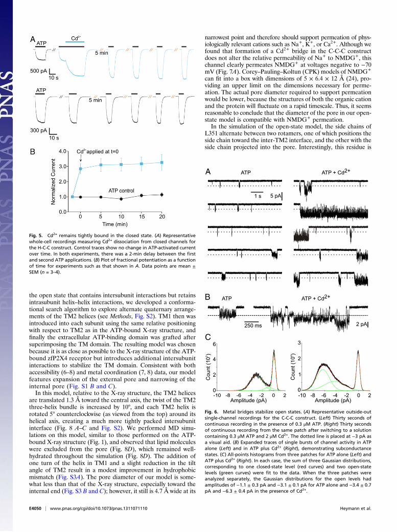
coordination of Cd2+ involving residues in TM1 and TM2 wouldbe predicted to occur in the open state, but partial and poten-tially stable coordination involving S345C and C348 could occureven in the closed state. In both apo and ATP-bound structures,these TM2 residues are equivalently positioned on the same faceof the helix, and there is precedence in other proteins in whichtwo residues on one face of an α-helix form a strong metal-binding site (17–19). In experiments in which both Cd2+ andATP were removed following potentiation of ATP-activatedcurrents, we observed that both H-C-C and C-C-C constructscould close (Fig. 3 A and C), indicating either that Cd2+ rapidlydissociates from the channel or that the bridge does not lock thechannel open per se, as was observed previously for the internalgate region of the Shaker Kv channel (20). To determine whetherCd2+ remains stably associated with the P2X2 receptor afterATP unbinds and channels close, we used a protocol in whichCd2+ was applied together with ATP to potentiate channel ac-tivity; once the channel closed after both Cd2+ and ATP wereremoved, we repeatedly challenged the cell with ATP alone toassess how long channel activity would remain potentiated (Fig.5A). In experiments with the H-C-C construct, we observed littleevidence of recovery from Cd2+-mediated potentiation even20 min after the removal of Cd2+ (Fig. 5), demonstrating stableinteraction of the metal with the closed channel. Using thisprotocol for the C-C-C construct, we also observed stable po-tentiation 15 min after removal of Cd2+.To investigate the mechanism of Cd2+ potentiation further, we
undertook single-channel recordings. Membrane patches in theoutside-out configuration of the patch-clamp technique wereisolated from cells expressing the C-C-C construct because thesechannels produced the most robust potentiation of ATP-activatedcurrents by Cd2+. Analysis was conducted on three patches inwhich channel activity was observed in the presence of ATP butnot in its absence. Two additional patches exhibited qualitativelysimilar results but were excluded from the analysis because in-frequent channel openings in the absence of ATP precluded thedefinitive identification of ATP-activated currents. Channels acti-vated by 0.3 μM ATP fluctuated rapidly among multiple currentlevels, with a predominant level of 3.1 ± 0.1 pA at −120 mV (Fig.
Fig. 3. Two metal bridges that potentiate ATP-activated currents. (A)Representative whole-cell currents for the H-C-C bridging construct in rP2X2receptor channels. EC20 concentrations of ATP (3 μM; black bars) and Cd2+
(20 μM; blue bars) were applied as shown. (B) Concentration–response rela-tionships for activation of rP2X2 H-C-C by ATP in the absence and presenceof 20 μM Cd2+. A control concentration of ATP (3 μM) was applied to eachcell, followed by a test concentration of ATP before the additional appli-cation of 20 μM Cd2+. Currents measured with test concentrations of ATP(before and after Cd2+) were normalized to the current amplitude measuredat the control concentration of ATP to generate the ATP-alone (black datapoints and curve) and ATP plus Cd2+ (blue data points and curve) relations.The ATP relation then was scaled (gray data points and curve) to show theshift in EC50. Data points are mean for three to seven measurements, anderror bars indicate SEM. (C) Representative whole-cell recordings for the C-C-C bridging construct in rP2X2 receptor channels. EC20 concentrations of ATP(2.6 μM; black bars) and Cd2+ (20 μM; blue bars) were applied as shown. (D)Concentration–response relationships for activation of rP2X2 C-C-C by ATP inthe absence and presence of 20 μM Cd2+. A different protocol was used inthe C-C-C construct because the large magnitude of potentiation necessi-tated the activation of currents in the absence of Cd2+ that were too small tobe reliably measured. For this construct, concentration–response relation-ships were generated independently for ATP alone (n = 4) or for ATP plus20 μM Cd2+ in the external solution (n = 3). The scaled relationship (gray datapoints and curve) was obtained by measuring fold potentiation at saturating[ATP] in a separate set of experiments (n = 4). Error bars indicate SEM. TheHill equation was fit to the data, and parameters are given in Table S1.
E4048 | www.pnas.org/cgi/doi/10.1073/pnas.1311071110 Heymann et al.

6, Left). The addition of 2 μM Cd2+ resulted in dramatic in-creases in both the open probability and unitary current ampli-tudes of the channel (Fig. 6, Right). The predominant currentamplitude in the single-channel current distribution increasedapproximately twofold in the presence of Cd2+, whereas in-dividual burst events, defined as channel openings not inter-rupted by a close event exceeding 50 ms in duration, increased7.5 ± 1.8-fold. The maximum observed burst duration in ATPalone was 471 ms, whereas in the presence of Cd2+ the longestmeasurable burst lasted 6,698 ms. The effect of Cd2+ on burstduration is an underestimation, because bursts of channel acti-vity during which a second channel also became active wereexcluded from the analysis, and such instances predominantlyoccurred during long bursts of channel activity. The observedeffect of Cd2+ on burst duration indicates that the bridge formedby Cd2+ stabilizes open states. The effect of Cd2+ on the unitaryconductance of the channel is intriguing, because it suggests thatthe metal bridge either stabilizes channel conformations in-frequently visited by the channel in its absence or that the metalbridge leads to a conformational change that results in an ap-proximate doubling of the current at all channel sublevels.We also explored whether the conducting states stabilized by
the C-C-C bridge might correspond to the dilated state of P2Xreceptors that gives rise to a change in the relative permeabilityof Na+ to N-methyl-D-glucamine (NMDG+) (21–23). Macroscopiccurrent–voltage (I–V) relationships were obtained using voltageramps in whole-cell recordings in which Na+ was the primaryinternal cation and NMDG+ was the primary external cation. Inprevious reports with related ionic conditions and protocols, thezero-current potential shifted in the positive direction in thecontinuous presence of ATP as the relative permeability of Na+
to NMDG+ changes (21–23). When Cd2+ was applied to theC-C-C construct, we observed large increases in ATP-activatedmacroscopic current for both inward and outward limbs of theI–V relationship, with no detectable change in the zero-currentpotential (Fig. 7). These results indicate that the relative per-meability of Na+:NMDG+ for the rP2X2 receptor does notchange when Cd2+ forms a bridge between TM1 and TM2, andthus the state stabilized by the bridge does not correspond to theproposed dilated state of P2X receptor channels.
A Structural Model for the Open State of zfP2X4 Receptors. Theresults presented thus far suggest that the intrasubunit inter-actions observed in both the apo and ATP-bound structures areremarkably consistent with our engineered metal bridges be-tween TM1 and TM2. However, the ATP-bound structure of thezfP2X4 receptor is nonnative in that intersubunit interactions arelargely absent within the TM domain. To generate a model for
Fig. 4. Subunit relationships of metal bridges. (A) Legend for concatenatedsubunit constructs. Each rectangle (made up of three squares) corresponds toone subunit of the trimer, and the residues at the three bridging positions(33, 345, and 348) are represented by their one-letter amino acid code ineach square of the rectangle. Bridging residues (H33, S345C, and C348) arecolored blue; nonbridging residues (H33Y, S345, and C348T) are coloredgray. (B–D) Concatenated H-C-C subunit constructs with one, two, or three
subunits containing all three residues necessary for bridging. Control cur-rents activated by ATP alone (black trace) or ATP plus Cd2+ (blue trace) aresuperimposed for comparison. (E–G) Concatenated H-C-C subunit constructswith the three residues necessary for bridging split between subunits to testfor intersubunit bridges. In each construct, one of the three bridging resi-dues was removed from subunit 1 and placed in the two adjacent subunits.Control currents activated by ATP alone (black trace) or ATP plus Cd2+ (bluetrace) are superimposed for comparison. (H) Bar graph summarizing theeffects of Cd2+ on each concatameric construct (n = 3–4). Measurements ofstatistical significance are based on unpaired Student t tests; *P ≤ 0.05, **P ≤0.005. See Table S1 for concentration–response relations for each construct.EC 50 concentrations of ATP and 20 μM Cd2+ were used for each construct. (I)Modeling of a Cd2+ bridge between S345C and H33 in the 3T construct ofrP2X2 receptor (7) using the X-ray structure of the apo zfP2X4 receptor. In 3TrP2X2 a native Cys at 348 and two additional Cys residues in the C and Ntermini were mutated to Thr. Bridging residues S345C and H33 (rP2X2numbering) are shown for only one subunit, with Cd2+ represented as a bluesphere. The side view shown here is from the side opposite that depicted inFig. 2.
Heymann et al. PNAS | Published online September 30, 2013 | E4049
PHYS
IOLO
GY
PNASPL
US
the open state that contains intersubunit interactions but retainsintrasubunit helix–helix interactions, we developed a conforma-tional search algorithm to explore alternate quaternary arrange-ments of the TM2 helices (see Methods, Fig. S2). TM1 then wasintroduced into each subunit using the same relative positioningwith respect to TM2 as in the ATP-bound X-ray structure, andfinally the extracellular ATP-binding domain was grafted aftersuperimposing the TM domain. The resulting model was chosenbecause it is as close as possible to the X-ray structure of the ATP-bound zfP2X4 receptor but introduces additional intersubunitinteractions to stabilize the TM domain. Consistent with bothaccessibility (6–8) and metal coordination (7, 8) data, our modelfeatures expansion of the external pore and narrowing of theinternal pore (Fig. S1 B and C).In this model, relative to the X-ray structure, the TM2 helices
are translated 1.3 Å toward the central axis, the twist of the TM2three-helix bundle is increased by 10°, and each TM2 helix isrotated 5° counterclockwise (as viewed from the top) around itshelical axis, creating a much more tightly packed intersubunitinterface (Fig. 8 A–C and Fig. S2). We performed MD simu-lations on this model, similar to those performed on the ATP-bound X-ray structure (Fig. 1), and observed that lipid moleculeswere excluded from the pore (Fig. 8D), which remained well-hydrated throughout the simulation (Fig. 8D). The addition ofone turn of the helix in TM1 and a slight reduction in the tiltangle of TM2 result in a modest improvement in hydrophobicmismatch (Fig. S3A). The pore diameter of our model is some-what less than that of the X-ray structure, especially toward theinternal end (Fig. S3 B and C); however, it still is 4.7 Å wide at its
narrowest point and therefore should support permeation of phys-iologically relevant cations such as Na+, K+, or Ca2+. Although wefound that formation of a Cd2+ bridge in the C-C-C constructdoes not alter the relative permeability of Na+ to NMDG+, thischannel clearly permeates NMDG+ at voltages negative to −70mV (Fig. 7A). Corey–Pauling–Koltun (CPK) models of NMDG+
can fit into a box with dimensions of 5 × 6.4 × 12 Å (24), pro-viding an upper limit on the dimensions necessary for perme-ation. The actual pore diameter required to support permeationwould be lower, because the structures of both the organic cationand the protein will fluctuate on a rapid timescale. Thus, it seemsreasonable to conclude that the diameter of the pore in our open-state model is compatible with NMDG+ permeation.In the simulation of the open-state model, the side chains of
L351 alternate between two rotamers, one of which positions theside chain toward the inter-TM2 interface, and the other with theside chain projected into the pore. Interestingly, this residue is
Fig. 5. Cd2+ remains tightly bound in the closed state. (A) Representativewhole-cell recordings measuring Cd2+ dissociation from closed channels forthe H-C-C construct. Control traces show no change in ATP-activated currentover time. In both experiments, there was a 2-min delay between the firstand second ATP applications. (B) Plot of fractional potentiation as a functionof time for experiments such as that shown in A. Data points are mean ±SEM (n = 3–4).
Fig. 6. Metal bridges stabilize open states. (A) Representative outside-outsingle-channel recordings for the C-C-C construct. (Left) Thirty seconds ofcontinuous recording in the presence of 0.3 μM ATP. (Right) Thirty secondsof continuous recording from the same patch after switching to a solutioncontaining 0.3 μM ATP and 2 μM Cd2+. The dotted line is placed at −3 pA asa visual aid. (B) Expanded traces of single bursts of channel activity in ATPalone (Left) and in ATP plus Cd2+ (Right), demonstrating subconductancestates. (C) All-points histograms from three patches for ATP alone (Left) andATP plus Cd2+ (Right). In each case, the sum of three Gaussian distributions,corresponding to one closed-state level (red curves) and two open-statelevels (green curves) were fit to the data. When the three patches wereanalyzed separately, the Gaussian distributions for the open levels hadamplitudes of −1.1 ± 0.3 pA and −3.1 ± 0.1 pA for ATP alone and −3.4 ± 0.7pA and −6.3 ± 0.4 pA in the presence of Cd2+.
E4050 | www.pnas.org/cgi/doi/10.1073/pnas.1311071110 Heymann et al.

equivalent to V343 in the rP2X2 receptor, where substitution ofCys results in the formation of a stable Cd2+ bridge that blocksthe channel (7). This Cd2+ bridge forms rapidly (rate constant =
106 M/s) in the presence of ATP, suggesting that bridging occursin the highly populated open state, and requires Cys in all threesubunits, implying that it forms at the central axis of the pore (7).L351 in the X-ray structure of the ATP-bound zfP2X4 receptorcannot coordinate Cd2+ because of the larger diameter of thepore at this position (L351 Cβ–Cβ distances of 11.2 Å; Fig. S1B).However, in our open-state model L351C residues in the alter-native rotamer, together with two water molecules, can coor-dinate Cd2+ in the triagonal bipyramidal geometry (Fig. 8F).
DiscussionThe objective of the present study was to evaluate both inter-subunit and intrasubunit interactions within the TM domain ofP2X receptors using both computational and functional ap-proaches. A key motivation for this work was the unexpectedabsence of intersubunit interactions in the recent X-ray structureof the ATP-bound zfP2X4 receptor, which raises the possibilitythat the structure is distorted (15) and brings into question theproposed mechanism of ATP activation. Indeed, our MD sim-ulations suggest that lipid molecules can diffuse through the in-tersubunit crevices to occupy and dehydrate the ion permeationpathway (Fig. 1), a scenario that is incompatible with ion con-duction. As noted previously (15), the predominance of hydro-carbon side chains in helical transmembrane domains implicatesnonspecific van der Waals and weak electrostatic interactions asthe main intraprotein forces for tertiary and quaternary struc-
Fig. 7. The Cd2+ metal bridge does not alter the relative permeability ofNa+ to NMDG+. (A) Representative whole-cell ATP-activated current–voltagerelationships in response to voltage ramps for the C-C-C construct in theabsence (blue trace) and presence (black trace) of Cd2+. EC20 [ATP] or EC20
[ATP] plus 20 μM Cd2+ were applied externally. The currents shown are thenet current activated by ATP alone or by ATP in the presence of Cd2+. Eachcurrent trace is the average of five consecutive voltage ramps from −100 to+40 mV (ramp duration was 2.3 s with 1-s intervals between ramps) aftersubtracting the current in the absence of ATP. (B) Bar graph displaying theaverage zero-current potential measured from three cells.
Fig. 8. A structural model for the open state of P2X receptors. (A) Comparison of the TM domain between the X-ray structure for the ATP-bound zfP2X4 re-ceptor (shown in light green) and the open-state model (with individual subunits colored in blue, gold, and gray) viewed from the external side of the membrane.The Cα atoms of the C terminus (residue G56) of TM1 and the N terminus (residue I335) of TM2 are shown as spheres. (B and C) Interfaces between adjacentsubunits for the ATP-bound zfP2X4 receptor and the open-state model, respectively, depicted in a surface rendering. Hypothetical boundaries of the lipid bilayerare represented by gray bands. (D) Snapshot of the TM domain of the open-state model and its adjacent lipid molecules in the restrained MD simulation, viewedfrom the external side of the membrane. In contrast to observations in the simulations of the ATP-bound zfP2X4 receptor, no lipid molecules entered the pore insimulations of the open-state model. (E) Continuous water chain in the pore of the model. (F) Cd2+ coordination by the L351C trimer and two water molecules.
Heymann et al. PNAS | Published online September 30, 2013 | E4051
PHYS
IOLO
GY
PNASPL
US
tural stability. In native membranes, the hydrophobic environ-ment provided by the acyl chains of lipid molecules enhanceshelix–helix packing and helps define the hydrophobic dimension.The lack of such a hydrophobic environment in the crystallinelattice is likely the reason for the poor intersubunit packing ofthe TM1 and TM2 helices and the apparent hydrophobic mis-match in the X-ray structure of the ATP-bound zfP2X4 receptor.Such distortions may be particularly problematic for membraneproteins such as P2X receptors, which contain large extracellulardomains that provide most of the crystal contacts, together withsmall transmembrane domains that contain only six helices in thetrimer (15).Although the intersubunit crevices are a striking feature of the
ATP-bound X-ray structure, only relatively modest structural re-arrangements are required to form an intersubunit interfacewithin the membrane. In our open-state model of the zfP2X4receptor, intersubunit interactions within two regions of the TMdomain were created by small rotations and translation of theTM2 helix toward the central axis (Fig. 8). The first interface iswithin the internal region of the TM domain, where residues onthree turns of the TM2 helix (L351, I355, and W358) from onesubunit make contact with L346, A347, and V354 on the adja-cent TM2 helix. The second is within the external end of the TMdomain, where Y45 in TM1 contacts L340 in TM2 of the adjacentsubunit. These contacts do not completely seal off the pore fromthe surrounding lipid membrane but diminish the crevices toopenings or portals (Fig. 8C), similar to what has been seen in theKcsA potassium channel (25) and the NavAb voltage-activatedsodium channel (26). In addition, our model exhibits a modestimprovement in hydrophobic mismatch (Fig. S3A).We also investigated the intrasubunit interactions depicted in
both the apo and ATP-bound X-ray structures (Fig. 2), and inthis case we identified metal bridges that are fully compatiblewith those structures. Our results demonstrate that S345C andC348 in TM2 and H/C33 in TM1 form a robust intrasubunit Cd2+
bridge that stabilizes the open state of the rP2X2 receptor (Figs.3–6). The X-ray structures position the equivalent of S345C andC348 on one face of the TM2 helix where they can coordinateCd2+ in either the apo or ATP-bound states (Fig. 2), consistentwith our functional results demonstrating that Cd2+ remains stablyassociated with the channel in the closed state (Fig. 5). Thosestructures also show that ATP binding triggers a relative rotationof the TM1/TM2 interface so that residue 33 in TM1 also canparticipate in coordinating the metal. Thus, our metal-bridgingresults substantiate intrasubunit helix interactions and theintrasubunit motions between TM1 and TM2 upon ATP bindingthat are depicted in the X-ray structures of the zfP2X4 receptor.When considered together with the modest modifications thatare required to decrease the size of the intersubunit crevices, theseresults support the proposed structural mechanism by which ATPbinding leads to an iris-like opening of P2X receptor channels (5).In our structural model for the open state, this structural rear-rangement would also be accompanied by a narrowing of the in-ternal pore to create intersubunit interactions between the TM2helices (Fig. 8).The metal bridges that we engineered between TM1 and TM2
also identify an internal region of the TM domain that is par-ticularly sensitive to modification, where the activity of thechannel can be tuned readily (Figs. 3–6). The relative motionsbetween TM1 and TM2 in this region are not very large (Fig. 2),but occupancy of the engineered metal-binding site has profoundconsequences for both the gating and conduction properties ofP2X receptors. In the case of the C-C-C bridge, Cd2+ occu-pancy results in an increase in burst duration, suggesting thatthe bridge stabilizes the open state. We also observed a substantialincrease in mean unitary conductance (Fig. 6), implying that thislocal region plays a key role in determining the permeationproperties of P2X receptor channels. The internal pore in our
open-state model of the zfP2X4 receptor is the narrowestregion of the pore, and it contains the most extensive intersubunitinteractions, providing an initial picture of the structure of thisimportant region of P2X receptor channels. Formation of inter-subunit interfaces with the internal region of TM2 is consistent withobservations that this region of TM2 is particularly sensitive tomutations (6, 27–29), and the model is fully compatible with metalbridges that have been described for the TM domain of rP2X2 re-ceptor channels. These bridges include an intersubunit metal bridgeformed at the threefold axis by V343C (Fig. 8F) (7), stable metalbridges formed by D349C (8), as well as the metal bridges pre-sented here that are located at the intrasubunit interface betweenthe TM1 and TM2 helices. Refinement of this working model toreduce further the hydrophobic mismatch, while retaining theconstraints imposed by the metal bridges discussed here, will helpto identify key intersubunit interfaces involved in channel gating.
MethodsEvaluation of the X-Ray Structure for ATP-Bound zfP2X4 Receptor by MDSimulations. Before being embedded in a lipid bilayer for MD simulations,the X-ray structure of ATP-bound zfP2X4 receptor was preprocessed as fol-lows. Using symmetry operations, the monomer entry in ATP-bound zfP2X4receptor was replicated into a threefold symmetric trimer. The trimer wasenergy minimized for 10,000 steps while the Cα atoms and the heavy atomsof the ATP molecules and the surrounding residues (N296, R298, K316, K70,K72, and T189) were restrained in their original positions with a force con-stant of 10 kcal−1·mol−1·Å−2.
A preequilibrated DMPC lipid bilayer was obtained from CHARMM-GUI(30), replicated, and trimmed to generate a membrane with 398 lipid mol-ecules solvated by 32,187 water molecules. The preprocessed structure of thezfP2X4 receptor then was inserted into the membrane, with lipid moleculeswithin 1 Å and water molecules within 3 Å removed. Sodium and chlorideions were added to neutralize the system and provide a physiological con-centration of salt. The resulting system contained the receptor along with332 lipid molecules, 32,004 water molecules, 93 sodium ions, and 90 chlorideions (corresponding to a salt concentration of 0.15 M). The system prepa-ration was done using VMD (31) and its Solvate and Ionize plugins.
The systemwas energy minimized for 10,000 steps and then was simulatedfor 79.44 ns, with the restraints stated above, but the restraining forceconstant was reduced to 1 kcal−1·mol−1·Å−2 after the first 21.98 ns. A secondsimulation was started from the snapshot at 12.15 ns of the restrainedsimulation and was run for 53.66 ns. In this simulation, all the previousrestraints were released. However, to ensure that the ATP molecules stayedin the binding pockets, distance restraints were introduced between pairs ofatoms involved in ATP–zfP2X4 receptor salt bridges and hydrogen bonds (5).
All simulations were performed using NAMD 2.9 (32) with temperature at310 K and pressure at 1 atm. The Langevin dynamics and Nosé–HooverLangevin piston methods were used for temperature and pressure coupling,respectively. CHARMM27 protein (33) and CHARMM36 lipid (34) force fieldswere used. The force field for ATP was from Pavelites et al. (35).
Modeling Cd2+ Bridges Between TM1 and TM2 of the zfP2X4 Receptor. Putativebridging residues (e.g., N35 and N353) were mutated into Cys or His using theMutator plugin of VMD. Sidechain dihedral angles of the introduced Cys orHis residueswere adjustedmanually so that their sulfur or nitrogen atomswereat possible positions to coordinate Cd2+. To eliminate possible steric clashes bythe mutations, each mutant was energy minimized for 10,000 steps, while Cαatoms were restrained with a force constant of 10 kcal−1·mol−1·Å−2.
Construction of an Open-State Model of the zfP2X4 Receptor. The TM2 helixfrom a single subunit in the energy-minimized ATP-bound zfP2X4 receptorstructure was used as the starting point for the conformational search togenerate the open-state model. This helix was translated and rotated so thatits helical axis was aligned with the z-axis and its center of mass was at theorigin of the coordinate system and is called “TM2mono” hereafter. Thez-axis represents the membrane normal. All translation and rotation oper-ations were done using VMD scripts.
A series of rotation, translation, and replication was performed onTM2mono to generate an ensemble of conformations of TM2 trimer uponwhich several filters were applied to select plausible models. The operationson TM2mono were carried out sequentially as follows: (i) rotation around itshelical axis (i.e., the z-axis) by an angle ρ; (ii) rotation around the x-axis by anangle θ; (iii) rotation around the z axis by an angle ϕ; (iv) translation along
E4052 | www.pnas.org/cgi/doi/10.1073/pnas.1311071110 Heymann et al.

the x-axis by a distance r; (v) replication to create a threefold symmetricTM2 trimer.
We constrained the search space to be centered around the ATP-boundX-ray structure by using the following ranges for the four degrees of freedom:ρ from −30° to 10° with increments of 10°; θ from 25° to 45° with incrementsof 5°; ϕ from −40° to 80° with increments of 10°; and r from 6–10 Å withincrements of 1 Å.
The first filter aimed to tighten the interfaces in the TM2 trimer. Wetherefore selected models with Cα–Cα distances for neighboring A344 resi-dues and for neighboring L351 residues that were shorter than in the ATP-bound X-ray structure.
The secondfilter was applied to avoid steric clashes on the one hand and toensure adequate intersubunit contacts on the other. We found the minimumCα–Cα distances between neighboring TM2 helices are 4.5 Å in the apostructure and 9.5 Å in the ATP-bound structure. We eliminated any model inwhich at least one intersubunit Cα–Cα distance was less than 4.5 Å or inwhich no single intersubunit Cα–Cα distance was less than 9.5 Å.
The third filter was used to select those with adequate pore sizes from thesurviving models. For each model, the pore-radius profile along the threefoldaxis was calculated using the HOLE program (36), and the minimum radius wasobtained. Models with minimum pore radius greater than 2 Å were selected.
The final model was chosen, by visual inspections, because the inter-TM2crevices were filled to the greatest extent. In this model ρ = −10°, θ = 45°, ϕ =30°, and r = 7 Å. The side chain dihedral angles of L351, V354, and W358were adjusted to open the pore further and improve the packing in theinterfaces. To the final model of the TM2 trimer, we added a TM1 helix ineach subunit by using its relative positioning with respect to TM2 as in theATP-bound X-ray structure. We did so by taking TM1 and TM2 in one subunitof the ATP-bound X-ray structure and then superimposing the TM2 portionon our model, one subunit at a time; the resulting TM1 helices in the threesubunits were the desired additions. Finally the ectodomain of the ATP-bound X-ray structure was grafted after the TM domain was superimposedon our model. A restrained MD simulation was run on this open-state model,similar to the simulation performed for the ATP-bound X-ray structure.
Channel Constructs. Mutations were introduced into an rP2X receptor con-struct (generously provided by David Julius, University of California, SanFrancisco) in which C9 and C430 were mutated to threonine (6), a constructtermed “2T.” The previously reported inhibitory Cd2+ bridge between H33and S345C (7) was studied in the 3T background, a construct that also con-tains the C348T mutation. The primary potentiating Cd2+ bridges studiedhere involved H33 or H33C in TM1 and both S345C and C348 in TM2 (H-C-Cand C-C-C, respectively), and these were studied in the 2T background. Cd2+
also has weak and rapidly reversible potentiating activity on the wild-typerP2X2 receptor channel; external application of 20 μM Cd2+ increases mac-roscopic currents activated by 15 μM ATP by 15 ± 3% (n = 3), and 100 μMCd2+ increases ATP-activated currents by 48 ± 8% (n = 3). Although we havenot determined the coordinating residues responsible for this weak andrapidly reversible potentiation in the wild-type channel, a simple explana-tion that would be consistent with the present study is that H33 and C348Ccan form a weak intrasubunit Cd2+ bridge that stabilizes the open state.Concatamers were constructed as described previously (7, 37, 38) and wereconfirmed by restriction digests and DNA sequencing. In addition, cell lysateswere evaluated with SDS/PAGE and Western blot analysis, which confirmedthat the most abundant species of rP2X2 receptors in HEK cells expressingthe concatenated trimeric construct corresponds to the molecular weightof a trimer (Fig. S4).
Cell Culture and Transfection. HEK293 cells were cultured in DMEM supple-mented with 10% (vol/vol) FBS and 10 mg/L gentamicin. All cell-culturereagents were obtained from GIBCO. Trypsin-treated HEK293 cells weretransiently transfected and seeded onto glass coverslips in six-well plates andwere placed in a 37 °C incubator with 95% air and 5% CO2. Transfectionswere performed using FuGENE6 Transfection Reagent (Promega). P2X re-ceptors were cotransfected with a GFP cDNA construct in pGreen-Lantern(Invitrogen) at ratios varying from 2:1–8:1. Recordings were conducted 24–60 hafter transfection.
Western Blot Analysis of Concatenated Constructs. Cells grown in T25 25-cm2
flasks were transfected with P2X receptor DNA using FuGene 6. Two daysafter transfection, cells were collected in Dulbecco’s PBS and then werecentrifuged at 500 × g for 3 min. The supernatant was discarded aftercentrifugation, and a lysis buffer containing 1% Triton X-100 and proteaseinhibitors was used to suspend the cell pellet. Cells in lysis solution weresonicated for 5 s on ice and incubated on ice for 30 min, with vortexing for5 s every 5 min. The cell lysate then was centrifuged at 13,000 × g for 20 minat 4 °C, and the supernatant was combined with LDS buffer, DTT, and2-mercaptoethnol. Samples were heated at 95 °C for 5 min and then werecentrifuged at 13,000 × g for 2 min. Proteins were separated in a 4–12%NuPage Bis-Tris gel (Invitrogen) using a running buffer containing (in mM):50 3-(N-morpholino)propanesulfonic acid, 50 Tris base, 3.46 SDS, and 1EDTA. SeeBlue Plus2 (Invitrogen) was used as the protein molecular-weightmarker. Protein in the gel was transferred to nitrocellulose membrane andprobed with goat anti-hP2X2 antibody (Santa Cruz).
Electrophysiology. All experiments were performed in transiently transfectedHEK293 cells under voltage-clamp (−60 mV for whole-cell and −120 mV forsingle-channel) recording using an Axopatch 200B patch-clamp amplifier(Axon Instruments, Inc.) and were digitized on-line using a Digidata 1321Ainterface board and pCLAMP 10.2 software (Axon Instruments, Inc.). Whole-cell currents were filtered at 5 kHz using eight-pole Bessel filters and weredigitized at 20 kHz. Single-channel recordings were filtered initially at 5 kHz,were digitized at 10–50 kHz, and were further filtered at 2 kHz for dataanalysis and at 1 kHz for presentation.
The whole-cell external solution contained (in mM): 140 NaCl, 5.4 KCl,2 CaCl2, 0.5 MgCl2, 10 Hepes, and 10 D-glucose, adjusted to pH 7.3 withNaOH. The internal solution contained (in mM): 140 NaCl, 10 EGTA and 10Hepes, adjusted to pH 7.0 with NaOH. The single-channel external solutioncontained (in mM): 147 NaCl, 1 CaCl2, 10 Hepes, and 11 D-glucose, adjustedto pH 7.4 with NaOH. The internal solution contained (in mM): 140 NaF,5 NaCl, 10 EGTA and 10 Hepes, adjusted to pH 7.0 with NaOH. NMDG+ per-meability experiments were conducted with an external solution containing(in mM): 150 NMDG+, 10 Hepes, adjusted to pH 7.3 with HCl. The internalsolution contained (in mM): 140 NaCl, 10 EGTA, 10 Hepes, adjusted to pH 7.0with NaOH. Voltage ramps were given from −100 mV to +40 mV. In eachexperimental condition, five consecutive current traces were averaged, andthen the average leak current was subtracted from the average ATP- andATP plus Cd2+-evoked currents.
Solution exchange was achieved using the Rapid Solution Changer RSC-200 (BioLogic), which has the capacity of switching between nine solutionswithin 40 ms depending on the size of the cell. ATP and Cd2+ solutions wereprepared daily and diluted to the desired concentration in external solutionimmediately before each experiment.
To generate the concentration–response relationships, a reference con-centration of ATP was applied before applying a test concentration, aspreviously described (6). The Hill equation was fit to the data according to
I=Imax ¼ ½ATP�n=�½ATP�n þ ECn50
�
where I is the normalized current at a given concentration of ATP, Imax is themaximum normalized current, EC50 is the concentration of ATP ([ATP])producing half-maximal currents, and n is the Hill coefficient.
Analysis of the duration of single-channel bursts was conducted inpCLAMP 10.2 (Axon Instruments, Inc.). Only patches in which no channelactivity was observed in the absence of ATP were analyzed. All eventsdetected by the software were inspected visually and were rejected if two ormore channel openings superimposed. Bursts of single-channel currents wereextracted from the original data file and transferred to SigmaPlot 10.0, whichwas used for generating the all-point histogram. Origin 8.1 was used forfitting the data with Gaussian distributions.
ACKNOWLEDGMENTS. We thank Andres Jara-Oseguera, Mark Mayer, MiguelHolmgren, Dmitriy Krepkiy, Jeet Kalia, Gilman Toombes, and other membersof the K.J.S. laboratory for helpful discussions. This work was supported by theIntramural Research Program of the National Institute of Neurological Disor-ders and Stroke, National Institutes of Health (NIH) (K.J.S.), NIH Pathway toIndependence Award NS070954 (to M.L.), and NIH Grants GM058187 and GM088187(to H.-X.Z.).
1. Khakh BS, North RA (2012) Neuromodulation by extracellular ATP and P2X receptors
in the CNS. Neuron 76(1):51–69.2. Jiang R, Taly A, Grutter T (2013) Moving through the gate in ATP-activated P2X re-
ceptors. Trends Biochem Sci 38(1):20–29.
3. Surprenant A, North RA (2009) Signaling at purinergic P2X receptors. Annu Rev
Physiol 71:333–359.4. Kawate T, Michel JC, BirdsongWT, Gouaux E (2009) Crystal structure of the ATP-gated
P2X(4) ion channel in the closed state. Nature 460(7255):592–598.
Heymann et al. PNAS | Published online September 30, 2013 | E4053
PHYS
IOLO
GY
PNASPL
US

5. Hattori M, Gouaux E (2012) Molecular mechanism of ATP binding and ion channelactivation in P2X receptors. Nature 485(7397):207–212.
6. Li M, Chang TH, Silberberg SD, Swartz KJ (2008) Gating the pore of P2X receptorchannels. Nat Neurosci 11(8):883–887.
7. Li M, Kawate T, Silberberg SD, Swartz KJ (2010) Pore-opening mechanism in trimericP2X receptor channels. Nat Commun 1:44.
8. Kracun S, Chaptal V, Abramson J, Khakh BS (2010) Gated access to the pore of a P2Xreceptor: Structural implications for closed-open transitions. J Biol Chem 285(13):10110–10121.
9. Kawate T, Robertson JL, Li M, Silberberg SD, Swartz KJ (2011) Ion access pathway tothe transmembrane pore in P2X receptor channels. J Gen Physiol 137(6):579–590.
10. Samways DS, Khakh BS, Dutertre S, Egan TM (2011) Preferential use of unobstructedlateral portals as the access route to the pore of human ATP-gated ion channels (P2Xreceptors). Proc Natl Acad Sci USA 108(33):13800–13805.
11. Jiang R, et al. (2011) Agonist trapped in ATP-binding sites of the P2X2 receptor. ProcNatl Acad Sci USA 108(22):9066–9071.
12. Du J, Dong H, Zhou HX (2012) Gating mechanism of a P2X4 receptor developed fromnormal mode analysis and molecular dynamics simulations. Proc Natl Acad Sci USA109(11):4140–4145.
13. Lörinczi E, et al. (2012) Involvement of the cysteine-rich head domain in activation anddesensitization of the P2X1 receptor. Proc Natl Acad Sci USA 109(28):11396–11401.
14. Jiang R, et al. (2012) Tightening of the ATP-binding sites induces the opening of P2Xreceptor channels. EMBO J 31(9):2134–2143.
15. Zhou HX, Cross TA (2013) Influences of membrane mimetic environments on mem-brane protein structures. Annu Rev Biophys 42:361–392.
16. Liang X, et al. (2013) Functional Identification of Close Proximity Amino Acid SideChains within the Transmembrane-Spanning Helixes of the P2X2 Receptor. PLoS ONE8(8):e70629.
17. Arnold FH, Haymore BL (1991) Engineered metal-binding proteins: Purification toprotein folding. Science 252(5014):1796–1797.
18. Suh SS, Haymore BL, Arnold FH (1991) Characterization of His-X3-His sites in alpha-helices of synthetic metal-binding bovine somatotropin. Protein Eng 4(3):301–305.
19. Taraska JW, Puljung MC, Olivier NB, Flynn GE, Zagotta WN (2009) Mapping thestructure and conformational movements of proteins with transition metal ion FRET.Nat Methods 6(7):532–537.
20. Holmgren M, Shin KS, Yellen G (1998) The activation gate of a voltage-gated K+channel can be trapped in the open state by an intersubunit metal bridge. Neuron21(3):617–621.
21. Browne LE, Compan V, Bragg L, North RA (2013) P2X7 receptor channels allow directpermeation of nanometer-sized dyes. J Neurosci 33(8):3557–3566.
22. Virginio C, MacKenzie A, Rassendren FA, North RA, Surprenant A (1999) Pore dilationof neuronal P2X receptor channels. Nat Neurosci 2(4):315–321.
23. Khakh BS, Bao XR, Labarca C, Lester HA (1999) Neuronal P2X transmitter-gated cationchannels change their ion selectivity in seconds. Nat Neurosci 2(4):322–330.
24. Villarroel A, Burnashev N, Sakmann B (1995) Dimensions of the narrow portion ofa recombinant NMDA receptor channel. Biophys J 68(3):866–875.
25. Zhou Y, Morais-Cabral JH, Kaufman A, MacKinnon R (2001) Chemistry of ion co-ordination and hydration revealed by a K+ channel-Fab complex at 2.0 A resolution.Nature 414(6859):43–48.
26. Payandeh J, Gamal El-Din TM, Scheuer T, Zheng N, Catterall WA (2012) Crystalstructure of a voltage-gated sodium channel in two potentially inactivated states.Nature 486(7401):135–139.
27. Silberberg SD, Li M, Swartz KJ (2007) Ivermectin Interaction with transmembranehelices reveals widespread rearrangements during opening of P2X receptor channels.Neuron 54(2):263–274.
28. Silberberg SD, Chang TH, Swartz KJ (2005) Secondary structure and gating re-arrangements of transmembrane segments in rat P2X4 receptor channels. J GenPhysiol 125(4):347–359.
29. Cao L, Broomhead HE, Young MT, North RA (2009) Polar residues in the secondtransmembrane domain of the rat P2X2 receptor that affect spontaneous gating,unitary conductance, and rectification. J Neurosci 29(45):14257–14264.
30. Jo S, Kim T, Iyer VG, Im W (2008) CHARMM-GUI: A web-based graphical user interfacefor CHARMM. J Comput Chem 29(11):1859–1865.
31. Humphrey W, Dalke A. Schulten K. (1996) VMD: Visual molecular dynamics. J MolGraph 14(1):33–38.
32. Phillips JC, et al. (2005) Scalable molecular dynamics with NAMD. J Comput Chem26(16):1781–1802.
33. Mackerell AD, Jr., Feig M, Brooks CL, 3rd (2004) Extending the treatment of backboneenergetics in protein force fields: Limitations of gas-phase quantum mechanics inreproducing protein conformational distributions in molecular dynamics simulations.J Comput Chem 25(11):1400–1415.
34. Klauda JB, et al. (2010) Update of the CHARMM all-atom additive force field for lipids:Validation on six lipid types. J Phys Chem B 114(23):7830–7843.
35. Pavelites JJ, Gao J, Bash PA, Mackerell AD (1997) A molecular mechanics force field forNAD+, NADH, and the pyrophosphate groups of nucleotides. J Comput Chem 18(2):221–239.
36. Smart OS, Neduvelil JG, Wang X, Wallace BA, SansomMS (1996) HOLE: A program forthe analysis of the pore dimensions of ion channel structural models. J Mol Graph14(6):354–360, 376.
37. Stoop R, et al. (1999) Contribution of individual subunits to the multimeric P2X(2)receptor: Estimates based on methanethiosulfonate block at T336C. Mol Pharmacol56(5):973–981.
38. Nagaya N, Tittle RK, Saar N, Dellal SS, Hume RI (2005) An intersubunit zinc bindingsite in rat P2X2 receptors. J Biol Chem 280(28):25982–25993.
E4054 | www.pnas.org/cgi/doi/10.1073/pnas.1311071110 Heymann et al.

Supporting InformationHeymann et al. 10.1073/pnas.1311071110
Fig. S1. Evaluation of the X-ray structure (Protein Data Bank ID code 4DW1) and our model of the ATP-bound zebra fish P2X4 (zfP2X4) receptor againstprevious accessibility and bridging results. (A) Rates of Ag+ modification of individual Cys residues (1, 2) mapped onto the transmembrane (TM) domain of theX-ray structure using the color scale shown. The TM domain is viewed from the external side of the membrane. (B) Compatibility of the X-ray structure (lightgray) and model (dark gray) for ATP-bound zfP2X4 receptors with the rat P2X2 (rP2X2) V343C inhibitory Cd2+ bridge. Side chains of L351 (equivalent to 343 inrP2X2) are shown in green. Cβ–Cβ distances are shown in red for the X-ray structure and in black for the model. (C) Rates of Ag+ modification of individual Cysresidues mapped onto the TM domain of the open-state model using the color scale in A. The TM domains are viewed from the external side of the membrane.
1. Li M, Chang TH, Silberberg SD, Swartz KJ (2008) Gating the pore of P2X receptor channels. Nat Neurosci 11(8):883–887.2. Li M, Kawate T, Silberberg SD, Swartz KJ (2010) Pore-opening mechanism in trimeric P2X receptor channels. Nat Commun 1:44.
Heymann et al. www.pnas.org/cgi/content/short/1311071110 1 of 4

Fig. S2. Changes in the TM domain in the open-state model relative to the ATP-bound X-ray structure after superimposing the Cα atoms of the I335 residuesin the three subunits. (A) Oblique view into the pore lined by the TM2 helices of the open-state model. The same coloring scheme is used in subsequent panels.The N terminus (residue I335) and C terminus (residue I359) of TM2 are shown as spheres at the Cα positions. (B) Decrease in distance between TM2 (asrepresented by the center of mass of its Cα atoms) and the threefold axis from 8.3 Å (light green line and light green helix) in the ATP-bound zfP2X4 receptorto 7.0 Å (red line and gold helix) in the open-state model, as seen from the external side of the membrane. From the N terminus to the C terminus, the decreasein distance to the threefold axis becomes greater, contributing to tighter inter-TM2 packing and a greater decrease in pore radius toward the internal end ofthe open-state model. (C) Increase in the twist of the TM2 three-helix bundle by 10°, as measured by the change in rotation angle of the C terminus in thelateral plane and illustrated by the coloring scheme used as in B. The increased twist further tightens the inter-TM2 packing and reduces the pore radius towardthe internal end of the open-state model. (D) Counterclockwise helical rotation of TM2 by 5° (as seen from the top), as illustrated by lines connecting G345 onTM2 and A44 on TM1 using the coloring scheme in B. This TM2 rotation places TM1 closer to the TM2 helix of a neighboring subunit.
Heymann et al. www.pnas.org/cgi/content/short/1311071110 2 of 4
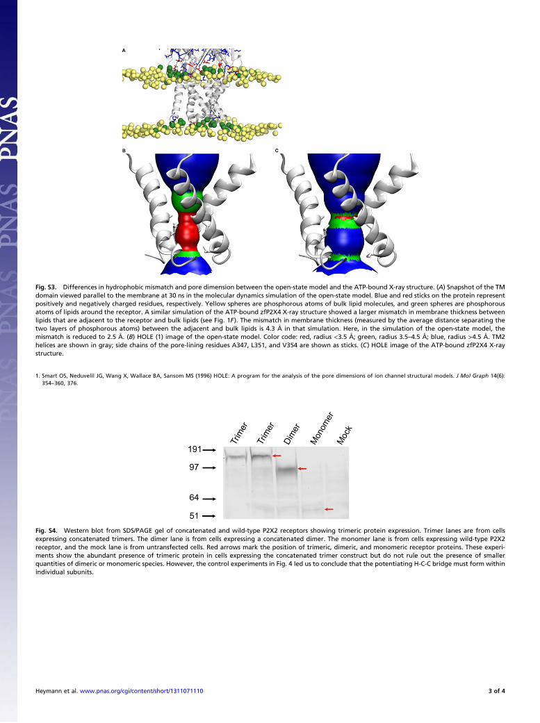
Fig. S3. Differences in hydrophobic mismatch and pore dimension between the open-state model and the ATP-bound X-ray structure. (A) Snapshot of the TMdomain viewed parallel to the membrane at 30 ns in the molecular dynamics simulation of the open-state model. Blue and red sticks on the protein representpositively and negatively charged residues, respectively. Yellow spheres are phosphorous atoms of bulk lipid molecules, and green spheres are phosphorousatoms of lipids around the receptor. A similar simulation of the ATP-bound zfP2X4 X-ray structure showed a larger mismatch in membrane thickness betweenlipids that are adjacent to the receptor and bulk lipids (see Fig. 1F). The mismatch in membrane thickness (measured by the average distance separating thetwo layers of phosphorous atoms) between the adjacent and bulk lipids is 4.3 Å in that simulation. Here, in the simulation of the open-state model, themismatch is reduced to 2.5 Å. (B) HOLE (1) image of the open-state model. Color code: red, radius <3.5 Å; green, radius 3.5–4.5 Å; blue, radius >4.5 Å. TM2helices are shown in gray; side chains of the pore-lining residues A347, L351, and V354 are shown as sticks. (C) HOLE image of the ATP-bound zfP2X4 X-raystructure.
1. Smart OS, Neduvelil JG, Wang X, Wallace BA, Sansom MS (1996) HOLE: A program for the analysis of the pore dimensions of ion channel structural models. J Mol Graph 14(6):354–360, 376.
Fig. S4. Western blot from SDS/PAGE gel of concatenated and wild-type P2X2 receptors showing trimeric protein expression. Trimer lanes are from cellsexpressing concatenated trimers. The dimer lane is from cells expressing a concatenated dimer. The monomer lane is from cells expressing wild-type P2X2receptor, and the mock lane is from untransfected cells. Red arrows mark the position of trimeric, dimeric, and monomeric receptor proteins. These experi-ments show the abundant presence of trimeric protein in cells expressing the concatenated trimer construct but do not rule out the presence of smallerquantities of dimeric or monomeric species. However, the control experiments in Fig. 4 led us to conclude that the potentiating H-C-C bridge must form withinindividual subunits.
Heymann et al. www.pnas.org/cgi/content/short/1311071110 3 of 4

Table S1. Estimated EC50 and Hill coefficients for ATP activation
Construct EC50, μM Hill coefficient
Wild type 15 ± 1.0 2.4 ± 0.3H-C-C 9.9 ± 2.0 1.3 ± 0.2H-C-C plus 20 μM Cd2+ 3.2 ± 0.2 1.3 ± 0.1C-C-C 5.7 ± 0.5 1.8 ± 0.2C-C-C plus 20 μM Cd2+ 3.7 ± 0.2 2.6 ± 0.3H-C-C concatamer B (1 subunit) 5.6 ± 0.1 1.4 ± 0.1H-C-C concatamer C (2 subunits) 5.7 ± 0.9 1.4 ± 0.2H-C-C concatamer D (3 subunits) 6.7 ± 1.3 0.9 ± 0.1Intersubunit concatamer E 11.5 ± 0.4 1.9 ± 0.1Intersubunit concatamer F 11.9 ± 1.6 1.6 ± 0.3Intersubunit concatamer G 13.9 ± 0.9 1.8 ± 0.2
Concentration–response relationships for activation by ATP were gener-ated from three to seven cells as described in Methods. Values for the wild-type channel are from Li et al. (1).
1. Li M, Chang TH, Silberberg SD, Swartz KJ (2008) Gating the pore of P2X receptor channels. Nat Neurosci 11(8):883–887.
Heymann et al. www.pnas.org/cgi/content/short/1311071110 4 of 4