integrated human/sars-cov-2 metabolic models present novel
TRANSCRIPT

Research Article
Integrated human/SARS-CoV-2 metabolic models presentnovel treatment strategies against COVID-19Bridget P Bannerman1,5 , Jorge Julvez3 , Alexandru Oarga3 , Tom L Blundell2, Pablo Moreno4, R Andres Floto1
The coronavirus disease 2019 (COVID-19) pandemic caused by the newcoronavirus (SARS-CoV-2) is currently responsible for more than 3milliondeaths in219countriesacross theworldandwithmore than140million cases. The absence of FDA-approved drugs against SARS-CoV-2has highlighted an urgent need to design new drugs. We developed anintegratedmodel of the human cell and SARS-CoV-2 to provide insightinto the virus’ pathogenic mechanism and support current therapeuticstrategies. We show the biochemical reactions required for the growthand general maintenance of the human cell, first, in its healthy state.We thendemonstrate how the entry of SARS-CoV-2 into the human cellcauses biochemical and structural changes, leading to a change of cellfunctions or cell death. A new computational method that predicts 20unique reactions as drug targets from our models and provides aplatform for future studieson viral entry inhibition, immune regulation,and drug optimisation strategies. The model is available in BioModels(https://www.ebi.ac.uk/biomodels/MODEL2007210001) and the soft-ware tool, findCPcli, that implements the computational method isavailable at https://github.com/findCP/findCPcli.
DOI 10.26508/lsa.202000954 | Received 10 November 2020 | Revised 26 July2021 | Accepted 27 July 2021 | Published online 5 August 2021
Introduction
SARS-COV-2, the causativeagentof COVID-19, belongs toagroupof virusescommonly knownasβ-coronavirus. This class of viruses is responsible formild-to-fatal respiratory tract infections in animals andbirds.Whereas thecommon cold is more commonly associated with the mild forms of thedisease, the previous MERS and SARS-2002 infections and, currently,COVID-19 belong to the group of fatal diseases. The genome of the virusresponsible for the ongoing COVID-19 disease, SARS-CoV-2, has ~80%sequence identity to SARS-CoV and is 96% identical at the whole-genomelevel to a bat coronavirus (Zhou et al, 2020). SARS-CoV-2 affects the lowerrespiratory tract cells and the upper cells in the pharyngeal region (Chenet al, 2020; Huang et al, 2020), and the viral infections range fromasymptomatic, mild, moderate, and severe cases. Previous studies inChina show that 86% of cases of infection and the contagiousnessof the virus were undocumented before travel restrictions were
imposed (Li et al, 2020). In addition, the interim results from theSolidarity international clinical trial conducted by the World HealthOrganization confirmed that only corticosteroids are effective againstsevere and critical cases of COVID-19. The report shows little or nobenefit from the other four treatments evaluated (remdesivir, hydrox-ychloroquine, lopinavir/ritonavir, and interferon) on overall mortalityagainst COVID-19 (Dyer, 2020; Pan et al, 2021). Therefore, there arestill many factors to unravel regarding the stages of infection andtransmissibility patterns of the virus to achieve good treatment man-agement strategies. Studies in France demonstrate the transmissionpotential of asymptomatic persons and suggest varying dynamics oftransmission in children (Danis et al, 2020). The human angiotensin-converting enzyme 2 (human-ACE-2 protein) has been identified as thecell receptor for both the SARS-2002 virus and SARS-CoV-2. The ACE-2enzyme, which has the primary function of controlling blood pressure,is usually found in the epithelial cells of the heart, lungs, kidneys, andintestine (Donoghue et al, 2000; Hamming et al, 2004; Liu et al, 2020).
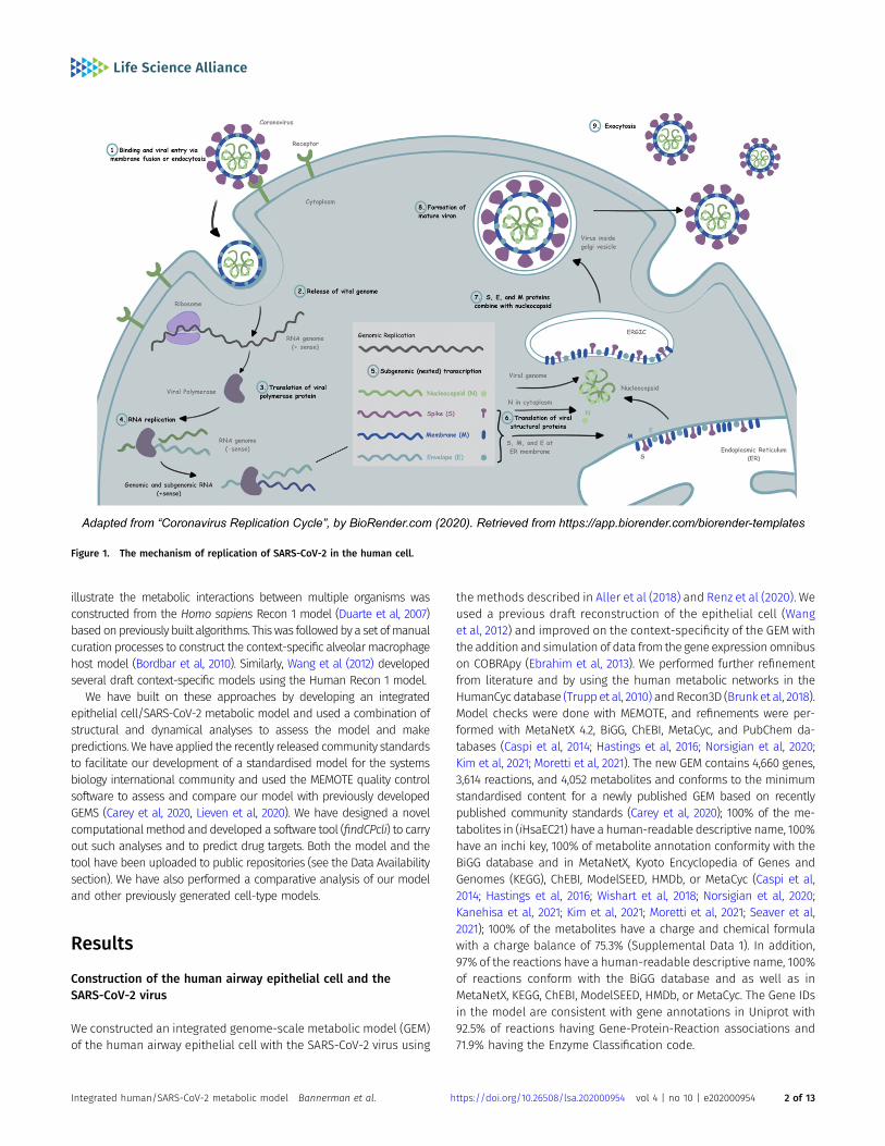
The mechanism of replication of SARS-CoV-2 in the human cellinvolves an initial binding and attachment of the spike (S) glyco-protein to the angiotensin-converting enzyme 2 (ACE2) receptor ofits host. During endocytosis, the virus’s genetic material is injectedinto the host cell, where it loses its protective envelope (Fig 1). Thesubsequent assembly and maturation of viral proteins lead to celldeath and proliferation of the virus within the human body.
The lack of FDA-approved drugs against COVID-19, coupled with thedifficulties encountered globally in containing the virus, prompted theWHO to declare the outbreak a pandemic in March 2020. This has led tointensified efforts around theworld tofight this disease. Previous studiesin drug target identification against viral diseases such as Zika, chi-kungunya, and dengue by Aller et al, (2018) introduced a system of in-tegrating the host’s macrophage and viral metabolic networks to predicta set of host reactions which, when constrained, can inhibit viral pro-duction. A recent study by Renz et al, (2020) demonstrates a similarapproachandpredicts drug targets against SARS-CoV-2. Targets of knownantiviral drugs predicted from both studies, using a macrophage met-abolic model (Bordbar et al, 2010) demonstrate the applicability of theintegrated human/virus metabolic modelling in drug target identifica-tion. The alveolar macrophage host model (Bordbar et al, 2010) used to
1Molecular Immunity Unit, Department of Medicine, University of Cambridge, Cambridge, UK 2Department of Biochemistry, University of Cambridge, Cambridge, UK3Department of Computer Science and Systems Engineering, University of Zaragoza, Zaragoza, Spain 4EMBL-EBI, European Bioinformatics Institute, Hinxton, UK 5TheCenter for Research and Interdisciplinarity, Paris, France
Correspondence: [email protected]; [email protected]
© 2021 Bannerman et al. https://doi.org/10.26508/lsa.202000954 vol 4 | no 10 | e202000954 1 of 13
on 2 October, 2021life-science-alliance.org Downloaded from http://doi.org/10.26508/lsa.202000954Published Online: 5 August, 2021 | Supp Info:
illustrate the metabolic interactions between multiple organisms wasconstructed from the Homo sapiens Recon 1 model (Duarte et al, 2007)basedonpreviously built algorithms. Thiswas followedby a set ofmanualcuration processes to construct the context-specific alveolar macrophagehost model (Bordbar et al, 2010). Similarly, Wang et al (2012) developedseveral draft context-specific models using the Human Recon 1 model.
We have built on these approaches by developing an integratedepithelial cell/SARS-CoV-2 metabolic model and used a combination ofstructural and dynamical analyses to assess the model and makepredictions.We have applied the recently released community standardsto facilitate our development of a standardised model for the systemsbiology international community and used the MEMOTE quality controlsoftware to assess and compare our model with previously developedGEMS (Carey et al, 2020, Lieven et al, 2020). We have designed a novelcomputationalmethod anddeveloped a software tool (findCPcli) to carryout such analyses and to predict drug targets. Both the model and thetool have been uploaded to public repositories (see the Data Availabilitysection). We have also performed a comparative analysis of our modeland other previously generated cell-type models.
Results
Construction of the human airway epithelial cell and theSARS-CoV-2 virus
We constructed an integrated genome-scale metabolic model (GEM)of the human airway epithelial cell with the SARS-CoV-2 virus using
the methods described in Aller et al (2018) and Renz et al (2020). Weused a previous draft reconstruction of the epithelial cell (Wanget al, 2012) and improved on the context-specificity of the GEM withthe addition and simulation of data from the gene expression omnibuson COBRApy (Ebrahim et al, 2013). We performed further refinementfrom literature and by using the human metabolic networks in theHumanCyc database (Trupp et al, 2010) andRecon3D (Brunk et al, 2018).Model checks were done with MEMOTE, and refinements were per-formed with MetaNetX 4.2, BiGG, ChEBI, MetaCyc, and PubChem da-tabases (Caspi et al, 2014; Hastings et al, 2016; Norsigian et al, 2020;Kim et al, 2021; Moretti et al, 2021). The new GEM contains 4,660 genes,3,614 reactions, and 4,052 metabolites and conforms to the minimumstandardised content for a newly published GEM based on recentlypublished community standards (Carey et al, 2020); 100% of the me-tabolites in (iHsaEC21) have a human-readable descriptive name, 100%have an inchi key, 100% of metabolite annotation conformity with theBiGG database and in MetaNetX, Kyoto Encyclopedia of Genes andGenomes (KEGG), ChEBI, ModelSEED, HMDb, or MetaCyc (Caspi et al,2014; Hastings et al, 2016; Wishart et al, 2018; Norsigian et al, 2020;Kanehisa et al, 2021; Kim et al, 2021; Moretti et al, 2021; Seaver et al,2021); 100% of the metabolites have a charge and chemical formulawith a charge balance of 75.3% (Supplemental Data 1). In addition,97% of the reactions have a human-readable descriptive name, 100%of reactions conform with the BiGG database and as well as inMetaNetX, KEGG, ChEBI, ModelSEED, HMDb, or MetaCyc. The Gene IDsin the model are consistent with gene annotations in Uniprot with92.5% of reactions having Gene-Protein-Reaction associations and71.9% having the Enzyme Classification code.
Figure 1. The mechanism of replication of SARS-CoV-2 in the human cell.
Integrated human/SARS-CoV-2 metabolic model Bannerman et al. https://doi.org/10.26508/lsa.202000954 vol 4 | no 10 | e202000954 2 of 13

Comparative analysis of integrated models of infected humanepithelial cell and the macrophage cell with SARS-CoV-2
For our study, we performed a comparative analysis of the essentialand unique reactions needed for the viability of the virus in the ep-ithelial cell/SARS-CoV-2 integratedmodel and the GEM constructed byRenz et al (2020). Our results showhow the virus heightens its virulencemechanisms by modifying the host’s defences within different cellcompartments. Consequently, we suggest treatment regimens basedon different stages of viral infection and replication.
Host-dependent metabolic pathways
We initially demonstrated the biochemical requirements for themaintenance of the human airway epithelial and macrophage cellsand used the integrated models to show the essential host reactionsneeded for the survival and viability of SARS-CoV-2 within the host’s cellcompartments. We have mapped the experimentally characterizedhuman/SARS-CoV-2 protein–protein interaction data fromGordon et al(2020) on the in silico virus-integrated human macrophage and epi-thelial cells. Of the 334 metabolic pathways in the human metabolicnetwork, 48 pathways including the biosynthesis and degradationpathways of amino acids, fatty acids, carbohydrates, amines, cofactors,and core components of the central mRNAmetabolism are hijacked bythe virus for its survival strategies (Fig 2).
The 48 metabolic pathways that were mapped to the protein–protein interaction network produced by Gordon et al (2020) arereferred to as PPi-pathway intersection nodes in this article (Table 1).
These include cysteine, methionine, and selenocysteine aminoacid biosynthetic pathways; C20 prostanoid hormone biosyn-thetic pathways; and vitamin D3 and vitamin K epoxide cycle. Thedegradation pathways identified include the lysine, tryptophan,methionine, fatty acid degradation, ceramide, and sphingolipidrecycling pathways; phospholipases degradation; and amine andheme degradation (Table 1).
Our results identify host dependency factors required for theSARS-CoV-2 virus infection, replication, survival, and viability withindifferent cell compartments and provide insight into novel treat-ment strategies.
Essential reactions for the host and viral metabolism
The Flux Balance Analysis (FBA) method (Orth et al, 2010) was usedto compute both the biomass maintenance of the cell in the ab-sence of virus and the maximum growth rate of the virus in the cell(host optimum and virus optimum conditions). We identified 52essential reactions in the macrophage (iAB-AMØ-1410) model and10 reactions in the epithelial cell model (iHsaEC21) essential for thevirus to propagate (Tables S1 and S2). It was also demonstrated that:(i) the maximal biomass maintenance of the macrophage cell in theabsence of virus was 0.0269 (Table S1) and 0.012 h−1 for the humanairway epithelial cell (Table S2); (ii) the maximum growth rate of thevirus in the macrophage cell was 0.0144 and 0.0181 h−1 in the humanairway epithelial cell. These numerical results mean that 0.0144 h−1
is the theoretical maximum of the growth rate of the virus in thehuman macrophage cell. If this flux is assigned to the viral growth
Figure 2. SARS-CoV-2 viral genome and host-dependent metabolic pathways.
Integrated human/SARS-CoV-2 metabolic model Bannerman et al. https://doi.org/10.26508/lsa.202000954 vol 4 | no 10 | e202000954 3 of 13

reaction, then flux variability analysis (FVA) (Orth et al, 2010) can beused to calculate the ranges of fluxes allowed for the remainingreactions in the cell while the virus is being replicated at its op-timum condition. The execution of FVA under such conditionsproduced zero biomass maintenance of the host cell, that is, boththe lower and upper flux bounds of the reaction indicate that thegrowth is zero. This means that if the virus is replicating at itsmaximum rate, then the cell will not be viable.
Bottleneck reactions and the prioritization of potential drugtargets
The bottleneck reactions identified by the findCPcli tool are uniquereactions of the metabolic network required for the growth andsurvival of the organism and, like chokepoint reactions, are po-tential drug targets (Yeh et al, 2004; Oarga et al, 2020). Althoughclassical chokepoint reactions identify reactions that are the only
Table 1. List of bottleneck and essential enzymes on the PPi-pathway intersection nodes.
Class Pathway Sub-pathway Humangene Sars gene Macrophage Epithelial
cell
Biosynthesis Amino acids L-selenocysteine biosynthesis SEPSECS Nsp8 Y N
Biosynthesis Amino acids Cysteine, and methionine MAT2B Nsp9 Y Y
Biosynthesis Fatty acids Fatty acid and long fatty acid biosynthesis SLC27A2 Nsp2 Y Y
Biosynthesis Fatty acids Fatty acid and long fatty acid biosynthesis ACSL3 Nsp7 Y N
Biosynthesis Fatty acids Stearate biosynthesis SLC27A2 Nsp2 Y Y
Biosynthesis Fatty acids Stearate biosynthesis ACSL3 Nsp7 Y Y
Biosynthesis Carbohydratebiosynthesis Glycan and oligosaccharide biosynthesis ALG11 Nsp4 Y Y
Biosynthesis Carbohydratebiosynthesis Glycan HS2ST1 Orf8a Y Y
Biosynthesis Carbohydratebiosynthesis Glycan MOGS Nsp7 Y Y
Biosynthesis Carbohydratebiosynthesis Glycan and oligosaccharide biosynthesis ALG5 ORF3a Y Y
Biosynthesis Carbohydratebiosynthesis
Glycan, oligosaccharide, and glycosaminoglycanbiosynthesis CHPF ORF8a Y Y
Biosynthesis Carbohydratebiosynthesis
Glycan, oligosaccharide, and glycosaminoglycanbiosynthesis HS6ST2 ORF8a Y Y
Biosynthesis Cofactors Vitamin D3 biosynthesis POR Nsp2 Y Y
Biosynthesis Cofactors Vitamin K epoxide cycle GGCX Mainprotease N Y
Biosynthesis Hormones C20 prostanoid biosynthesis PTGES2 Nsp7 Y Y
Biosynthesis tRNA charging tRNA charging TARS2 Mainprotease Y Y
Degradation Amino acids L-lysine degradation AASS Mainprotease N Y
Degradation Amino acids L-tryptophan degradation POR Nsp2 Y Y
Degradation Amino acids L-methionine degradation MAT2B Mainprotease Y Y
Degradation Fatty acids Ceramide and sphingolipid recycling SLC27A2 Nsp2 Y Y
Degradation Fatty acids Ceramide and sphingolipid recycling POR Nsp2 Y Y
Degradation Fatty acids Ceramide and sphingolipid recycling ACSL3 Nsp7 Y Y
Degradation Fatty acids Phospholipases PLD3 ORF8a Y Y
Degradation Amine degradation Dopamine degradation COMT Nsp7 Y Y
Degradation Hormones Heme HMOX1 ORF3a Y Y
Degradation Hormones melatonin POR Nsp2 Y Y
Degradation Hormones Adrenalin COMT Nsp2 Y Y
Degradation Hormones L-dopa degradation COMT Nsp2 Y Y
Integrated human/SARS-CoV-2 metabolic model Bannerman et al. https://doi.org/10.26508/lsa.202000954 vol 4 | no 10 | e202000954 4 of 13

producers or the only consumers of a given metabolite and con-sider just the model structure, we improve on this approach byusing both the structural and dynamical information. FVA is used tocompute flux bounds of the reactions, and in turn, to determinewhether a given reaction is reversible or not. Reversibility will beused to obtain the sets of metabolites that can be produced andconsumed by the reactions, and thus, to compute flux-dependentbottleneck reactions. This approach has been applied to the in-tegrated Human/SARS-CoV-2 metabolic model within the airwayepithelial cell and the macrophage cell to predict potential drugtargets against SARS-CoV-2.
We initially identified 1,595 bottleneck reactions required for thevirus’ maintenance and replication in the human macrophage cell;these include pathways in lipidmetabolism, coenzyme transport andmetabolism, energy production and conversion, and amino acid andnucleotide transport andmetabolism (Table S3). In the humanairwayepithelial cell, 1,598 bottleneck reactions were initially identified;these include the biosynthesis and degradation pathways of aminoacids, fatty acids, carbohydrates, amines, cofactors, and some compo-nents of the central mRNA metabolism (Table S4).
Because each bottleneck reaction should be balanced by at leastone other reaction that produces or consumes that metabolite, wehave excluded reactions in the model with dead-end metabolites.The bottleneck reactions are potential drug targets as they areindispensable for the maintenance and replication of the viruswithin the host. To rank the potential drug targets identified, weprioritised enzymes for unique reactions that occur at the nodes ofintersection between the bottleneck and essential reactions andthe results from the human/virus protein–protein interactionnetwork (Gordon et al, 2020) (Table 1). The following steps weretaken to label the reactions which occur at the nodes of inter-sections between the referenced points above: (1) we initiallyidentified both the bottleneck and essential reactions to the viruswithin the model, (2) we highlighted the interactions between theviral proteins and the human metabolic enzymes, and (3) we se-lected the reactions from the human/virus interactions which arealso present in the list of bottleneck and/or essential reactions.
The enzymes of the reactions at the nodes of intersection are (1)host dependency factors identified by the model as necessary forthe survival of the virus and (2) proteins from experimental datasetswith high-confidence virus–human protein–protein interaction data;we refer to these points of intersection as PPi-pathway inter-section nodes.
The PPi-pathway intersection (P-Pi) nodes identified are presentin biosynthesis pathways such as the cysteine and S-adenosyl-L-methionine biosynthetic pathways. In both pathways, the enzymeS-adenosylmethionine synthase (MAT2B) catalyses the phosphor-ylation reaction of methionine to S-adenosyl-L-methionine. Duringinfection, the viral protein Nsp9 is seen to react withMAT2B (Gordonet al, 2020) (Fig 3A–C). Another viral protein, Nsp8, also interacts withthe enzyme O-phosphoseryl-tRNA(Sec) selenium transferase (SEPSECS),which catalyses the last step of the L-selenocysteine biosynthesispathway (Fig 4).
P-Pi nodes also occur in a network of various fatty acid andstearate biosynthetic pathways with Nsp2 interacting with the verylong-chain acyl-CoA synthetase (SLC27A2) (Fig 5A and B). The viralprotein, Nsp2, also interacts with POR in other pathways including
vitamin D3 biosynthesis (Fig 5C) and in L-tryptophan degradation,ceramide, and sphingolipid recycling pathways (Table 1).
In carbohydrate metabolism, a P-Pi node is identified at theglycan and oligosaccharide biosynthetic pathways, and specificallywhere two mannose residues are added in α (1→2) linkages to thenascent oligosaccharide and catalysed by the enzyme ALG11. Theviral protein Nsp4 interacts with ALG11 during the infection of theSARS-CoV-2 virus (Fig 6). Another viral protein, Nsp7, interacts withACSL3 in the γ-linolenate biosynthesis (Fig 7A). Nsp7 also reacts withACSL3 and ORF8a interacts with HS2ST1 (Fig 7B), a key enzyme in-volved in the heparan sulfate biosynthesis pathway. The first enzymeof the N-linked oligosaccharide processing pathway, mannosyl-oligosaccharide α-1,2-glucosidase (MOGS), also interacts with Nsp7and ORF8a (Fig 7C).
P-Pi nodes specific to the human macrophage cell include theO-phosphoseryl-tRNA(Sec) selenium transferase in the L-seleno-cysteine biosynthetic pathway, which interacts with the viral pro-tein Nsp8. The alkylglycerone-phosphate synthase/Nps7 P-Pi node,which is present in the phospholipid/plasmalogen biosyntheticpathway is also specific to the macrophage cell. Alternatively,PPi-pathway intersection nodes common to both human airwayepithelial cell and the macrophage cell include the MAT2B/Nsp9intersection pathways present in the cysteine metabolism andL-methionine degradation. P-Pi nodes specific to the human epi-thelial cell include the peptidyl-glutamate 4-carboxylase present inthe Vitamin K epoxide cycle and the α-aminoadipic semialdehydesynthase enzyme in the L-lysine degradation pathway.
Discussion
Metabolic pathway perturbations in the human cell due to COVID-19reflect the viral entry and infection of SARS-CoV-2 and the immuneregulation changes in the human body. We have used in silicomodels to study the interactions of SARS-CoV-2 in the host andpropose new treatment management regimens. We have built onstudies using the human alveolar macrophage model iAB-AMØ-1410(Bordbar et al, 2010) as host cells and SARS-CoV-2 (Renz et al, 2020,2021); influenza ((Aller et al 2018)), and tuberculosis as pathogens anddeveloped a new integrated model of the human epithelial cell andSARS-CoV-2.
The initial draft epithelial cell model was based on the humanRecon1 model and consists of 1,206 reactions, 994 metabolites, and0 genes (Wang et al, 2012). Our revised iHsaEC21 epithelial cellmodel contains 4,660 genes, 3,614 reactions, and 4,052 metabolitesinitially scored 34% on MEMOTE (Lieven et al, 2020). After furtherrefinements using Recon3D, HumanCyc, MetaNetX 4.2, BiGG, ChEBI,MetaCyc, and PubChem databases (Caspi et al, 2014; Hastings et al,2016; Norsigian et al, 2020; Moretti et al, 2021) including manualcurations with literature, iHsaEC21 now has a MEMOTE quality scoreof 51% (Supplemental Data 1). Although the iAB-AMØ-1410 (Bordbaret al, 2010) was also developed from the human Recon1 model,there was further refinement with literature, which attributed to itsquality score of 45% (Supplemental Data 2) compared with the draftepithelial cell model fromWang et al (2012) which had a low-qualityscore of 19% (Supplemental Data 3).
Integrated human/SARS-CoV-2 metabolic model Bannerman et al. https://doi.org/10.26508/lsa.202000954 vol 4 | no 10 | e202000954 5 of 13

Previous studies have also demonstrated the role of the ACE2 asthe receptor for both the SARS-CoV and the SARS-CoV-2. The ACE2cells are expressed in the human airway epithelial cells (Hamminget al, 2004; Liu et al, 2020). In this study, we have constructed anintegrated human epithelial cell and SARS-CoV-2 to provide insightinto the infection patterns of the virus in the human body.
Our in silico comparative analyses of the SARS-CoV-2 viral in-fection between two different conditions (infected human macro-phage and airway epithelial cells) show the requirements of viabilityof the virus between these two conditions. Our results complementprevious efforts to propose drug targets and repurposing strategiesincluding the SARS-CoV-2-Human Protein–Protein Interaction Map
Gordon et al, (2020 and studies from Steward, 2020, which identifiedhost dependency factors facilitating virus infection. We also provideadditional resources to the COVID-19 disease map (Ostaszewski et al,2020). In addition, we have designed a new algorithm (Oarga et al,2020) and used the dynamic information of the human/virus modelsto predict new treatment regimens. Here, we demonstrate the fluxchanges from lipid metabolism, cofactor biosynthetic pathways, re-dox balance, and immune regulation indicative of pathogenic reac-tions arising from the COVID-19 viral infections.
Inhibition of viral entry and replication
The components of the plasma membrane, such as cholesterol andsphingolipid-rich lipid, Abu-Farha et al, (2020) are involved in viruspenetration, entry, replication, and infection (Wang et al, 2008; Abu-Farha et al, 2020). In this study, we demonstrate current therapeuticstrategies that interfere with different stages of the viral cycle bytargeting lipidmetabolism and proposing new treatment strategies.
The dynamical changes of flux metabolism in our in-silico virusoptimal models show a significant increase in viral infection. Fourreactions involved in the biosynthesis of fatty acids with predictednon-zero fluxes in the host model exhibit an average increase of190% in their maximum fluxes in the viral model (the maximumFigure 4. PPi-pathway intersection node—Nsp8.
Figure 3. PPi-pathway intersection node—Nsp9.(A) Methionine degradation. (B) Methionine salvage cycle. (C) Cysteine biosynthesis.
Integrated human/SARS-CoV-2 metabolic model Bannerman et al. https://doi.org/10.26508/lsa.202000954 vol 4 | no 10 | e202000954 6 of 13

increase is 298%). The average increase of 32 reactions in lipidmetabolism with non-zero fluxes in the host model is 277% (themaximum increase is 498%). Concerning sphingolipid metabolism,14 of 15 reactions with non-zero fluxes in the host model exhibit anaverage increase of 228% (the maximum increase is 298%) andsimilar increases in phospholipases and palmitic acid biosynthesis(Fig 5A and B). We show an average increase of 190% in cholesteroland fatty acid metabolism during viral infection and demonstratethe essentiality of these pathways to SARS-CoV-2. Previous studieshave shown that cholesterol and fatty acids are main componentsof the viral membranes and needed for viral replication (Heaton &Randall, 2011); therefore, drugs inhibiting these pathways such asAM580, statins, and fibrate (Fievet & Staels, 2009) will be essentialfor both early and late stages of COVID-19.
Sphingolipids are composed of both hydrophobic and hydro-philic units and play a large role in the endocytic or exocytic viralentry processes into the cell (Dimitrov, 2004). The pH-dependentendocytic process is further enhanced by the presence of clathrin, aprotein present in the plasma membrane, the Golgi apparatus andin the cytoplasm, whereas the exocytic route involves viral crossingthrough the plasma membrane at neutral pH. Our results show athreefold increase in sphingolipid metabolism during viral infec-tion (Fig 5); we hypothesise that drugs inhibiting sphingolipidmetabolism and/or the endocytosis process will inhibit infection ofSARS-CoV-2. Indeed, a sphingosine kinase-2 (SphK2) inhibitor,opaganib, which has proved beneficial in the treatment of COVID-19,is currently in global phase 2/3 clinical trials and in US phase 2
studies. Previous studies also demonstrate that chloroquine andhydroxychloroquine elevate the pH of endosomes in the cells anddirectly inhibit endocytosis and the exocytic process (Munro et al, 1997;Devaux et al, 2020). More recent studies have also shown thatartemisinin-inhibited endocytosis (Hoppe et al, 2004; Uzun & Toptas,2020). A recent study by Abu-Farha described other lipid-modifyingdrugs including LJ-001, arbidol, methyl-B-cyclodextrin (Blaising et al,2013; de Wilde et al, 2014; Mazzon & Marsh, 2019). We highlight criticalreactions as drug targets for lipid metabolism for the SARS-CoV-2 virus(Table S2) and in our PPi-pathway intersection nodes (Table 1).
Our lists of bottleneck, essential reactions, and PPi-pathwayintersection nodes also include critical points in the biosynthe-sis of phospholipids. We show that these reactions are essential forviral infection and replication (Table 1) and propose targeting thephospholipase enzyme or the interacting Nsp2 protein to inhibitviral replication. Our results support previous studies from Mülleret al (2018) that targeting the phospholipase enzyme could inhibitthe early stage of COVID-19.
Redox homeostasis and antioxidant therapy
Redox homeostasis refers to the ability of the cell to maintain itsbalance amidst infections and other unstable cellular environmentalfactors. Delgado-Roche and Mesta (2020) have described oxidativestress as a key player in severe acute respiratory syndrome coronavirus(SARS-CoV) infection with cytokine production. Foyer and Noctor (2005)have previously shown that antioxidants, such as glutathione and
Figure 5. PPi-pathway intersection node—Nsp2.(A) Fatty acid oxidation. (B) Stearate biosynthesis. (C) Vitamin D3 biosynthesis.
Integrated human/SARS-CoV-2 metabolic model Bannerman et al. https://doi.org/10.26508/lsa.202000954 vol 4 | no 10 | e202000954 7 of 13

ascorbate, are important metabolites for the cellular redox state. Ourstudieshave identifiedkey target enzymes involved in themetabolismofglutathione and ascorbic acid as bottleneck and essential reactions,including glutathione synthase, glutathione peroxidase, and ascorbicacid oxidase (Table S2). We also demonstrate an increase in the flux ofthese enzymatic reactions on infection of the virus. In a recent study,
Horowitz et al (2020) previously demonstrated how the use of high doseoral and/or IV glutathione on severe outcomes of SARS-CoV-2 led tofavourable treatment outcomes. Other studies have shown that steroidssuch as dexamethasone and Methylprednisolone are used to treatsevere cases of COVID-19. Because of the possible side effects of steroidtreatment, wepropose theuseof glutathioneas therapy for severe cases
Figure 6. PPi-pathway intersection node—Nsp4.
Integrated human/SARS-CoV-2 metabolic model Bannerman et al. https://doi.org/10.26508/lsa.202000954 vol 4 | no 10 | e202000954 8 of 13

of COVID-19 in the aged population and other severe caseswith cytokinestorm syndrome.
Immune regulation
SARS-CoV-2 can proliferate unhindered in infected cells because of thelack of immunity in humans (Felsenstein et al, 2020). The result is celldeath, a release of viral particles to the extracellular environment and ageneral hyperactivity of the immune system in some patients with severeCOVID-19 disease and subsequent lung inflammation and cytokinesyndrome. Immunocompromised patients or those with underlyingsymptoms such as diabetes, hypertension, and transplantation aremostaffected (Zhonget al, 2020). Although clinical trials are ongoingworldwidewith various antivirals and immune-modulating treatments, there iscurrently limited knowledge on the host dependency factors responsiblefor the individual outcomes of the disease. Our results provide insightinto the immune evasion strategies of the SARS-CoV-2 virus; we dem-onstrate changes in the flux metabolism of vitamin D and tryptophanmetabolismduring viral infection. Vitamin D is important for bone growthand turnover and a low vitamin D status is associated with increasedsusceptibility to upper respiratory tract infections (Mitchell, 2020). Pre-vious studies have shown that supplementation of vitamin D preventsacute respiratory tract infections (Martineau et al, 2017). Our resultshighlight vitamin D as an essential reaction in the PPi-pathway inter-section nodes and we show the viral protein Nsp2 interaction withkey enzymes in the vitamins D and C metabolism pathways (Fig 5C).SARS-CoV-2 viral infection causesmetabolic perturbations of vitamin Dmetabolism in the host resulting in disruptions in cellular homeo-stasis. We propose support therapy management strategies wherevitaminD supplements are provided to all COVID-19 patients. Our results
also show that tryptophan, melatonin and prostaglandins, importantcompounds for immunity andhomeostasis (Gitto et al, 2011; Platten et al,2019), are affected by the infection of SARS-CoV-2 andwe provide insightinto the viral mechanism of action within the human body.
In summary, we have provided a platform for drug target pre-diction against COVID-19, and future studies on viral entry inhibi-tion, antioxidant therapy, and immune regulation.
Materials and Methods
For our modelling analyses, we based our reconstruction in the in-tegrated macrophage cell with the SARS-CoV-2 virus model, previouslyconstructed by Renz et al (2020) to develop a new human airwayepithelial cell integrated with the SARS-CoV-2 virus. We predicted thereactions which were critical for the survival of the virus in both cellcompartments (macrophage and epithelial cells); our results provideinsights into COVID-19 treatment management strategies.
GEM reconstruction, curation, and simulations
Weobtained an automated draft reconstruction of the human airwayepithelial cell (Wang et al, 2012) and evaluated the metabolicfunctions and reactions collected in the draft reconstruction againstorganism-specific literature. We used the gene expression datasetsof the human airway epithelial cell (Deprez et al, 2019 Preprint; VieiraBraga et al, 2019) to curate, annotate and improve on the model (Fig8). We manipulated and simulated the model, including the additionof the gene expression data, with COBRApy (Ebrahim et al, 2013) andGLPK (https://www.gnu.org/software/glpk/). We obtained additional
Figure 7. Pi-pathway intersection nodes - Nsp7 and Orf8a in carbohydrate and fatty acid metabolism.(A) PPi-pathway intersection node—Nsp7. (B) PPi-Pathway intersection node—Orf8a. (C) PPi-pathway intersection node—Nsp7 and Orf8a.
Integrated human/SARS-CoV-2 metabolic model Bannerman et al. https://doi.org/10.26508/lsa.202000954 vol 4 | no 10 | e202000954 9 of 13

reactions, gene-to-reaction associations, and pathways (that werenot in the automated model) from HumanCyc (Trupp et al, 2010) todevelop a new genome-scale metabolic model (GEM) of the humanairway epithelial cell. We improved on the new reconstruction bymapping the genes and reactions of the GEM to Recon3D, a standardhuman metabolic model, MetaNetX 4.2, BiGG, ChEBI, MetaCyc, andPubChem databases (Caspi et al, 2014; Hastings et al, 2016; Norsigianet al, 2020; Moretti et al, 2021) and produced a revised human airwayepithelial cell iHsaEC21 (Fig 8). The revised model reconstruction,iHsaEC21, can be instantiated without error on the COBRA software(version 0.16.0) (Ebrahim et al, 2013).
Model optimisation
To improve on the quality of the model, we removed the reactionswith dead-end metabolites, which were previously found in theautomated model. This was followed by iteratively assessing themodel and accounting for the cell-specific metabolic and exchangereactions in the epithelial cell model. We compared and performedvarious iterations of the new GEM with Recon3D, MetaCyc, MetaNetX4.2, BiGG, ChEBI, and PubChem databases (Caspi et al, 2014; Hastingset al, 2016; Norsigian et al, 2020; Kim et al, 2021; Moretti et al, 2021). Asa result, we added charge and formulae to 1,473 compounds on thenew GEM. During the iterations and optimisation of the model, weperformed quality control checks on MEMOTE, a standardised genome-scale metabolic model testing software (Leiven et al, 2020).
Integration of the human airway epithelial cell model and theSARS-CoV-2 virus
We integrated the viral biomass maintenance function, previously de-veloped for themacrophage cell (Renz et al. 2020, 2021) into thenewGEM
to produce an integrated model of iHsaEC21+SARS-CoV-2. We interro-gated the new model to identify the host-dependency factors for theSARS-CoV-2 virus by using the novel software tool findCPcli.
The new GEM is encoded in the Systems Biology Markup Lan-guage (SBML) (Keating et al, 2020) (iHsaEC21); i for in silico, Hsa forH.sapiens, and EC for airway epithelial cell published in 2021. iHsaEC21consists of 3,752 reactions, 3,914 metabolites, 4,660 genes, and 48metabolic pathways.
To assess and predict the performance of the models, we madeuse of FBA and FVA (Orth et al, 2010). FBA is a computational methodthat can be applied efficiently to genome-scale models to estimatethe fluxes of reactions at a steady state. It is based on the solutionof a linear programming problem that maximizes an objectivefunction of interest subject to a set of constraints on the fluxes ofthe reactions. The linear programming problem associated with FBAcan be expressed as follows:
max c⋅v;
s:t: S⋅v = 0;
L ≤ v ≤ U;
where v is the vector of fluxes, c represents the objective functioncoefficients, S is the stoichiometry matrix, and L and U are lower andupper bounds on the fluxes. Thus, c v is the objective function, whichusually refers to the growth rate of the organism and S v = 0 representsthe balance of fluxes at steady state.
FVA is also based on the solution of linear programmingproblems, and its main use is the computation of ranges of fluxesthat are compatible with given flux constraints. For instance, if thegrowth rate predicted by FBA is μmax, then the range of fluxes of agiven reaction i that are compatible with such growth rate can be
Figure 8. Development of an integrated model of theHuman Airway Epithelial cell and SARS-CoV-2.
Integrated human/SARS-CoV-2 metabolic model Bannerman et al. https://doi.org/10.26508/lsa.202000954 vol 4 | no 10 | e202000954 10 of 13

obtained by minimizing andmaximizing the following programmingproblem:
min=max vi;
s:t: S⋅v = 0;
L ≤ v ≤ U;
vgrowth = μmax;
where vgrowth is the flux of the reaction associated with growth and viis the flux of reaction i. FBA and FVA were computed on themetabolicnetwork of the host, bothwith andwithout the reactionmodelling theproduction of the virus, by using the Python toolbox COBRApy.
Bottleneck reactions, like chokepoint reactions, are required forthe reaction synthesis and the removal of these reactions will causean accumulation or depletion of the metabolites; thus, they rep-resent potential drug targets. The software tool findCPcli was de-veloped to compute bottleneck reactions on genome-scale modelsby considering the structural and dynamic information of themodels. The dynamic information is considered as follows: (a) FVAis run to compute lower and upper flux bounds of the reactions; (b)the obtained flux bounds are used to identify reversible and non-reversible; and (c) this directionality of reactions is used to de-termine consumer and producer reactions, and in turn, bottleneckreactions. In addition to the computation of bottleneck reactions,findCPcli can also compute and remove dead-end metabolites, findessential reactions, and update the flux bounds of the reactionsaccording to the results of FVA.
Data Availability
Themodel has been deposited as an SBML Level 3 Version 1 (Hucka et al,2018)filewith FBC extension and theminimal information required in theannotation of models (MIRIAM) (Juty et al, 2012) in BioModels (https://www.ebi.ac.uk/biomodels/MODEL2007210001) (Malik-Sheriff et al, 2020).The source code of findCPcli together with its documentation is availableat https://github.com/findCP/findCPcli. The tool requires Python 3.5 (orhigher) and can be installed with pip, the standard package installer forPython. The pathway maps were created with the pathway collagesoftware (Paley et al, 2016) and the essential reactions are available as asmart table on HumanCyc (https://biocyc.org/group?id=biocyc17-29351-3833343490).
Supplementary Information
Supplementary Information is available at https://doi.org/10.26508/lsa.202000954.
Acknowledgements
The authors of this manuscript are supported by The Wellcome Trust107032AIA (RA Floto, BP Bannerman) The UK Cystic Fibrosis Trust (Innovation
Hub grant 001) (RA Floto), and the Spanish Ministry of Science, Innovation,and Universities (J Julvez and A Oarga). The authors would like to thank JoChukualim for the graphics and illustrations. The authors would also like tothank Charlotte Passemar for slides with preliminary data demonstrating theexpression of ACE2 in epithelial cells.
Author Contributions
BP Bannerman: conceptualization, data curation, software, formalanalysis, investigation, methodology, and writing—original draft,review, and editing.J Julvez: data curation, software, formal analysis, investigation, andwriting—original draft, review, and editing.A Oarga: data curation, software, formal analysis, and writing—original draft, review, and editing.TL Blundell: formal analysis and writing—review and editing.P Moreno: data curation, formal analysis, investigation, and writing—review and editing.RA Floto: formal analysis, investigation, and writing—review andediting.
Conflict of Interest Statement
The authors declare that they have no conflict of interest.
References
Abu-Farha M, Thanaraj TA, Qaddoumi MG, Hashem A, Abubaker J, Al-Mulla F(2020) The role of lipid metabolism in COVID-19 virus infection and asa drug target. Int J Mol Sci 21: 3544. doi:10.3390/ijms21103544
Aller S, Scott A, Sarkar-Tyson M, Soyer OS (2018) Integrated human-virusmetabolic stoichiometric modelling predicts host-based antiviraltargets against Chikungunya, Dengue and Zika viruses. J R Soc Interf 15:20180125. doi:10.1098/rsif.2018.0125
Blaising J, Levy PL, Polyak SJ, Stanifer M, Boulant S, Pecheur EI (2013) Arbidolinhibits viral entry by interfering with clathrin-dependent trafficking.Antiviral Res 100: 215–219. doi:10.1016/j.antiviral.2013.08.008
Bordbar A, Lewis NE, Schellenberger J, Palsson BØ, Jamshidi N (2010) Insightinto human alveolar macrophage and M. tuberculosis interactions viametabolic reconstructions. Mol Syst Biol 6: 422. doi:10.1038/msb.2010.68
Brunk E, Sahoo S, Zielinski DC, Altunkaya A, Drager A, Mih N, Gatto F, Nilsson A,Preciat Gonzalez GA, Aurich MK, et al (2018) Recon3D enables a three-dimensional view of gene variation in human metabolism. NatBiotechnol 36: 272–281. doi:10.1038/nbt.4072
Carey MA, Drager A, Beber ME, Papin JA, Yurkovich JT (2020) Communitystandards to facilitate development and address challenges inmetabolic modeling. Mol Syst Biol 16: e9235. doi:10.15252/msb.20199235
Caspi R, Altman T, Billington R, Dreher K, Foerster H, Fulcher CA, Holland TA,Keseler IM, Kothari A, Kubo A, et al (2014) The MetaCyc database ofmetabolic pathways and enzymes and the BioCyc collection ofpathway/genome databases. Nucleic Acids Res 42: D459–D471.doi:10.1093/nar/gkt1103
Chen Y, Liu Q, Guo D (2020) Emerging coronaviruses: Genome structure,replication, and pathogenesis. J Med Virol 92: 418–423. doi:10.1002/jmv.25681
Danis K, Epaulard O, Benet T, Gaymard A, Campoy S, Botelho-Nevers E,Bouscambert-Duchamp M, Spaccaferri G, Ader F, Mailles A, et al (2020)
Integrated human/SARS-CoV-2 metabolic model Bannerman et al. https://doi.org/10.26508/lsa.202000954 vol 4 | no 10 | e202000954 11 of 13

Cluster of coronavirus disease 2019 (COVID-19) in the French alps,February 2020. Clin Infect Dis 71: 825–832. doi:10.1093/cid/ciaa424
Delgado-Roche L, Mesta F (2020) Oxidative stress as key player in severeacute respiratory syndrome coronavirus (SARS-CoV) infection. ArchMed Res 51: 384–387. doi:10.1016/j.arcmed.2020.04.019
Deprez M, Zaragosi L, Truchi M, Garcia SR, Arguel MJ, Lebrigand K, Paquet A,Pee’r D, Marquette C-H, Leroy S, et al (2019) A single-cell atlas ofthe human healthy airways. BioRxiv doi:10.1101/2019.12.21.884759(Preprint posted December 23, 2019).
Devaux CA, Rolain J-M, Colson P, Raoult D (2020) New insights on the antiviraleffects of chloroquine against coronavirus: What to expect for COVID-19? Int J Antimicrobial Agents 55: 105938. doi:10.1016/j.ijantimicag.2020.105938
de Wilde AH, Jochmans D, Posthuma CC, Zevenhoven-Dobbe JC, vanNieuwkoop S, Bestebroer TM, van den Hoogen BG, Neyts J, Snijder EJ(2014) Screening of an FDA-approved compound library identifies foursmall-molecule inhibitors of Middle East respiratory syndromecoronavirus replication in cell culture. Antimicrob Agents Chemother58: 4875–4884. doi:10.1128/AAC.03011-14
Dimitrov DS (2004) Virus entry: Molecular mechanisms and biomedicalapplications. Nat Rev Microbiol 2: 109–122. doi:10.1038/nrmicro817
Donoghue M, Hsieh F, Baronas E, Godbout K, Robinson K, Jeyaseelan R,Breitbart RE, Acton S (2000) A novel angiotensin-convertingenzyme–related carboxypeptidase (ACE2) converts angiotensin I toangiotensin 1-9. Clin Res 87: e1–e9. doi:10.1161/01.res.87.5.e1
Duarte NC, Becker SA, Jamshidi N, Thiele I, Mo ML, Vo TD, Srivas R, Palsson BØ(2007) Global reconstruction of the human metabolic network basedon genomic and bibliomic data. Proc Natl Acad Sci U S A 104: 1777–1782.doi:10.1073/pnas.0610772104
Dyer O (2020) Covid-19: Remdesivir has little or no impact on survival, WHOtrial shows. BMJ 371: m4057. doi:10.1136/bmj.m4057
Ebrahim A, Lerman JA, Palsson BO, Hyduke DR (2013) COBRApy: COnstraints-based reconstruction and analysis for Python. BMC Syst Biol 7: 74.doi:10.1186/1752-0509-7-74
Felsenstein S, Herbert JA, McNamara PS, Hedrich CM (2020) COVID-19:Immunology and treatment options. Clin Immunol 215: 108448.doi:10.1016/j.clim.2020.108448
Fievet C, Staels B (2009) Combination therapy of statins and fibrates in themanagement of cardiovascular risk. Curr Opin Lipidol 20: 505–511.doi:10.1097/mol.0b013e328332e9ef
Foyer CH, Noctor G (2005) Redox homeostasis and antioxidant signaling: Ametabolic interface between stress perception and physiologicalresponses. Plant Cell 17: 1866–1875. doi:10.1105/tpc.105.033589
Gitto E, Aversa S, Reiter RJ, Barberi I, Pellegrino S (2011) Update on the use ofmelatonin in pediatrics. J Pineal Res 50: 21–28. doi:10.1111/j.1600-079X.2010.00814.x
Gordon DE, Jang GM, Bouhaddou M, Xu J, Obernier K, White KM, O’Meara MJ,Rezelj VV, Guo JZ, Swaney DL, et al (2020) A SARS-CoV-2 proteininteraction map reveals targets for drug repurposing. Nature 583:459–468. doi:10.1038/s41586-020-2286-9
Hamming I, Timens W, Bulthuis ML, Lely AT, Navis G, van Goor H (2004) Tissuedistribution of ACE2 protein, the functional receptor for SARScoronavirus. A first step in understanding SARS pathogenesis. J Pathol203: 631–637. doi:10.1002/path.1570
Hastings J, Owen G, Dekker A, Ennis M, Kale N, Muthukrishnan V, Turner S,Swainston N, Mendes P, Steinbeck C (2016) ChEBI in 2016: Improvedservices and an expanding collection of metabolites. Nucleic AcidsRes 44: D1214–D1219. doi:10.1093/nar/gkv1031
Heaton NS, Randall G (2011) Multifaceted roles for lipids in viral infection.Trends Microbiol 19: 368–375. doi:10.1016/j.tim.2011.03.007
Hoppe HC, van Schalkwyk DA, Wiehart UI, Meredith SA, Egan J, Weber BW(2004) Antimalarial quinolines and artemisinin inhibit endocytosis in
Plasmodium falciparum. Antimicrob Agents Chemother 48: 2370–2378.doi:10.1128/AAC.48.7.2370-2378.2004
Horowitz RI, Freeman PR, Bruzzese J (2020) Efficacy of glutathione therapy inrelieving dyspnea associated with COVID-19 pneumonia: A report of 2cases. Respir Med Case Rep 30: 101063. doi:10.1016/j.rmcr.2020.101063
Huang C, Wang Y, Li X, Ren L, Zhao J, Hu Y, Zhang L, Fan G, Xu J, Gu X, et al (2020)Clinical features of patients infected with 2019 novel coronavirus inWuhan, China. Lancet 395: 497–506. doi:10.1016/S0140-6736(20)30183-5
Hucka M, Bergmann FT, Drager A, Hoops S, Keating SM, Le Novère N, Myers CJ,Olivier BG, Sahle S, Schaff JC, et al (2018) The systems biology markupLanguage (SBML): Language specification for level 3 version 2 core. JIntegr Bioinform 15: 20170081. doi:10.1515/jib-2017-0081
Juty N, Le Novère N, Laibe C (2012) Identifiers.org and MIRIAM registry:Community resources to provide persistent identification. NucleicAcids Res 40: D580–D586. doi:10.1093/nar/gkr1097
Kanehisa M, Furumichi M, Sato Y, Ishiguro-Watanabe M, Tanabe M (2021)KEGG: Integrating viruses and cellular organisms. Nucleic Acids Res 49:D545–D551. doi:10.1093/nar/gkaa970
Keating SM, Waltemath D, Konig M, Zhang F, Drager A, Chaouiya C, BergmannFT, Finney A, Gillespie CS, Helikar T, et al (2020) SBML level 3: Anextensible format for the exchange and reuse of biological models.Mol Syst Biol 16: e9110. doi:10.15252/msb.20199110
Kim S, Chen J, Cheng T, Gindulyte A, He J, He S, Li Q, Shoemaker BA, ThiessenPA, Yu B, et al (2021) PubChem in 2021: New data content and improvedweb interfaces. Nucleic Acids Res 49: D1388–D1395. doi:10.1093/nar/gkaa971
Norsigian CJ, Pusarla N, McConn JL, Yurkovich JT, Drager A, Palsson BO, King Z(2020) BiGG models 2020: Multi-strain genome-scale models andexpansion across the phylogenetic tree. Nucleic Acids Res 48:D402–D406. doi:10.1093/nar/gkz1054
Li R, Pei S, Chen B, Song Y, Zhang T, Yang W, Shaman J (2020) Substantialundocumented infection facilitates the rapid dissemination of novelcoronavirus (SARS-CoV2). Science 368: 489–493. doi:10.1126/science.abb3221
Lieven C, Beber ME, Olivier BG, Bergmann FT, Ataman M, Babaei P, Bartell JA,Blank LM, Chauhan S, Correia K, et al (2020) MEMOTE for standardizedgenome-scale metabolic model testing. Nat Biotechnol 38: 272–276.doi:10.1038/s41587-020-0446-y
Liu Y, Qu HQ, Qu J, Tian L, Hakonarson H (2020) Expression pattern of theSARS-CoV-2 entry genes ACE2 and TMPRSS2 in the respiratory tract.Viruses 12: 1174. doi:10.3390/v12101174
Malik-Sheriff RS, Glont M, Nguyen TVN, Tiwari K, Roberts MG, Xavier A, Vu MT,Men J, Maire M, Kananathan S, et al (2020) BioModels-15 years ofsharing computational models in life science. Nucleic Acids Res 48:D407–D415. doi:10.1093/nar/gkz1055
Martineau AR, Jolliffe DA, Hooper RL, Greenberg L, Aloia JF, Bergman P,Dubnov-Raz G, Esposito S, Ganmaa D, Ginde AA, et al (2017) Vitamin Dsupplementation to prevent acute respiratory tract infections:Systematic review and meta-analysis of individual participant data.BMJ 356: i6583. doi:10.1136/bmj.i6583
Mazzon M, Marsh M (2019) Targeting viral entry as a strategy for broad-spectrum antivirals. F1000Res 8: 1628. doi:10.12688/f1000research.19694.1
Mitchell F (2020) Vitamin-D and COVID-19: Do deficient risk a pooreroutcome? Lancet Diabetes Endocrinol 8: 570. doi:10.1016/S2213-8587(20)30183-2
Moretti S, Tran VDT, Mehl F, Ibberson M, Pagni M (2021) MetaNetX/MNXref:Unified namespace for metabolites and biochemical reactions in thecontext of metabolic models. Nucleic Acids Res 49: D570–D574.doi:10.1093/nar/gkaa992
Müller C, Hardt M, Schwudke D, Neuman BW, Pleschka S, Ziebuhr J (2018)Inhibition of cytosolic phospholipase A2α impairs an early step of
Integrated human/SARS-CoV-2 metabolic model Bannerman et al. https://doi.org/10.26508/lsa.202000954 vol 4 | no 10 | e202000954 12 of 13

coronavirus replication in cell culture. J Virol 92: e01463–17.doi:10.1128/JVI.01463-17
Munro R, Morrison E, McDonald AG, Hunter JA, Madhok R, Capell HA (1997)Effect of disease modifying agents on the lipid profiles of patientswith rheumatoid arthritis. Ann Rheum Dis 56: 374–377. doi:10.1136/ard.56.6.374
Oarga A, Bannerman B, Julvez J (2020) Lecture notes in computer science.Growth dependent computation of chokepoints in metabolicnetworks. In Computational Methods in Systems Biology, Vol. 12314.Cham, Switzerland: Springer.
Orth JD, Thiele I, Palsson BØ (2010) What is flux balance analysis? NatBiotechnol 28: 245–248. doi:10.1038/nbt.1614
Ostaszewski M, Mazein A, Gillespie ME, Kuperstein I, Niarakis A, Hermjakob H,Pico AR, Willighagen EL, Evelo CT, Hasenauer J, et al (2020) COVID-19Disease Map, building a computational repository of SARS-CoV-2virus-host interaction mechanisms. Sci Data 7: 136. doi:10.1038/s41597-020-0477-8
Paley S, O’Maille PE, Weaver D, Karp PD (2016) Pathway collages: Personalizedmulti-pathway diagrams. BMC Bioinformatics 17: 529. doi:10.1186/s12859-016-1382-1
Pan H, Pan H, Peto R, Henao-Restrepo AM, Preziosi MP, Sathiyamoorthy V,Abdool Karim Q, Alejandria MM, Hernandez Garcıa C, Kieny MP, et al(2021) Repurposed antiviral drugs for covid-19: Interim WHO solidaritytrial results. N Engl J Med 384: 497–511. doi:10.1056/NEJMoa2023184
Platten M, Nollen EAA, Rohrig UF, Fallarino F, Opitz CA (2019) Tryptophanmetabolism as a common therapeutic target in cancer,neurodegeneration and beyond. Nat Rev Drug Discov 18: 379–401.doi:10.1038/s41573-019-0016-5
Renz A, Widerspick L, Drager A (2020) FBA reveals guanylate kinase as apotential target for antiviral therapies against SARS-CoV-2.Bioinformatics 36: i813. doi:10.1093/bioinformatics/btaa813
Renz A, Widerspick L, Drager A (2021) Genome-scale metabolic model ofinfection with SARS-CoV-2 mutants confirms guanylate kinase asrobust potential antiviral target. Genes (Basel) 12: 796. doi:10.3390/genes12060796
Seaver SMD, Liu F, Zhang Q, Jeffryes J, Faria JP, Edirisinghe JN, Mundy M, Chia N,Noor E, Beber ME, et al (2021) The ModelSEED Biochemistry Databasefor the integration of metabolic annotations and the reconstruction,comparison and analysis of metabolic models for plants, fungi andmicrobes. Nucleic Acids Res 49: D575–D588. doi:10.1093/nar/gkaa1143
Steward J (2020) Host pathways in coronavirus replication and COVID-19pre-clinical drug target identification using proteomicand chemoinformatic analysis. Drug Target Rev.
https://www.drugtargetreview.com/article/58628/host-pathways-in-coronavirus-replication-and-covid-19-pre-clinical-drug-target-identification-using-proteomic-and-chemoinformatic-analysis/
Trupp M, Altman T, Fulcher CA, Caspi R, Krummenacker M, Paley S, Karp PD(2010) Beyond the genome (BTG) is a (PGDB) pathway genomedatabase: HumanCyc. Genome Biol 11: O12. doi:10.1186/gb-2010-11-s1-o12
Uzun T, Toptas O (2020) Artesunate: Could be an alternative drug tochloroquine in COVID-19 treatment? Chin Med 15: 54. doi:10.1186/s13020-020-00336-8
Vieira Braga FA, Kar G, Berg M, Carpaij OA, Polanski K, Simon LM, Brouwer S,Gomes T, Hesse L, Jiang J, et al (2019) A cellular census of human lungsidentifies novel cell states in health and in asthma. Nat Med 25:1153–1163. doi:10.1038/s41591-019-0468-5
Wang H, Yang P, Liu K, Guo F, Zhang Y, Zhang G, Jiang C (2008) SARScoronavirus entry into host cells through a novel clathrin- andcaveolae-independent endocytic pathway. Cell Res 18: 290–301.doi:10.1038/cr.2008.15
Wang Y, Eddy JA, Price ND (2012) Reconstruction of genome-scale metabolicmodels for 126 human tissues using mCADRE. BMC Syst Biol 6: 153.doi:10.1186/1752-0509-6-153
Wishart DS, Feunang YD, Marcu A, Guo AC, Liang K, Vazquez-Fresno R, Sajed T,Johnson D, Li C, Karu N, et al (2018) HMDB 4.0: The humanmetabolomedatabase for 2018. Nucleic Acids Res 46: D608–D617. doi:10.1093/nar/gkx1089
Yeh I, Hanekamp T, Tsoka S, Karp PD, Altman RB (2004) Computationalanalysis of plasmodium falciparum metabolism: Organizing genomicinformation to facilitate drug discovery. Genome Res 14: 917–924.doi:10.1101/gr.2050304
Zhong J, Tang J, Ye C, Dong L (2020) The immunology of COVID-19: Is immunemodulation an option for treatment? Lancet Rheumatol 2: e428–e436.doi:10.1016/S2665-9913(20)30120-X
Zhou P, Yang XL, Wang XG, Hu B, Zhang L, ZhangW, Si HR, Zhu Y, Li B, Huang CL,et al (2020) A pneumonia outbreak associated with a new coronavirusof probable bat origin. Nature 579: 270–273. doi:10.1038/s41586-020-2012-7
License: This article is available under a CreativeCommons License (Attribution 4.0 International, asdescribed at https://creativecommons.org/licenses/by/4.0/).
Integrated human/SARS-CoV-2 metabolic model Bannerman et al. https://doi.org/10.26508/lsa.202000954 vol 4 | no 10 | e202000954 13 of 13