intact-protein based sample preparation strategies …
TRANSCRIPT

INTACT-PROTEIN BASED SAMPLE PREPARATION STRATEGIESFOR PROTEOME ANALYSIS IN COMBINATION WITHMASS SPECTROMETRY
Hong Wang and Sam Hanash*Department of Pediatrics, University of Michigan, Ann Arbor, Michigan 48109-0656
Received 03 October 2003; received (revised) 30 January 2004; accepted 05 February 2004
Published online in Wiley InterScience (www.interscience.wiley.com) DOI 10.1002/mas.20018
The complexity of tissue and cell proteomes and the vast dynamicrange of protein abundance present a formidable challenge foranalysis that no one analytical technique can overcome. As aresult, there is a need to integrate technologies to achieve thehigh-resolution and high-sensitivity analysis of complex biolo-gical samples. The combined technologies of separation scienceand biological mass spectrometry (Bio-MS) are the currentworkhorse in proteomics, and are continuing to evolve tomeet theneeds for high sensitivity and high throughput. They are reliedupon for protein quantification, identification, and analysis ofpost-translationalmodifications (PTMs). The standard techniqueof two dimensional poly-acrylamide gel electrophoresis (2DPAGE) offers relatively limited resolution and sensitivity for thesimultaneous analysis of all cellular proteins, with only the mosthighly abundant proteins detectable in whole cell or tissue-derived samples. Hence, many alternative strategies are beingexplored. Numerous sample preparation procedures are cur-rently available to reduce sample complexity and to increase thedetectability of low-abundance proteins. Maintaining proteinsintact during sample preparation has important advantagescompared with strategies that digest proteins at an early step.These strategies include the ability to quantitate and recoverproteins, and the assessment of PTMs. A review of current intactprotein-based strategies for protein sample preparation prior tomass spectrometry (MS) is presented in the context of bio-medically driven applications. # 2004 Wiley Periodicals, Inc.,Mass Spec Rev 24:413–426, 2005Keywords: protein separation; sample preparation; proteomeanalysis
I. INTRODUCTION
The sequencing of the human and other important genomes hasopened the door for proteomics by providing a sequence-basedframework for mining the proteome of healthy and diseased cellsand tissues (Chalmers & Gaskell, 2000; Mann, Hendrickson, &Pandey, 2001; Pasa-Tolic et al., 2002; Yarmush & Jayaraman,2002; Aebersold & Mann, 2003; Bauer & Kuster, 2003; Hanash,
2003; Lin, Tabb, & Yates, 2003). Major applications ofproteomics include: (1) expression profiling to determine theidentity, abundance, modification state, and sub-cellular locali-zation of proteins, all of which are context-dependent; (2) deter-mination of protein-interaction networks; and (3) elucidation ofprotein structure. With the emergence of soft ionizationtechniques such as fast atom bombardment (FAB), matrix-assisted laser desorption ionization (MALDI), and electrosprayionization (ESI) more than a decade ago (Barber et al., 1981;Karas & Hillenkamp, 1988; Fenn et al., 1989), biological massspectrometry (Bio-MS) has become a standard tool for proteinanalysis. Biological samples subjected to proteomic analysisconsist of three major types: (1) tissues, (2) cell populations, and(3) biological fluids. A common feature of biological samples istheir extraordinary complexity because of the high multi-dimensionality of their protein constituents, which differ in theircellular and subcellular distribution; their occurrence in com-plexes; their charge, molecular mass, and hydrophobicity; andtheir expressed level and their post-translational modification(PTM). It is, therefore, unrealistic that any one analyticaltechnique would be well suited to deal with all the proteincomplexities. As a result, various schemes are currently beingimplemented to reduce the complexity of biological samplesprior to analysis by mass spectrometry (MS). Desirableobjectives include extending the detection, quantification, andidentification to low-abundance proteins, assessment of proteindistribution among cells, and subcellular structures and assess-ment of their PTM.
Innovations in MS continue to have a substantial impact onproteomics. Nano-electrospray techniques (Wilm & Mann,1996; Shevchenko et al., 1997) combined with a hybrid quad-rupole time-of-flight mass spectrometer tandem mass analyzer(ESI Q-TOF MSMS) enable extensive fragmentations to producecollision-induced dissociation (CID) spectra that allow unam-biguous protein identification by peptide sequence tags throughprotein sequence database searches. High-throughput proteomicanalysis may also be performed with a MALDI Q-TOF MSMStandem instrument (Loboda et al., 2000; Shevchenko et al., 2000)and MALDI-TOF-TOF MSMS tandem MS (Medzihradszkyet al., 2000). A new ion source for Fourier-transform ioncyclotron resonance mass spectrometry (FTICR-MS) enablesquick changes between MALDI and ESI modes (Baykut et al.,2002). A new concept of a sample inlet technique, micro-fabricated fluidic, and array systems have been coupled with MS
Mass Spectrometry Reviews, 2005, 24, 413– 426# 2004 by Wiley Periodicals, Inc.
————*Correspondence to: Sam Hanash, Department of Pediatrics, Uni-
versity of Michigan, 1150 West Medical Center Drive, MSRB 1, Room
A520, Ann Arbor, MI 48109-0656. E-mail: [email protected]
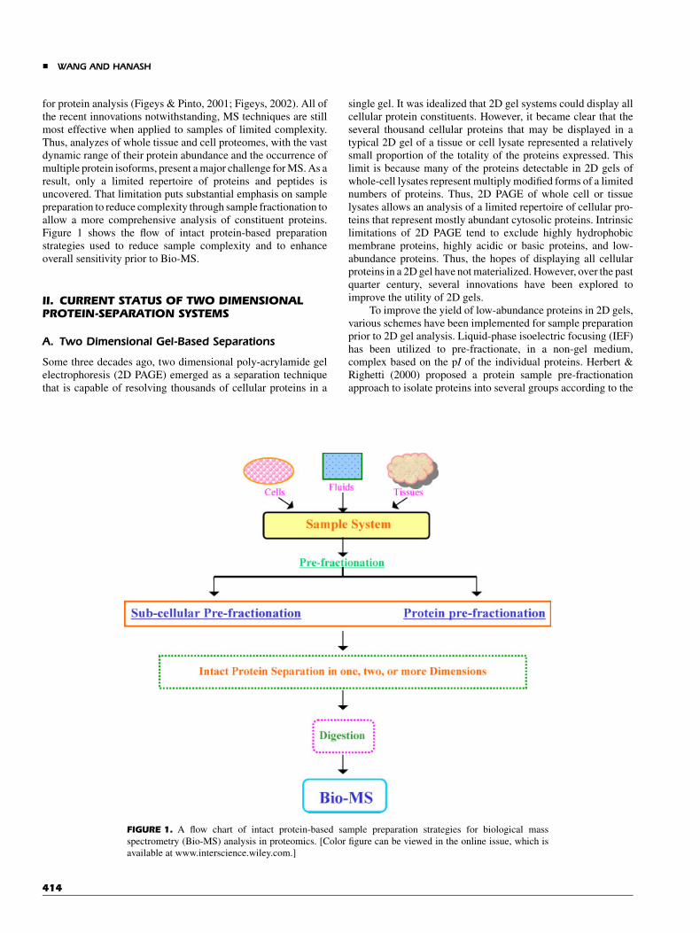
for protein analysis (Figeys & Pinto, 2001; Figeys, 2002). All ofthe recent innovations notwithstanding, MS techniques are stillmost effective when applied to samples of limited complexity.Thus, analyzes of whole tissue and cell proteomes, with the vastdynamic range of their protein abundance and the occurrence ofmultiple protein isoforms, present a major challenge for MS. As aresult, only a limited repertoire of proteins and peptides isuncovered. That limitation puts substantial emphasis on samplepreparation to reduce complexity through sample fractionation toallow a more comprehensive analysis of constituent proteins.Figure 1 shows the flow of intact protein-based preparationstrategies used to reduce sample complexity and to enhanceoverall sensitivity prior to Bio-MS.
II. CURRENT STATUS OF TWO DIMENSIONALPROTEIN-SEPARATION SYSTEMS
A. Two Dimensional Gel-Based Separations
Some three decades ago, two dimensional poly-acrylamide gelelectrophoresis (2D PAGE) emerged as a separation techniquethat is capable of resolving thousands of cellular proteins in a
single gel. It was idealized that 2D gel systems could display allcellular protein constituents. However, it became clear that theseveral thousand cellular proteins that may be displayed in atypical 2D gel of a tissue or cell lysate represented a relativelysmall proportion of the totality of the proteins expressed. Thislimit is because many of the proteins detectable in 2D gels ofwhole-cell lysates represent multiply modified forms of a limitednumbers of proteins. Thus, 2D PAGE of whole cell or tissuelysates allows an analysis of a limited repertoire of cellular pro-teins that represent mostly abundant cytosolic proteins. Intrinsiclimitations of 2D PAGE tend to exclude highly hydrophobicmembrane proteins, highly acidic or basic proteins, and low-abundance proteins. Thus, the hopes of displaying all cellularproteins in a 2D gel have not materialized. However, over the pastquarter century, several innovations have been explored toimprove the utility of 2D gels.
To improve the yield of low-abundance proteins in 2D gels,various schemes have been implemented for sample preparationprior to 2D gel analysis. Liquid-phase isoelectric focusing (IEF)has been utilized to pre-fractionate, in a non-gel medium,complex based on the pI of the individual proteins. Herbert &Righetti (2000) proposed a protein sample pre-fractionationapproach to isolate proteins into several groups according to the
FIGURE 1. A flow chart of intact protein-based sample preparation strategies for biological mass
spectrometry (Bio-MS) analysis in proteomics. [Color figure can be viewed in the online issue, which is
available at www.interscience.wiley.com.]
& WANG AND HANASH
414

pI within multi-compartment electrolyzers (MCE) that aredelimited by immobilized isoelectric membranes with pH valuesof 3.0, 4.0, 5.0, 6.0, and 10.5. They applied this liquid-phase IEFmethod to pre-fractionate Escherichia coli whole-cell extractsand human plasma. Proteins in each fraction were subsequentlyseparated by narrow-pH range 2D PAGE. In plasma separationsbecause albumin was concentrated within the membranesbetween pH 5.6 and 6.1, the acidic and basic chambers wereboth free of albumin; that method resulted in an increase in thenumber of highly acidic and basic proteins in the fractionatedsample compared to whole plasma. Pedersen applied the sametechnology to fractionate alkaline proteins from Saccharomyescerevisiae solubilized-membrane protein mixtures within theMCE compartment between pH 7.5 and 10.5. The concentratedalkaline fraction was subjected to narrow-pH range 2D PAGEfollowed by MALDI-TOF MS (Pedersen et al., 2003). A total of93 unique proteins were identified in this pH 7–10.5 fraction,including 30 low-abundance proteins with the codon adaptationindex (CAI) below 0.2, 20 integral membrane proteins, and10 membrane-associated proteins.
Zuo and Speicher developed a microscale solution IEF(msol-IEF) device that consisted of six to seven separationchambers bound by the immobilized isoelectric membranes topre-fractionate mouse serum into a series of well-defined poolsprior to subsequent analysis with 2D PAGE (Zuo et al., 2002).After IEF, each chamber contained only proteins with a pIbetween the pH of the boundary membranes of that chamber.That pre-fractionation method fractionated complex proteinsamples into very narrow ranges (<0.5 pH units) with anenhanced ability to analyze low-abundance proteins. Six- to 30-fold greater protein loads were practical for non-albuminfractions in the subsequent narrow-pH range 2D gels, andthat in turn increased the dynamic range of protein analysis.That method was also used to pre-fractionate the whole-cellextract of a human breast-cancer cell line into sevendiscrete pools, including four sequential 0.5 pH range fractionsin the pH 4.5–6.5 region, which contained the majority of cellularproteins (Zuo & Speicher, 2002). These four pH pools (0.5 pHunits) were applied onto the narrow pH range gels for furtherseparation.
Alternatively, liquid-phase IEF in the Rotofor system (Bio-Rad Laboratory) with 20 IEF cells has been utilized to pre-fractionate human cerebrospinal fluid (CSF) prior to 2D PAGE(Davidsson et al., 2002). Proteins in selected IEF fractions werefurther resolved on SYPRO-Ruby-stained 2D PAGE gels. It wasfound that more protein spots were detected in 2D gels from pre-fractionated CSF compared with direct 2D PAGE separations ofCSF. Some low-abundance proteins, including cystatin C, IgM-kappa, b-2 microglobulin, alpha-1-acid glycoprotein, acetyl-coenzyme A carboxylase-alpha, and hemopexin, were identifiedin pre-fractionated but not in non-fractionated CSF. Low-abundance forms of post-translationally modified proteins, as inthe case of alpha-1-acid glycoprotein and alpha-2-HS glycopro-tein, could be enriched, thus improving overall resolution andsensitivity. Similarly, Zuo et al. (2002) utilized an IEF method tofractionate human-breast cancer-cell lysates prior to 2D PAGE,with improved results.
Gorg et al. (2002) revisited the use of flat-bed IEF ingranulated Sephadex gels, namely as a pre-fractionation
procedure that was applied to mouse-liver proteins. Ten gelfractions were simply extracted with a spatula, and each fractionwas directly applied onto the surface of corresponding narrow-range IPG strips and subjected to further separation by 2D PAGE.Proteins in the Sephadex gel fractions migrated electrophoreti-cally onto the IPG gel with high efficiency and without anysample dilution. One milligram of mouse-liver proteins was pre-fractionated in this fashion, and neither protein precipitation norhorizontal or vertical streaking was observed in the subsequentnarrow pH range 2D gels. As a result, the considerably greaternumbers of protein spots detectable in 2D gels indicatedenrichment in low abundance proteins.
These improvements in resolution and sensitivity arepromising, and stem from the ability to apply greater amountsof proteins. The gains are obviously achieved at a certain cost-namely, that the rather complicated procedure of 2D gels is madeeven more complicate from the need to integrate data and imagesacross multiple 2D gels. There is a need for a critical assessmentof reproducibility with such schemes as well as an assessment ofthe extent to which certain proteins may be subjected tomodifications that could have a negative impact on sampleanalysis.
B. Two Dimensional Liquid-Based Separations
There is a great deal of interest at the present time in developinggel-free systems for protein analysis because of their potential formultiplexing (Liu et al., 2002; Wang & Hanash, 2003). Ananalogy may be made to DNA sequencing, notably as utilized inthe genome project, which received a considerable boost whenthe switch from gel-based approaches to a gel-free technologytook place. Multi-modular combinations of HPLC, liquid-phaseIEF, and capillary electrophoresis (CE) provide various optionsto develop high-resolution orthogonal 2D liquid phase-basedstrategies for the separation of complex mixtures of proteins.Such strategies include size-exclusion chromatography (SEC)–CE or SEC–reversed-phase liquid chromatography (RPLC) wasused by Jorgenson’s group to fractionate protein mixtures inEscherichia coli lysates (Larmann et al., 1993; Opiteck et al.,1998). Le Coutre et al. (2000) analyzed Escherichia colimembrane proteins with affinity chromatography, followed byon-line RPLC–MS. Feng et al. (2001) reported the use of ion-exchange chromatography (IEC) followed by on-line eight-channel parallel RPLC–ESI-MS to purify recombinant proteinsin a high-throughput fashion. A major advantage of liquidseparations is that proteins are maintained in solution that allowson-line intact protein characterization by MS as well as proteinrecovery. Our group developed a novel 2D IEF-RPLC system tofractionate or resolve large numbers of cellular proteins. Theseprotein fractions were recovered and applied to protein biochipsto determine their antigenicity in cancer (Wall et al., 2000;Madoz-Gurpide et al., 2001; Wang & Hanash, 2003). Thecapacity of the 2D separation system in practice is limited toresolving no more than 10,000 protein forms according toGiddings’ model, if each dimension has a capacity of 100; thatcapacity may not be sufficient to achieve complete resolution of acell or tissue proteome. It is, therefore, beneficial to reducesample complexity as much as possible.
PROTEOME ANALYSIS WITH MASS SPECTROMETRY &
415

III. STRATEGIES TO REDUCE SAMPLECOMPLEXITY BY ENRICHMENT FORSPECIFIC CELL POPULATIONS ANDSUB-CELLULAR STRUCTURES
A. Reducing Cellular Heterogeneity
The heterogeneous nature of tissue samples makes their directproteomic analysis difficult, particularly when the cell type ofinterest is under-represented in the sample. A traditional way toreduce cellular heterogeneity prior to analysis of tissue samples isto disaggregate the tissue by treatment with collagenase or byother means, followed by a separate analysis of specific cellpopulations of interest. This traditional approach is still widelyrelied upon, particularly when large numbers of cells are needed.An elegant approach to reduce cellular heterogeneity and toextract a particular cell population in a tissue is laser capturemicrodissection (LCM), a technology that was developed at theNational Cancer Institute (Emmert-Buck et al., 1996). It has beensuccessfully used to isolate single cells within a tissue section(Emmert-Buck et al., 1996). Cells can be selected according totheir phenotypic and functional characteristics. The majorlimitation of the LCM approach for proteomics is the labor-intensive nature to extract a sufficient number of cells forproteomic analysis.
Palmer-Toy et al. (2000) described a rapid and sensitivemethod to obtain an abridged protein-expression profile frommicrodissected human-breast tissue cells by direct acquisition ofMALDI-TOF mass spectra from LCM transfer films. Four cellpopulations, including normal stromal cells, normal epithelialcells, ductal carcinoma in situ, and invasive ductal carcinoma,were isolated from a single frozen section of human breast byLCM, and were subjected to direct MALDI-TOF analysis.Distinct mass spectra were obtained from 1,250 cells from eachof the four cell types. The stromal cells revealed severalprominent peaks in the 4.5–7.0 kDa range. Those peaks wereattenuated or absent in the spectra from cells of epithelialderivation. A series of high-mass peaks from 45 to 60 kDadistinguished the invasive carcinoma spectrum from that ofnormal epithelium and from the stromal spectrum as well. Xuet al. (2002) reported a comparison of mass spectra obtained fromhuman breast tissue that contained invasive mammary carcinomaand normal breast epithelium, using LCM MALDI-TOF MS.More than 40 peaks were identified that significantly differed inintensity between invasive mammary carcinoma and normalbreast epithelium. Bhattacharya used LCM to procure cancercells from archived human lung tissue that contained an adeno-carcinoma and squamous cell carcinoma. The captured cancercells were mixed with matrix solution, and that solution wasdeposited on the MALDI target for direct MALDI-TOF MSanalysis (Bhattacharya, Gal, & Murray, 2003). The resultsshowed that half of the observed peaks in the mass range between1,000 and 4,000 Da were indicative of either adenocarcinoma orsquamous cell carcinoma, and may be used as a fingerprint forthose cancer types.
Lawrie et al. (2001) used LCM to selectively microdissecttumor cells from colon cancer tissues. The protein mixturesrecovered from the captured cells were separated and character-ized by 2D PAGE/Bio-MS. A method that utilized an adjacent
stained section to guide the dissection of an unstained region ofinterest was used to overcome problems that resulted from animmuno-histochemical marking of the dissected cells; thatstaining has detrimental effects on protein analysis by 2D PAGE(Wong et al., 2000). Mouledous et al. applied this approach tounstained rat brain tissues to precisely dissect nuclei from speci-fically defined brain regions. Proteins from the captured tissuecells were extracted and separated by 2D PAGE, and selectedspots were identified by Bio-MS (Mouledous et al., 2003).
A potentially spectacular way to reduce tissue heterogeneityis direct, in situ, mass spectrometric analysis of cellularconstituents of a tissue—as pioneered by Caprioli’s group. Withthis approach, imaging MS is undertaken for the analysis ofpolypeptide expression in tissue sections, where the spatial arrayof specific polypeptides present in neighboring cells is profiled byMALDI-TOF MS (Caprioli, Farmer, & Gile, 1997; Stoeckli et al.,2001). Imaging mass spectra for different cell populations ofinterest are determined and are compared with each other andbetween healthy and disease tissues. In a study of humanglioblastoma, tumor cells displayed many protein differencescompared to normal tissue. For example, a protein of molecularmass 4,964 Da was localized to the outer area of tumors and wasidentified as thymosin b4 (Tb4), an immuno-regulatory peptidethat has ability to sequester cytoplasmic monomeric actin. Thisconcept offers the tantalizing prospect that imaging MS may beused, for example, intra-operatively to assess the surgicalmargins of excised tumors (Chaurand, Schwartz, & Caprioli,2002). The major limitation of this approach, as with otherapproaches for direct mass spectrometric analysis of complextissue samples, is the difficulty to identify the protein for whichmolecular mass is detected.
B. Enrichment in Sub-Cellular Structures
Sub-cellular fractionation strategies include a variety ofestablished and innovative approaches that are made particularlyeffective in combination with Bio-MS to profile proteinconstituents. With traditional approaches, generally the sampleis first subjected to homogenization to obtain a free suspension ofintact, individual organelles by means of low-speed centrifuga-tion. The nuclei, together with cell debris and unbroken cells, areremoved as a pellet. The supernatant that contains the cytosol andother organelles in suspension is subjected to sub-cellular frac-tionation. Density-gradient centrifugation is the most popularapproach to efficiently perform sub-cellular fractionation. Thesub-cellular fraction(s) of interest is enriched, and is subjected tofurther separation of constituent proteins, coupled with Bio-MS.
1. Density-Gradient Centrifugation forSub-Cellular Fractionation
Fractionation by gradient centrifugation, based on the sedimen-tation velocity of organelles in gradient medium such as sucroseand percoll, has been frequently applied to the fractionation ofsub-cellular organelles, such as Golgi and mitochondria.Bergeron’s group has utilized sucrose density-gradient centrifu-gation to isolate different organelles. In studies of the Golgiorganelle, membrane proteins were resolved by 1D SDS
& WANG AND HANASH
416

electrophoresis. Bands of interest were analyzed by MALDI-TOF MS or Q-TOF MS/MS. A total of 81 membrane proteins,including a novel Golgi-associated protein of 34 kDa (GPP34),were unambiguously identified (Dominguez et al., 1999; Bellet al., 2001). An abundance of trafficking proteins was un-covered, such as KDEL receptors, p24 family members,SNAREs, Rabs, ARF-guanine nucleotide exchange factor, andSCAMPs. Hanson and Lescuyer used sucrose density-gradientcentrifugation to analyze mitochondrial proteins in combinationwith 2D PAGE and Bio-MS. Functional information on proteincomplexes within human brain mitochondria was obtained(Hanson et al., 2001). The human mitochondrial proteomemap, using placenta as the source tissue, was recently constructedand a large number of proteins were identified, including novelones (Lescuyer et al., 2003). Andersen and his colleaguesreported their direct study of the human sub-nuclear proteome byusing a combination of sonication and sucrose density-gradientcentrifugation to fractionate nucleoli from HeLa cell nuclei,followed by 1D or 2D gel protein separation and Bio-MS analysis(Andersen et al., 2002). A total of 271 proteins were identified,and more than 30% of the nucleolar proteins were encoded bynovel or uncharacterized human genes.
Murayama et al. (2001) described a novel approach that usesfreeze-thawing to produce a density-gradient solution of Nyco-denz for the one-step fractionation of organelles from rat liver andsubsequent analysis of fractions by 2D PAGE. An alternativetechnique that used differential centrifugation and hypotoniclysis was applied to separate lysosomes from endosomes and pre-lysosomal compartments (Schafer & Heizmann, 1996). Thisapproach resulted in a pure lysosomal fraction that containedhigh specific activities of lysosomal enzymes, and an endosomalfraction that contained endosomes at different stages withoutdetrimental effects on the quality of the isolated fractions. Thissub-cellular pre-fractionation technique is applicable to a varietyof human cell populations.
2. Immune-Based Sub-Cellular Fractionation
Immune-based techniques use the high specificity of antibodiesto capture sub-cellular organelles that contained the cognateantigen. Shevchenko et al. (1997) used this approach to isolatetrans-Golgi network (TGN)-derived apical and basolateraltransport vesicles, followed by 2D PAGE and Bio-MS. Twoproteins that belong to the p23/p24 family of putative cargoreceptors for vesicular trafficking were identified, and caveolin-2was also characterized as a constituent of basolateral transportvesicles. This approach can also be used in combination withgradient centrifugation to further reduce protein cross-contam-ination from different organelles, such as endoplasmic reticulum,Golgi membrane, and plasma-membrane proteins.
3. Free-Flow Electrophoresis forSub-Cellular Fractionation
Free-flow electrophoresis (FFE) is a revitalized old techniquethat is based on differences in electrophoretic mobility betweenvarious components in mixtures, ranging from polypeptides andsub-cellular organelles to cells. Thus, a variety of sub-cellular
organelles may be separated on the basis of their unique chargedensity. For example, lysosomes of human skin fibroblasts wereefficiently isolated by FFE after differential centrifugation of thecell lysate suspended in isotonic sucrose (Harms, Kern, &Schneider, 1980). Marsh et al. (1987) described a rapid sub-cellular pre-fractionation approach that combined density-gradient centrifugation with FFE to isolate endosomes from avariety of tissue culture cells. The post-nuclear supernatants weresubjected to FFE. Endosomes and lysosomes migrated togetheras a single anodally deflected peak separated from most otherorganelles such as plasma membrane, mitochondria, endoplas-mic reticulum, and Golgi. The endosomes were further resolvedfrom lysosomes by centrifugation in a percoll density gradient. A70-fold enrichment of endosomes was achieved relative to theinitial homogenate. FFE was also used to separate flagellarpocket-derived membranes from other endosomal and lysosomalorganelles of African trypanosomes (Grab, Webster, & Lonsdale-Eccles, 1998).
Immune free-flow electrophoresis (IFFE) combines theadvantages of electrophoretic separation with the high selectivityof antigen–antibody binding. It relies on the altered electro-phoretic mobility of a sub-cellular organelle complexed to aspecific antibody against the cytoplasmic domain of one of itsintegral membrane proteins when the electrophoresis buffer pH isadjusted to 8.0, close to the pI of immunoglobulin (Ig). Thus, Ig-coupled organelles can be separated from other structures byFFE. Mohr and Volkl applied IFFE to the isolation and analysis ofperoxisomes as well as of microsomal fractions obtained bydifferential centrifugation of a rat liver homogenate (Volkl, Mohr,& Fahimi, 1999; Mohr & Volkl, 2002).
Another variation on the theme, density-gradient electro-phoresis (DGE), combines the principle of FFE with densitygradients (Tulp, Verwoerd, & Pieters, 1993). After homogeniza-tion, organelle mixtures are layered within a sucrose or Ficollgradient that is subjected to electrophoresis. Endosomal andlysosomal organelles, being negatively charged, migrate pre-ferentially towards the anode and are separated from other sub-cellular organelles. Tulp et al. (1998) applied high-resolutionDGE for the sub-cellular fractionation of late endosomes, earlyendosomes, lysosomes, endoplasmic reticulum, plasma mem-brane, clathrin-coated pits, proteasomes, and clathrin-coatedvesicles from the postnuclear supernatant, by using a novel low-conductivity buffer. A DGE protocol was developed that alloweda one-step separation of plasma membrane, Golgi/TGN (Lindner,2001) and endosomes for the quantitative analysis of vesiculartransport from the Golgi/TGN compartment to the plasmamembrane and endosomes (Lindner, 2001).
IV. FRACTIONATION OF PROTEINS BASED ONTHEIR CHEMICAL AND PHYSICAL PROPERTIESREDUCES COMPLEXITY AND ENHANCES THEYIELD OF LOW-ABUNDANCE PROTEINS
The protein complexity of biological samples may be simplifiedthrough fractionation based on different protein properties,including sequential solubilization, selective precipitation, andaffinity purification, or through various chromatography-based
PROTEOME ANALYSIS WITH MASS SPECTROMETRY &
417

methods (Issaq et al., 2002; Liu et al., 2002; Wang & Hanash,2003).
A. Chromatography-Based Fractionations
Virtually all chromatographic modalities have been used for thepre-fractionation of biological samples in order to achieve anenhanced resolution of proteins in individual fractions. RP-HPLC has been applied to fractionate protein mixtures of tissuelysates, and each fraction was presented to 2D PAGE for furtherseparation (Badock et al., 2001; Van Den Bergh et al., 2003).Some low-abundance proteins were enriched in 2D gels and wereidentified by Bio-MS (Badock et al., 2001). In another study, RP-HPLC pre-fractionation was applied to visual cortex tissuelysates prior to analysis. Some protein spots that were notobserved in total tissue lysates were visualized and identified(Van Den Bergh et al., 2003).
SEC has also been used as a pre-fractionation technique. Forexample, the human lens proteins crystallins become extensivelymodified with aging, and the characterization of these modifiedproteins is of significance because they are the likely precursorsof cataract. In one study, the soluble crystallins were firstfractionated into a-, b-, and g-crystalins by SEC (Zhang, Smith,& Smith, 2001). All of the b-crystallins, including three acidicsubunits (bA1, bA3, bA4) and three basic subunits (bB1, bB2,bB3), were collected into one fraction, and were furtherfractionated by RP-HPLC. ThebA4 andbB1 RP-HPLC fractionswere separated by 2D PAGE, followed by the characterization ofthe spots of interest. IEC separates protein mixtures based oncharge in a non-denaturing environment. Proteins with similar pIand strongly associated proteins are co-eluted in the samefraction. In one study, IEC pre-fractionation simplified thecomplexity of whole cell lysates for the analysis of multi-proteincomplexes by 2D PAGE, and also resulted in protein enrichmentfor subsequent mass spectrometric analysis (Butt et al., 2001).
Chromatofocusing (CF) is a type of IEC that separatesproteins according to their pI. Proteins bound to the gel matrix areeluted with a specific poly-buffer in the order of decreasing pI.Fountoulakis et al. used CF to fractionate and enrich Haemophi-lus influenzae protein mixtures. Proteins were further separatedby 2D PAGE. Seventy new proteins were identified in the CFpools, many of which occurred in low abundance and were notdetectable by the direct analysis of lysates by 2D PAGE(Fountoulakis et al., 1998). Similarly, that same group usedhydrophobic interaction chromatography (HIC) to separateHaemophilus influenzae proteins based on their hydrophobicity,followed by 2D PAGE and MALDI-TOF MS to identify novelproteins (Fountoulakis, Takacs, & Takacs, 1999).
B. Liquid-Phase Electrophoresis Prior to SDS–PAGE
FFE has been used to fractionate protein mixtures based on pI.Hoffmann and his colleagues used continuous FFE to isolatecytosolic proteins from the human colon carcinoma cell line LIM1215 into 96 fractions, followed by SDS–PAGE gel separation.The resolved proteins were identified by peptide fragmentsequencing, using on-line capillary LC-MSMS (Hoffmann et al.,2001). The experimental relative molecular mass (Mr) and pI of
identified proteins were in good agreement with the theoreticalvalues calculated from the amino acid sequence.
Bae et al. applied the Gradiflow technique, another liquid-phase IEF separation applicable to proteins with pI> 9.0 (Lockeet al., 2002) to fractionate proteins from H. pylori whole celllysates to obtain two groups of basic proteins followed by furtherseparation with SDS–PAGE. Sixteen bands were selected forprotein characterization by MALDI-TOF MS (Bae et al., 2003).Seventeen basic proteins were identified, five of which (HP1216,HP1283, Cag3, Cag13, and KataA) with predicted pIs between8.97 and 9.69 were not identified on either pH 6–11 or pH 9–12 IPG 2D PAGE gels without sample pre-fractionation.
C. Liquid-Phase IEF Prior to HybridMulti-Dimensional Separation
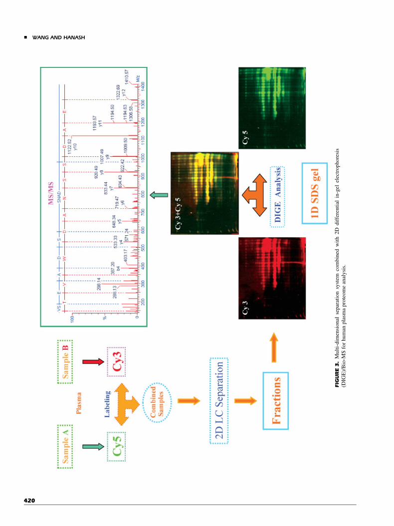
An orthogonal high-resolution multi-dimensional separationsystem developed in our laboratory, using liquid-phase IEF asthe pre-fractionation approach followed by separation with RP-HPLC and SDS–PAGE, was applied to cancer cell-line lysates orbio-fluids. This system was coupled with protein biochips andBio-MS to probe the human cancer proteome, as illustrated inFigure 2 (Madoz-Gurpide et al., 2001). The advantage of thecombination of liquid-phase IEF with RP-HPLC is that proteinsin hundreds of individual fractions can be arrayed directly andused as targets for a variety of probes. Constituent proteins inreactive fractions of interest are subjected to further separation onSDS gels followed by protein characterization of bands ofinterest by capillary LC ESI Q-TOF MS/MS (Nam et al., 2003).We have also used this separation system in combination withfluorescence detection to quantitatively profile the human serumproteome (Wang et al., 2002). Two independent serum samples,such as a study sample and a control, are labeled with differentfluorescence reagents such as Cy3 and Cy5, and combined priorto three-dimensional protein separation. The resolved proteinsare visualized in SDS gels, and protein bands of interest areanalyzed by either MALDI-TOF MS or capillary LC ESI Q-TOFMS/MS (Fig. 3).
D. Affinity and Immunocapture
Affinity- and immunocapture-based methods represent a well-established approach to enrich protein subsets of interest. Withthese approaches, complexity may be reduced to such a largeextent that a simple one-dimensional separation procedure maybe sufficient to resolve captured protein constituents. For exa-mple, Pandey et al. (2000) used immunoprecipitation techniquesto precipitate tyrosine-phosphorylated proteins and to probesignaling-related cell-surface receptors by 1D SDS gel/Bio-MS.Journet used mannose-6-phosphate (M-6-P) receptors (MPRs) asthe affinity chromatography separation media to specificallypurify M-6-P proteins from soluble human U937 cell lysosomalhydrolase. The captured proteins were separated by 2D PAGE,and were analyzed with either MALDI-TOF MS or ESI Q-TOFMS/MS. Twenty-two proteins were identified, among which16 were well-known lysosomal hydrolases, such as proteinase 3,cathepsin A, cathepsin D, cathepsin S, Dnase II,b-glucuronidase,and acid ceramidase (Journet et al., 2002). Phosphospecific
& WANG AND HANASH
418

FIG
URE2.
Mu
lti-
dim
ensi
on
alse
par
atio
nsy
stem
cou
ple
dw
ith
pro
tein
bio
chip
and
Bio
-MS
.A:C
ello
r/an
d
tiss
ue
lysa
tes
are
sep
arat
edin
to2
0fr
acti
on
sb
yis
o-e
lect
ric
focu
sin
g;
(B)
ind
ivid
ual
frac
tio
ns
are
furt
her
reso
lved
by
rever
sed
-ph
ase
HP
LC
;(C
)al
iqu
ots
of
sep
arat
edp
rote
ins
are
arra
yed
on
tog
lass
slid
esfo
r
sub
seq
uen
tp
rob
ing
,u
nco
ver
ing
inth
isca
sesp
ott
edp
rote
ins
that
reac
tw
ith
anti
bo
die
sin
aca
nce
rsu
bje
ct’s
seru
m.D
:In
div
idu
alfr
acti
on
so
fin
tere
star
efu
rth
eran
aly
zed
by
mas
ssp
ectr
om
etry
,sh
ow
ing
inth
isca
sea
mas
ssp
ectr
um
for
ap
epti
de
dig
est
fro
ma
spo
tted
pro
tein
.
PROTEOME ANALYSIS WITH MASS SPECTROMETRY &
419
FIG
URE3.
Mu
lti-
dim
ensi
on
alse
par
atio
nsy
stem
com
bin
edw
ith
2D
dif
fere
nti
alin
-gel
elec
tro
ph
ore
sis
(DIG
E)/
Bio
-MS
for
hu
man
pla
sma
pro
teo
me
anal
ysi
s.
& WANG AND HANASH
420
antibody-based immunoaffinity chromatography and immobi-lized metal-based affinity chromatography (IMAC) have beenused to enrich phosphoproteins and to decipher the phosphopro-teome with Bio-MS-based strategies (Kalume, Molina, &Pandey, 2003). Heparin affinity chromatography was used toenrich low-abundance human fetal brain protein mixtures, andthe eluted affinity-specific fractions were resolved by 2D PAGE.Approximately 70 enriched unique proteins that belong to severalclasses, including proteasome components, dihydropirimidi-nase-related proteins, and T-complex protein I components, andenzymes with various catalytic activities were identified byMALDI-TOF MS (Karlsson et al., 1999). Shefcheck, Yao, &Fenselau (2003) used heparin affinity chromatography tofractionate cytosolic protein mixtures of human MCF-7 breastcancer cells into three fractions prior to 2D PAGE. A strikinglevel of enrichment is achieved for low-abundance proteins ineach fraction, and 300 proteins were visible in 2D gel patterns ofthe three fractions. Those 300 proteins could not be detected innon-fractionated cytosol.
E. Depletion/Enrichment of Proteins FromHuman Plasma/Serum
There is an increasing interest focused on the human plasma/serum proteome because of the great relevance of plasma/serumproteins to biomedicine, including diagnostics and therapeuticmonitoring (Adkins et al., 2002). However, high-abundanceproteins in plasma/serum, including albumin, immunoglobulins(IgG and IgA), antitrypsin, transferring, and haptoglobin,interfere with the proteomic analysis of low-abundance proteinsbecause of the consequent loss of resolution in 2D PAGE orchromatographic separations. Thus, most of the interesting low-abundance proteins (�1 ng/mL level) are not detected. There-fore, it is advantageous to specifically remove high-abundanceproteins in a sample pre-fractionation step prior to proteinanalysis. Specific removal of high-abundance proteins willdeplete approximately 85–90% of the total protein mass fromhuman plasma/serum. Pieper et al. (2003) reported an elegantquantitative strategy to selectively profile low-abundance pro-teins in human plasma by a multi-component immunoaffinitychromatography approach, based on antibody–antigen interac-tions, to deplete 10 high-abundance proteins from plasma.Affinity-purified polyclonal antibodies (pAbs) were used as thestationary phase in the column to specifically capture high-abundance proteins albumin, IgG, IgA, transferrin, a-1-anti-trypsin, haptoglobin, a-2-macroglobulin, hemopexin, a-1-acidglycoprotein, and a-2-HS glycoprotein. This specific step ofselective immunodepletion provided an enriched pool of low-abundance proteins in the flow-through fraction from the columnfor subsequent 2D PAGE/Bio-MS analysis. An increment of 350low-abundance proteins was visualized after depletion.
Wang & Hanash (2003) developed a simple and rapidimmunoaffinity-based method, using an affinity spin-tube filter todeplete high-abundance proteins or to enrich low-abundanceprotein biomarkers in human serum. The affinity spin-tube filtercontains protein G, coupled with antibodies against either high-abundance proteins for the depletion of proteins like albumin andIgG, or specific proteins of interest for the enrichment of low-
abundance protein biomarkers such as fatty acid synthase (FAS).The flow-through fraction or the eluate was recovered andsubjected to 2D PAGE for further separation. Of total albumin88% was depleted from the serum with enhanced detection of theremaining proteins. Total protein recovery was >95%.
Rothemund et al. (2003) used the Gradiflow technique todeplete albumin from human plasma on the basis of its pI andMr. Human plasma proteins were fractionated into four groups:albumin, proteins with pI greater than albumin, proteins with Mrhigher than albumin, and proteins with Mr lower than albumin.The albumin-depleted fractions were separated by 2D PAGE toallow the detection of low-abundance proteins. A chain of proteinspots that lay beneath albumin in non-fractionated plasma wererevealed, and allowed the identification of C4B-binding proteinachain. One advantage of using Gradiflow for the depletion ofalbumin is its ability to separate proteins by Mr to allow someproteins to be separated from albumin even though their pIs wereclose to that of albumin. The Human Proteome Organization(http://WWW.HUPO.org) is currently assessing the merits ofvarious depletion protocols as part of the Human PlasmaProteome Project.
V. FRACTIONATION OF PROTEINS BASED ONTHEIR POST-TRANSLATIONAL MODIFICATIONS
PTMs, such as glycosylation, phosphorylation, sulfation, andacetylation, modulate important biological activity of proteinsduring cellular processes. Identification of sites of protein PTMsand the quantitative analysis of modified proteins would provideinsight into biological functions. There are currently two majoraffinity chromatography-based techniques coupled with Bio-MSfor the analysis of proteins. One is the IMAC and the other is theisotope-coded affinity tagging (ICAT) technique (Fiacre et al.,2002; Kalume, Molina, & Pandey, 2003; Mann & Jensen, 2003).These approaches are outside the scope of this review. However,from the biomedical applications point of view, there is still asubstantial need to develop highly efficient approaches for thequantitative analysis of PTMs.
VI. PROTEIN-TAGGING STRATEGIES
Protein-tagging techniques have great utility to capture proteinsubsets and/or to enhance sensitivity. They have been applied tothe study of multi-protein complexes (Bauer & Kuster, 2003),comprehensive profiling of subcellular proteomes (Shin et al.,2003), and for quantitative profiling of overall protein expression(Unlu, Morgan, & Minden, 1997).
A. Biotin Tags to Profile Cell-Surface Proteins
Cell-surface proteins are involved in a multitude of intercellularand extracellular functions. However, the global proteomicanalysis of this compartment has been quite challenging becauseof the intrinsic features of its protein constituents, including highhydrophobicity and low expression levels. Thus, the develop-ment of efficient sample-preparation techniques to isolate intact
PROTEOME ANALYSIS WITH MASS SPECTROMETRY &
421
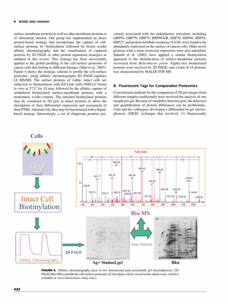
surface membrane proteins as well as other membrane proteins isof substantial interest. Our group has implemented an intactprotein-based strategy that incorporates the capture of cell-surface proteins by biotinylation followed by biotin–avidinaffinity chromatography and the visualization of capturedproteins by 2D PAGE or other protein separation strategies asoutlined in this review. This strategy has been successfullyapplied to the global profiling of the cell-surface proteome ofcancer cells that belong to different lineages (Shin et al., 2003).Figure 4 shows the strategic scheme to profile the cell-surfaceproteome, using affinity chromatography-2D PAGE-capillaryLC-MS/MS. The surface proteins of viable, intact cells aresubjected to biotinylation with EZ-Link sulfo-NHS-LC-biotinin vitro at 378C for 10 min, followed by the affinity capture ofsolubilized biotinylated surface-membrane proteins with amonomeric avidin column. The enriched biotinylated proteinsmay be visualized in 2D gels as intact proteins to allow theelucidation of their differential expression and assessment oftheir PTMs. Alternatively, they may be fractionated with a liquid-based strategy. Interestingly, a set of chaperone proteins pre-
viously associated with the endoplasmic reticulum, includingGRP94, GRP78, GRP75, HSP90A/B, HSP70, HSP60, HSP54,HSP27, and protein disulfide isomerase A3/A6, were found to beabundantly expressed on the surface of cancer cells. Other novelproteins with a more restricted expression were also identified.Sabarth et al. (2002) have applied a similar biotinylationapproach to the identification of surface-membrane proteinsrecovered from Helicobacter pylori. Eighty-two biotinylatedproteins were resolved by 2D PAGE, and a total of 18 proteinswas characterized by MALDI-TOF MS.
B. Fluorescent Tags for Comparative Proteomics
Conventional methods for the comparison of 2D gel images fromdifferent samples traditionally have involved the analysis of onesample per gel. Because of variability between gels, the detectionand quantification of protein differences can be problematic.Unlu and his colleagues developed a differential in-gel electro-phoresis (DIGE) technique that involved: (1) fluorescently
FIGURE 4. Affinity chromatography prior to two dimensional poly-acrylamide gel electrophoresis (2D
PAGE)/Bio-MS to profile the cell-surface proteome. [Color figure can be viewed in the online issue, which is
available at www.interscience.wiley.com.]
& WANG AND HANASH
422

tagging two protein samples with two different fluorescent dyes(such as Cy3 and Cy5), (2) separating them on the same 2D gel(2D DIGE), (3) fluorescence imaging of the gel into two images,and superimposing the images to detect any differentialexpression between the two protein samples (Unlu, Morgan, &Minden, 1997). This method improves the reproducibility andreliability of differential protein expression analysis betweensamples, and is particularly useful in comparative studies ofnormal and diseased tissues. Several groups have used the 2DDIGE technique in various studies, such as analysis of mouse-liver homogenates treated with N-acetyl-p-aminophenol (APAP)(Tonge et al., 2001), elucidation of the effect of ErbB-2 over-expression on breast cancer cells (Gharbi et al., 2002), andanalysis of bacteria under different growth conditions (Gadeet al., 2003). Yan et al. (2002) used this technique to study theEscherichia coli proteome after benzoic acid treatment. A total of179 differentially expressed protein spots were identified. Thoseproteins included enzymes, stress-related, and substrate-bindingproteins (e.g., amino acids, maltose, ribose, and TRP repressor).
VII. PERSPECTIVE AND CONCLUSION
It has become obvious exceedingly that there are no simpleuniversal strategies for the comprehensive analysis of complexproteomes. There are specialized strategies—each with someadvantages and some disadvantages. The merits of thesestrategies must be weighed in relation to the contemplatedspecific applications. In this review, we have presented intactprotein-based strategies that are intended to reduce the complex-ity of biological samples prior to MS. Such strategies havesubstantial versatility because they allow the detection, identi-fication, and recovery of proteins of interest. It should be pointedout that a relatively small subset of proteins, particularly largehydrophobic membrane proteins, may defy analysis with such astrategy, and their investigation may be better-suited to otherapproaches. However, these proteins likely represent a smallproportion of the proteome of complex organisms. For mostproteins, and in particular for biological fluids, it may be possiblein the future to eliminate the need for digestion altogether, forexample, by interfacing intact protein-separation strategiesdirectly with MS, where proteins are first assessed with respectto their intact mass followed by their fragmentation that processis to derive their identity as well their PTMs. Additionally,microfluidics and nanotechnologies have yet to make an impacton proteomics, despite their substantial appeal. Nevertheless, itmay also be envisioned that such technologies will make, in thenot so distant future, as much of an impact on proteomics as MShas and we look forward to substantial miniaturization andautomation of intact protein-based strategies for the analysis ofcomplex proteomes.
VIII. ABBREVIATION LIST
1D one dimension2D two dimension2D PAGE two dimensional poly-acrylamide gel
electrophoresis
Bio-MS biological mass spectrometryCE capillary electrophoresisCF chromatofocusingCID collision-induced dissociationCSMPs cell-surface membrane proteinsCy3 1-(5-carboxypentyl)-10-propylindocarbocya-
nine halide N-hydroxysuccinimidyl esterCy5 1-(5-carboxypentyl)-10-methylindodicarbocya-
nine halide N-hydroxysuccinimidyl esterCSF cerebrospinal fluidDGE density-gradient electrophoresisDIGE differential in-gel electrophoresisESI electro-spray ionizationFAB fast atom bombardmentFFE free-flow electrophoresisFTICR Fourier-transform ion cyclotron resonanceGRP78 78 kDa glucose-regulated proteinHIC hydrophobic interaction chromatographyHSP90B heat-shock protein HSP 90-betaHUPO Human Proteome OrganizationICAT isotope-coded affinity tagIEC ion-exchange chromatographyIEF isoelectric focusingIFFE immune free-flow electrophoresisIMAC immobilized metal affinity chromatographyLC liquid chromatographyLCM laser-capture microdissectionLC-MSMS liquid-chromatography combined with
tandem mass spectrometryM-6-P mannose-6-phosphateMALDI-TOF matrix-assisted laser
desorption/ionization-time-of-flightMCE multi-compartment electrolyzerMS mass spectrometryMSMS tandem mass spectrometryPTMs post-translational modificationsQ-TOF Quadrupole-time-of-flightRPLC reversed-phase liquid chromatographySEC size-exclusion chromatography
REFERENCES
Adkins JN, Varnum SM, Auberry KJ, Moore RJ, Angell NH, Smith RD,
Springer DL, Pounds JG. 2002. Toward a human blood serum proteome:
Analysis by multidimensional separation coupled with mass spectro-
metry. MCP 1:947–955.
Aebersold R, Mann M. 2003. Mass spectrometry-based proteomics. Nature
422:198–207.
Andersen JS, Lyon CE, Fox AH, Leung AKL, Lam YW, Steen H, Mann M,
Lamond AI. 2002. Directed proteomic analysis pf the human nucleolus.
Curr Biol 12:1–11.
Badock V, Steinhusen U, Bommert K, Otto A. 2001. Prefractionation of
protein samples for proteome analysis using reversed-phase high-
performance liquid chromatography. Electrophoresis 22:2856–2864.
Bae SH, Harris AG, Hains PG, Chen H, Garfin DE, Hazell SL, Paik YK, Walsh
BJ, Cordwell SJ. 2003. Strategies for the enrichment and identification
of basic proteins in proteome projects. Proteomics 3:569–579.
PROTEOME ANALYSIS WITH MASS SPECTROMETRY &
423

Barber M, Bordoli RS, Sedgwick RD, Tyler AN. 1981. Fast atom
bombardment of solids (FAB): A new ion source for mass spectrometry.
J Chem Soc Chem Commun 270:325–327.
Bauer A, Kuster B. 2003. Affinity purfication-mass spectrometry: Powerful
tools for the characterization of protein complexes. Eur J Biochem
270:570–578.
Baykut G, Funchser J, Witt M, Weiss G, Gosteli C. 2002. A combined ion
source for fast switching between electrospray and matrix-assisted laser
desorption/ionization in Fourier transform ioncyclotron resonance mass
spectrometry. Rapid Commun Mass Spectrom 16:1631–1641.
Bell AW, Ward MA, Freeman HN, Choudhary JS, Blackstock WP, Lewis AP,
Fazel A, Gushue JN, Paiement J, Palcy S, Chevet E, Lafreniere-Roula
M, Solari R, Thomas DY, Rowley A, Bergeron JM. 2001. Proteomics
characterization of abundant Golgi membrane proteins. J Biol Chem
276(7):5152–5165.
Bhattacharya SH, Gal AA, Murray KK. 2003. Laser capture microdissec-
tion MALDI for direct analysis of archival tissue. J Proteome Res 2:
95–98.
Butt A, Davison MD, Smith GJ, Young JA, Gaskell SJ, Oliver SG, Beynon RJ.
2001. Chromatographic separations as a prelude to two-dimensional
electrophoresis in proteomics analysis. Proteomics 1:42–53.
Caprioli RM, Farmer TB, Gile J. 1997. Molecular imaging of biological
samples: Localization of peptides and proteins using MALDI-TOF MS.
Anal Chem 69:4751–4760.
Chalmers MJ, Gaskell SJ. 2000. Advances in mass spectrometry for proteome
analysis. Curr Opin Biotechnol 11:384–390.
Chaurand P, Schwartz SA, Caprioli RM. 2002. Imaging mass spectrometry: A
new tool to investigate the spatial organization of peptides and proteins
in mammalian tissue sections. Curr Opin Chem Biol 6:676–681.
Davidsson P, Folkesson S, Christiansson M, Lindbjer M, Dellheden B,
Blennow K, Westman-Brinkmalm A. 2002. Identification of proteins in
human cerebrospinal fluid using liquid-phase isoelectric focusing as a
prefractionation step followed by two-dimensional gel electrophoresis
and matrix-assisted laser desorption/ionization mass spectrometry.
Rapid Commun Mass Spectrom 16:2083–2088.
Dominguez M, Fazel A, Dahan S, Lovell J, Hermo L, Claude A, Melancon P,
Bergeron JJ. 1999. Fusogenic domains of Golgi membranes are
sequestered into specialized regions of the stack that can be released
by mechanical fragmentation. J Cell Biol 145:673–688.
Emmert-Buck MR, Bonner RF, Smith PD, Chuaqui RF, Zhuang Z, Goldstein
SR, Weiss RA, Liotta LA. 1996. Laser capture microdissection. Science
274(5289):998–1001.
Feng B, Patel AH, Keller PM, Selmmon JR. 2001. Fast characterization of
intact proteins using a high-throughput eight-channel parallel liquid
chromatography/mass spectrometry system. Rapid Commun Mass
Spectrom 15(10):821–826.
Fenn JB, Mann M, Meng CK, Wong SF, Whitehouse CM. 1989. Electrospray
ionization for mass spectrometry of large biomolecules. Science
246(4920):64–71.
Fiacre SB, McClelland ML, Schulenburg PT, Burke DJ, Ross MM,
Shabanowitz J, Hunt DF, White FM. 2002. Phosphoproteome analysis
by mass spectrometry and its application to Saccharomyces cerevisiae.
Nat Biotechnol 20:301–305.
Figeys D. 2002. Adapting arrays and lab-on-a-chip technology for
proteomics. Proteomics 2:373–382.
Figeys D, Pinto D. 2001. Proteomics on a chip: Promising developments.
Electrophoresis 22:208–216.
Fountoulakis M, Takacs MF, Takacs B. 1999. Enrichment of low-copy-
number gene products by hydrophobic interaction chromatography.
J Chromatogr A 80b:157–168.
Fountoulakis M, Langen H, Gray C, Takacs B. 1998. Enrichment and
purification of proteins of Haemophilus influenzae by chromatofocus-
ing. J Chromatogr A 833:279–291.
Gade D, Thiermann J, Markowsky D, Rabus R. 2003. Evaluation of two-
dimensional difference gel electrophoresis for protein profiling. Soluble
proteins of the marine bacterium Pirellula sp. strain 1. J Mol Microbiol
Biotechnol 5:240–251.
Gharbi S, Gaffney P, Yang A, Zvelebi lMJ, Cramer R, Waterfield MD, Timms
JF. 2002. Evaluation of two-dimensional differential gel electrophoresis
for proteomic expression analysis of a model breast cancer cell system.
MCP 2:91.
Grab DJ, Webster P, Lonsdale-Eccles JD. 1998. Analysis of trypanosomal
endocytic organelles using preparative free-flow electrophoresis.
Electrophoresis 19:1162–1170.
Gorg A, Boguth G, Kopf A, Reil G, Parlar H, Weiss W. 2002. Sample
prefractionation with Sephadex isoelectric focusing prior to narrow pH
range two-dimensional gels. Proteomics 2:1652–1657.
Hanash S. 2003. Disease proteomics. Nature 422(6928):226–232.
Hanson BJ, Schulenberg B, Patton WF, Capaldi RA. 2001. A novel
subfractionation approach for mitochondrial proteins: A three-dimen-
sional mitochondrial proteome map. Electrophoresis 22:950–959.
Harms E, Kern H, Schneider JA. 1980. Human lysosomes can be purified from
diploid skin fibroblasts by free-flow electrophoresis. Proc Natl Acad Sci
USA 77:6139–6143.
Herbert B, Righetti PG. 2000. A turning point in proteome analysis: Sample
prefractionation via multicompartment electrolyzers with isoelectric
membranes. Electrophoresis 21:3639–3648.
Hoffmann P, Ji H, Moritz RL, Connolly LM, Frecklington DF, Layton MJ,
Eddes JS, Simpson RJ. 2001. Continuous free-flow electrophoresis
separation of cytosolic proteins from the human colon carcinoma cell
line LIM 1215: A non two-dimensional gel electrophoresis-based
proteome analysis strategy. Proteomics 1:807–818.
Issaq HJ, Conrads TP, Janini GM, Veenstra TD. 2002. Methods for frac-
tionation, separation, and profiling pf proteins and peptides. Electro-
phoresis 23:3048–3061.
Journet A, Chapel A, Kieffer S, Roux F, Garin J. 2002. Proteomics analysis of
human lysosomes: Application to monocytic and breast cancer cells.
Proteomics 2:1026–1040.
Kalume DE, Molina H, Pandey A. 2003. Tackling the phosphoproteome:
Tools and strategies. Curr Opin Chem Biol 7:64–69.
Karas M, Hillenkamp F. 1988. Laser desorption ionization of proteins with
molecular masses exceeding 10000 Da. Anal Chem 60:1199–2301.
Karlsson K, Cairns N, Lubec G, Fountoulakis M. 1999. Enrichment of human
brain proteins by heparin chromatography. Electrophoresis 20:2970–
2976.
Larmann JP, Jr., Lemmo AV, Moore AW, Jr., Jorgenson JW. 1993. Two-
dimensional separations of peptides and proteins by comprehensive
liquid chromatography–capillary electrophoresis. Electrophoresis
14(5-6):434–447.
Lawrie LC, Curran S, McLeod HL, Fothergill JE, Murray GI. 2001.
Application of laser capture microdissection and proteomics in colon
cancer. Mol Pathol 54:253–258.
Le Coutre J, Whitelegge JP, Gross A, Turk E, Wright EM, Kaback HR, Faull
KF. 2000. Proteomics on full-length membrane proteins using mass
spectrometry. Biochemistry 39(15):4237–4242.
Lescuyer P, Strub JM, Luche S, Diemer H, Martinez P, Van Dorsselaer A,
Lunardi J, Rabilloud T. 2003. Progress in the definition of a reference
human mitochondrial proteome. Proteomics 3:157–167.
Lin D, Tabb DL, Yates JR. 2003. Large-scale protein identification using mass
spectrometry. Biochim Biophys Acta 1646:1–10.
Lindner R. 2001. One-step separation of endocytic organelles, Golgi/trans-
Golgi network and plasma membrane by density gradient electrophor-
esis. Electrophoresis 22:386–393.
Liu H, Berger SJ, Chakraborty AB, Plumb RS, Cohen SA. 2002.
Multidimensional chromatography coupled to electrospray ionization
time-of-flight mass spectrometry as an alternative to two-dimensional
& WANG AND HANASH
424

gels for the identification and analysis of complex mixtures of intact
proteins. J Chromatogr B Analyt Technol Biomed Life Sci 782:267–
289.
Loboda AV, Krutchinsky AN, Bromirski M, Ens W, Standing KG. 2000. A
tandem quadrupole/time-of-flight mass spectrometer with a matrix-
assisted laser desorption/ionization source: Design and performance.
Rapid Commun Mass Spectrom 14:1047–1057.
Locke VL, Gibson TS, Thomas TM, Corthals GL, Rylatt DB. 2002. Gradiflow
as a prefractionation tool for two-dimensional electrophoresis. Pro-
teomics 2:1254–1260.
Madoz-Gurpide J, Wang H, Misek DE, Brichory F, Hanash SM. 2001. Protein
based microarrays: A tool for probing the proteome of cancer cells and
tissues. Proteomics 1:1279–1287.
Mann M, Hendrickson RC, Pandey A. 2001. Analysis of proteins and
proteomes by mass spectrometry. Annu Rev Biochem 70:437–473.
Mann M, Jensen ON. 2003. Proteomic analysis of post-translational modi-
fications. Nat Biotechnol 21:255–261.
Marsh M, Schmid S, Kern H, Harms E, Male P, Mellman I, Helenius A. 1987.
Rapid analytical and preparative isolation of functional endosomes by
free flow electrophoresis. J Cell Biol 104:875–886.
Medzihradszky KF, Campbell JM, Baldwin MA, Falick AM, Juhasz P, Vestal
ML, Burlingame AL. 2000. The characteristics of peptides collision-
induced dissociation using a high-performance MALDI-TOF/TOF
tandem mass spectrometer. Anal Chem 72:552–558.
Mohr H, Volkl A. 2002. Isolation of peroxisomal subpopulations from mouse
liver by immune free-flow electrophoresis. Electrophoresis 23:2130–
2137.
Mouledous L, Hunt S, Harcourt R, Harry J, Williams KL, Gutstein HB. 2003.
Navigated laser capture microdissection as an alternative to direct
histological staining for proteomic analysis of brain samples.
Proteomics 3:610–615.
Murayama K, Fujimura T, Morita M, Shindo N. 2001. One-step subcellular
fractionation of rat liver tissue using a Nycodenz density gradient
prepared by freezing-thawing and two-dimensional sodium dodecyl
sulfate electrophoresis profiles of the main fraction of organelles.
Electrophoresis 22:2872–2880.
Nam MJ, Madoz-Gurpide J, Wang H, Lescure P, Schmalbach CE, Zhao R,
Misek DE, Kuick R, Brenner DE, Hanash SM. 2003. Molecular
profiling of the immune response in colon cancer using protein
microarrays: Occurrence of autoantibodies to ubiquitin C-terminal
hydrolase L3. Proteomics 3:2108–2115.
Opiteck GJ, Ramirez SM, Jorgenson JW, Moseley MA III. 1998.
Comprehensive two-dimensional high-performance liquid chromato-
graphy for the isolation of overexpressed proteins and proteome
mapping. Anal Biochem 258:349–361.
Palmer-Toy DE, Sarracino DA, Sgroi D, LeVangie R, Leopold PE. 2000.
Direct acquisition of matrix-assisted laser desorption/ionization time-
of-flight mass spectra from laser capture microdissected tissues. Clin
Chem 46:1513–1516.
Pandey A, Podtelejnikov AV, Blagoev B, Bustelo XR, Mann M, Lodish
HF. 2000. Analysis of receptor signaling pathways by mass spectro-
metry: Identification of vav-2 as a substrate of the epidermal and
platelet-derived growth factor receptors. Proc Natl Acad Sci USA 97:
179–184.
Pasa-Tolic L, Lipton MS, Masselon CD, Anderson GA, Shen Y, Tolic N,
Smith RD. 2002. Gene expression profiling using advanced mass
spectrometric approaches. J Mass Spectrom 37:1185–1198.
Pedersen SK, Harry JL, Sebastian L, Baker J, Traini MD, McCarthy JT,
Manoharan A, Wilkins MR, Gooley AA, Righetti PG, Packer NH,
Williams KL, Herbert BR. 2003. Unseen proteome: Mining below the
tip of the iceberg to find low abundance and membrane proteins.
J Proteome Res 2:303–311.
Pieper R, Su Q, Gatlin CL, Huang ST, Anderson NL, Steiner S. 2003. Multi-
component immunoaffinity subtraction chromatography: An innovative
step towards a comprehensive survey of the human plasma proteome.
Proteomics 3:422–432.
Rothemund DL, Locke VL, Liew A, Thomas TM, Wasinger V, Rylatt DB.
2003. Depletion of the highly abundant protein albumin from human
plasma using the Gradiflow. Proteomics 3:279–287.
Sabarth N, Lamer S, Zimny-Arndt U, Junglut PR, Meyer TF, Bumann D.
2002. Identificaiton of surface proteins of Helicobacter pylori by
selective biotinylation, affinity purification, and two-dimensional gel
electrophoresis. J Biol Chem 277:27896–27902.
Schafer BW, Heizmann CW. 1996. The S100 family of EF-hand calcium-
binding proteins: Functions and pathology. Trends Biochem Sci 21:
134–140.
Shefcheck K, Yao X, Fenselau C. 2003. Fractionation of cytosolic
proteins on an immobilized heparin column. Anal Chem 75(7):1691–
1698.
Shevchenko A, Keller P, Scheiffele P, Mann M, Simons K. 1997.
Identification of components of trans-Golgi network-derived transport
vesicles and detergent-insoluble complexes by nanoelectrospray
tandem mass spectrometry. Electrophoresis 18:2591–2600.
Shevchenko A, Loboda A, Shevchenko A, Ens W, Standing KG. 2000.
MALDI quadrupole time-of-flight mass spectrometry: A powerful tool
for proteomic research. Anal Chem 72(4):2132–2141.
Shin BK, Wang H, Yim AM, Le Naour F, Brichory F, Jang JH, Zhao R,
Puravs E, Tra J, Michael CW, Misek DE, Hanash SM. 2003. Global
profiling of the cell surface proteome of cancer cells uncovers an
abundance of proteins with chaperone function. J Biol Chem 278(9):
7607–7616.
Stoeckli M, Chaurand P, Hallahan DE, Caprioli RM. 2001. Imaging mass
spectrometry: A new technology for the analysis of protein expression
in mammalian tissues. Nat Med 7(4):493–496.
Tonge R, Shaw J, Middleton B, Rowlinson R, Rayner S, Young J, Pognan F,
Hawkins E, Currie I, Davison M. 2001. Validation and development of
fluorescence two-dimensional differential gel electrophoresis proteo-
mics technology. Proteomics 1:377–396.
Tulp A, Verwoerd D, Pieters J. 1993. Application of an improved density
gradient electrophoresis apparatus to the separation of proteins, cells,
and subcellular organelles. Electrophoresis 14:1295–1031.
Tulp A, Fernandez-Borja M, Verwoerd D, Neefjes J. 1998. High-
resolution density gradient electrophoresis of subcellular organelles
and proteins under nondenaturing conditions. Electrophoresis 19:
1288–1293.
Unlu M, Morgan ME, Minden JS. 1997. Difference gel electrophoresis: A
single gel method for detecting changes in protein extracts. Electro-
phoresis 18:2071–2077.
Van Den Bergh G, Clerens S, Vandesande F, Arckens L. 2003. Reversed-
phase high-performance liquid chromatography prefractionation prior
to two-dimensional difference gel electrophoresis and mass spectro-
metry identifies new differentially expressed proteins between striate
cortex of kitten and adult cat. Electrophoresis 24:1471–1481.
Volkl A, Mohr H, Fahimi HD. 1999. Peroxisome subpopulations of the rat
liver. J Histochem Cytochem 47:1111–1118.
Wall DB, Kachman MT, Gong S, Hinderer R, Parus S, Misek DE, Hanash SM,
Lubman DM. 2000. Isoelectric focusing nonporous RP HPLC: A two-
dimensional liquid-phase separation method for mapping of cellular
proteins with identification using MALDI-TOF mass spectrometry.
Anal Chem 72(6):1099–1111.
Wang H, Hanash S. 2003. Multi-dimensional liquid based separations in
proteomics. J Chromatogr B Analyt Technol Biomed Life Sci 787(1):
11–18.
Wang H, Zhao R, Hinderer R, Misek DE, Hanash S. 2002. An orthogonal high
resolution three-dimension separation system coupled with DIGE for
quantitative analysis of serum proteins. MCP 1:734.
Wilm M, Mann M. 1996. Analytical properties of the nanoelectrospray ion
source. Anal Chem 68:1–8.
PROTEOME ANALYSIS WITH MASS SPECTROMETRY &
425

Wong MH, Saam JR, Stappenbeck TS, Rexer CH, Gordon JI. 2000. Genetic
mosaic analysis based on Cre recombinase and navigated laser capture
microdissection. Proc Natl Acad Sci USA 97:12601–12606.
Xu BJ, Caprioli RM, Sanders ME, Jensen RA. 2002. Direct analysis of laser
capture microdissected cells by MALDI mass spectrometry. J Am Soc
Mass Spectrom 13:1292–1297.
Yan JX, Devenish AT, Wait R, Stone T, Lewis S, Fowler S. 2002. Fluorescence
two-dimensional difference gel electrophoresis and mass spectrometry
based proteomic analysis of Escherichia coli. Proteomics 2:1682–1698.
Yarmush ML, Jayaraman A. 2002. Advances in proteomic technologies.
Annu Rev Biomed Eng 4:439–473.
Zhang Z, Smith DL, Smith JB. 2001. Multiple separations facilitate
identification of protein variants by mass spectrometry. Proteomics
1(8):1001–1009.
Zuo X, Speicher DW. 2002. Comprehensive analysis of complex proteomes
using microscale solution isoelectrofocusing prior to narrow pH range
two-dimensional electrophoresis. Proteomics 2:58–68.
Zuo X, Hembach P, Echan L, Speicher DW. 2002. Enhanced analysis of
human breast cancer proteomes using micro-scale solution isoelec-
trofocusing combined with high resolution 1-D and 2-D gels. J Chro-
matogr B Analyt Technol Biomed Life Sci 782:253–265.
& WANG AND HANASH
426