insurance regulation and legislation - society of actuaries
TRANSCRIPT

Quinine and Caffeine Effects on 45Ca
Movements in Frog Sartorius Muscle
ALLEN ISAACSON and ALEXANDER SANDOW
From the Division of Physiology, Institute for Muscle Disease, Inc., New York I0021
ABSTRACT 1 mM caffeine, which produces only twitch potentiation and not contracture in frog sartorius muscle, increases both the uptake and release of ~Ca in this muscle by about 50 %, thus acting like higher, contracture-pro- ducing concentrations but less intensely. Quinine increases the rate of release of ~Ca from frog sartorius but not from the Achilles tendon. The thresholds for the quinine effect on 4~Ca release and contracture tension are about 0.1 and 0.5 mM, respectively, at pH 7.1. Quinine (2 mM) also doubles the uptake of ~Ca by normally polarized muscle. However, there are variable effects of quinine upon ~Ca uptake in potassium-depolarized muscle. Quinine (2 raM), increases the Ca, Na, and water content of muscle while decreasing the K content. Both caffeine (1 raM) and quinine (2 mM) act to release ~Ca from muscles that have been washed in Ringer's solution from which Ca was omitted and to which EDTA (5 inM) was added. These results, correlated with those of others, indicate that a basic effect of caffeine and quinine on muscle is to directly release activator Ca ~+ from the sarcoplasmic reticulum in proportion to the drug concentration. The drugs may also enhance the depolarization-induced Ca release caused by extra K + or an action potential. In respect to the myo- plasmic Ca 2+ released by direct action of the drugs, a relatively high concen- tration is required to activate even only threshold contracture, but a much lower concentration, added to that released during excitation-contraction coupling, is associated with the condition causing considerable twitch potentia- tion.
Caffeine and quinine affect frog skeletal muscle similarly in some respects. In low concentrations (caffeine, 1 mM; quinine, 0.1 raM) they increase the tension output of the twitch; i.e., they are twitch potentiators (for review, see Sandow, 1964, 1965), whereas in somewhat higher concentrations they not only potentiate the twitch bu t also engender contracture, though with con- siderable variability (caffeine: Axelsson and Thesleff, 1958; Bianchi, 1961 ; Frank, 1960, 1962; Feinstein, 1963; Sandow and Brust, 1966; quinine : Krayer and George, 1951 ; Benoit et al., 1964).
Bianchi (1961) found that movements of 45Ca in frog sartorius muscles were increased by caffeine at relatively high concentrations (2.5 and 5.0 mM). He
2~o 9
The Journal of General Physiology
on April 5, 2019jgp.rupress.org Downloaded from http://doi.org/10.1085/jgp.50.8.2109Published Online: 1 September, 1967 | Supp Info:

OllO T H E J O U R N A L O F G E N E R A L P H Y S I O L O G Y " V O L U M E 5 ° " I967
therefore suggested that such movements reflected a release into the myoplasm of Ca *+ which had been bound at either some superficial membrane or the sarcoplasmic reticulum of the fiber and that this Ca ~+ acted on the myo- fibrils to produce contracture effects. Mechanical effects, appeared consist- ently in Bianchi's tests only when 5 rnM caffeine, acting conjointly with 80 rn~ KC1, strengthened and prolonged the K depolarization contracture. Feinstein (1963) confirmed the effect of caffeine on 45Ca movements. He also found, as had Axelsson and Thesleff (1958) and Frank (1960), that caffeine produced definite c0ntracture when acting alone in concentrations as low as 2.5 raM; and these results indicated clearly, as could only be inferred from Bianchi's work, that the evident ability of caffeine to release Ca 2+ into the myoplasm could act in the absence of a K contracture to cause an inde- pendent contracture of its own. Feinstein showed, furthermore, that certain local anesthetics which suppressed caffeine contracture also inhibited the caffeine-induced 45Ca movements in muscle, in harmony with the suggested mechanism of the contracture.
Tha t the release of free Ca 2+ into the myoplasm could cause caffeine con- tracture is strongly indicated by the many studies showing, in general, that Ca 2+ activates contraction in various myofibrillar systems (Heilbrunn and Wiercinski, 1947; Weber et al., 1963; Portzehl, Caldwell, and Ruegg, 1964; Podolsky and Costantin, 1964). Furthermore, there is considerable evidence that in the intact muscle activator Ca 2+ originates from a store of bound Ca within the sarcoplasmic ret iculum (for review, see Sandow, 1965) and such a source of Ca 2+ for caffeine contracture, in particular, is indicated by the finding of Herz and Weber (1965) that caffeine causes a release of at least a part (about 30%) of the total Ca sequestered in the vesicles of isolated sarco- plasmic reticulum.
Although the cited studies on Ca movements explicitly at tempted to explain the mechanism by which caffeine causes contracture, they omitted considera- tion of its action as a twitch potenfiator. Moreover, they lacked any study of Ca flux when the alkaloid acted at such low concentration as to cause poten- tiation but not contracture, thus leaving open the questions whether there was a threshold concentration for caffeine to cause increase in Ca movement, and whether the potentiation produced in company with contracture by the higher caffeine concentrations was an effect of the associated increase in Ca movement or of some other action of caffeine. To define a concentration of caffeine, however, that always produces potentiation but never contracture, is rather difficult, because, as already indicated, the sensitivity of muscle in producing caffeine contracture is variable. Thus, Bianchi (1961) reported that 5 rnM caffeine, acting alone, caused either no contracture at all (although different but comparable muscles showed large increases in Ca movement), or a slight contracture (in a separate group of muscles tested only for con-

A. ISAACSON AND A. SANDOW Quirting and Caffeine Effects on Muscle Calcium 2111
tracture), and this difference evidently depended on a seasonal variation of his frogs. Axelsson and Thesleff (1958) obtained contractures regularly in the relatively low concentration of "0.50 × 10 -8 w/v , " i.e. essentially 2.6 raM; but they used sartorii from Rana esculenta while Bianchi tested Rana pipiens sartorii, and thus there may be a species variation factor among amphibia (as there certainly is between amphibia and mammals (Gutmann and Sandow, 1965; Isaacson and Sandow, 1967)). Yet Caputo (1966) has reported that the drug causes threshold contracture in about 9.5 rnM concentration in R. pipiens semitendinosus muscle, while Frank (1962), using this concentration of the drug, obtained strong contractures in the R. pipiens toe muscle, suggesting that there may be variability in respect to different skeletal muscles of the same species. Furthermore, a muscle exposed at room temperature to the ordinarily subcontracture concentration of 1.5 mM caffeine will promptly produce a very strong contracture when its temperature is suddenly lowered to a value below 13°C (Sakai, 1965). In any case, many experiments in our own laboratory show that the threshold concentration necessary for caffeine to cause contracture of Rana pipiens sartorii at room temperature is generally about 2.0-3.0 mM (Feinstein, 1963; Sandow and Brust, 1966). Hence, to determine whether or not an increase in Ca flux plays some role in caffeine twitch potentiation, we have worked with the drug in a concentration of 1 mM which always causes potentiation but never produces contracture (see also Brust, 1965, footnote 2).
As for the mechanism by which quinine causes its mechanical effects, this has hardly been studied, and, in particular, it is not known whether it pro- duces any changes in Ca movement like those produced by caffeine.
In view of the above, we have studied the indicated questions and have found that caffeine in the subcontracture concentration of 1 raM, and quinine in general, produce movements of Ca qualitatively like those produced by the previously studied higher concentrations of caffeine. However, by cor- relating our results for caffeine with those of others, and from our own results with quinine, we find certain quantitative differences resulting from action of the drugs at different concentrations, and these are shown to be significant in explaining the role of Ca 2+ in the mechanisms by which the two alkaloids
cause contracture and potentiate the twitch. A preliminary report of some of this work has already appeared (Isaacson, 1966).
M E T H O D S
Paired sartorius muscles were dissected from medium sized frogs, Rana pipiens. For isotope experiments, the muscles, at about 110 % of rest length in the animal, were tied with 000 nylon monofilament thread to fine glass tubes. In some experiments, frog Achilles tendons were similarly mounted and used as controls to study possible effects of quinine on connective tissue Ca within the muscle (Bianchi, 1961). After

2II2 T H E J O U R N A L O F O I ~ N E R A L P H Y S I O L O G Y • V O L U M E 5 0 • 1967
dissection, the muscles were equilibrated for at least 30 min (generally 1 hr) in Ringer's solution made from deionized water, containing 108.4 m_u NaC1, 1.6 rnu KC1, 1.0 mu CaC12, and generally 2 mM Tris(hydroxymethyl)aminomethane buff- fered to pH 7.3. Oxygen was bubbled through the glass tubes during the entire experiment especially to promote mixing of Ca in process of flux and to satisfy respira- tory needs. In some experiments, zero Ca Ringer's solution was used, which was the same as the Ringer solution above, except for the omission of CaC12. When this Ringer's solution containing EDTA (ethylenediaminetetraaceticacid) was needed, the disodium EDTA salt was dissolved in it and neutralized with concentrated NaOH.
We employed the isotope procedures described by Bianchi (1961) and further dis- cussed by us (Isaacson and Sandow, 1967). For muscle uptake of 4~Ca, 10 rain ex- posures to the isotope were used, followed by blotting (on Whatman No. 42 filter paper), and rinsing three times in 10 ml of Ringer's (to remove solution adhering to the muscle and rod). Then after a final gentle blotting, the muscles were trans- ferred to seven consecutive test tubes (each containing 5 ml of Ringer's solution). The collection times for the consecutive washouts were 2, 6, 14, 30, 60, 90, and 120 rain. For the 4~Ca uptake experiments with caffeine (1 mu) we corrected (Bianchi, 1961) both control and experimental muscle ash counts obtained at the end of the 120 min washout, for loss of ~Ca from the slow component during the 120 rain wash- out (correction factor is 1.32 corresponding to the average time constant of 438 rain we found on similar muscles during 4 hr washouts). However, it should be noted that this correction procedure does not affect the magnitude of the caffeine effect relative to the controls. Indeed, our later data on quinine (2 mM) effects on 46Ca uptake are presented in terms of the counts in the muscle ash and are uncorrected for the loss of 45Ca during the 120 min washout.
In the efflux experiments, muscles were loaded with ~ Ca for 3 hr and then handled as mentioned before for uptake, except that a 4 hr washout was used. For washout, a series of 25 test tubes, each containing 2 ml of Ringer's solution, was used. Muscles were transferred twice at intervals of 5 rain and thereafter at intervals of 10 min. At completion of the washout, muscles were lightly blotted and their wet weights were measured on a torsion balance. Then they were placed into tared silica crucibles and dried for 16 hr at 90°C, after which dry weights were obtained. Fi- nally, they were ashed for 16 hr at 500°C in a muffle furnace. To the ash, 5 ml of 0.1 N HC1 were added, and the crucibles then were shaken for 3 hr. This procedure following washout also applies to the uptake experiments, in which, however, we used only a 120 min washout.
We detected radioactivity with a Tracerlab windowless gas flow proportional counter. Planchets containing either 2 ml of the washout Ringer or 2 ml of the acidified ash solution were prepared. The muscle ash samples were added to planchets containing 2 ml dried Ringer's solution, so that no correction was needed for self- absorption. Similar procedures were used to count samples of the 4nCa solution. All planchets contained a disc of lens tissue paper and 2 drops of concentrated Zephiran chloride (Winthrop Laboratories, New York); this promoted uniform spreading and sticking of the 45Ca when the planchets were dried before counting.
Washout and rate coefficient curves were prepared (Bianchi, 1961) to describe the

A. ISAACSON AND A. SANDOW Quinine and Caffeine Effects on Muscle Calcium 2 x 13
decline in tissue radioactivity. The rate coefficient, in units of per cent per minute, AC
is defined as 100% X Cm-A-------~ where AC is the radioactivity lost from the tissue
during the time interval At, and Cm is the mean radioactivity in the tissue between the time t and t + At. In some graphs, scatter was reduced by calculating a relative rate coefficient, wherein the relatively constant, slow average release found during the 3rd hour of desaturation was generally taken as 100 % and the later rate coeffi- cients were calculated as a percentage of the 3rd hr value.
In some experiments, records of mechanical responses were obtained from muscles dissected with the pelvic bone attached and mounted in a Lucite chamber containing 50 ml of Ringer's solution. Conventional massive stimulation (Sandow and Isaac- son, 1966) and tension-recording procedures were used.
To prevent erratic outflux curves caused by spontaneous twitching (Bianchi, 1961), procaine in amounts of 2 mg/100 ml (0.07 raM) was added to all Ringer's solutions. This procedure is analogous to Bianchi's general use of cocaine to stabilize the muscle preparations and uses the same concentration of procaine (2 rag/100 ml) that Feinstein (1963) likewise found suitable for 45Ca studies on the effects of higher concentrations of caffeine and local anesthetics (including procaine) on frog muscle. We obtained ~Ca C12 in HC1 solution from Oak Ridge National Labora- tory and neutralized it with N a O H before use. We added ~Ca to give about I /~c/ml activity in the labeled Ringer solution.
In these experiments we used the following organic chemical forms: caffeine, as the free base (Eastman Organic Chemicals); procaine hydrochloride (Merck, USP, lot number 60191); quinine hydrochloride (S. B. Penick & Co., New York), N. F., lot number LSX-444 and also from Merck, N. F. X1, lot number 63145; and in some related experiments quinidine sulfate (S. B. Penick), USP, lot number L U X 591. In general, we measured the pH of our Ringer's solution prior to and after the addition of these compounds. In each ease, although the buffering capacity of the Tris (HC1) buffer at pH 7.2 was limited (as shown for example that when 5 mM Na2EDTA was added we had to add NaOH to maintain pH 7.2 in the Ringer solution), the organic compounds at the concentrations we used had in general little effect on the pH; e.g., there was only about a 0.1 pH decrease when 2 mM quinine hydrochloride was added to our normal Ringer's solution. Hence, for the compounds tested, the buffer used was adequate to avoid significant changes in pH during an experiment. Also, little change in 45Ca outflow was seen during the exposure of muscles to pure Ringer's solution with pH values -4- 1 pH unit from pH 7.2. However, if quinine effects were tested at the indicated pH values, we noted considerable differences in the magnitude of quinine action at the different pH values.
All experiments were done at room temperature of about 23°C.
R E S U L T S
I. Effect of 1 ma~ Caffeine on 4SCa In•ux
Fig. 1 shows tha t 1 rnM caffeine, which produces twi tch po ten t ia t ion bu t no t con t rac ture , increases the 10 rain 45Ca influx in frog sartorius muscles by 51

,')ii 4 T H E J O U R N A L O F G E N E R A L P H Y S I O L O G Y • V O L U M E 5 ° • i967
4- 11% (n = 8). (Dispersions are given by standard error.) The absolute values for the Ca uptake by normal and caffeine-treated muscles are given in Table I. Also included in Fig. 1 are data calculated from Bianchi (1961) and Feinstein (1963) showing the relative effect of 2.5 and 5 m u caffeine on 45Ca influx. It is apparent from this figure that the caffeine-mediated increment in 45Ca influx increases with the caffeine concentration over the range covered and that evidently the threshold for this effect on Ca influx is below the 1 concentration of our tests.
T A B L E I
l0 MIN 46Ca UPTAKE* AND INFLUX~ IN FROG SARTORIUS MUSCLE (n = 8), -4-sz
(c) (E)
R i n g e r ' s 1 m ~ ca f fe ine (E - c)
0.00802 (umole Ca) 0.01211 (umO_lge Ca) 0.00410 (amole Ca) "4-0.00108 \ g "4-0.00112 "4-0.00089 k
O.O. / .moleCa (..mole_ q (. mole "4-0.0060 \ ~ e t ~ ] "4-0.0052 ~, cmLsec ] "4-0.0049 \ c---m-~-sec ]
p<o.ol§
3 100% 151% 514-11%
* Uptake is corrected for loss of 4BCa from slow component during 120 min washout (correction factor is 1.32 corresponding to an average time constant of 438 min). ;t Influx calculated from uptake using 300 cm2/g wet weight (Bianchi and Shanes, 1959) as the value of surface membrane per unit weight of muscle. § P is calculated from t test for paired variates.
II. Effect of I m~ Caffeine on 45Ca Outflow
Fig. 2 shows the average effect on 45Ca outflow of 1 mM caffeine when added after 180 min washout of initial radioactivity in the muscle in normal Ringer 's solution. Caffeine (1 raM) thus increases 4~Ca outflow (plotted as a rate co- efficient) by 40 4- 10%, or by about the same increment as is produced on influx.
We calculated an average normal Ca efflux for the muscles in Fig. 2 (in- cluding data from an additional pair of muscles) from the product of the average rate coefficients (expressed as a fraction) during 130-180 min of wash- out and the total estimated exchangeable Ca in the slow components deter- mined from the outflow curves for these muscles (Bianchi, 1961); this product was divided by 300 cm~/g (Bianchi and Shanes, 1959) and by 60 sec/min to give an efflux. This normal effiux equalled (n -- 6) 0.0536 4- 0.0131 /~/~mole Ca/cm~-sec, and was not significantly different from the influx shown in Table I.

A. ISAAC$ON AND A. SANDOW Quinine and Caffeine Effects on Muscle Calcium 2115
III. Effect of 1 m~ Caffeine on 45Ca Release after Muscles Have Been Exposed to 5 ma~ E l ) T A
Fig. 2 shows a release of 45Ca from muscles treated with 1 rn~ caffeine. This result does not indicate the source of this 45Ca, however, for either a superficial
2OO
180
.-I
160 r r O Z
140
X 120
_.i 1.1. Z H 100 o r~
Z 70
W
W 5O tY
Z H 50
I0
l
//l /
I I I I I 0 I 2 5 4 5
CAFFEINE (raM)
FIovm~ I. The average per cent increase, above controls, in l0 min 4N]a influx rate in frog sartorius muscles as a function of the caffeine concentration in the Ringer solution. The points plotted for 2.5 and 5.0 mM caffeine were calculated from the papers of Bianchi (1961) and Feinstein (1963).
or intracellular store of Ca could conceivably be acted upon by 1 m u caffeine. This is especially difficult to resolve in view of the ready penetrability of this muscle by caffeine, the time constant for release being about 6 rain (Bianchi, 1962). To determine the origin of the ~SCa released by caffeine, muscles were washed in zero Ca Ringer's solution for 100 rain, then placed in zero Ca Ringer's solution containing 5 m u EDTA (ethylenediaminetetraacetic acid) for the next 80 rain, and then placed into zero Ca solution containing 1 m u

2II6 T H E J O U R N A L O F G E N E R A L P H Y S I O L O G Y • V O L U M E 5 0 • 1967
caffeine and 5 m~ EDTA. Since EDTA is a powerful chelator of Ca and does not penetrate into the intracellular space of the fibers (Bianchi, 1965) during the time they are exposed to it (for longer times, see Feinstein, 1966), it acts to completely remove superficially located Ca and thus prepares the muscle to test selectively for possible action of an agent on intraceUular Ca. Fig. 3 shows the results of such an experiment when the average rate coefficient of six pairs of muscles is plotted, the control muscles having been desaturated for 180 rain in normal Ringer's solution containing 1 m_~ Ca ~+ and then exposed to 1 n ~ caffeine. These procedures are similar to those used by
I.g I -
Z
I - Z Lu U It
I.d 0
UJ
0.40
0.30
0.20
0.10
AVERAGE STANDARD ERROR
t '°'' >
ImM CAFFEINE
•
I I I I I I I
120 140 160 180 200 220 240
T I M E ( MIN )
FIGURE 2. Average effect on the 45Ca release of frog sartorii (plotted as a rate coefficient) of 1 rr~ caffeine added at 180 rain to normal Ringer's solution.
Bianchi (1961). However, while he used caffeine in 5 n ~ concentration, we used it at 1 rmi, and we made a direct comparison between the effect of caffeine on 45Ca release from paired muscles pretreated with EDTA and their control muscles in normal Ringer's solution; whereas he compared EDTA control with caffeine ~ EDTA-treated muscles.
Fig. 3 shows the effect of EDTA on the release of 45Ca, and the rapidity and transitory nature of this effect suggest that EDTA has only removed a super- ficial fraction of the muscle's Ca, consistent with its lack of penetration into the intracellular space of muscle (Bianchi, 1965). But, Fig. 3 shows that the addition of I ram caffeine releases a fraction of the Ca even after EDTA treat- ment. Moreover, this effect of caffeine is somewhat greater after treatment with EDTA than it is in normal Ringer's solution. Thus, like Bianchi in evaluating his results (1961) relating to the effect of higher concentrations of caffeine, we conclude that 1 m_~ caffeine acts to release Ca from an intra- cellular store inaccessible to EDTA and therefore unaffected by it.
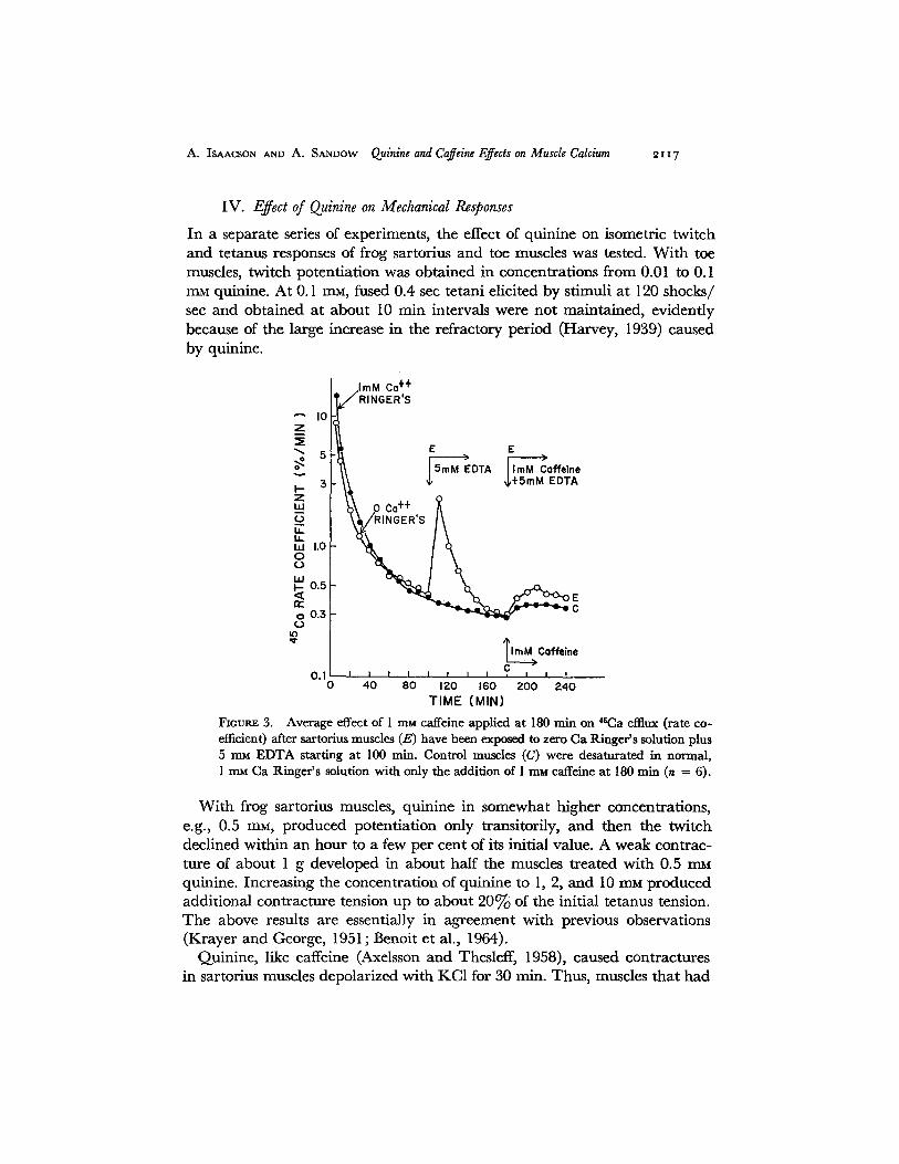
A. ISAACSON AND A. SANDOW Quinine and Caffeine Effects on Muscle Calcium ~ t 17
IV. Effect of Quinine on Mechanical Responses
In a separate series of experiments, the effect of quinine on isometric twitch and tetanus responses of frog sartorius and toe muscles was tested. With toe muscles, twitch potentiation was obtained in concentrations from 0.01 to 0.1 rnM quinine. At 0.1 raM, fused 0.4 sec tetani elicited by stimuli at 120 shocks/ see and obtained at about 10 rain intervals were not maintained, evidently because of the large increase in the refractory period (Harvey, 1939) caused by quinine.
[ .lmM Co ++ I V RINGER'S
"-'z= l°II 5 • ,E >
o~ ' ~\ |SmM EDTA /'raM C0ffe|ne 3 \ \ ~ ~,+5mM EDTA
I--
I.L EL ¢,0 ~k RINGER'S / \
w 0.5
o0 .3 ~ ~ ~ 4 ~ O
'¢ TI mM>Coffein e 0 . I I I I I I I I I C I I I
0 40 80 120 160 200 ;>40 TIME (MIN)
Fioums 3. Average effect of i m~i caffeine applied at 180 min on 4~Ca efflux (rate co- efficient) after sartorius muscles (E) have been exposed to zero Ca Ringer 's solution plus 5 mM EDTA starting at 100 min. Control muscles (C) were desaturated in normal, 1 m~ Ca Ringer 's solution with only the addition of 1 ram caffeine at 180 rain (n = 6).
With frog sartorius muscles, quinine in somewhat higher concentrations, e.g., 0.5 rrL% produced potentiation only transitorily, and then the twitch declined within an hour to a few per cent of its initial value. A weak contrac- ture of about 1 g developed in about half the muscles treated with 0.5 mM quinine. Increasing the concentration of quinine to 1, 2, and 10 mM produced additional contracture tension up to about 20c~ of the initial tetanus tension. The above results are essentially in agreement with previous observations (Krayer and George, 1951; Benoit et al., 1964).
Quinine, like caffeine (Axelsson and Thesleff, 1958), caused contractures in sartorius muscles depolarized with KC1 for 30 min. Thus, muscles that had

21x8 T H E J O U R N A L O F G E N E R A L P H Y S I O L O G Y • V O L U M E 5 ° • ~967
been depolarized by 80 rnM KC1 in Ringer's solution and had produced the associated transient contracture, produced contracture anew on addition of quinine (2 rn~) to the medium, which developed slowly during the next 30 rain and generally produced, at maximum, a tension of about 40% of that previously produced in the KC1 contracture. Similar results have been ob- tained with muscles depolarized for 30 min by 95 mM K 2SO 4 (buffered to pH 7.2 with 2 rn~ Tris HC1) and then exposed to the isotonic K2SO, solution containing 1.6 mM quinine. Thus quinine contracture probably is not medi- ated by way of a surface membrane depolarization. Contradictory results on the effectiveness of quinine in producing contracture after potassium depolar- ization have been reported by Benoit et al. (1964) in work with frog toe muscle; however, Conway and Sakai (1960) state results in this regard which agree with our observations.
In confirmation of the findings of Benoit et al. (1964), 2 InM quinine has been shown to produce contracture even in zero Ca Ringer's. In this experi- ment twitch and tetanus responses of a sartorius muscle were abolished after exposure for about 1 hr to zero Ca plus 2 mM EDTA Ringer's solution. At that time 2 m_~ quinine was added and a generally normal contracture was obtained. Thus, as is the case for caffeine (Axelsson and Thesleff, 1958), ex- ternal Ca 2+ is not needed to produce quinine contracture.
V. Effect of Quinine on Release of 45Ca from Muscle and Tendon
In view of the previously mentioned similarities between the effects of quinine and caffeine, the following experiments were performed to see whether qui- nine released Ca from muscle. Fig. 4 shows the average effects of 0.1, 0.5, and 2 rnM quinine on 4~Ca release from frog sartorius muscles, plotted as a relative rate coefficient. At each of these concentrations, an increase in outflow occurs when the quinine is added after 180 rain of desaturation of 46Ca from the muscles. Expressed as a percentage of either a portion (for 0.1 raM), or the entire 3rd hr average rate coefficient, the peak *SCa release rises to 121, 420, and 610% after 0.1, 0.5, and 2 m~ quinine respectively. At all concentrations tested the increase in 45Ca release was sustained with little decrease during the 60 min of exposure to the quinine.
Similar experiments with frog Achilles tendon showed no effect of 0.5 or 2 mM quinine on 4~Ca release. Therefore we conclude that in the whole muscle the action of quinine is confined to the muscle fibers. This specificity of quinine for Ca in muscle fibers is like that previously shown for caffeine (Bianchi, 1961).
VI. Effect o/Quinine on Release o/4~Ca after Muscles Have Been Exposed to 5 rn~ E D TA
The results shown in Fig. 4, like those in Fig. 2 for caffeine, are inconclusive as to the source of the 45Ca emerging from the muscle fibers. To get further

A. ISAACSON AND A. S.~mow Quinine and Caffeine Effects on Muscle Calcium 2119
in format ion on this question, sartorius muscles were t rea ted wi th zero Ca Ringer ' s solution conta in ing 5 mM E D T A after 100 man of washout in zero Ca Ringer ' s solution, and then a t 180 rain qu in ine hydroch lo r ide (2 mM) was added to the zero ca lc ium Ringer ' s solution wi th 5 rnM E D T A . Fig. 5 shows the results of such an expe r imen t in which the average ra te coefficients of six
800
700
600 I-- Z I,l.l
500
0 u.I
"~ =~ ~oo W o
~ ~oo o ~
0 i t )
200 175 150 125 I00
3 R° HOUR AVERAGE
i I i I t t t t J I I •
- 60 -40 -20 0 20 40 60
TIME IN QUININE (MIN)
2.0 mM QUININE (3)
0.5 rnM QUIN~INE (5)
0.1 mM QUININE (4)
Figures 4. The average effects of 0.1, 0.5, and 2 mu quinine on 45Ca release (plotted as a relative rate coefficient) from frog sartorius muscles. The s~- for the normalized 100% points, the 3rd hr average, was about 10%. For the muscles in 0.1 mat quinine, the 100% level was taken as the average of the 170 and 180 rain collections; i.e., the two collections just preceding the addition of the quinine.
pairs of muscle are p lo t ted ; the con t ro l muscles were washed in no rma l R inge r ' s solution con ta in ing 1 mM C a and finally were exposed to R inger ' s solut ion plus 2 mM quin ine a t 180 min.
Like caffeine (Fig. 3), qu in ine releases 4~Ca f rom muscles previously t rea ted wi th E D T A to r emove superficial calcium. Indeed , it p roduces a somewhat g rea te r re la t ive increase in the ra te coefficient of 45Ca f rom muscles af ter E D T A t r ea tmen t than f rom muscles in no rma l Ringer ' s solution. As was con-

2120 THE JOURNAL OF GENERAL PHYSIOLOGY • VOLUME 50 • 1967
eluded ior caffeine, these results indicate that 2 mM quinine acts to release Ca from an intracellular store inaccessible to EDTA.
VII . Effect of Quinine on 45Ca Uptake, Total Ca, Na, K, and Water Content
In an experiment similar to that used to measure 45Ca uptake in the presence of caffeine, we found that 2 mM quinine increased the 10 rain 45Ca influx 111 4- 10% (n = 4) over the normal 46Ca influx. The normal influx in this experi- ment, uncorrected for loss of 4~Ca from the slow component during the 120 rain desaturation before analysis for 45Ca in the muscle, was 0.0451 4- 0.0066 /z#mole Ca/cm2-sec.
~" 10 Z
~ 1.0
hi ~n~ 0'5
~.~ O.B
0.1 0
ImM Ca ++ RINGER'S
E E [ 2mM Quinine
EDTA ,,~+ 5rnM EDTA
o co++ I / ,0ERS
[~.M._~ uinine C
1 4 1 181 i , l 1 , l , 0 0 IZO 160 2 0 0 240
TIME ( MIN )
FIGUEE 5. Average effect of quinine (2 rnM) applied at 180 rain on ~sCa efftux (rate coefficient) after frog sartorins muscles (E) have been exposed to E D T A (5rr~) in zero Ca Ringer 's solution start- ing at 100 rain. Control muscles (C) were desaturated in normal, I mM Ca Ringer 's solution with only the addition of 2 m_M quinine at 180 rain (n -- 6, sE are shown for later portions of efftux).
Total calcium analyses (Table II) were performed with a Perkin-Elmer absorption spectrophotometer, as described by Carvalho (1966), in a dupli- cate series of experiments to see whether 2 mM quinine had any effect on total Ca in muscle. In this experiment, five pairs of muscles were equilibrated in normal Ca Ringer's solution and the experimental muscles were then placed in Ringer's solution plus 2 mM quinine for 10 rain, after which they were transferred through seven sets of test tubes containing normal Ringer's solu- tion for a total of 120 rain. Total Ca analyses gave 2.07 4- 0.006 for normal and 2.44 4- 0.10 #moles Ca/g (n = 5) for quinine-treated muscles. This in- crease of 18°-/o is statistically significant (t test for paired variates, 0.01 < P < 0.05). This increase of 18°-/o in total Ca in quinine-treated muscle is much smaller than the increase of I 11 ~ in 45Ca uptake by muscles similarly exposed to quinine for 10 min and then washed out for 2 hr in normal Ringer's solu- tion. Thus, it is likely that the increase in *SCa content represents mostly an increase in exchange of 4°Ca for *~Ca in quinine-treated muscles. However,

A. ISAACgON AND A. SANDOW Quinine and Caffeine Effects on Muscle Calcium 2121
the possibility remains, as noted in similar experiments with caffeine (Feinstein, 1963), that a larger increase in total muscle Ca may occur during the 10 rain exposure to quinine. The subsequent 2 hr washout in normal Ringer's solution may possibly obscure effects of quinine on total muscle Ca.
Table II also contains data on total Na, K, and water content of the muscles analyzed for total Ca after exposure to quinine for 10 rain. Standards were prepared for Na and K containing 1 ram of either Na (in K) or K (in Na) to approximate the interference with absorption spectroscopy caused by having
T A B L E I I
C A T I O N AND W A T E R C O N T E N T OF N O R M A L AND Q U I N I N E - T R E A T E D F R O G S A R T O R I U S M U S C L E S
C o n c e n t r a t i o n
No. of muscles Experimental conditions* Ca Na K
Water c o n t e n t
Per cent of total m u s c l e
Control (normal Ringer ' s )
~tmoles/g wet wt. 4- (s z)
2.07 32.5 74.8 (4-0.06) (4-1.1) (4-2.1)
Exper imenta l (10 min in Ring- 2.44 49.1 54.4 er 's + 2 m~ quinine) (4-0.10) (4-3.2) (4-3.9)
10 (Experimental - - control) + 0 . 3 7 + 1 6 . 6 --20.4 (-4-0.12) (-4-2.7) (-4-2.7)
[P<O.05] :1: [P<:O.O1] ~. [P<O.O1]$
79.1 (~o.~)
82.0 (!o.5)
+3.0 (4-0.5)
[P<O.O1]$
* Control and exper imental muscles were equi l ibra ted for about 2 hr in normal Ringer ' s pr ior to the 10 rain e x p o s u r e to quinine (experimental) after which t ime all muscles were washed out in normal Ringer ' s for an addi- t ional 2 hr. ~t P is based on the sE of differences from paired muscles.
a mixture of Na and K in the samples of muscle ash (Carvalho, 1966). Quinine can be seen to cause a 51% gain in Na (16.6 #moles/g) and a 27% decrease in K ( -20 .4 #moles/g) in the experimental muscles relative to the muscles soaked in normal Ringer's solution. Likewise, the water content is increased by 3.7% in the quinine-treated muscles. All these changes are statistically significant (P < 0.01).
VIII. Effect of 2 mM Quinine on 46Ca Uptake or Release in Muscles Depolarized with Potassium
In view of the effectiveness of quinine in producing contracture in muscles depolarized for 30 rain by KC1 (80 mM added to Ringer's solution), similar experiments were performed in which 45Ca uptake was measured. Paired muscles were first exposed for 10-30 rain in Ringer's solution plus 80 m_u KC1; then the experimental muscles were placed for 10 rain in 4~Ca-labeled Ringer's

2 I ~ 2 T H E J O U R N A L O F G E N E R A L P H Y S I O L O G Y • V O L U M E 5 0 • I967
solution plus 80 rnM KCI containing 2 raM quinine. The control muscles were exposed to the same labeled high potassium Ringer's solution, but without quinine. After the soak in ~SCa the muscles were washed in normal Ringer's for 120 rain and the radioactivity remaining in the muscles was determined as described earlier. There was no significant difference between the average 4~Ca content of five pairs of experimental and control muscles after a 10 rain presoak in high potassium Ringer's solution. Similar results were obtained in an experiment with a 30 min presoak in high potassium Ringer's; however, there was a tendency for the quinine-treated muscles to take up less ~SCa than their controls (experimental - control = -0.006 :t: 0.004 #mole Ca/g [n = 5, P = 0.2] ).
Corresponding experiments on release are of interest and preliminary results indicate that in muscles labeled with 4SCa for 3 hr, quinine (I raM) consistently caused the usual sustained two- to threefold increase in the rate coefficient of 45Ca release, regardless of whether the muscles had been previously depolar- ized with K 2SO 4 (95 mM).
D I S C U S S I O N
A major presumption underlying the interpretation of the present results in general, and earlier ones especially regarding caffeine (Bianchi, 1961; Feinstein, 1963; Isaacson and Sandow, 1967), is that the increased movement of Ca ~+ caused by the alkaloids reflects the presence of a proportionately in- creased level of myoplasmic Ca $+ which has been internally released and which is responsible for the ability of the drugs to produce contracture and other mechanical effects. The drugs might also increase the permeability of the membrane to Ca ~+. This is unlikely for caffeine since Feinstein (1963) ob- served no change in Ca content of frog sartorii subjected to 5 ~ caffeine for 10 rain and then washed for 2 hr; but it might be true for quinine since our comparable experiments indicated that this alkaloid did increase the muscles' Ca content. However, such permeability changes, in general, should play no role in producing at least the contractures of interest, for our results and those of others (Frank, 1960, 1962; Bianchi, 1961; Feinstein, 1963; Benoit et al., 1964; Caputo, 1966) showing that both drugs cause contracture and in- creased 4~Ca movement in muscles exposed to zero calcium, even after EDTA treatment, strongly indicate that contracture production does not depend on the entrance of Ca ~+ from the external medium at all, but on release of the ion internally. Furthermore, the findings that caffeine (Herz and Weber, 1965) and quinine also (for details, see later) both release a portion of the Ca sequestered within extracted vesicles of the sarcoplasmic reticulum (SR), suggest a definite mechanism for the supposed internal action of the drugs.
We have found that 1 mM caffeine, which does not produce contracture, does cause increases in both *SCa inflow and outflow, and in outflow even

A. ISAACSON AND A. SANDOW Quinine and Caffeine Effects on Muscle Calcium 2123
after t reatment with EDTA, which are all qualitatively like those produced by caffeine at higher concentrations. Thus we infer that 1 rnM caffeine causes an internal release of Ca ~+ qualitatively like that it produces at higher con- centrations. This indicates that the inability of caffeine at this concentration to produce contracture is not because it was acting below some absolute thres- hold connected with its action, as such, to cause release of Ca 2+. But the cor- relation of our results on influx with those obtained by others at higher con- centrations, as shown by Fig. I, indicates that the level of free Ca *+ produced in the myoplasm is lower, the smaller the concentration of the caffeine. A similar result appears in respect to release of 45Ca, for calculation shows that in Bianchi's (1961) work, 5 mM caffeine caused the rate coefficient of release to increase over the normal by 3.2 times, but the corresponding increase in our work with 1 rnM caffeine was only 1.4 times. Thus we infer that the reason contracture is always absent in muscles subjected to 1 m_~ caffeine is that the level of myoplasmic Ca *+ it produces is below the threshold of about 10 -7 u generally needed to activate contraction, as demonstrated in various other contractile systems (for review see Sandow, 1965; Weber, 1966). Our analysis suggests, furthermore, that the levels of this Ca s+ obtained at 2.5 and 5.0 mM and still higher caffeine concentrations are generally sufficiently great to either attain or exceed this threshold. Our results, however, do not explain the great variability in the sensitivity of muscles to produce caffeine contracture.
As for causing twitch potent iat ion--and, incidentally, also production of the special increased speed of contraction obtained during the earliest part of both isotonic (Sandow and Seaman, 1964) and isometric (Sandow and Preiser, 1964) twitches (see also Sandow, Taylor, and Preiser, 1965; Sandow, 1965; Sandow and Brust, 1966)--1 mM caffeine might act so that, during excitation-contraction (E-C) coupling, there would be increased permeability or alteration of some other function of the transverse tubules involving Ca 2+, or increased release of Ca 2+ from the sarcoplasmic reticulum (SR), thus super- normally raising the level of activator Ca ~+ which would prolong the active state and augment the twitch. But available evidence is inadequate to decide whether such processes occur. However, the amount of Ca 2+ we infer is already released in the resting muscle by 1 rnM caffeine, even though sub- threshold for contracture, would be available to add on to that normally released by E-C coupling during a twitch and thus provide the supernormal amount of free Ca 2+ needed to produce the various features of a potentiated response. It is possible that this mechanism may also account for the positive inotropic effect of caffeine on cardiac muscle (Nayler, 1963; de Gubareff and Sleator, 1965), especially since Nayler (1963) found that caffeine increased both inflow and outflow of 46Ca in hearts. At any rate, according to this mechanism for potentiating the twitch, there would be more extra Ca 2+ and therefore greater potentiation, the greater the caffeine concentration; and

~i24 T H E J O U R N A L O F G E N E R A L P H Y S I O L O G Y • V O L U M E 5 0 • 1 9 6 7
this, at least up to 3 mM caffeine, is indeed the case (Sandow and Brust, 1966). We attempted to obtain further information regarding this point by using procaine, in experiments like those of Feinstein (1963), to inhibit the increased movement of 45Ca caused by I m u caffeine. The procaine was in 0.7 rnu concentration (thus, it was in essentially the same ratio to the 1 mM concen- tration of our caffeine as Feinstein had employed (1 mg/ml) in relation to his 5 m u caffeine), and we found that it considerably delayed and reduced the magnitude of the increase in 45Ca efflux produced by the 1 m u caffeine. How- ever, the effects of the procaine on the influx of Ca caused by 1 m u caffeine were erratic. Futhermore, in attempting to relate the clear effect of procaine on the caffeine-induced Ca efflux to its effect on caffeine twitch potentiation, it has been found in various experiments in this laboratory (unpublished results) that procaine itself, depending on its concentration and time of action, either depresses or potentiates the twitch. Thus, although it is of interest that procaine inhibits the Ca el:flux produced by 1 m u caffeine, more work is needed to permit clear evaluation of our results with procaine in regard to caffeine potentiation of the twitch. In any case, the general features of poten- tiation by caffeine have been attributed to its capacity to lower the mechanical threshold (Etzensperger and Gasciolli, 1963; Sandow, Taylor, Isaacson, and Sequin, 1964; Sandow, Taylor, and Preiser, 1965). In view of the preceding discussion, this effect could be due to the presence of the free myoplasmic Ca 2+ produced by caffeine in the resting muscle, or to some effect on the SR or the T tubule that causes a given depolarization to release a supernormal amount of Ca ~+ from the SR into the myoplasm.
In considering our results involving quinine we must note that it potentiates the twitch by a dual action on electromechanical coupling: it not only lowers the mechanical threshold, like caffeine, but it also considerably prolongs the action potential, this being in contrast to the slight and mechanically in- significant ability of caffeine, especially at only 1 rnu concentration, to pro- long the action potential (Sandow, Taylor, Isaacson, and Sequin, 1964; Isaacson, Taylor, and Sandow, unpublished results). Our present results show, however, that quinine, though acting at much smaller concentrations, pro- duces effects which, in general, are like those produced by caffeine; i.e., it causes contracture (not only in normal but also in Ca-deprived and depolar- ized muscles), increases Ca flux of various types (including the increase occur- ring in presence of EDTA), and produces certain concentration changes of muscle Na and K (and, anomalously, of Ca also: compare Feinstein's (1963) results with caffeine). Thus, we conclude that these effects reflect the in- fluence on electromechanical coupling that quinine has in common with caffeine; i.e., the reduction in the mechanical threshold. Furthermore, we infer that the mechanism by which quinine reduces the mechanical threshold is essentially similar to that by which caffeine reduces the threshold; i.e., as

A. ISAACSON AND A. SANDOW (~uinine and Caffeine Effects on Muscle Calcium 2x25
indicated above, either by increasing the effectiveness of a depolarization in releasing myoplasmic Ca 2+, or by a release of Ca ~+ from the SR in the un- stimulated muscle.
In the case of quinine acting directly on the SR, it would be necessary that it be able to penetrate into the muscle like caffeine. But, this should be true, for quinine is a weak base with pKa = 8.4 (Schanker et al., 1957), and when dissolved in Ringer's solution at about pH 7.2 it should have about 10% of its molecules uncharged and therefore penetrable. Concordant with our views as to the mechanisms by which quinine causes contracture are the results of Ashley (1965) and Ashley et al. (1965) on crab fibers, which show that injections of the Ca chelator EGTA (ethylene glycol bis(Cl-amino-ethyl- ether)-N,N'-tetraacetic acid) delay and markedly reduce contractures pro- duced by external application of quinine (and of caffeine, as well). Benoit (personal communication, 1966) has suggested that the site where quinine releases intracellular Ca may be different from that where caffeine acts. This is based on the observations in Benoit et al. (1964) that quinine contractures in zero Ca Ringer's solution can still be obtained after caffeine contractures have been abolished by repeated application of the caffeine. In respect to this question, quinine evidently releases activator Ca from reticular vesicles, as does caffeine, since it causes contraction (as does caffeine, also) of extracted models of muscle fibers, provided they contain reticular material (Hasselbach and Weber, 1955). Bondani and Karler (1966) showed that quinidine (2 raM) causes release of Ca bound (evidently passively; i.e., independently of adeno- sinetriphosphate [ATP]) by microsomes of skeletal and cardiac muscle. Fur- thermore, according to Carvalho 1 1 mM quinine or quinidine at pH 7.0 can release about 33°/v of the Ca bound by SR in the presence of ATP. However, these alkaloids appear to exert an action more general than that of caffeine, for they also reduce the binding by the SR of Mg and K. Moreover, caffeine has no effect on at least the ATP-dependent fraction of the SR's bound Ca, although other types of experiments (Weber, 1966) have demonstrated that caffeine affects a relatively small labile fraction of the total Ca that can be maximally loaded into SR preparations. Thus, though both caffeine and quinine (or quinidine) may act on Ca bound to SR, they probably affect differently characterized fractions of the SR's Ca-binding sites, and this may account for the quantitative differences between the actions of the two drugs on muscle.
General consideration of our results with both quinine and caffeine indicates that our conclusions on the Ca-dependence of both contracture and twitch potentiation of the drug-treated muscles are in general harmony with those derived from experiments involving artificial addition of Ca 2+ to activate
1 Carvalho, A. 1967. Unpublished results.

2126 T H E J O U R N A L O F G E N E R A L P H Y S I O L O G Y • V O L U M E 5 ° ° i967
various contrac t i le systems, e i ther ex t rac ted (Weber et al., 1964) or in tac t (Caldwell and Walster , 1963; Por tzehl , Caldwell , and Ruegg, 1964; H e l l a m and Podolsky, 1966). This suggests tha t essentially the same mechanisms of Ca d e p e n d e n c e which de t e rmine ac t iva t ion by art if icially a d d e d Ca ~+ occu r in in tac t fibers when, u n d e r the act ion of the drugs, the Ca 2+ needed to ac t iva te the i r con t rac t ion is ob t a ined by release f rom the no rma l s tore of Ca seques- tered wi th in the sarcoplasmic re t icu lum.
We are grateful for the aid given by Dr. A. P. Carvalho in connection with atomic absorption spectroscopic analyses and for the general technical assistance of Mr. Harold Marcus and Mr. John Polk. This work was supported by grants from the Muscular Dystrophy Associations of America, Inc., and from the United States Public Health Service (NB-04262-04).
Received for publication 25 August 1966.
R E F E R E N C E S
ASHLE'Z, C. C. 1965. Doctoral Dissertation. University of Bristol, England. A s a ~ v , C. C., P. C. CALDWSLL, A. G. Low~, C. D. RmaARDS, and H. SCHIR~mR.
1965. The amount of injected EGTA needed to suppress the contractile responses of single Maia muscle fibres and its relation to the amount of calcium released during contraction. J. Physiol., (London). 179:32P.
AXELSSON, J., and S. TrmSLEFF. 1958. Activation of the contractile mechanism in striated muscle. Acta Physiol. Scand. 44:55.
BENOIT, P. H., N. CARPEm, and J. PRZVBYSLAWSKL 1964. Sur la contracture pro- voqude par la quinine chez le muscle strid de GrenouiUe. J. Physiol., (Paris). 56:289.
BIANCm, C. P. 1961. The effect of caffeine on radiocalcium movement in frog sar- torius. J. Gen. Physiol. 44:845.
BIANCar, C. P. 1962. Kinetics of radiocaffeine uptake and release in frog sartorius. J. Pharmacol. Exptl. Therap. 138:41.
BroncHI, C. P. 1965. The effect of EDTA and SCN on radiocalcium movement in rectus abdominis muscle during contractures induced by calcium removal. J . Pharmacol. Exptl. Therap. 147:360.
BXANCm, C. P., and A. M. SHAN~.S. 1959. Calcium influx in skeletal muscle at rest, during activity, and during potassium contracture. J. Gen. Physiol. 42:803.
BONDANI, A., and R. KARI~R. 1966. Effect of drugs on the in vitro uptake of Ca++ by microsomes. The Pharmacologist. 8:184.
BRUST, M. 1965. Combined effects of nitrate and caffeine on contractions of skeletal muscle. Am. J. Physiol. 208:431.
CALDW~LL, P. C., and G. WAt,STER. 1963. Studies on the microinjection of various substances into crab muscle fibres. J. Physiol., (London). 169:353.
CA•UTO, C. 1966. Caffeine- and potassium-induced contractures of frog striated muscle fibers in hypertonic solutions. J. Gen. Physiol. 50:129.
CARVALHO, A. 1966. Binding of cations by microsomes from rabbit skeletal muscle. J. Cellular Physiol. 67:73.

A. ISAACSON AND A. SANDOW Quinine and Caffeine Effects on Muscle Calcium 2t27
CONWAY, D., and T. SAKAI. 1960. Caffeine contracture. Proc. Natl. Acad. Sci. U. S. 46:897.
DE GImAREFF, T., and W. SLEATOR, JR. 1965. Effects of caffeine on mammalian atrial muscle, and its interaction with adenosine and calcium. J. Pharmacol. Exptl. Therap. 148:202.
ETZENSP~ROER, J., and A. GASCIOT.Lt. 1963. Action de la caf~ine sur les contractures de depolarisation produites par le potassium chez le muscle stri6 de GrenouiUe. Compt. Rend. Soc. Biol. 157:1776.
F~ms~m, M. B. 1963. Inhibition of caffeine rigor and radiocalcium movements by local anesthetics in frog sartorius muscle. J. Gen. Physiol. 47:151.
FP.mSTEm, M. B. 1966. Inhibition of muscle rigor by EDTA and EGTA. Life Sci. 5:2177.
FgANK, G. B. 1960. Effects of changes in extracellular calcium concentration on the potassium-induced contracture of frog's skeletal muscle, or. Physiol., (London). 151:518.
FR~'~a, G. B. 1962. Utilization of bound calcium in the action of caffeine and cer- tain multivalent cations on skeletal muscle. J. Physiol., (London). 163:254.
GUTMANN, E., and A. S~DOW. 1965. Caffeine induced contracture and potentiation of contraction in normal and denervated rat muscle. Life Sd. 4:1149.
HARWY, A. M. 1939. The actions of quinine on skeletal muscle. J. Physiol., (London). 95:45.
HASSELBACH, W., and A. WEBER. 1955. Models for the study of the contraction of muscle and cell protoplasm. Pharmacol. Rev. 7:97.
HEI~RUNN, L. V., and F. J. WmRcms~. 1947. The action of various cations on muscle protoplasm. J. Cellular Comp. Physiol. 29:15.
H~LLAM, D. C., and R. J. PoDomav. 1966. The relation between calcium concen- tration and isometric force in skinned frog muscle fibers. Fed. Pro¢. 25:466.
HERZ, R., and A. WEBER. 1965. Caffeine inhibition of calcium uptake by muscle reticulum. Fed. Proc. 24:208.
ISAACSON, A. 1966. Caffeine and quinine effects on 4~Ca movements in muscle. Fed. Proc. 25:308.
ISAACSON, A., and A. S~qDOW. 1967. Caffeine effects on radiocalcium movement in normal and denervated rat muscle. J. Pharmacol. Exptl. Therap. 155:376.
KRA~R, O., and H. W. GEORGE. 1951. Studies on veratrum alkaloids. XV. The quinine-like effect of veratramine upon the single twitch and upon the "veratrine response" of the sartorius muscle of the frog. J. Pharmacol. Exptl. Therap. 103:249.
NA'C~R, W. G. 1963. Effect of caffeine on cardiac contractile activity and radio- calcium movement. Am. J. Physiol. 204:969.
PODOmXY, R. J., and L. L. COSTANTm. 1964. Regulation by calcium of the contrac- tion and relaxation of muscle fibers. Fed. Proc. 23:933.
PORTZEHL, H., P. C. CALDWELL, and J. C. R~-~Gc. 1964. The dependence of contrac- tion and relaxation of muscle fibres from the crab Maia squinado on the internal concentration of free calcium ions. Biochim. Biophys. Acta. 79:581.
SAKAI, T. 1965. The effect of temperature and caffeine on activation of the con- tractile mechanism in the striated muscle fibres. Jikeikai Med. J. 12:88.

~i28 T H E J O U R N A L O F G E N E R A L P H Y S I O L O G Y • V O L U M E 5 ° • i967
S A N D O W ~ A. 1964. Potentiation of muscular contraction. Arch. Phys. Med. Rehabil. 45:62.
SANDOW, A. 1965. Excitation-contraction coupling in skeletal muscle. Pharmacol. Rev. 17:265.
SANDOW, A., and M. BRUST. 1966. Caffeine potentiation of twitch tension in frog sartorius muscle. Biochem. Z. 345:232.
SANDOW, A., and A. ISAACSON. 1966. Topochemical factors in potentiation of con- traction by heavy metal cations. J. Gen. Physiol. 49:937.
SANDOW, A., and H. t~ISER. 1964. Muscular contraction as regulated by the action potential. Science. 146:1470.
SANDOW, A., and T. SEAMAN. 1964. Muscle shortening velocity in normal and po- tentiated contractions. Life Sci. 3:91.
SANDOW, A., S. R. TAYLOR, A. ISAACSON, and J. J. SEQuin. 1964. Electromechanical coupling in potentiation of muscular contraction. Science. 143:577.
SANDOW, A., S. R. TAYLOR, and H. PR~.ISER. 1965. Role of the action potential in excitation-contraction coupling. Fed. Proc. 24:1116.
SCI~ANKER, L. S., P. A. SHORE, B. B. BRODm, and C. A. M. HOOBEN. 1957. Absorp- tion of drugs from the stomach. I. The rat. d. Pharmacol. Expa. Therap. 120:528.
WEBER, A. 1966. Energized calcium transport and relaxing factors. Current Topics in Bioencrgetics. 1:203.
WEBER, A., R. HERZ, and I. REISS. 1963. On the mechanism of the relaxing effect of fragmented sarcoplasmic reticulum, d. Gen. Physiol. 46:679.
WEBER, A., R. HERZ, and I. REISS. 1964. The regulation of myofibrillar activity by calcium. Proc. Roy, Soc. (London), Set. B. 160:489.