insulin resistance, cognitive impairment and ...cdn.intechweb.org/pdfs/19803.pdf · insulin...
TRANSCRIPT
20
Insulin Resistance, Cognitive Impairment and Neurodegeneration: Roles of Nitrosamine
Exposure, Diet and Lifestyles
Suzanne de la Monte, Ming Tong and Jack R. Wands Rhode Island Hospital and the Warren Alpert Medical School of Brown University
USA
1. Introduction
In Western societies, Alzheimer's disease (AD) is epidemic (de la Monte, Neusner et al. 2009). For decades, the prevailing theories about AD pathogenesis focused on the roles of hyper-phosphorylated and ubiquitinated cytoskeletal proteins in neuronal perikarya and dystrophic fibers, and increased expression and abnormal processing of amyloid-beta precursor protein (AβPP), leading to AβPP-Aβ peptide deposition in neurons, plaques, and vessels, as mediators of neuronal loss, neuritic pathology, synaptic disconnection, and loss of plasticity. Apart from aging, which is the most dominant risk factor, it has not been possible to define convincing roles for AβPP-Aβ or phospho-tau accumulations as principal driving forces of neurodegeneration that could account for all of the protean manifestations of AD, including neuro-inflammation, mitochondrial dysfunction, white matter fiber loss, and neurotransmitter deficiencies. This point is particularly relevant with respect to sporadic AD, which accounts for the vast majority of cases. However, it is entirely possible that the focal points of 30+ years of intense research on the accumulation, abnormal processing, and misfolding of essentially two proteins are somewhat misplaced since these process could be the end-result rather than the cause of the pathophysiological processes that trigger and propagate the neurodegeneration cascade. Growing evidence supports the concept that AD is actually a metabolic disease in which, neurodegeneration is mediated by impairments in brain glucose utilization and energy production that begin early in the course of disease, and worsen with its progression (Frolich, Blum-Degen et al. 1998; Hoyer 2002; Hoyer 2004; Rivera, Goldin et al. 2005; Steen, Terry et al. 2005). AD-associated reductions in glucose utilization and energy metabolism are accompanied by increased oxidative stress and neuro-inflammation, which appear to be driven by insulin and insulin-like growth factor (IGF) resistance in the brain. AD can be regarded as a brain-specific form of diabetes mellitus, i.e. Type 3 diabetes. This chapter reviews the data showing that sporadic AD in humans is associated with progressive impairments in brain insulin and IGF signaling, and contrasts the roles of peripheral insulin resistance disease states versus primary brain insulin/IGF resistance using data from both human and experimental animal studies. Epidemiological and experimental evidence supporting exposures, particularly to environmental toxins such as nitrosamines, rather than only genetic factors as causal in the pathogenesis of AD are discussed. Dietary and
www.intechopen.com
Alzheimer’s Disease Pathogenesis-Core Concepts, Shifting Paradigms and Therapeutic Targets 460
lifestyle changes that could reduce risk for developing insulin resistance diseases, including diabetes mellitus, obesity, non-alcoholic steatohepatitis and neurodegeneration, are discussed as public health strategies that must be considered in earnest.
2. Alzheimer's disease and brain glucose metabolism
Metabolic abnormalities in AD have been linked to deficits in brain insulin and insulin-like growth factor (IGF) signaling through pathways that are critical for maintaining neuronal survival, energy production, gene expression, and plasticity (Frolich, Blum-Degen et al. 1998). Thorough consideration of the pleitropic actions of insulin and IGFs helps to clarify how impairments in these signal transduction networks could account for many, if not all of the critical features of AD-type neurodegeneration, including increased: 1) activity of kinases that aberrantly phosphorylate tau; 2) expression of AβPP and accumulation of AβPP-Aβ; 3) oxidative and endoplasmic reticulum (ER) stress; 4) the generation of reactive oxygen and reactive nitrogen species that damage proteins, RNA, DNA, and lipids; 5) mitochondrial dysfunction; and 6) activation of pro-inflammatory and pro-death cascades. Moreover, impairment or inhibition of insulin/IGF signaling disrupts cholinergic homeostasis, thereby compromising one of the most important neurotransmitter systems utilized for neuronal plasticity, memory, and cognition.
3. Brain insulin and insulin-like growth factor problems in AD
The concept that AD could represent a metabolic disease stemmed from the findings that deficits in cerebral glucose utilization are detectable in the early stages of disease (Adolfsson, Bucht et al. 1980; Fujisawa, Sasaki et al. 1991; Caselli, Chen et al. 2008; Mosconi, Pupi et al. 2008; Mosconi, Mistur et al. 2009; Langbaum, Chen et al. 2010), and as AD progresses, so do the metabolic abnormalities (Hoyer and Nitsch 1989; Hoyer, Nitsch et al. 1991). Although several studies provided initial clues that insulin resistance could be an important feature of AD (Frolich, Blum-Degen et al. 1998; Hoyer 2002; Hoyer 2004), the human postmortem brain studies were critical for definitively establishing that both insulin resistance and insulin deficiency in the brain were consistent and fundamental abnormalities in AD (Rivera, Goldin et al. 2005; Steen, Terry et al. 2005). Gene expression and receptor binding studies showed that in AD, brain insulin gene expression, insulin levels, insulin receptor levels, and insulin receptor binding were all impaired. Insulin resistance is manifested by reduced levels of insulin receptor binding and decreased responsiveness to insulin/IGF stimulation, while the trophic factor deficiency is associated with reduced levels of insulin polypeptide and gene expression in brain and cerebrospinal fluid (Blum-Degen, Frolich et al. 1995; Blass, Gibson et al. 2002; Hoyer 2004; Hoyer 2004; Rivera, Goldin et al. 2005; Steen, Terry et al. 2005). To consolidate these concepts, we proposed that AD be referred to as, “Type 3 diabetes” because it represents the conglomerate effects of Type 1 (insulin deficiency) and Type 2 (insulin resistance) diabetes, whereby the consequential metabolic abnormalities are largely restricted to the brain (Rivera, Goldin et al. 2005; Steen, Terry et al. 2005). It has been well-established that insulin signaling mechanisms parallel, interact, or cross-talk with related insulin-like growth factor (IGF) pathways in various organ-systems, including the brain (de la Monte and Wands 2005). Despite apparent redundancy, insulin, IGF-1 and IGF-2 stimulations have differential effects on various target cells in the brain, and at
www.intechopen.com
Insulin Resistance, Cognitive Impairment and Neurodegeneration: Roles of Nitrosamine Exposure, Diet and Lifestyles 461
different stages of development, maturation, and aging (de la Monte and Wands 2005). Ordinarily, insulin and IGF signaling networks overlap and complement one another, enabling deficits in one pathway to be compensated for by increased activation of one or both of the others, and thereby provide a kind of safety net for supporting critical neuronal and glial functions in the brain. Therefore, with regard to neurodegenerative diseases, it was of interest to determine if the impairments in insulin signaling networks in AD were selective, or whether they broadly affected IGF pathways as well. The human postmortem studies demonstrated that, like insulin, AD is associated with significant impairments in IGF-1 and IGF-2 signaling mechanisms in the brain, and that as dementia progresses, so do the deficits in IGF signaling networks (Rivera, Goldin et al. 2005; Steen, Terry et al. 2005). AD-associated deficits in IGF signaling are marked by reduced expression of IGF trophic factors and receptors, and decreased binding to IGF receptors, reflecting IGF deficiency and resistance. Since the impairments in IGF signaling mechanisms develop and progress simultaneously with declines in insulin signaling, the scaffolding needed to support a broad range of functions, including cerebral glucose utilization, neuronal survival, myelin maintenance, metabolism, neurotransmitter function, and plasticity gets destroyed, practically en masse. Consequences include reductions in the expression of choline acetyltransferase, tau, and glyceraldehyde-3-phosphate dehydrogenase (GAPDH) genes, which mediate cholinergic/cognitive, neuronal cytoskeletal, and metabolic functions (Rivera, Goldin et al. 2005). Insulin/IGF resistance-mediated impairments in energy metabolism promote oxidative stress, accumulation of reactive oxygen species (ROS), DNA damage, and mitochondrial dysfunction, all of which drive pro-apoptosis, pro-inflammatory, and pro-AβPP-Aβ cascades.
4. Impaired insulin/IGF signaling and tau pathology in AD
Neuronal cytoskeletal lesions that correlate with severity of dementia in AD include, neurofibrillary tangles, dystrophic neurites, and neuropil threads, which contain aggregated and ubiquitinated insoluble fibrillar tau (Duyckaerts, Delatour et al. 2009; Takashima 2009). In AD, hyperphosphorylation of tau, a microtubule-associated protein, is mediated by
inappropriate activation of kinases, such as glycogen synthase kinase 3 (GSK-3). Hyper-phosphorylated tau is rendered highly susceptible to misfolding and self-aggregating into insoluble fibrils, generating dementia-associated cytoskeletal lesions (Iqbal, Liu et al. 2009). These degenerative processes lead to disruption of the neuronal cytoskeletal network and synaptic disconnection (Iqbal, Liu et al. 2009). In addition, pre-fibrillar tau aggregates to form soluble oligomers that are neurotoxic and can cause synaptic disconnection and neuronal death (Takashima 2010). Ubiquitination of hyper-phosphorylated tau (Arnaud, Robakis et al. 2006), along with dysfunction of the ubiquitin-proteasome system (Oddo 2008), result in further accumulation of insoluble fibrillar tau, oxidative stress, ROS generation, and cell loss in AD (Mandelkow, Stamer et al. 2003). Brain insulin/IGF resistances contribute to the development and accumulation of dementia-associated neuronal cytoskeletal lesions (de la Monte, Ganju et al. 2000; de la Monte, Neely et al. 2001; de la Monte and Wands 2002; Xu, Eun Yeon et al. 2003) because tau gene expression and phosphorylation are regulated by insulin and IGF (Schubert, Brazil et al. 2003; Schubert, Gautam et al. 2004), and brain insulin and IGF resistances decrease signaling through phosphoinositol-3-kinase (PI3K), Akt (Schubert, Brazil et al. 2003; Schubert, Gautam
et al. 2004), and Wnt/-catenin (Doble and Woodgett 2003), which result in increased
www.intechopen.com
Alzheimer’s Disease Pathogenesis-Core Concepts, Shifting Paradigms and Therapeutic Targets 462
activation of GSK-3 (Nishimura, Yu et al. 1999; De Ferrari and Inestrosa 2000; Fraser, Yu et al. 2001; Mudher, Chapman et al. 2001; Grilli, Ferrari Toninelli et al. 2003). In addition, tau hyper-phosphorylation in AD is mediated by inhibition of protein phosphatases 1 and 2A (Hanger, Seereeram et al. 2009; Iqbal, Liu et al. 2009; Morales, Farias et al. 2010), which are regulated by insulin/IGF. Besides hyper-phosphorylation, tau pathology in AD is mediated by impaired tau gene expression due to reduced insulin and IGF signaling (de la Monte, Chen et al. 2003). Consequences include failure to generate sufficient amounts of normal soluble tau protein, vis-a-vis accumulation of hyper-phosphorylated insoluble fibillar tau, and attendant exacerbation of cytoskeletal collapse, neurite retraction, and synaptic disconnection.
5. Insulin/IGF resistance and amyloid-beta (Aβ) accumulation and neurodegeneration
Dysregulated expression and processing of amyloid precursor protein (AβPP) results in accumulation of neurotoxic AβPP-Aβ (Aβ) oligomeric fibrils and insoluble aggregated fibrils (plaques) in the brain. These lesions arise secondary to increased AβPP gene expression and abnormal AβPP proteolysis, leading to the generation of 40 or 42 amino acid length Aβ peptides that can aggregate. In sporadic AD, which accounts for 90% or more of the cases, the causes of Aβ accumulation and toxicity are not known, but over the past few years, the role of impaired insulin/IGF signaling has come into consideration. One argument is that Aβ toxicity causes insulin resistance. The opposing argument is that brain insulin resistance with attendant oxidative stress and neuro-inflammation, promotes Aβ accumulation and toxicity. The findings that insulin stimulation promotes trafficking of Aβ from the trans-Golgi network to the plasma membrane, stimulates Aβ secretion (Watson, Peskind et al. 2003), and inhibits Aβ intracellular accumulation (Gasparini, Gouras et al. 2001; Gasparini, Netzer et al. 2002) support the latter position. Moreover, experimental data indicate that impaired insulin signaling disrupts the processing of AβPP and clearance of Aβ (Messier and Teutenberg 2005), which again supports a causal role for brain insulin resistance. On the other hand, it is important to recognize that AD, and all other neurodegenerative diseases represent the effects of cascades that cause diseases to progress over time. Therefore, it is essential that we understand both initiating and propagating factors in disease because treatments can be preventive or interventional. The findings that: 1) Aβ disrupts insulin signaling by competing with insulin, or reducing the affinity of insulin binding to its own receptor (Ling, Martins et al. 2002; Xie, Helmerhorst et al. 2002); 2) AβPP oligomers inhibit neuronal transmission of insulin-stimulated signals by desensitizing insulin receptors; and 3) intracellular AβPP-Aβ interferes with PI3 kinase activation of Akt-
mediated survival signaling and increases GSK-3 activation (promoting tau hyper-phosphorylation), suggest that dysregulation of AβPP expression and processing can exacerbate insulin resistance.
6. Insulin/IGF resistance, oxidative stress, and metabolic dysfunction in AD
Deficits in cerebral glucose utilization and energy metabolism occur very early in the course of AD (Iwangoff, Armbruster et al. 1980; Sims, Bowen et al. 1980; Hoyer 2004). Glucose uptake and utilization in brain are dependent upon glucose transport by glucose transporter 4 (GLUT4), which is stimulated by insulin. Therefore, impairments in brain insulin
www.intechopen.com
Insulin Resistance, Cognitive Impairment and Neurodegeneration: Roles of Nitrosamine Exposure, Diet and Lifestyles 463
responsiveness could account for the deficits in brain glucose utilization and energy metabolism in AD. Deficiencies in energy metabolism correlate with the increased levels of oxidative stress, mitochondrial dysfunction, and pro-inflammatory cytokine activation in AD brains (Hoyer and Lannert 1999; Hoyer, Lee et al. 2000; de la Monte and Wands 2002). Oxidative stress leads to increased generation and accumulation of reactive oxygen (ROS) and reactive nitrogen species (RNS) that attack subcellular organelles, yielding DNA, RNA, lipid, and protein adducts that further compromise the structural and functional integrity of neurons. Consequences include disruption of the neuronal cytoskeleton, synaptic disconnection, neurotransmitter deficits, impaired neuronal plasticity, and reduced neuronal survival. Mitochondrial dysfunction reduces ATP production, increases ROS, and triggers neuro-inflammatory responses through the activation of microglia and astrocytes. This cascade is propagated by neuro-inflammation inhibiting insulin/IGF signaling, and increasing stress, organelle dysfunction, pro-apoptosis signaling, AβPP expression, and aberrant AβPP cleavage with AβPP-Aβ deposition and toxic fibril formation (Lorenzo and Yankner 1996; Niikura, Hashimoto et al. 2002; Chen, Xu et al. 2003; Tsukamoto, Hashimoto et al. 2003; Blasko, Stampfer-Kountchev et al. 2004; Eikelenboom and van Gool 2004; Tuppo and Arias 2005). Persistence of oxidative stress leads to constitutive activation of kinases e.g. GSK-3β, that promote hyper-phosphorylation of tau. Therefore, oxidative stress and energy imbalances stemming from brain insulin/IGF resistance contribute to the cascade of neuronal loss, AβPP toxicity, tau cytoskeletal pathology, and neuro-inflammation (de la Monte and Wands 2005; Rivera, Goldin et al. 2005; de la Monte, Tong et al. 2006).
7. Peripheral versus central mediators of brain insulin/IGF deficiency and resistance
The molecular and biochemical abnormalities in AD brains closely mimic those present in diabetes mellitus; however, until recently, the vast majority of sporadic AD cases had no association with diabetes. In fact, prior to 1980, the epidemiological trends for AD (increasing prevalence) were opposite those of diabetes mellitus (declining as a cause of death) (de la Monte, Neusner et al. 2009). Even today, most cases of AD arise in the absence of type 2 diabetes, obesity, or metabolic syndrome, indicating that insulin/IGF resistance and deficiency can selectively involve the brain. Similarly, brain-only insulin and/or IGF resistance with cognitive impairment and AD-type neurodegeneration can be produced by intracerebroventricular (ICV) administration of small interfering (si)-RNA duplexes targeting the insulin or IGF receptors (see below), genetic depletion of insulin/IGF receptors, or ICV treatment with streptozotocin (STZ), which destroys insulin and IGF receptor bearing cells (Biju and Paulose 1998; Lannert and Hoyer 1998; Hoyer, Lannert et al. 1999; Hoyer, Lee et al. 2000; Nitta, Murai et al. 2002; Weinstock and Shoham 2004; Lester-Coll, Rivera et al. 2006; Grunblatt, Salkovic-Petrisic et al. 2007; Labak, Foniok et al. 2010; de la Monte, Tong et al. 2011). These studies support a primary role for brain insulin/IGF resistance and deficiency as the cause of AD-type neurodegeneration. Within the past 2-3 decades, morbidity and mortality rates have trended upward for diabetes and other insulin resistance diseases, including metabolic syndrome (dyslipidemic states), non-alcoholic steatohepatitis (NASH), and obesity, despite improvements in medical treatment (de la Monte, Neusner et al. 2009). At the same time, the increasing overlap between cognitive impairment or AD and peripheral insulin resistance diseases has raised concerns about the potential contributions or even causal roles of obesity and diabetes mellitus in
www.intechopen.com
Alzheimer’s Disease Pathogenesis-Core Concepts, Shifting Paradigms and Therapeutic Targets 464
neurodegeneration and dementia (Qiu, De Ronchi et al. 2007; de la Monte, Neusner et al. 2009). Correspondingly, a number of human and experimental animal studies have demonstrated significant associations between peripheral insulin resistance states, obesity, or diabetes mellitus and brain insulin/IGF resistance, mild cognitive impairment (MCI), dementia, or AD (Craft 2005; Rivera, Goldin et al. 2005; Steen, Terry et al. 2005; Craft 2006; de la Monte, Tong et al. 2006; Lester-Coll, Rivera et al. 2006; Craft 2007). The debate over whether AD and cognitive impairment are caused by peripheral, brain, or both forms of insulin/IGF resistance is on-going, and essentially focused on three main questions: 1) Do T2DM and other peripheral insulin resistance states cause neurodegeneration, including AD? 2) How do T2DM and other peripheral insulin resistance disease states serve as co-factors in cognitive impairment and neurodegeneration? and 3) Is AD fundamentally a brain-specific form of diabetes mellitus? These questions are addressed below.
7.1 Potential roles of obesity and T2DM in cognitive impairment and neurodegeneration Epidemiologic studies have shown that individuals with glucose intolerance, deficits in insulin secretion, or T2DM have a significantly increased risk for developing mild cognitive impairment (MCI) or AD-type dementia. Longitudinal studies also support roles for T2DM (Pasquier, Boulogne et al. 2006; Verdelho, Madureira et al. 2007) and obesity/dyslipidemic disorders (Martins, Hone et al. 2006) in the development of MCI, dementia, or AD (Haan and Wallace 2004; Luchsinger and Mayeux 2004; Ristow 2004; Launer 2005; Pasquier, Boulogne et al. 2006; Luchsinger, Reitz et al. 2007; Whitmer 2007). Despite these provocative observations, and an independent study demonstrating higher frequencies of peripheral insulin resistance in AD than in normal aging (Janson, Laedtke et al. 2004), postmortem human brain studies detected similar rates of AD among diabetics and controls (Nelson, Smith et al. 2008). Moreover, although experimental chronic high fat diet (HFD) feeding and diet induced obesity (DIO) with T2DM cause cognitive impairment with deficits in spatial learning and memory (Winocur and Greenwood 2005; Winocur, Greenwood et al. 2005), the resulting brain atrophy with brain insulin resistance, neuro-inflammation, oxidative stress, and deficits in cholinergic function are relatively mild and lack most of the important structural lesions that characterize AD pathology (Moroz, Tong et al. 2008; Lyn-Cook, Lawton et al. 2009). Therefore, observations in both humans and experimental models suggest that while obesity or T2DM can be associated with cognitive impairment, brain atrophy, and some aspects of AD-type neurodegeneration, these peripheral insulin resistance disease states are not sufficient to cause AD. Instead, the findings suggest that T2DM, obesity, and probably other peripheral/systemic insulin resistance states serve as co-factors that contribute to the pathogenesis or progression of neurodegeneration. The significance of this conclusion is that therapeutic strategies designed to treat T2DM, obesity, and systemic insulin resistance could help slow the progress or reduce the severity of AD, but will not likely prevent it altogether. Correspondingly, studies have already shown that treatment with hypoglycemic or insulin sensitizer agents can be protective for reducing the incidence and severity of AD (Luchsinger 2010).
7.2 Factors mediating cognitive impairment and neurodegeneration with systemic insulin resistance syndromes Since it is clear that peripheral insulin resistance disease states can significantly and adversely alter cognitive function and contribute to the progression of AD-type neurodegeneration, efforts should be made to understand which aspects of peripheral
www.intechopen.com
Insulin Resistance, Cognitive Impairment and Neurodegeneration: Roles of Nitrosamine Exposure, Diet and Lifestyles 465
insulin/IGF resistance are responsible for these effects. The rationale is that preventive measures could include treatments that ameliorate specific consequences of peripheral insulin resistance diseases, and the use of biomarkers that identify individuals who are at increased risk for developing MCI or AD. Moreover, treatments that restore peripheral insulin responsiveness could be used as neuroprotective agents as well. The ensuing discussion focuses on the roles of cerebral vascular disease and neurotoxic lipids.
7.2.1 Cerebral microvascular disease How T2DM, obesity, and peripheral insulin resistance contribute to MCI, dementia, and neurodegeneration is not well understood. Factors under investigation include, chronic hyperglycemia, peripheral insulin resistance, oxidative stress, accumulation of advanced glycation end-products, increased expression and activation of insulin degrading enzyme, increased production of pro-inflammatory cytokines, and cerebral microvascular disease (Whitmer 2007). The potential contributions of cerebral microvascular disease require particular attention because chronic ischemic injury is an established mechanism of neurodegeneration, and diabetes mellitus causes significant microvascular disease. Chronic ischemic encephalopathy in AD ranges from multifocal ischemic lesions, to infarcts strategically localized in structures that are typically targeted for AD neurodegeneration, to leukoaraiosis with attrition of white matter fibers (Etiene, Kraft et al. 1998). Magnetic resonance imaging (MRI) studies have shown that lacunes and ischemic atrophy of medial temporal lobe structures (AD targets) increase with duration and progression of T2DM (Korf, White et al. 2006). Hyper-insulinemia and inheritance of the ApoE-ε4 allele also contribute to cerebrovascular disease and increase risk of AD progression. Insulin resistance and hyperinsulinemia can injure blood vessels and cause intimal thickening, scarring, and leakiness (Huang, Zou et al. ; Haudenschild, Van Sickle et al. 1981; Hotta, Taguma et al. 1996; Kubota, Kubota et al. 2003; Kincaid-Smith 2004; Matsumoto, Nakao et al. 2005). A recent study demonstrated that chronic ischemic leukoencephalopathy and leukoaraiosis in cerebral autosomal dominant arteriopathy with subacute infarcts and leukoencephalopathy (CADASIL) is associated with white matter microvascular insulin and IGF resistance (Brennan-Krohn, Salloway et al. 2010). White matter atrophy and fiber degeneration in CADASIL are associated with marked narrowing of vessel lumens, fibrotic destruction of vessel walls, and perivascular tissue attrition. In effect, the loss of vascular elasticity/compliance combined with compromised flow over time lead to chronic ischemic leukoencephalopathy. It is noteworthy that one of the earliest gross abnormalities in AD is white matter atrophy and fiber loss (de la Monte 1989). These findings suggest that, in addition to peripheral insulin resistance, systemic diseases that promote end-arterial resistance, e.g. hypertension could contribute to neurodegeneration by promoting cerebral microvascular disease. In fact, these mechanisms probably underlie the pathophysiology of vascular dementia. Future studies are needed to determine the degree to which reduced cerebral microvascular expression of insulin and IGF receptors correlates with cognitive impairment and neurodegeneration in AD and T2DM.
7.2.2 Neurotoxic lipids and neurodegeneration Several studies have shown that cognitive impairment and neuropsychiatric dysfunction occur with liver disease caused by various agents, including alcohol abuse, obesity, chronic Hepatitis C virus infection, Reyes syndrome, or nitrosamine exposure (Schmidt, Gallo et al.
www.intechopen.com
Alzheimer’s Disease Pathogenesis-Core Concepts, Shifting Paradigms and Therapeutic Targets 466
2005; Elwing, Lustman et al. 2006; Weiss and Gorman 2006; Karaivazoglou, Assimakopoulos et al. 2007; Loftis, Huckans et al. 2008; Perry, Hilsabeck et al. 2008; Kopelman, Thomson et al. 2009). These disease states are linked by the presence of hepatic steatosis or steatohepatitis with hepatic insulin resistance, endoplasmic reticulum (ER) stress, and increased generation of cytotoxic sphingolipids, including ceramides (de la Monte, Tong et al. 2006; Lester-Coll, Rivera et al. 2006; Moroz, Tong et al. 2008; Lyn-Cook, Lawton et al. 2009; Tong, Neusner et al. 2009; Tong, Longato et al. 2010). Mechanistically, inflammation, superimposed on disease states that promote lipid storage in hepatocytes, results in progressive ER stress, oxidative damage, mitochondrial dysfunction, and lipid peroxidation, which together promote hepatic insulin resistance (Capeau 2008; Kraegen and Cooney 2008). Hepatic insulin resistance stimulates lipolysis (Kao, Youson et al. 1999), and lipolysis leads to increased generation of toxic lipids e.g. ceramides, which further impair insulin signaling, mitochondrial function, and cell viability (Holland and Summers 2008; Kraegen and Cooney 2008; Langeveld and Aerts 2009). Ceramides are lipid signaling molecules (Summers 2006) that cause insulin resistance (Liu,
Obeid et al. 1997; Chalfant, Kishikawa et al. 1999; Arboleda, Huang et al. 2007) by activating
pro-inflammatory cytokines (Summers 2006; Van Brocklyn 2007; Bryan, Kordula et al. 2008)
and inhibiting signaling through PI3 kinase-Akt (Hajduch, Balendran et al. 2001; Bourbon,
Sandirasegarane et al. 2002; Powell, Hajduch et al. 2003; Nogueira, Anhe et al. 2008). With
steatohepatitis, hepatic and peripheral insulin resistance are accompanied by local and
peripheral increases in ceramide levels (de la Monte, Tong et al. 2006; Lester-Coll, Rivera et
al. 2006; Moroz, Tong et al. 2008; Tong, Neusner et al. 2009; Tong, Longato et al. 2010).
Further studies showed that in vitro treatments with toxic ceramides cause brain insulin
resistance, oxidative stress, and metabolic dysfunction (Tong and de la Monte 2009), and
that in vivo administration (i.p.) of toxic ceramides causes cognitive-motor deficits, brain
insulin resistance, oxidative stress, and neurodegeneration, similar to the findings in AD-
type neurodegeneration. These findings led to the hypothesis that, in the settings of obesity,
T2DM, and various peripheral insulin resistance states, cognitive impairment can be
mediated via a liver-brain axis of neurodegeneration (de la Monte, Longato et al. 2009; de la
Monte, Longato et al. 2009; de la Monte, Tong et al. 2010). We propose that livers with
steatohepatitis, insulin resistance, and ER stress produce toxic lipids, in particular
ceramides, that traffic through the circulation, and due to their lipid-soluble nature, cross
the blood-brain barrier and exert neurotoxic and neurodegenerative effects by impairing
brain and cerebral microvascular insulin signaling. This mechanism could account for the
parallel and overlapping epidemics of peripheral systemic (T2DM, obesity, non-alcoholic
steatohepatitis, metabolic syndrome) and brain (AD) insulin resistance diseases (de la
Monte, Neusner et al. 2009).
7.3 Primary brain insulin/IGF resistance as the key pathogenic factor in AD-type neurodegeneration The above discussion provides ample evidence that peripheral insulin resistance diseases can significantly contribute to the pathogenesis of neurodegeneration mediated through cerebral micro-vascular disease and/or liptoxicity effectuated via a liver-brain axis. However, on the whole, the neuro-behavioral, structural, biochemical, and molecular abnormalities produced by peripheral insulin resistance disease states are relatively modest compared with AD, reinforcing the concept that additional factors are needed to cause the
www.intechopen.com
Insulin Resistance, Cognitive Impairment and Neurodegeneration: Roles of Nitrosamine Exposure, Diet and Lifestyles 467
full-blown disease. To better understand the underlying problems associated with AD and why peripheral insulin resistance diseases are themselves not sufficient to cause AD per se, the salient abnormalities in human AD brains requires scrutiny. Moreover, studies designed to produce states of brain but not peripheral insulin resistance must be examined and compared to both AD and the secondary effects of diabetes mellitus and other peripheral insulin resistance diseases.
7.3.1 Alzheimer's is a brain diabetes mellitus (type 3 diabetes) A convincing argument could be made that AD, in its pure configuration, represents a brain form of diabetes mellitus (Rivera, Goldin et al. 2005; Steen, Terry et al. 2005) given that it is associated with progressive brain insulin/IGF resistance in the absence of T2DM, obesity, or peripheral insulin resistance (Rivera, Goldin et al. 2005; Steen, Terry et al. 2005; Craft 2006; Craft 2007). Moreover, postmortem studies demonstrated that the molecular, biochemical, and signal transduction abnormalities in AD are nearly identical to those that occur in both T1DM and T2DM (Nicolls 2004; Rivera, Goldin et al. 2005; Steen, Terry et al. 2005; Pasquier, Boulogne et al. 2006; Marchesini and Marzocchi 2007; Papandreou, Rousso et al. 2007; Pessayre 2007; Yeh and Brunt 2007). This led us to suggest the term, ‘Type 3 diabetes’ to better characterize the nature and pathophysiology of AD (Rivera, Goldin et al. 2005; Steen, Terry et al. 2005). These studies beg the question as to whether inhibition of brain insulin/IGF signaling is sufficient to cause neurodegeneration, particularly of the AD type? Alternatively, are factors other than brain insulin/IGF resistance required to mediate neurodegeneration? In this regard, one point that seems to get lost from the discussion is that IGF resistance and deficiency are part of the whole story, as was well documented in the human brain studies. With this in mind, data generated by siRNA inhibition of insulin, IGF-1, or IGF-2 receptor genes are presented to demonstrate the degree to which their adverse effects mimic AD. In addition, results of brain diabetes models produced by administration of streptozotocin are discussed in relation to human sporadic AD.
7.3.2 Experimental targeting of neuronal insulin/IGF receptors — in vitro model of neuronal insulin/IGF resistance
7.3.2.1 Methods
In vitro model. Primary cerebellar granule neuron cultures were generated from postnatal day 6 (P6) Long Evans rats (de la Monte and Tong 2009). Cerebellar granule neurons are a suitable model because the cells are non-transformed, they can be efficiently transfected with non-viral vectors, and they are highly dependent upon insulin and IGF signaling for function. In addition, the cerebellar cortex is a target of neurodegeneration in various diseases including, AD (Cole, Neal et al. 1993; Ishii, Sasaki et al. 1997; Larner 1997; Wegiel, Wisniewski et al. 1999). Freshly isolated cerebellar granule cells were transfected with commercially prepared small interfering RNA duplexes (si-RNA) targeting insulin receptor (siInR) [INSR NM_017071], IGF-1 receptor (siIGF-1R) [IGF1R NM_052807], IGF-2R (siIGF-IIR) [IGF2R NM_012756], or no specific gene sequences (Scrambled; si-Scr) [NM D-001210-01-20] using the Amaxa "v" nucleofector cell line reagents and the Amaxa nucleofector apparatus (Amaxa, Inc. Gaithersburg, MD). With this approach, we achieved 75-90% transfection efficiencies as determined by fluorescence microscopic imaging of cells co-transfected with recombinant plasmid expressing green fluorescent protein (GFP). Cells were seeded into 96-well plates to measure viability, mitochondrial function, and immunoreactivity, or 6-well plates to measure
www.intechopen.com
Alzheimer’s Disease Pathogenesis-Core Concepts, Shifting Paradigms and Therapeutic Targets 468
gene expression after 24 or 48 hours in culture. Cell density was determined by labeling the cultures with 10 µg/ml Hoechst H33342 in Tris-buffered saline (TBS), and measuring fluorescence (Ex360 nm/Em460 nm) in a Spectramax M5 microplate reader (Molecular Dynamics, Inc., Sunnyvale, CA). Mitochondrial function was measured using the 3-[4,5-dimethylthiazol-2-yl]-2,5-diphenyltetrazolium bromide (MTT) assay as previously described (de la Monte, Neely et al. 2001). Absorbances were measured at 540 nm in Spectramax M5 microplate reader. Gene expression and immunoreactivity were measured by quantitative reverse transcriptase polymerase chain reaction (qRT-PCR) amplification and cellular enzyme-linked immunosorbant assays. Gene expression. Gene expression was measured by quantitative reverse transcriptase polymerase chain reaction (qRT-PCR) analysis, as described (Cantarini, de la Monte et al. 2006; Gundogan, Elwood et al. 2008). In brief, cells were lysed in Qiazol reagent and total RNA was isolated using the EZ1 RNA universal tissue kit and the BIO Robot EZ1 (Qiagen, Inc., Valencia, CA). RNA was reverse transcribed using random oligodeoxynucleotide primers and the AMV First Strand cDNA synthesis kit. The cDNA templates were used in qPCR amplifications with gene specific primer pairs published elsewhere (Gundogan, Elwood et al. 2008). Primers were designed using MacVector 10 software (MacVector, Inc., Cary, NC) and their target specificity was verified using NCBI-BLAST (Basic Local Alignment Search Tool). Amplified signals from triplicate reactions were detected and analyzed using the Mastercycler ep realplex instrument and software (Eppendorf AG, Hamburg, Germany). Relative mRNA abundance was calculated from the ng ratios of specific mRNA to 18S rRNA measured in the same samples. Inter-group statistical comparisons were made using calculated mRNA/18S rRNA ratios. Immunoreactivity. Cellular enzyme-linked immunosorbant assays (ELISAs) were used to measure immunoreactivity directly in fixed cultured cells (96-well plates) (de la Monte, Ganju et al. 1999). The main modification of the original protocol was that immunoreactivity was detected with Amplex Red fluorophor (Ex 579/Em 595) instead of a colorimetric reagent. Cell density was assessed by staining the cells with Hoechst H33342 and measuring fluorescence (Ex360 nm/Em460 nm) in a Spectramax M5 microplate reader. The calculated ratios of fluorescence immunoreactivity (fluorescence light units; FLU) to H33342 were used for inter-group comparisons. Eight replicate cultures were analyzed in each experiment. Statistics. Inter-group (N=8-12/group) comparisons were made using one-way analysis of variance (ANOVA) with the Tukey post-hoc test. Statistical analyses were performed using the GraphPad Prism 5 software (San Diego, CA) and significant P-values (<0.05) are indicated over the graphs. Reagents. Rabbit, mouse, or goat generated monoclonal or polyclonal antibodies to tau, phospho-tau (AT8-S199, S202, T205), glial fibrillary acidic protein (GFAP), choline
acetyltransferase (ChAT), Amyloid- precursor protein (APP), APP-amyloid- peptide
(APP-A), acetylcholinesterase (AChE), and Hu (neuronal marker) were purchased from Chemicon (Tecumsula, CA), CalBiochem (Carlsbad, CA), Molecular Probes (Eugene, OR), Invitrogen (Carlsbad, CA), or Abcam (Cambridge, MA). All other immunodetection reagents were purchased from Vector Laboratories (Burlingame, CA) or Molecular Probes (Eugene, OR). Histochoice fixative was purchased from Amresco, Inc (Solon, OH). QIAzol Lysis Reagent for RNA extraction and QuantiTect SYBR Green PCR Mix were obtained from Qiagen, Inc (Valencia, CA). The AMV 1st Strand cDNA Synthesis kit was purchased from Roche Applied Science (Indianapolis, IN). Synthetic oligonucleotides used in quantitative
www.intechopen.com
Insulin Resistance, Cognitive Impairment and Neurodegeneration: Roles of Nitrosamine Exposure, Diet and Lifestyles 469
polymerase chain reaction (qPCR) assays were purchased from Sigma-Aldrich Co (St. Louis, MO). Fine chemicals were purchased from CalBiochem (Carlsbad, CA) or Sigma-Aldrich (St. Louis, MO).
7.3.2.2 Results
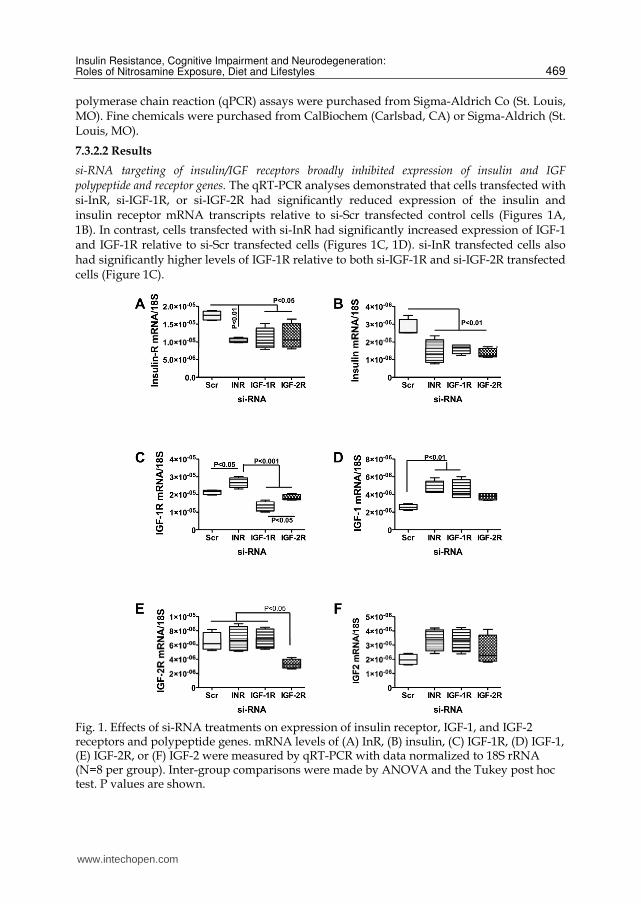
si-RNA targeting of insulin/IGF receptors broadly inhibited expression of insulin and IGF polypeptide and receptor genes. The qRT-PCR analyses demonstrated that cells transfected with si-InR, si-IGF-1R, or si-IGF-2R had significantly reduced expression of the insulin and insulin receptor mRNA transcripts relative to si-Scr transfected control cells (Figures 1A, 1B). In contrast, cells transfected with si-InR had significantly increased expression of IGF-1 and IGF-1R relative to si-Scr transfected cells (Figures 1C, 1D). si-InR transfected cells also had significantly higher levels of IGF-1R relative to both si-IGF-1R and si-IGF-2R transfected cells (Figure 1C).
Fig. 1. Effects of si-RNA treatments on expression of insulin receptor, IGF-1, and IGF-2 receptors and polypeptide genes. mRNA levels of (A) InR, (B) insulin, (C) IGF-1R, (D) IGF-1, (E) IGF-2R, or (F) IGF-2 were measured by qRT-PCR with data normalized to 18S rRNA (N=8 per group). Inter-group comparisons were made by ANOVA and the Tukey post hoc test. P values are shown.
www.intechopen.com
Alzheimer’s Disease Pathogenesis-Core Concepts, Shifting Paradigms and Therapeutic Targets 470
Cells transfected with si-IGF-1R had significantly reduced levels of IGF-1R relative to all
other groups (Figure 1C) and higher levels of IGF-1 relative to si-Scr controls (Figure 1D).
Cells transfected with si-IGF-2R had significantly lower mean levels of IGF-2R compared
with the other groups (Figure 1E). IGF-1 (Figure 1D) and IGF-2 (Figure 1F) polypeptide
expressions were generally higher in the experimental si-RNA groups relative to control,
but the differences were only statistically significant with respect to IGF-1 mRNA levels in
si-InR and si-IGF-1R transfected cells. Therefore, InR and insulin gene expression were
inhibited in CNS neurons that were transfected with si-RNA targeting insulin, IGF-1, or
IGF-2 receptors, while IGF-1R expression was selectively suppressed by transfection with
si-IGF-1R, and IGF-2R expression was selectively inhibited in cells transfected with si-
IGF-2R. With regard to the polypeptide genes, as noted, insulin gene expression was
suppressed by si-InR, si-IGF-1R, or si-IGF-2R, whereas IGF-1 and IGF-2 mRNAs were
generally increased relative to control. The latter could reflect a compensatory response to
down regulation of the receptors. The aggregate results suggest that in cerebellar granule
neurons, InR expression overlaps with IGF-1R and IGF-2R, and that some sub-
populations express of IGF-1R and IGF-2R, while others may only express IGF-2R.
Moreover, the findings suggest that the insulin polypeptide gene is expressed in insulin
receptor bearing cells, whereas IGF-1 and IGF-2 are probably expressed in several
different neuronal populations.
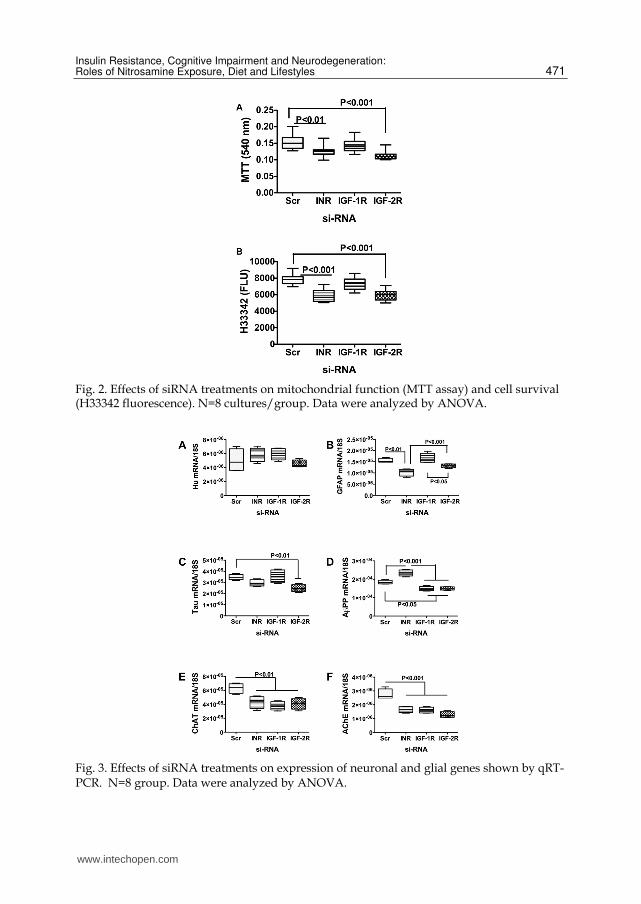
si-RNA targeting InR or IGF-2R impairs cell survival and mitochondrial function in
neuronal cultures. Mitochondrial function was measured with the MTT assay, and cell
density was measured by Hoechst H33342 fluorescence. MTT activity, corrected for cell
density, was significantly lower in si-InR and si-IGF-2R relative to si-Scr transfected cultures
(Figure 2A). In addition, the mean cell densities in cultures transfected with si-InR or si-IGF-
2R were reduced relative to si-Scr transfected control cells (Figure 2B). Therefore, inhibition
of signaling through the insulin or IGF-2 receptors impairs CNS neuronal viability and
mitochondrial function.
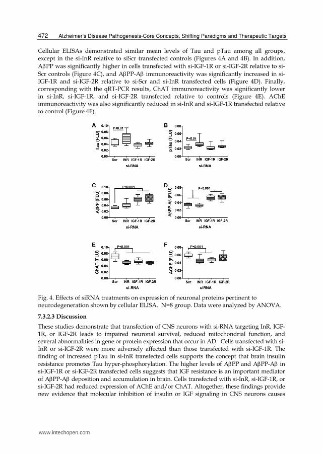
Effects of si-RNA transfection on neuronal and astrocytic biomarkers. We used qRT-PCR
analysis to measure gene expression corresponding to Hu and glial fibrillary acidic protein
(GFAP), reflecting neuronal and astrocyte cell populations. The higher levels of Hu relative
to GFAP reflect the predominantly neuronal nature of the cells isolated from cerebella
(Figures 3A and 3B). Hu expression in si-InR, si-IGF-1R, and si-IGF-2R transfected cultures
was similar to that measured in si-Scr control cells (Figure 3A). In contrast, GFAP
expression was significantly reduced in si-InR transfected relative to all other groups, and in
si-IGF-2R relative to si-IGF-1R transfected cells (Figure 3B). GFAP expression in cells
transfected with si-IGF-1R was similar to control.
Inhibition of insulin and IGF receptor gene expression mediate neurodegeneration. We
investigated the extent to which neuronal transfection with si-RNA targeting insulin, IGF-1
or IGF-2 receptor altered Tau, ChAT, APP, or AChE gene expression, as occur in AD. Tau
gene expression was significantly reduced in cells transfected with si-IGF-2R relative to
control (Figure 3C), corresponding with the somewhat reduced expression of Hu in the
same cells (Figure 3A). In addition, APP mRNA was increased in si-InR transfected cells,
and reduced in si-IGF-1R and si-IGF-2R transfected relative to si-Scr and si-InR transfected
cells (Figure 3D). ChAT and AChE mRNA levels were significantly reduced in si-InR, si-
IGF-1R, and si-IGF-2R transfected relative to si-Scr controls (Figure 3E and 3F).
www.intechopen.com
Insulin Resistance, Cognitive Impairment and Neurodegeneration: Roles of Nitrosamine Exposure, Diet and Lifestyles 471
Fig. 2. Effects of siRNA treatments on mitochondrial function (MTT assay) and cell survival (H33342 fluorescence). N=8 cultures/group. Data were analyzed by ANOVA.
Fig. 3. Effects of siRNA treatments on expression of neuronal and glial genes shown by qRT-PCR. N=8 group. Data were analyzed by ANOVA.
www.intechopen.com
Alzheimer’s Disease Pathogenesis-Core Concepts, Shifting Paradigms and Therapeutic Targets 472
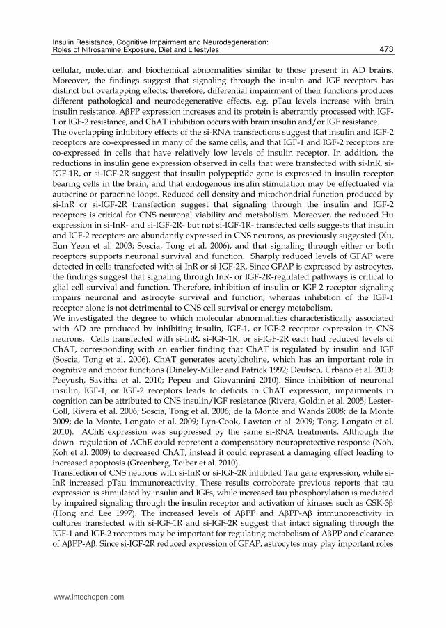
Cellular ELISAs demonstrated similar mean levels of Tau and pTau among all groups, except in the si-InR relative to siScr transfected controls (Figures 4A and 4B). In addition,
APP was significantly higher in cells transfected with si-IGF-1R or si-IGF-2R relative to si-
Scr controls (Figure 4C), and APP-A immunoreactivity was significantly increased in si-IGF-1R and si-IGF-2R relative to si-Scr and si-InR transfected cells (Figure 4D). Finally, corresponding with the qRT-PCR results, ChAT immunoreactivity was significantly lower in si-InR, si-IGF-1R, and si-IGF-2R transfected relative to controls (Figure 4E). AChE immunoreactivity was also significantly reduced in si-InR and si-IGF-1R transfected relative to control (Figure 4F).
Fig. 4. Effects of siRNA treatments on expression of neuronal proteins pertinent to neurodegeneration shown by cellular ELISA. N=8 group. Data were analyzed by ANOVA.
7.3.2.3 Discussion
These studies demonstrate that transfection of CNS neurons with si-RNA targeting InR, IGF-1R, or IGF-2R leads to impaired neuronal survival, reduced mitochondrial function, and several abnormalities in gene or protein expression that occur in AD. Cells transfected with si-InR or si-IGF-2R were more adversely affected than those transfected with si-IGF-1R. The finding of increased pTau in si-InR transfected cells supports the concept that brain insulin
resistance promotes Tau hyper-phosphorylation. The higher levels of APP and APP-A in si-IGF-1R or si-IGF-2R transfected cells suggests that IGF resistance is an important mediator
of APP-A deposition and accumulation in brain. Cells transfected with si-InR, si-IGF-1R, or si-IGF-2R had reduced expression of AChE and/or ChAT. Altogether, these findings provide new evidence that molecular inhibition of insulin or IGF signaling in CNS neurons causes
www.intechopen.com
Insulin Resistance, Cognitive Impairment and Neurodegeneration: Roles of Nitrosamine Exposure, Diet and Lifestyles 473
cellular, molecular, and biochemical abnormalities similar to those present in AD brains. Moreover, the findings suggest that signaling through the insulin and IGF receptors has distinct but overlapping effects; therefore, differential impairment of their functions produces different pathological and neurodegenerative effects, e.g. pTau levels increase with brain
insulin resistance, APP expression increases and its protein is aberrantly processed with IGF-1 or IGF-2 resistance, and ChAT inhibition occurs with brain insulin and/or IGF resistance. The overlapping inhibitory effects of the si-RNA transfections suggest that insulin and IGF-2 receptors are co-expressed in many of the same cells, and that IGF-1 and IGF-2 receptors are co-expressed in cells that have relatively low levels of insulin receptor. In addition, the reductions in insulin gene expression observed in cells that were transfected with si-InR, si-IGF-1R, or si-IGF-2R suggest that insulin polypeptide gene is expressed in insulin receptor bearing cells in the brain, and that endogenous insulin stimulation may be effectuated via autocrine or paracrine loops. Reduced cell density and mitochondrial function produced by si-InR or si-IGF-2R transfection suggest that signaling through the insulin and IGF-2 receptors is critical for CNS neuronal viability and metabolism. Moreover, the reduced Hu expression in si-InR- and si-IGF-2R- but not si-IGF-1R- transfected cells suggests that insulin and IGF-2 receptors are abundantly expressed in CNS neurons, as previously suggested (Xu, Eun Yeon et al. 2003; Soscia, Tong et al. 2006), and that signaling through either or both receptors supports neuronal survival and function. Sharply reduced levels of GFAP were detected in cells transfected with si-InR or si-IGF-2R. Since GFAP is expressed by astrocytes, the findings suggest that signaling through InR- or IGF-2R-regulated pathways is critical to glial cell survival and function. Therefore, inhibition of insulin or IGF-2 receptor signaling impairs neuronal and astrocyte survival and function, whereas inhibition of the IGF-1 receptor alone is not detrimental to CNS cell survival or energy metabolism. We investigated the degree to which molecular abnormalities characteristically associated with AD are produced by inhibiting insulin, IGF-1, or IGF-2 receptor expression in CNS neurons. Cells transfected with si-InR, si-IGF-1R, or si-IGF-2R each had reduced levels of ChAT, corresponding with an earlier finding that ChAT is regulated by insulin and IGF (Soscia, Tong et al. 2006). ChAT generates acetylcholine, which has an important role in cognitive and motor functions (Dineley-Miller and Patrick 1992; Deutsch, Urbano et al. 2010; Peeyush, Savitha et al. 2010; Pepeu and Giovannini 2010). Since inhibition of neuronal insulin, IGF-1, or IGF-2 receptors leads to deficits in ChAT expression, impairments in cognition can be attributed to CNS insulin/IGF resistance (Rivera, Goldin et al. 2005; Lester-Coll, Rivera et al. 2006; Soscia, Tong et al. 2006; de la Monte and Wands 2008; de la Monte 2009; de la Monte, Longato et al. 2009; Lyn-Cook, Lawton et al. 2009; Tong, Longato et al. 2010). AChE expression was suppressed by the same si-RNA treatments. Although the down--regulation of AChE could represent a compensatory neuroprotective response (Noh, Koh et al. 2009) to decreased ChAT, instead it could represent a damaging effect leading to increased apoptosis (Greenberg, Toiber et al. 2010). Transfection of CNS neurons with si-InR or si-IGF-2R inhibited Tau gene expression, while si-InR increased pTau immunoreactivity. These results corroborate previous reports that tau expression is stimulated by insulin and IGFs, while increased tau phosphorylation is mediated by impaired signaling through the insulin receptor and activation of kinases such as GSK-3 (Hong and Lee 1997). The increased levels of APP and APP-A immunoreactivity in cultures transfected with si-IGF-1R and si-IGF-2R suggest that intact signaling through the IGF-1 and IGF-2 receptors may be important for regulating metabolism of APP and clearance of APP-A. Since si-IGF-2R reduced expression of GFAP, astrocytes may play important roles
www.intechopen.com
Alzheimer’s Disease Pathogenesis-Core Concepts, Shifting Paradigms and Therapeutic Targets 474
in APP and APP-A processing and clearance in the brain. On the other hand, cultures transfected with si-InR had increased APP gene expression, similar to the findings in AD (Rivera, Goldin et al. 2005; Steen, Terry et al. 2005). This effect could be mediated by increased oxidative stress caused by reduced energy metabolism and increased phospho-tau. Previous studies showed that oxidative stress was sufficient to increase CNS neuronal phospho-Tau and APP-A (Chen, Xu et al. 2003). Together, the findings herein suggest that inhibition of insulin, IGF-1, and IGF-2 receptor expression and function have complimentary but overlapping roles in mediating the molecular pathogenesis of neurodegeneration, including AD. In addition, the results suggest that therapeutic measures should target all 3 receptors, since impairments of their signaling pathways would likely mediate distinct but overlapping components of the neurodegeneration cascade.
7.3.3 Experimental targeting of neuronal insulin/IGF receptors — in vivo model of brain insulin/IGF resistance
7.3.3.1 Methods
In vivo siRNA delivery. To evaluate the roles of insulin and IGF receptor resistance in relation to neurodegeneration in vivo, we generated a model in which P2 Long Evans rats were given a single intracerebroventricular (ICV) injection of si-InR [INSR NM_017071], si-IGF-1R [IGF1R NM_052807], si-IGF-2R [IGF2R NM_012756], or si-Scr (negative control) [NM D-001210-01-20]. For each rat, 0.4 nmol si-RNA plus 100 ng recombinant green fluorescent
protein (GFP) expressing plasmid were combined with 10 l of Dharmafect reagent, and injected into the right frontal region over the lateral ventricle using a Hamilton syringe with a 26-gauge needle as previously described (de la Monte, Jhaveri et al. 2007). GFP expression, which was under the control of a CMV promoter, was used to monitor success of the transfections. All rats survived the procedure, and none of them exhibited aberrant behaviors or adverse effects such as failure to thrive, poor grooming, reduced physical activity, or weight loss. To monitor effectiveness of the transfections, rats (N=4 per group) were sacrificed at regular
intervals after ICV gene delivery, and GFP expression was measured in whole brain by qRT-
PCR analysis. Those studies demonstrated peak levels of GFP expression 2-3 days after ICV
gene delivery, followed by a gradual decline in GFP mRNA levels. However, GFP
expression persisted over the time course of the experiment as previously reported (de la
Monte, Jhaveri et al. 2007). Gene expression and histological studies were performed with
temporal lobe tissue. Upon sacrifice, temporal lobes were either immersion fixed in Histofix,
embedded in paraffin, and used to make hematoxylin and eosin (H&E) stained histological
sections (5 µm-thick), or they were snap-frozen in a dry ice/methanol bath and stored at -
80°C for mRNA and protein studies. Our protocol was approved by the Institutional
Animal Care and Use Committee at Lifespan-Rhode Island Hospital, and it conforms to
guidelines set by the National Institutes of Health.
Morris water maze testing. We used Morris water maze tests to assess long-term effects of ICV si-InR, si-IGF-1R and si-IGF-2R on spatial learning and memory, corresponding to hippocampal function (Hamm, Dixon et al. 1992; McLay, Freeman et al. 1999; Gomez-Pinilla, So et al. 2001). Beginning on P24, rats were subjected to 3 daily trials in which latency (seconds) required to locate and land on the platform was recorded. On Day 1, the platform was visible, but on Days 2-4, the platform was submerged. On Days 3 and 4, the water entry quadrants were randomized for each trial. The data were analyzed using area-under-the-
www.intechopen.com
Insulin Resistance, Cognitive Impairment and Neurodegeneration: Roles of Nitrosamine Exposure, Diet and Lifestyles 475
curve (AUC) calculations corresponding to performance over the 3 trials each day (de la Monte, Tong et al. 2006; Lester-Coll, Rivera et al. 2006). Gene and protein expression. Gene and protein expression were measured by qRT-PCR analysis (see above) and direct binding ELISA. For ELISAs, temporal lobe homogenates were prepared in radioimmunoprecipitation assay (RIPA) buffer containing protease and phosphatase inhibitors (Lester-Coll, Rivera et al. 2006). Protein concentrations were determined using the BCA assay. 50 ng protein samples in Tris buffered saline, pH 7.4 (TBS) were adsorbed to flat-bottom surfaces of 96-well polystyrene plates, overnight at 4°C. Non-specific binding sites were masked by a 3-hour room temperature incubation with 300
l/well of TBS + 0.05% Tween 20 + 3% BSA. Samples were then incubated with 0.1-0.5
g/ml primary antibody for 1 h at 37°C. Immunoreactivity was detected with horseradish peroxidase (HRP)-conjugated secondary antibody and Amplex Red soluble fluorophore (de la Monte and Tong 2009). Fluorescence was measured (Ex 530/Em 590) in a SpectraMax M5 microplate reader. Parallel negative control assays had primary, secondary, or both antibodies omitted. Between steps, reactions were rinsed 3 times with TBS + 0.05% Tween 20 using a Nunc ELISA plate washer. Levels of immunoreactivity were normalized to protein content in the wells, which was measured using NanoOrange reagent. Statistical analysis. Inter-group comparisons were made using repeated measures one-way ANOVA and the Dunnett post-hoc significance test for comparisons with the si-Scr control group. Statistical analyses were performed using the GraphPad Prism 5 software (San Diego, CA) and P<0.05 was considered significant.
7.3.3.2 Results
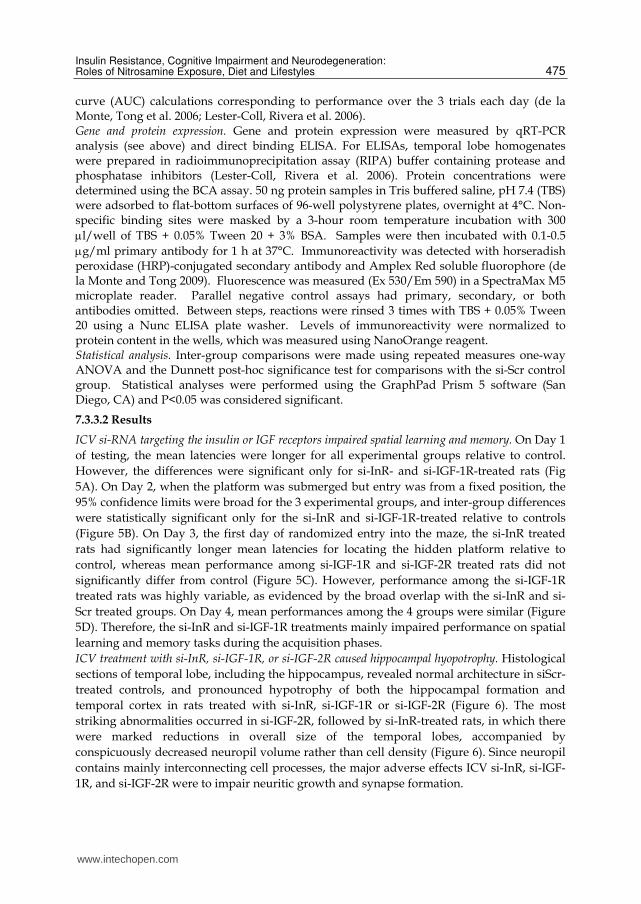
ICV si-RNA targeting the insulin or IGF receptors impaired spatial learning and memory. On Day 1
of testing, the mean latencies were longer for all experimental groups relative to control.
However, the differences were significant only for si-InR- and si-IGF-1R-treated rats (Fig
5A). On Day 2, when the platform was submerged but entry was from a fixed position, the
95% confidence limits were broad for the 3 experimental groups, and inter-group differences
were statistically significant only for the si-InR and si-IGF-1R-treated relative to controls
(Figure 5B). On Day 3, the first day of randomized entry into the maze, the si-InR treated
rats had significantly longer mean latencies for locating the hidden platform relative to
control, whereas mean performance among si-IGF-1R and si-IGF-2R treated rats did not
significantly differ from control (Figure 5C). However, performance among the si-IGF-1R
treated rats was highly variable, as evidenced by the broad overlap with the si-InR and si-
Scr treated groups. On Day 4, mean performances among the 4 groups were similar (Figure
5D). Therefore, the si-InR and si-IGF-1R treatments mainly impaired performance on spatial
learning and memory tasks during the acquisition phases.
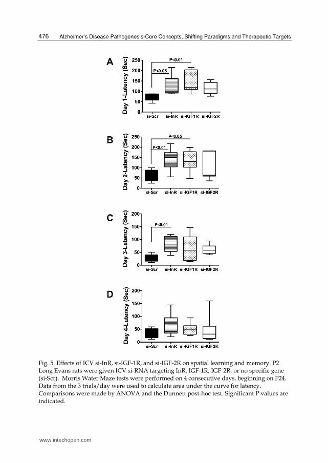
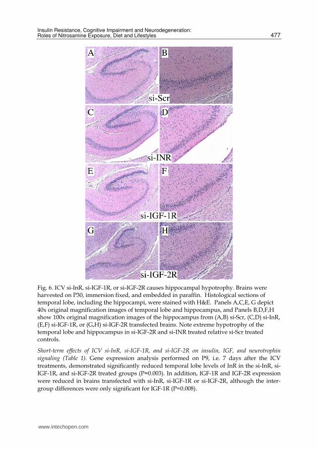
ICV treatment with si-InR, si-IGF-1R, or si-IGF-2R caused hippocampal hyopotrophy. Histological
sections of temporal lobe, including the hippocampus, revealed normal architecture in siScr-
treated controls, and pronounced hypotrophy of both the hippocampal formation and
temporal cortex in rats treated with si-InR, si-IGF-1R or si-IGF-2R (Figure 6). The most
striking abnormalities occurred in si-IGF-2R, followed by si-InR-treated rats, in which there
were marked reductions in overall size of the temporal lobes, accompanied by
conspicuously decreased neuropil volume rather than cell density (Figure 6). Since neuropil
contains mainly interconnecting cell processes, the major adverse effects ICV si-InR, si-IGF-
1R, and si-IGF-2R were to impair neuritic growth and synapse formation.
www.intechopen.com
Alzheimer’s Disease Pathogenesis-Core Concepts, Shifting Paradigms and Therapeutic Targets 476
Fig. 5. Effects of ICV si-InR, si-IGF-1R, and si-IGF-2R on spatial learning and memory. P2
Long Evans rats were given ICV si-RNA targeting InR, IGF-1R, IGF-2R, or no specific gene (si-Scr). Morris Water Maze tests were performed on 4 consecutive days, beginning on P24. Data from the 3 trials/day were used to calculate area under the curve for latency. Comparisons were made by ANOVA and the Dunnett post-hoc test. Significant P values are indicated.
www.intechopen.com
Insulin Resistance, Cognitive Impairment and Neurodegeneration: Roles of Nitrosamine Exposure, Diet and Lifestyles 477
Fig. 6. ICV si-InR, si-IGF-1R, or si-IGF-2R causes hippocampal hypotrophy. Brains were harvested on P30, immersion fixed, and embedded in paraffin. Histological sections of temporal lobe, including the hippocampi, were stained with H&E. Panels A,C,E, G depict 40x original magnification images of temporal lobe and hippocampus, and Panels B,D,F,H show 100x original magnification images of the hippocampus from (A,B) si-Scr, (C,D) si-InR, (E,F) si-IGF-1R, or (G,H) si-IGF-2R transfected brains. Note extreme hypotrophy of the temporal lobe and hippocampus in si-IGF-2R and si-INR treated relative si-Scr treated controls.
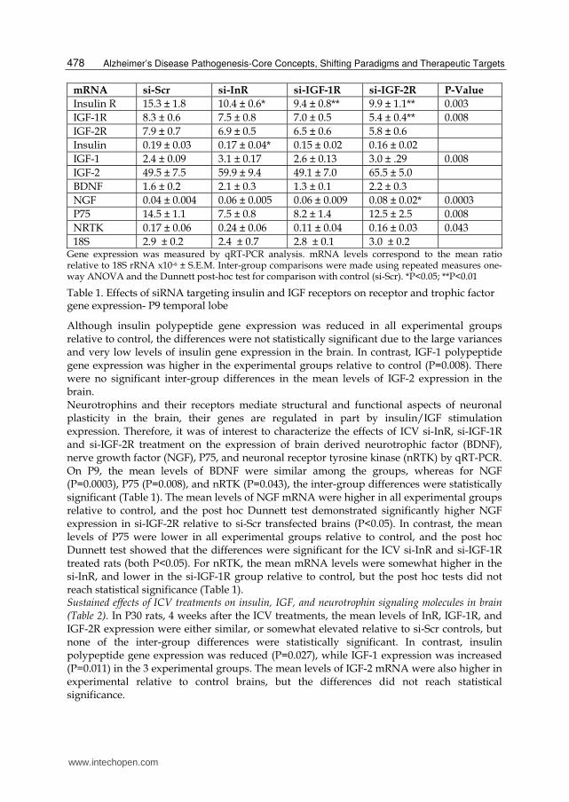
Short-term effects of ICV si-InR, si-IGF-1R, and si-IGF-2R on insulin, IGF, and neurotrophin
signaling (Table 1). Gene expression analysis performed on P9, i.e. 7 days after the ICV
treatments, demonstrated significantly reduced temporal lobe levels of InR in the si-InR, si-
IGF-1R, and si-IGF-2R treated groups (P=0.003). In addition, IGF-1R and IGF-2R expression
were reduced in brains transfected with si-InR, si-IGF-1R or si-IGF-2R, although the inter-
group differences were only significant for IGF-1R (P=0.008).
www.intechopen.com
Alzheimer’s Disease Pathogenesis-Core Concepts, Shifting Paradigms and Therapeutic Targets 478
mRNA si-Scr si-InR si-IGF-1R si-IGF-2R P-Value
Insulin R 15.3 ± 1.8 10.4 ± 0.6* 9.4 ± 0.8** 9.9 ± 1.1** 0.003
IGF-1R 8.3 ± 0.6 7.5 ± 0.8 7.0 ± 0.5 5.4 ± 0.4** 0.008
IGF-2R 7.9 ± 0.7 6.9 ± 0.5 6.5 ± 0.6 5.8 ± 0.6
Insulin 0.19 ± 0.03 0.17 ± 0.04* 0.15 ± 0.02 0.16 ± 0.02
IGF-1 2.4 ± 0.09 3.1 ± 0.17 2.6 ± 0.13 3.0 ± .29 0.008
IGF-2 49.5 ± 7.5 59.9 ± 9.4 49.1 ± 7.0 65.5 ± 5.0
BDNF 1.6 ± 0.2 2.1 ± 0.3 1.3 ± 0.1 2.2 ± 0.3
NGF 0.04 ± 0.004 0.06 ± 0.005 0.06 ± 0.009 0.08 ± 0.02* 0.0003
P75 14.5 ± 1.1 7.5 ± 0.8 8.2 ± 1.4 12.5 ± 2.5 0.008
NRTK 0.17 ± 0.06 0.24 ± 0.06 0.11 ± 0.04 0.16 ± 0.03 0.043
18S 2.9 ± 0.2 2.4 ± 0.7 2.8 ± 0.1 3.0 ± 0.2 Gene expression was measured by qRT-PCR analysis. mRNA levels correspond to the mean ratio relative to 18S rRNA x10-6 ± S.E.M. Inter-group comparisons were made using repeated measures one-way ANOVA and the Dunnett post-hoc test for comparison with control (si-Scr). *P<0.05; **P<0.01
Table 1. Effects of siRNA targeting insulin and IGF receptors on receptor and trophic factor gene expression- P9 temporal lobe
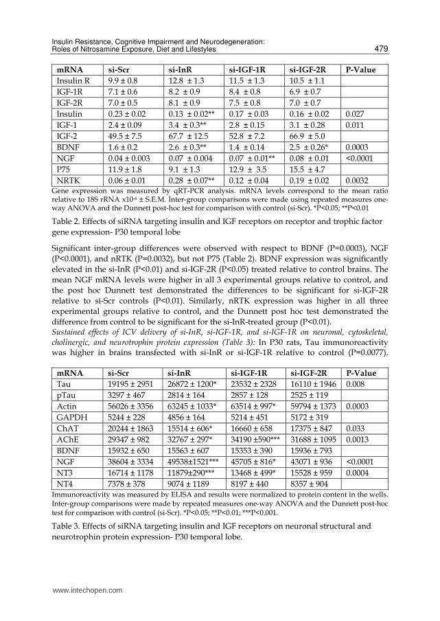
Although insulin polypeptide gene expression was reduced in all experimental groups relative to control, the differences were not statistically significant due to the large variances and very low levels of insulin gene expression in the brain. In contrast, IGF-1 polypeptide gene expression was higher in the experimental groups relative to control (P=0.008). There were no significant inter-group differences in the mean levels of IGF-2 expression in the brain. Neurotrophins and their receptors mediate structural and functional aspects of neuronal plasticity in the brain, their genes are regulated in part by insulin/IGF stimulation expression. Therefore, it was of interest to characterize the effects of ICV si-InR, si-IGF-1R and si-IGF-2R treatment on the expression of brain derived neurotrophic factor (BDNF), nerve growth factor (NGF), P75, and neuronal receptor tyrosine kinase (nRTK) by qRT-PCR. On P9, the mean levels of BDNF were similar among the groups, whereas for NGF (P=0.0003), P75 (P=0.008), and nRTK (P=0.043), the inter-group differences were statistically significant (Table 1). The mean levels of NGF mRNA were higher in all experimental groups relative to control, and the post hoc Dunnett test demonstrated significantly higher NGF expression in si-IGF-2R relative to si-Scr transfected brains (P<0.05). In contrast, the mean levels of P75 were lower in all experimental groups relative to control, and the post hoc Dunnett test showed that the differences were significant for the ICV si-InR and si-IGF-1R treated rats (both P<0.05). For nRTK, the mean mRNA levels were somewhat higher in the si-InR, and lower in the si-IGF-1R group relative to control, but the post hoc tests did not reach statistical significance (Table 1). Sustained effects of ICV treatments on insulin, IGF, and neurotrophin signaling molecules in brain (Table 2). In P30 rats, 4 weeks after the ICV treatments, the mean levels of InR, IGF-1R, and IGF-2R expression were either similar, or somewhat elevated relative to si-Scr controls, but none of the inter-group differences were statistically significant. In contrast, insulin polypeptide gene expression was reduced (P=0.027), while IGF-1 expression was increased (P=0.011) in the 3 experimental groups. The mean levels of IGF-2 mRNA were also higher in experimental relative to control brains, but the differences did not reach statistical significance.
www.intechopen.com
Insulin Resistance, Cognitive Impairment and Neurodegeneration: Roles of Nitrosamine Exposure, Diet and Lifestyles 479
mRNA si-Scr si-InR si-IGF-1R si-IGF-2R P-Value
Insulin R 9.9 ± 0.8 12.8 ± 1.3 11.5 ± 1.3 10.5 ± 1.1
IGF-1R 7.1 ± 0.6 8.2 ± 0.9 8.4 ± 0.8 6.9 ± 0.7
IGF-2R 7.0 ± 0.5 8.1 ± 0.9 7.5 ± 0.8 7.0 ± 0.7
Insulin 0.23 ± 0.02 0.13 ± 0.02** 0.17 ± 0.03 0.16 ± 0.02 0.027
IGF-1 2.4 ± 0.09 3.4 ± 0.3** 2.8 ± 0.15 3.1 ± 0.28 0.011
IGF-2 49.5 ± 7.5 67.7 ± 12.5 52.8 ± 7.2 66.9 ± 5.0
BDNF 1.6 ± 0.2 2.6 ± 0.3** 1.4 ± 0.14 2.5 ± 0.26* 0.0003
NGF 0.04 ± 0.003 0.07 ± 0.004 0.07 ± 0.01** 0.08 ± 0.01 <0.0001
P75 11.9 ± 1.8 9.1 ± 1.3 12.9 ± 3.5 15.5 ± 4.7
NRTK 0.06 ± 0.01 0.28 ± 0.07** 0.12 ± 0.04 0.19 ± 0.02 0.0032 Gene expression was measured by qRT-PCR analysis. mRNA levels correspond to the mean ratio relative to 18S rRNA x10-6 ± S.E.M. Inter-group comparisons were made using repeated measures one-way ANOVA and the Dunnett post-hoc test for comparison with control (si-Scr). *P<0.05; **P<0.01
Table 2. Effects of siRNA targeting insulin and IGF receptors on receptor and trophic factor
gene expression- P30 temporal lobe
Significant inter-group differences were observed with respect to BDNF (P=0.0003), NGF
(P<0.0001), and nRTK (P=0.0032), but not P75 (Table 2). BDNF expression was significantly
elevated in the si-InR (P<0.01) and si-IGF-2R (P<0.05) treated relative to control brains. The
mean NGF mRNA levels were higher in all 3 experimental groups relative to control, and
the post hoc Dunnett test demonstrated the differences to be significant for si-IGF-2R
relative to si-Scr controls (P<0.01). Similarly, nRTK expression was higher in all three
experimental groups relative to control, and the Dunnett post hoc test demonstrated the
difference from control to be significant for the si-InR-treated group (P<0.01). Sustained effects of ICV delivery of si-InR, si-IGF-1R, and si-IGF-1R on neuronal, cytoskeletal, cholinergic, and neurotrophin protein expression (Table 3): In P30 rats, Tau immunoreactivity was higher in brains transfected with si-InR or si-IGF-1R relative to control (P=0.0077).
mRNA si-Scr si-InR si-IGF-1R si-IGF-2R P-Value
Tau 19195 ± 2951 26872 ± 1200* 23532 ± 2328 16110 ± 1946 0.008
pTau 3297 ± 467 2814 ± 164 2857 ± 128 2525 ± 119
Actin 56026 ± 3356 63245 ± 1033* 63514 ± 997* 59794 ± 1373 0.0003
GAPDH 5244 ± 228 4856 ± 164 5214 ± 451 5172 ± 319
ChAT 20244 ± 1863 15514 ± 606* 16660 ± 658 17375 ± 847 0.033
AChE 29347 ± 982 32767 ± 297* 34190 ±590*** 31688 ± 1095 0.0013
BDNF 15932 ± 650 15563 ± 607 15353 ± 390 15936 ± 793
NGF 38604 ± 3334 49538±1521*** 45705 ± 816* 43071 ± 936 <0.0001
NT3 16714 ± 1178 11879±290*** 13468 ± 499* 15528 ± 959 0.0004
NT4 7378 ± 378 9074 ± 1189 8197 ± 440 8357 ± 904 Immunoreactivity was measured by ELISA and results were normalized to protein content in the wells. Inter-group comparisons were made by repeated measures one-way ANOVA and the Dunnett post-hoc test for comparison with control (si-Scr). *P<0.05; **P<0.01; ***P<0.001.
Table 3. Effects of siRNA targeting insulin and IGF receptors on neuronal structural and
neurotrophin protein expression- P30 temporal lobe.
www.intechopen.com
Alzheimer’s Disease Pathogenesis-Core Concepts, Shifting Paradigms and Therapeutic Targets 480
-actin expression was higher in si-InR and si-IGF-1R relative to si-Scr transfected brains (P=0.0003). ICV delivery of si-InR, si-IGF-1R, or si-IGF-2R reduced temporal lobe levels of ChAT (P=0.036). In contrast, AChE immunoreactivity was elevated in si-InR, si-IGF-1R, and si-IGF-2R transfected brains (P=0.0013). Corresponding with the qRT-PCR analyses, NGF expression was increased in the si-InR, si-IGF-1R, and si-IGF-2R treated relative to control brains (P<0.0001). The mean levels of NT3 immunoreactivity were lower in the 3 experimental groups relative to control (P=0.0004). There were no significant inter-group differences in the mean levels of pTau, GAPDH, BDNF, or NT4 in P30 rat brains.
7.3.3.3 Discussion
We used ICV injections to transfect brains with si-RNA duplex molecules targeting InR, IGF-1R, IGF-2R, or no specific genes (si-Scr). Previously, we used the same approach to generate a model of neurodegeneration, and demonstrated sustained transgene expression throughout the brain and in all cell types (de la Monte, Jhaveri et al. 2007). Morris water maze tests demonstrated significant impairments in spatial learning in the si-InR, si-IGF-1R, and si-IGF-2R treated rats. Histopathological studies showed that the deficits in spatial learning were associated with reduced sizes of the hippocampi and temporal lobes, and hypotrophy of the neuropil, which contains interconnecting cell processes and synapses. Major abnormalities in AD include dendritic spine atrophy and synapse disruption. Since insulin and IGFs modulate neuronal plasticity and neurite outgrowth (de la Monte 2009), these findings suggest that AD-associated impairments in spatial learning and memory could be mediated by brain insulin and IGF resistance. Gene expression studies showed that, in addition to reducing expression of the target genes, ICV si-InR, si-IGF-1R and si-IGF-2R each inhibited expression of the other two non-targeted receptors. The most likely explanation for this phenomenon is that cells in the temporal lobe express all 3 receptors at once. The inhibitory effects of the si-RNA treatments were most pronounced for si-InR. This finding, together with fact that all 3 experimental treatments resulted in hypotrophy of the temporal lobe and hippocampus, suggest that the adverse effects of the si-RNA treatments were mainly due to inhibition of insulin signaling. Although GFP expression persisted in the brains due to continued presence of the plasmid,
it is unlikely that the si-RNA molecules remained intact 1 or 2 days after transfection. The
absence of any significant reduction in InR, IGF-1R, or IGF-2R expression at P30 indicates
that the effects of the si-RNA treatments were transient with respect to gene expression.
Therefore, the sustained effects of the si-RNA treatments on temporal lobe structure and
function were mediated by short-term inhibition of gene expression during the critical
postnatal period of synaptic plasticity and development. The relatively modest inhibitory
effects on insulin and IGF polypeptides indicate that the structural and functional
impairments caused by ICV si-InR, si-IGF-1R, or si-IGF-2R were mediated by insulin/IGF
resistance rather than withdrawal of endogenously produced trophic factors. We investigated the effects of the si-RNA treatments on neurotrophin and neurotrophin receptor expression because these molecules mediate neurite outgrowth, plasticity, neuronal survival, and other function, and insulin/IGF modulate expression and function of neurotrophins in brain (Maisonpierre, Belluscio et al. 1990; Cowansage, LeDoux et al. 2010). The studies demonstrated that the main adverse effect of the si-RNA treatments was to reduce expression of p75 neurotrophin receptor on P9. Otherwise, BDNF, NGF, p75 neurotrophin receptor, and nRTK were either increased or similarly expressed compared
www.intechopen.com
Insulin Resistance, Cognitive Impairment and Neurodegeneration: Roles of Nitrosamine Exposure, Diet and Lifestyles 481
with si-Scr controls. ELISA studies demonstrated similar or increased expression of BDNF, NGF, and neurotrophin 4 (NT4) in P30 temporal lobes of experimental si-RNA treated rats, but significantly reduced expression of NT3 in both si-InR and si-IGF-1R treated rats. Since all neurotrophins, including NGF, BDNF, NT3 and NT4/5 function by binding to the p75 neurotrophin receptor (Frebel and Wiese 2006), early inhibition of p75 expression would have impaired neurotrophin signaling and could account for both the spatial learning deficits and hypotrophy of the temporal lobe/hippocampus in the si-RNA treated rats. NT3 regulates neuronal survival, synaptic plasticity, and neurotransmission, particularly in the hippocampus (Pae, Marks et al. 2008). Therefore, persistent reductions in NT3 expression represent local trophic factor withdrawal, which could have contributed to the cognitive impairment detected by Morris water maze testing. Although insulin/IGF regulation of neurotrophin and neurotrophin expression has not yet been characterized, the findings herein suggest that neurotrophin expression and function may be downstream of insulin/IGF signaling pathways in the brain. In previous studies, inhibition of brain insulin and IGF signaling reduced expression of tau, ChAT, and GAPDH, increased pTau, and had variable effects on AChE expression (Soscia, Tong et al. 2006; de la Monte and Wands 2008; Tong, Longato et al. 2010). Corresponding with the normalized levels of InR and IGF-1R expression at P30, tau and GAPDH were not reduced, and pTau was not increased, but the impairments in cholinergic homeostasis were sustained in temporal lobes of si-InR and/or si-IGF-1R treated rats. ChAT is a major neurotransmitter that mediates cognitive function and plasticity throughout life (Perry, Piggott et al. 1993), and evidence suggests that besides insulin/IGF stimulation (Shi, Rabin et al. 1998), cholinergic function can be regulated by neurotrophins, including NT3 (Robertson, Baratta et al. 2006). It is noteworthy that in both si-InR and si-IGF-1R, ChAT and NT3 expression were reduced to greater extents than in si-IGF-2R treated brains. These in vivo experiments demonstrate that sustained structural and functional developmental abnormalities can be produced by transient ICV transfection with siRNA molecules targeting insulin, IGF-1, or IGF-2 receptors. The long-term effects include deficits in spatial learning and memory, striking abnormalities in temporal lobe structure, and inhibition of mechanisms needed for cholinergic homeostasis. While these results are exciting and very much validate a role for primary brain insulin and IGF resistance as mediators of neurodegeneration and dementia, the structural changes in neurons and disorganization of the cortical laminar architecture that characterize AD-type neurodegeneration were not detected. This suggests that additional factors such as ongoing oxidative injury, are likely required to propagate the neurodegeneration cascade.
8. Experimental type 3 diabetes
The human studies provide convincing evidence that AD is associated with progressive insulin and IGF resistance and deficiency, accompanied by oxidative stress, deficits in energy metabolism, cholinergic function, neuronal survival, and synaptic plasticity in the brain. Given the fact that most cases of sporadic AD are not associated with obesity or diabetes mellitus, the said abnormalities must originate primarily in the brain. The siRNA experiments targeting the insulin or IGF receptors showed that inhibition of signaling through these receptors is sufficient to cause neurodegeneration and significantly impair a range of functions as observed in AD. On the other hand, the deficits, although sustained, were relatively static rather than progressive, suggesting that another “hit” was needed to propagate the cascade of neurodegeneration.
www.intechopen.com
Alzheimer’s Disease Pathogenesis-Core Concepts, Shifting Paradigms and Therapeutic Targets 482
8.1 Toxin-induced brain diabetes mimics sporadic Alzheimer’s disease Analysis of postmortem human brains demonstrated progressive insulin and IGF resistance and deficiency with increasing severity of AD and dementia, which correlated with increased indices of oxidative stress and DNA damage, and reduced cholinergic
homeostasis. Neuroinflammation and APP gene expression were increased early in the course of AD, but they either stabilized or declined as disease progressed. None of the subjects in those studies had diabetes mellitus, non-alcoholic steatohepatitis, or obesity. Perhaps the best model of sporadic AD is the one generated by ICV streptozotocin (STZ) treatment. STZ is a pro-diabetes drug that when injected systemically causes Type 1 diabetes mellitus (insulin deficiency) with higher doses, and Type 2 diabetes (insulin resistance) at lower doses. The i.p. or i.v. administration of STZ also causes mild hepatic steatosis and neurodegeneration (Biju and Paulose 1998; Szkudelski 2001; Bolzan and Bianchi 2002; Koulmanda, Qipo et al. 2003), but low dose ICV STZ treatment causes cognitive impairment with brain insulin resistance, brain insulin deficiency, and AD-type neurodegeneration, and not diabetes mellitus or hepatic steatosis (Biju and Paulose 1998; Hoyer, Lannert et al. 1999; Nitta, Murai et al. 2002; Weinstock and Shoham 2004; Lester-Coll, Rivera et al. 2006). STZ-induced neurodegeneration is characterized by impairments in brain insulin and IGF signaling, brain atrophy, impaired energy metabolism, mitochondrial dysfunction, oxidative
stress, DNA damage, and increased APP, expression, APP-A accumulation, neuroinflammation, and tau phosphorylation. Therefore, limited exposure to a single pro-diabetes drug causes neurodegeneration with all of the characteristic features of AD. Moreover, depending upon dose and route of administration, STZ treatment produces insulin resistance diseases that target the CNS, liver, pancreas, or skeletal muscle with variable degrees of overlap, or else the brain could represent the primary focus of disease.
8.2 Environmental/dietary causes of insulin/IGF resistance syndromes: Nitrosamine exposures mediate Alzheimer’s, diabetes mellitus, and hepatic steatosis Sporadic AD or Type 3 diabetes is overwhelmingly a brain-specific disease that leads to total
body wasting and eventual arrival at a vegetative state. The closest experimental model of
sporadic AD is that produced by ICV STZ, which does not cause systemic diabetes mellitus,
i.e. Type 1 or Type 2. This very point begs the question of, what relevance could STZ
possibly have in relation to the pathogenesis AD, diabetes, or hepatic steatosis in humans?
After all, human are not generally exposed to STZ. The probable answer was uncovered by
the realization that STZ is a nitrosamine-related compound, and that over the past several
decades, Western societies have been assaulted by continuous and growing exposures to
environmental and food-related nitrosamines. The epidemiological trends for AD, diabetes
mellitus, obesity, and non-alcoholic steatohepatitis all show substantial increases in
incidence, prevalence, morbidity, and mortality rates that correspond more with exposures
than genetics as underlying causes (de la Monte, Neusner et al. 2009). For unclear reasons,
although the connection between nitrosamine exposure and diabetes was entertained years
ago (Berne, Gunnarsson et al. 1974; Portha, Giroix et al. 1980; Helgason, Ewen et al. 1984;
Boucher, Ewen et al. 1994; Dahlquist 1994; Essien and Akpan 2006), that concept seems to
have been missed, and therefore has not yet been incorporated into the conceptualization
about the origins of our insulin resistance diseases epidemic.
We conducted experiments to determine if low, sub-mutagenic doses of nitrosamines that
are found in processed foods, e.g. N-nitrosodiethylamine (NDEA), could cause insulin
www.intechopen.com
Insulin Resistance, Cognitive Impairment and Neurodegeneration: Roles of Nitrosamine Exposure, Diet and Lifestyles 483
resistance diseases. The results were alarming because low-dose and very limited exposures
to NDEA cause T2DM, non-alcoholic steatohepatitis, visceral obesity, cognitive impairment,
and AD-type neurodegeneration with peripheral, hepatic, and brain insulin resistance
(Tong, Neusner et al. 2009; Tong, Longato et al. 2010), similar to the effects of STZ.
Therefore, depending on the structure of the compound, dose, and route of administration,
exposures to nitrosamine-related chemicals can cause insulin resistance diseases in multiple
different target organs, including brain. In addition to providing evidence that the relatively
recent epidemics of sporadic AD, T2DM, and non-alcoholic steatohepatitis/metabolic
syndrome could be mediated by environmental or dietary exposures (de la Monte, Neusner
et al. 2009), these studies demonstrate that insulin resistance diseases with essentially the
same underlying cellular abnormalities, can develop in various organs and tissues. This
phenomenon could account for the overlapping increases in prevalence rates of various
insulin resistance diseases within the past several decades, as well as the very frequent but
incomplete overlap between AD and obesity, T2DM, and NASH (Janson, Laedtke et al.
2004), which did not exist prior to 1980, and is not accounted for by aging of the population
(de la Monte, Neusner et al. 2009).
8.3 The liver-brain-axis of neurodegeneration hypothesis Experiments with STZ and NDEA provided clear evidence that low dose exposures to nitrosamines can reproduce the molecular, biochemical, neuropathological, and neurobehavioral abnormalities observed in human sporadic cases of AD, and also cause a broad spectrum of peripheral insulin resistance diseases, including visceral obesity, diabetes, and non-alcoholic steatohepatitis (de la Monte, Tong et al. 2006; Lester-Coll, Rivera et al. 2006; de la Monte and Tong 2009; Tong, Neusner et al. 2009). Subsequent studies showed that, the adverse effects of NDEA were exacerbated by chronic high fat diet feeding (de la Monte, Tong et al. 2009; Tong, Longato et al. 2010), and that i.p. treatment with NDEA was more effective in causing neurodegeneration than ICV NDEA. Further consideration of the data revealed that the degrees of steatohepatitis, and the levels of toxic lipids, i.e. ceramides produced in liver following NDEA ± high fat diet exposures were critical variables in the equation. These findings led us to the liver-brain- axis of neurodegeneration hypothesis (de la Monte, Longato et al. 2009; de la Monte, Longato et al. 2009), which states that, chronic injury to the liver that results in steatohepatitis, hepatic insulin resistance, and ER stress, leads to dysregulated lipid metabolism and attendant elaboration of toxic lipids, including ceramides and other sphingolipids. The toxic lipids get released into the circulation due to impaired membrane integrity or cell death. Their lipid-soluble nature enables the toxic lipids to cross the blood brain barrier and exert their adverse effects on brain insulin signaling pathways that mediate neuronal and glial survival, energy metabolism, neurotransmitter function, and synaptic plasticity. This hypothesis is supported by preliminary data showing that the same profiles of sphingolipids in steatotic livers are represented in peripheral blood, and that lipids isolated from peripheral blood in insulin-resistance disease states, when applied to brain slice cultures, cause neurodegeneration with insulin resistance. Experimental in vitro and in vivo exposures to chemically defined toxic ceramides also cause neurodegeneration with brain insulin resistance (Tong and de la Monte 2009; de la Monte, Tong et al. 2010). In addition to providing a mechanism whereby environmental toxins in processed and preserved foods, and agricultural products grown with heavy doses of nitrate-containing fertilizers may be responsible for our current and
www.intechopen.com
Alzheimer’s Disease Pathogenesis-Core Concepts, Shifting Paradigms and Therapeutic Targets 484
growing insulin resistance diseases epidemic that is becoming pandemic with exportation of our diets and lifestyles, the liver-brain-axis hypothesis suggests how the lipotoxicity associated with visceral obesity, type 2 diabetes, and NASH could serve as co-factors in the pathogenesis of AD-type neurodegeneration.
9. Western diet and insulin/IGF resistance mediated neurodegeneration
Since aging is essential and unequivocally the most dominant risk factor for AD, critical
changes associated with the loss of youthful compensatory and protective mechanisms must
be responsible for the acquired permissiveness to neurodegeneration. Mechanisms of aging
are still under investigation. However, strong data from organisms that span the phylogenetic
tree, indicate a role for caloric restriction as preventive, and excess caloric intake as a promoter
of aging. One image I find impossible to dismiss, is the one displaying two primates of
approximately the same age, side by side: one monkey was chronically fed with a low calorie
diet, and the other was maintained on a high calorie diet. The differences in appearance and
behavior were striking. The lean monkey appeared youthful and spunky, whereas the obese
monkey looked aged and sluggish (Colman, Anderson et al. 2009). In addition to obesity, high
caloric intake increased the rates of diabetes, cardiovascular disease, cancer, and brain atrophy
in the colony (Colman, Anderson et al. 2009; Cruzen and Colman 2009). Moreover, diets high
in trans fatty acids, which are abundantly present in processed foods, caused abdominal
obesity and insulin resistance (Kavanagh, Jones et al. 2007). Similar effects of nutrient poor,
calorically dense diets have been demonstrated in humans (Hodge, Dowse et al. 1995; Tran,
Komatsu et al. 2001; Hursting, Lavigne et al. 2003; Newby, Muller et al. 2003). In humans,
caloric restriction also retards aging and reduces rates of chronic disease; therefore, this simple
measure may also help to reduce risk for neurodegeneration (Qin, Chachich et al. 2006; Kim,
Nguyen et al. 2007; Wang, Fivecoat et al. 2010).
Beyond avoidance of excessive caloric intake, growing evidence suggests that the source of
calories is also of concern. A number of reports suggest that aging and pathophysiological
states that contribute to chronic diseases are promoted by diets low in nutrient value, and high
in simple sugars (fructose), starches, and trans fatty acids. Added to this is the problem
associated with consumption of heavily processed foods and exposure to nitrosamines.
Unfortunately, the vast majority of foods consumed in the USA are processed and contain
preservatives, and over time, our exposures to nitrosamines through food processing and
preservation and the heavy use of fertilizers in agriculture have increased sharply and in
parallel with the insulin resistance disease epidemics (de la Monte, Neusner et al. 2009). The
implications with regard to strategies for disease prevention are obvious. The implementation
of measures to protect citizens of this country while not destroying lifestyle conveniences
brought by modernization of culture is possible for those who are informed and proactive, but
will be challenging for individuals who are unwilling to make proper choices. The larger
concern is the rapid rate in which our insulin resistance disease epidemic is becoming
pandemic secondary to the exportation of inexpensive Western convenience foods.
10. Sedentary lifestyles contribute to brain insulin/IGF resistance
Attempts to curb the obesity epidemic and associated health risks have focused on
implementing lifestyle changes that promote healthful eating and regular exercise. Cross-
www.intechopen.com
Insulin Resistance, Cognitive Impairment and Neurodegeneration: Roles of Nitrosamine Exposure, Diet and Lifestyles 485
cultural studies have shown that adoption of Western, go-easy lifestyles with reduced
physical activity results in higher rates of obesity (Hodge, Dowse et al. 1995), and increased
obesity correlates with progressive hyperlipidemia (Berns, de Vries et al. 1989). In addition,
sedentary lifestyles among children lead to increased energy intake in the forms of fat,
sweets, salty snacks, and carbonated beverages, and reduced intake of fruits and vegetables
(Coon and Tucker 2002). In contrast, increased physical activity in the form or aerobic
exercise prevents the long-term weight gain that is associated with aging (Littman, Kristal et
al. 2005), and reduces risk for both cardiovascular events and cognitive impairment (Nash
and Fillit 2006). More recently, clinical research demonstrated that regular physical exercise
can reduce the risks for cognitive impairment and neurodegeneration (Radak, Hart et al.), or
help maintain (Plassman, Williams et al. ; Deslandes, Moraes et al. 2009) or improve
(Rolland, Abellan van Kan et al. ; Weih, Abu-Omar et al. 2009; Baker, Frank et al. 2010)
cognitive function in the elderly. Experimentally, physical exercise was demonstrated to
reduce the AβPP-Aβ burden in transgenic mouse brains (Adlard, Perreau et al. 2005),
although other studies suggest that improved cognitive performance and neurotrophin
function were mediated by physical activity with environmental enrichment (Wolf,
Kronenberg et al. 2006). These results provide convincing evidence that regular aerobic
physical exercise can help prevent neurodegeneration and preserve cognitive function. The
therapeutic effects of aerobic physical exercise are likely mediated by weight loss with
reduced peripheral insulin resistance, increased cerebrovascular insulin/IGF responsiveness
with attendant improvements in blood flow, nutrient delivery, and metabolism, and
decreased production of toxic lipids that promote neurodegeneration and brain insulin
resistance.
11. Conclusions
After 30+ years of intense research, new data have emerged supporting the concept that AD
is heterogeneous in nature, and mediated by brain insulin and IGF resistance with
reductions in glucose utilization and energy metabolism, increased oxidative stress, and
neuro-inflammation. Impairments in brain insulin and IGF signaling contribute to neuronal
loss, synaptic disconnection, tau hyperphosphorylation, AβPP-Aβ accumulation, and
metabolic disturbances. Peripheral insulin resistance disease states, including diabetes
mellitus, obesity, and non-alcoholic steatohepatitis, contribute to brain insulin/IGF
resistance and neurodegeneration, but alone, they are not sufficient to cause AD. This
conclusion is supported by experiments in which brain insulin or IGF resistance were
produced by ICV delivery of siRNA duplexes targeting the insulin or IGF receptors. AD
fundamentally represents a brain-specific form of diabetes i.e. Type 3 diabetes that can, and
often does overlap with peripheral insulin resistance diseases. Analysis of experimental
streptozotocin (STZ) treatment models showed that insulin resistance and insulin deficiency
states could be produced in peripheral organs, including liver, and/or the brain. The
realization that STZ is a nitrosamine, led to further studies revealing that NDEA, which is
one of the nitrosamines present in processed and preserved foods, or can be produced in our
bodies by exposure to nitrites, also causes the full spectrum of insulin resistance diseases
that are epidemic in the USA. Moreover, experimental results showed that when combined
with chronic high fat dietary intake, the severity and extent of insulin resistance diseases are
www.intechopen.com
Alzheimer’s Disease Pathogenesis-Core Concepts, Shifting Paradigms and Therapeutic Targets 486
worsened substantially. Due to the common link of steatohepatitis with increased
dysregulation of lipid metabolism and generation of toxic lipids, we proposed that AD-type
neurodegeneration in the context of peripheral insulin resistance diseases is mediated via a
liver-brain axis. In essence, toxic lipids generated in liver, enter the circulation, cross the
blood-brain barrier, and cause brain insulin resistance by inhibiting signal transduction
through the insulin receptor. This concept provides a mechanism whereby peripheral
insulin resistance diseases could serve as co-factors in the pathogenesis of AD and cognitive
impairment. Dietary and lifestyle measures to prevent insulin resistance diseases, including
sporadic AD, were discussed, with particular emphasis on avoiding processed and
preserved foods that permeate Western diets.
12. Acknowledgements
The work in our laboratory is been generously supported by grants AA-11431, AA-12908, and AA-16260 from the National Institutes of Health. The author has no competing financial interests.
13. References
Adlard, P. A., V. M. Perreau, et al. (2005). "Voluntary exercise decreases amyloid load in a transgenic model of Alzheimer's disease." J Neurosci 25(17): 4217-4221.
Adolfsson, R., G. Bucht, et al. (1980). "Hypoglycemia in Alzheimer's disease." Acta Med Scand 208(5): 387-388.
Arboleda, G., T. J. Huang, et al. (2007). "Insulin-like growth factor-1-dependent maintenance of neuronal metabolism through the phosphatidylinositol 3-kinase-Akt pathway is inhibited by C2-ceramide in CAD cells." Eur J Neurosci 25(10): 3030-3038.
Arnaud, L., N. K. Robakis, et al. (2006). "It may take inflammation, phosphorylation and ubiquitination to 'tangle' in Alzheimer's disease." Neurodegener Dis 3(6): 313-319.
Baker, L. D., L. L. Frank, et al. (2010). "Effects of aerobic exercise on mild cognitive impairment: a controlled trial." Arch Neurol 67(1): 71-79.
Berne, C., R. Gunnarsson, et al. (1974). "Letter: Diabetogenic nitrosamines?" Lancet 1(7849): 173-174.
Berns, M. A., J. H. de Vries, et al. (1989). "Increase in body fatness as a major determinant of changes in serum total cholesterol and high density lipoprotein cholesterol in young men over a 10-year period." American journal of epidemiology 130(6): 1109-1122.
Biju, M. P. and C. S. Paulose (1998). "Brain glutamate dehydrogenase changes in streptozotocin diabetic rats as a function of age." Biochem Mol Biol Int 44(1): 1-7.
Blasko, I., M. Stampfer-Kountchev, et al. (2004). "How chronic inflammation can affect the brain and support the development of Alzheimer's disease in old age: the role of microglia and astrocytes." Aging Cell 3(4): 169-176.
Blass, J. P., G. E. Gibson, et al. (2002). "The role of the metabolic lesion in Alzheimer's disease." J Alzheimers Dis 4(3): 225-232.
Blum-Degen, D., L. Frolich, et al. (1995). "Altered regulation of brain glucose metabolism as a cause of neurodegenerative disorders?" J Neural Transm Suppl 46: 139-147.
www.intechopen.com
Insulin Resistance, Cognitive Impairment and Neurodegeneration: Roles of Nitrosamine Exposure, Diet and Lifestyles 487
Bolzan, A. D. and M. S. Bianchi (2002). "Genotoxicity of streptozotocin." Mutat Res 512(2-3): 121-134.
Boucher, B. J., S. W. Ewen, et al. (1994). "Betel nut (Areca catechu) consumption and the induction of glucose intolerance in adult CD1 mice and in their F1 and F2 offspring." Diabetologia 37(1): 49-55.
Bourbon, N. A., L. Sandirasegarane, et al. (2002). "Ceramide-induced inhibition of Akt is mediated through protein kinase Czeta: implications for growth arrest." J Biol Chem 277(5): 3286-3292.
Brennan-Krohn, T., S. Salloway, et al. (2010). "Glial Vascular Degeneration in CADASIL." Journal of Alzheimer's disease : JAD.
Bryan, L., T. Kordula, et al. (2008). "Regulation and functions of sphingosine kinases in the brain." Biochim Biophys Acta 1781(9): 459-466.
Cantarini, M. C., S. M. de la Monte, et al. (2006). "Aspartyl-asparagyl beta hydroxylase over-expression in human hepatoma is linked to activation of insulin-like growth factor and notch signaling mechanisms." Hepatology 44(2): 446-457.
Capeau, J. (2008). "Insulin resistance and steatosis in humans." Diabetes Metab 34(6 Pt 2): 649-657.
Caselli, R. J., K. Chen, et al. (2008). "Correlating cerebral hypometabolism with future memory decline in subsequent converters to amnestic pre-mild cognitive impairment." Arch Neurol 65(9): 1231-1236.
Chalfant, C. E., K. Kishikawa, et al. (1999). "Long chain ceramides activate protein phosphatase-1 and protein phosphatase-2A. Activation is stereospecific and regulated by phosphatidic acid." J Biol Chem 274(29): 20313-20317.
Chen, G. J., J. Xu, et al. (2003). "Transient hypoxia causes Alzheimer-type molecular and biochemical abnormalities in cortical neurons: potential strategies for neuroprotection." J Alzheimers Dis 5(3): 209-228.
Cole, G., J. W. Neal, et al. (1993). "The distribution of amyloid plaques in the cerebellum and brain stem in Down's syndrome and Alzheimer's disease: a light microscopical analysis." Acta Neuropathol 85(5): 542-552.
Colman, R. J., R. M. Anderson, et al. (2009). "Caloric restriction delays disease onset and mortality in rhesus monkeys." Science 325(5937): 201-204.
Coon, K. A. and K. L. Tucker (2002). "Television and children's consumption patterns. A review of the literature." Minerva pediatrica 54(5): 423-436.
Cowansage, K. K., J. E. LeDoux, et al. (2010). "Brain-derived neurotrophic factor: a dynamic gatekeeper of neural plasticity." Curr Mol Pharmacol 3(1): 12-29.
Craft, S. (2005). "Insulin resistance and cognitive impairment: a view through the prism of epidemiology." Arch Neurol 62(7): 1043-1044.
Craft, S. (2006). "Insulin resistance syndrome and Alzheimer disease: pathophysiologic mechanisms and therapeutic implications." Alzheimer Dis Assoc Disord 20(4): 298-301.
Craft, S. (2007). "Insulin resistance and Alzheimer's disease pathogenesis: potential mechanisms and implications for treatment." Curr Alzheimer Res 4(2): 147-152.
Cruzen, C. and R. J. Colman (2009). "Effects of caloric restriction on cardiovascular aging in non-human primates and humans." Clinics in geriatric medicine 25(4): 733-743, ix-x.
www.intechopen.com

Alzheimer’s Disease Pathogenesis-Core Concepts, Shifting Paradigms and Therapeutic Targets 488
Dahlquist, G. (1994). "Non-genetic risk determinants of type 1 diabetes." Diabete & metabolisme 20(3): 251-257.
De Ferrari, G. V. and N. C. Inestrosa (2000). "Wnt signaling function in Alzheimer's disease." Brain Res Brain Res Rev 33(1): 1-12.
de la Monte, S. M. (1989). "Quantitation of cerebral atrophy in preclinical and end-stage Alzheimer's disease." Ann Neurol 25(5): 450-459.
de la Monte, S. M. (2009). "Insulin resistance and Alzheimer's disease." BMB Rep 42(8): 475-481.
de la Monte, S. M., G. J. Chen, et al. (2003). "Neuronal thread protein regulation and interaction with microtubule-associated proteins in SH-Sy5y neuronal cells." Cell Mol Life Sci 60(12): 2679-2691.
de la Monte, S. M., N. Ganju, et al. (2000). "Partial rescue of ethanol-induced neuronal apoptosis by growth factor activation of phosphoinositol-3-kinase." Alcohol Clin Exp Res 24(5): 716-726.
de la Monte, S. M., N. Ganju, et al. (1999). "Microtiter Immunocytochemical ELISA Assay: A Novel and Highly Sensitive Method of Quantifying Immunoreactivity." Biotecniques 26(6): 1073-1076.
de la Monte, S. M., A. Jhaveri, et al. (2007). "Nitric oxide synthase 3-mediated neurodegeneration after intracerebral gene delivery." J Neuropathol Exp Neurol 66(4): 272-283.
de la Monte, S. M., L. Longato, et al. (2009). "The liver-brain axis of alcohol-mediated neurodegeneration: role of toxic lipids." Int J Environ Res Public Health 6(7): 2055-2075.
de la Monte, S. M., L. Longato, et al. (2009). "Insulin resistance and neurodegeneration: roles of obesity, type 2 diabetes mellitus and non-alcoholic steatohepatitis." Curr Opin Investig Drugs 10(10): 1049-1060.
de la Monte, S. M., T. R. Neely, et al. (2001). "Ethanol impairs insulin-stimulated mitochondrial function in cerebellar granule neurons." Cell Mol Life Sci 58(12-13): 1950-1960.
de la Monte, S. M., A. Neusner, et al. (2009). "Epidemilogical Trends Strongly Suggest Exposures as Etiologic Agents in the Pathogenesis of Sporadic Alzheimer's Disease, Diabetes Mellitus, and Non-Alcoholic Steatohepatitis." J Alzheimers Dis.
de la Monte, S. M. and M. Tong (2009). "Mechanisms of Nitrosamine-Mediated Neurodegeneration: Potential Relevance to Sporadic Alzheimer's Disease." J Alzheimers Dis.
de la Monte, S. M., M. Tong, et al. (2011). "si-RNA inhibition of brain insulin or insulin-like growth factor receptors causes developmental cerebellar abnormalities: relevance to fetal alcohol spectrum disorder." Molec Brain: (In Press).
de la Monte, S. M., M. Tong, et al. (2009). "Nitrosamine exposure exacerbates high fat diet-mediated type 2 diabetes mellitus, non-alcoholic steatohepatitis, and neurodegeneration with cognitive impairment." Mol Neurodegener 4: 54.
de la Monte, S. M., M. Tong, et al. (2006). "Therapeutic rescue of neurodegeneration in experimental type 3 diabetes: relevance to Alzheimer's disease." J Alzheimers Dis 10(1): 89-109.
de la Monte, S. M., M. Tong, et al. (2010). "Ceramide-mediated insulin resistance and impairment of cognitive-motor functions." J Alzheimers Dis 21(3): 967-984.
www.intechopen.com
Insulin Resistance, Cognitive Impairment and Neurodegeneration: Roles of Nitrosamine Exposure, Diet and Lifestyles 489
de la Monte, S. M. and J. R. Wands (2002). "Chronic gestational exposure to ethanol impairs insulin-stimulated survival and mitochondrial function in cerebellar neurons." CMLS, Cell Mol Life Sci 59: 882-893.
de la Monte, S. M. and J. R. Wands (2005). "Review of insulin and insulin-like growth factor expression, signaling, and malfunction in the central nervous system: relevance to Alzheimer's disease." J Alzheimers Dis 7(1): 45-61.
de la Monte, S. M. and J. R. Wands (2008). "Alzheimer's disease is type 3 diabetes-evidence reviewed." J Diabetes Sci Technol 2(6): 1101-1113.
Deslandes, A., H. Moraes, et al. (2009). "Exercise and mental health: many reasons to move." Neuropsychobiology 59(4): 191-198.
Deutsch, S. I., M. R. Urbano, et al. (2010). "Cholinergic abnormalities in autism: is there a rationale for selective nicotinic agonist interventions?" Clin Neuropharmacol 33(3): 114-120.
Dineley-Miller, K. and J. Patrick (1992). "Gene transcripts for the nicotinic acetylcholine receptor subunit, beta4, are distributed in multiple areas of the rat central nervous system." Brain Res Mol Brain Res 16(3-4): 339-344.
Doble, B. W. and J. R. Woodgett (2003). "GSK-3: tricks of the trade for a multi-tasking kinase." J Cell Sci 116(Pt 7): 1175-1186.
Duyckaerts, C., B. Delatour, et al. (2009). "Classification and basic pathology of Alzheimer disease." Acta Neuropathol 118(1): 5-36.
Eikelenboom, P. and W. A. van Gool (2004). "Neuroinflammatory perspectives on the two faces of Alzheimer's disease." J Neural Transm 111(3): 281-294.
Elwing, J. E., P. J. Lustman, et al. (2006). "Depression, anxiety, and nonalcoholic steatohepatitis." Psychosom Med 68(4): 563-569.
Essien, A. D. and J. O. Akpan (2006). "Diabetogenic property of charred meat on litters of pregnant albino rats." Nigerian journal of physiological sciences : official publication of the Physiological Society of Nigeria 21(1-2): 37-41.
Etiene, D., J. Kraft, et al. (1998). "Cerebrovascular Pathology Contributes to the Heterogeneity of Alzheimer's Disease." J Alzheimers Dis 1(2): 119-134.
Fraser, P. E., G. Yu, et al. (2001). "Presenilin function: connections to Alzheimer's disease and signal transduction." Biochem Soc Symp(67): 89-100.
Frebel, K. and S. Wiese (2006). "Signalling molecules essential for neuronal survival and differentiation." Biochem Soc Trans 34(Pt 6): 1287-1290.
Frolich, L., D. Blum-Degen, et al. (1998). "Brain insulin and insulin receptors in aging and sporadic Alzheimer's disease." J Neural Transm 105(4-5): 423-438.
Fujisawa, Y., K. Sasaki, et al. (1991). "Increased insulin levels after OGTT load in peripheral blood and cerebrospinal fluid of patients with dementia of Alzheimer type." Biol Psychiatry 30(12): 1219-1228.
Gasparini, L., G. K. Gouras, et al. (2001). "Stimulation of beta-amyloid precursor protein trafficking by insulin reduces intraneuronal beta-amyloid and requires mitogen-activated protein kinase signaling." J Neurosci 21(8): 2561-2570.
Gasparini, L., W. J. Netzer, et al. (2002). "Does insulin dysfunction play a role in Alzheimer's disease?" Trends Pharmacol Sci 23(6): 288-293.
Gomez-Pinilla, F., V. So, et al. (2001). "Spatial learning induces neurotrophin receptor and synapsin I in the hippocampus." Brain Res 904(1): 13-19.
www.intechopen.com
Alzheimer’s Disease Pathogenesis-Core Concepts, Shifting Paradigms and Therapeutic Targets 490
Greenberg, D. S., D. Toiber, et al. (2010). "Acetylcholinesterase variants in Alzheimer's disease: from neuroprotection to programmed cell death." Neuro-degenerative diseases 7(1-3): 60-63.
Grilli, M., G. Ferrari Toninelli, et al. (2003). "Alzheimer's disease linking neurodegeneration with neurodevelopment." Funct Neurol 18(3): 145-148.
Grunblatt, E., M. Salkovic-Petrisic, et al. (2007). "Brain insulin system dysfunction in streptozotocin intracerebroventricularly treated rats generates hyperphosphorylated tau protein." J Neurochem 101(3): 757-770.
Gundogan, F., G. Elwood, et al. (2008). "Impaired placentation in fetal alcohol syndrome." Placenta 29(2): 148-157.
Haan, M. N. and R. Wallace (2004). "Can dementia be prevented? Brain aging in a population-based context." Annu Rev Public Health 25: 1-24.
Hajduch, E., A. Balendran, et al. (2001). "Ceramide impairs the insulin-dependent membrane recruitment of protein kinase B leading to a loss in downstream signalling in L6 skeletal muscle cells." Diabetologia 44(2): 173-183.
Hamm, R. J., C. E. Dixon, et al. (1992). "Cognitive deficits following traumatic brain injury produced by controlled cortical impact." J Neurotrauma 9(1): 11-20.
Hanger, D. P., A. Seereeram, et al. (2009). "Mediators of tau phosphorylation in the pathogenesis of Alzheimer's disease." Expert Rev Neurother 9(11): 1647-1666.
Haudenschild, C. C., W. Van Sickle, et al. (1981). "Response of the aorta of the obese Zucker rat to injury." Arteriosclerosis 1(3): 186-191.
Helgason, T., S. W. Ewen, et al. (1984). "N-nitrosamines in smoked meats and their relation to diabetes." IARC scientific publications(57): 911-920.
Hodge, A. M., G. K. Dowse, et al. (1995). "Modernity and obesity in coastal and Highland Papua New Guinea." International journal of obesity and related metabolic disorders : journal of the International Association for the Study of Obesity 19(3): 154-161.
Holland, W. L. and S. A. Summers (2008). "Sphingolipids, insulin resistance, and metabolic disease: new insights from in vivo manipulation of sphingolipid metabolism." Endocr Rev 29(4): 381-402.
Hong, M. and V. M. Lee (1997). "Insulin and insulin-like growth factor-1 regulate tau phosphorylation in cultured human neurons." J Biol Chem 272(31): 19547-19553.
Hotta, O., Y. Taguma, et al. (1996). "Possible relationship between hyperinsulinemia and glomerular hypertrophy in nephrosclerosis." Ren Fail 18(2): 271-278.
Hoyer, S. (2002). "The brain insulin signal transduction system and sporadic (type II) Alzheimer disease: an update." J Neural Transm 109(3): 341-360.
Hoyer, S. (2004). "Causes and consequences of disturbances of cerebral glucose metabolism in sporadic Alzheimer disease: therapeutic implications." Adv Exp Med Biol 541: 135-152.
Hoyer, S. (2004). "Glucose metabolism and insulin receptor signal transduction in Alzheimer disease." Eur J Pharmacol 490(1-3): 115-125.
Hoyer, S. and H. Lannert (1999). "Inhibition of the neuronal insulin receptor causes Alzheimer-like disturbances in oxidative/energy brain metabolism and in behavior in adult rats." Ann N Y Acad Sci 893: 301-303.
www.intechopen.com
Insulin Resistance, Cognitive Impairment and Neurodegeneration: Roles of Nitrosamine Exposure, Diet and Lifestyles 491
Hoyer, S., H. Lannert, et al. (1999). "Damaged neuronal energy metabolism and behavior are improved by Ginkgo biloba extract (EGb 761)." J Neural Transm 106(11-12): 1171-1188.
Hoyer, S., S. K. Lee, et al. (2000). "Inhibition of the neuronal insulin receptor. An in vivo model for sporadic Alzheimer disease?" Ann N Y Acad Sci 920: 256-258.
Hoyer, S. and R. Nitsch (1989). "Cerebral excess release of neurotransmitter amino acids subsequent to reduced cerebral glucose metabolism in early-onset dementia of Alzheimer type." J Neural Transm 75(3): 227-232.
Hoyer, S., R. Nitsch, et al. (1991). "Predominant abnormality in cerebral glucose utilization in late-onset dementia of the Alzheimer type: a cross-sectional comparison against advanced late-onset and incipient early-onset cases." J Neural Transm Park Dis Dement Sect 3(1): 1-14.
Huang, K., C. C. Zou, et al. "Carotid intima-media thickness and serum endothelial marker levels in obese children with metabolic syndrome." Arch Pediatr Adolesc Med 164(9): 846-851.
Hursting, S. D., J. A. Lavigne, et al. (2003). "Calorie restriction, aging, and cancer prevention: mechanisms of action and applicability to humans." Annual review of medicine 54: 131-152.
Iqbal, K., F. Liu, et al. (2009). "Mechanisms of tau-induced neurodegeneration." Acta Neuropathol 118(1): 53-69.
Ishii, K., M. Sasaki, et al. (1997). "Reduction of cerebellar glucose metabolism in advanced Alzheimer's disease." J Nucl Med 38(6): 925-928.
Iwangoff, P., R. Armbruster, et al. (1980). "Glycolytic enzymes from human autoptic brain cortex: normal aged and demented cases." Mech Ageing Dev 14(1-2): 203-209.
Janson, J., T. Laedtke, et al. (2004). "Increased risk of type 2 diabetes in Alzheimer disease." Diabetes 53(2): 474-481.
Kao, Y., J. H. Youson, et al. (1999). "Effects of insulin on lipid metabolism of larvae and metamorphosing landlocked sea lamprey, Petromyzon marinus." Gen Comp Endocrinol 114(3): 405-414.
Karaivazoglou, K., K. Assimakopoulos, et al. (2007). "Neuropsychological function in Greek patients with chronic hepatitis C." Liver Int 27(6): 798-805.
Kavanagh, K., K. L. Jones, et al. (2007). "Trans fat diet induces abdominal obesity and changes in insulin sensitivity in monkeys." Obesity 15(7): 1675-1684.
Kim, D., M. D. Nguyen, et al. (2007). "SIRT1 deacetylase protects against neurodegeneration in models for Alzheimer's disease and amyotrophic lateral sclerosis." EMBO J 26(13): 3169-3179.
Kincaid-Smith, P. (2004). "Hypothesis: obesity and the insulin resistance syndrome play a major role in end-stage renal failure attributed to hypertension and labelled 'hypertensive nephrosclerosis'." J Hypertens 22(6): 1051-1055.
Kopelman, M. D., A. D. Thomson, et al. (2009). "The Korsakoff syndrome: clinical aspects, psychology and treatment." Alcohol Alcohol 44(2): 148-154.
Korf, E. S., L. R. White, et al. (2006). "Brain aging in very old men with type 2 diabetes: the Honolulu-Asia Aging Study." Diabetes Care 29(10): 2268-2274.
Koulmanda, M., A. Qipo, et al. (2003). "The effect of low versus high dose of streptozotocin in cynomolgus monkeys (Macaca fascilularis)." Am J Transplant 3(3): 267-272.
www.intechopen.com
Alzheimer’s Disease Pathogenesis-Core Concepts, Shifting Paradigms and Therapeutic Targets 492
Kraegen, E. W. and G. J. Cooney (2008). "Free fatty acids and skeletal muscle insulin resistance." Curr Opin Lipidol 19(3): 235-241.
Kubota, T., N. Kubota, et al. (2003). "Lack of insulin receptor substrate-2 causes progressive neointima formation in response to vessel injury." Circulation 107(24): 3073-3080.
Labak, M., T. Foniok, et al. (2010). "Metabolic changes in rat brain following intracerebroventricular injections of streptozotocin: a model of sporadic Alzheimer's disease." Acta Neurochir Suppl 106: 177-181.
Langbaum, J. B., K. Chen, et al. (2010). "Hypometabolism in Alzheimer-affected brain regions in cognitively healthy Latino individuals carrying the apolipoprotein E epsilon4 allele." Arch Neurol 67(4): 462-468.
Langeveld, M. and J. M. Aerts (2009). "Glycosphingolipids and insulin resistance." Prog Lipid Res.
Lannert, H. and S. Hoyer (1998). "Intracerebroventricular administration of streptozotocin causes long-term diminutions in learning and memory abilities and in cerebral energy metabolism in adult rats." Behav Neurosci 112(5): 1199-1208.
Larner, A. J. (1997). "The cerebellum in Alzheimer's disease." Dement Geriatr Cogn Disord 8(4): 203-209.
Launer, L. J. (2005). "Diabetes and brain aging: epidemiologic evidence." Curr Diab Rep 5(1): 59-63.
Lester-Coll, N., E. J. Rivera, et al. (2006). "Intracerebral streptozotocin model of type 3 diabetes: relevance to sporadic Alzheimer's disease." J Alzheimers Dis 9(1): 13-33.
Ling, X., R. N. Martins, et al. (2002). "Amyloid beta antagonizes insulin promoted secretion of the amyloid beta protein precursor." J Alzheimers Dis 4(5): 369-374.
Littman, A. J., A. R. Kristal, et al. (2005). "Effects of physical activity intensity, frequency, and activity type on 10-y weight change in middle-aged men and women." International journal of obesity 29(5): 524-533.
Liu, B., L. M. Obeid, et al. (1997). "Sphingomyelinases in cell regulation." Semin Cell Dev Biol 8(3): 311-322.
Loftis, J. M., M. Huckans, et al. (2008). "Depressive symptoms in patients with chronic hepatitis C are correlated with elevated plasma levels of interleukin-1beta and tumor necrosis factor-alpha." Neurosci Lett 430(3): 264-268.
Lorenzo, A. and B. A. Yankner (1996). "Amyloid fibril toxicity in Alzheimer's disease and diabetes." Ann N Y Acad Sci 777: 89-95.
Luchsinger, J. A. (2010). "Type 2 diabetes, related conditions, in relation and dementia: an opportunity for prevention?" J Alzheimers Dis 20(3): 723-736.
Luchsinger, J. A. and R. Mayeux (2004). "Cardiovascular risk factors and Alzheimer's disease." Curr Atheroscler Rep 6(4): 261-266.
Luchsinger, J. A., C. Reitz, et al. (2007). "Relation of diabetes to mild cognitive impairment." Arch Neurol 64(4): 570-575.
Lyn-Cook, L. E., Jr., M. Lawton, et al. (2009). "Hepatic ceramide may mediate brain insulin resistance and neurodegeneration in type 2 diabetes and non-alcoholic steatohepatitis." J Alzheimers Dis 16(4): 715-729.
Maisonpierre, P. C., L. Belluscio, et al. (1990). "NT-3, BDNF, and NGF in the developing rat nervous system: parallel as well as reciprocal patterns of expression." Neuron 5(4): 501-509.
www.intechopen.com
Insulin Resistance, Cognitive Impairment and Neurodegeneration: Roles of Nitrosamine Exposure, Diet and Lifestyles 493
Mandelkow, E. M., K. Stamer, et al. (2003). "Clogging of axons by tau, inhibition of axonal traffic and starvation of synapses." Neurobiol Aging 24(8): 1079-1085.
Marchesini, G. and R. Marzocchi (2007). "Metabolic syndrome and NASH." Clin Liver Dis 11(1): 105-117, ix.
Martins, I. J., E. Hone, et al. (2006). "Apolipoprotein E, cholesterol metabolism, diabetes, and the convergence of risk factors for Alzheimer's disease and cardiovascular disease." Mol Psychiatry 11(8): 721-736.
Matsumoto, H., T. Nakao, et al. (2005). "Insulin resistance contributes to obesity-related proteinuria." Intern Med 44(6): 548-553.
McLay, R. N., S. M. Freeman, et al. (1999). "Tests used to assess the cognitive abilities of aged rats: their relation to each other and to hippocampal morphology and neurotrophin expression." Gerontology 45(3): 143-155.
Messier, C. and K. Teutenberg (2005). "The role of insulin, insulin growth factor, and insulin-degrading enzyme in brain aging and Alzheimer's disease." Neural Plast 12(4): 311-328.
Morales, I., G. Farias, et al. (2010). "Neuroimmunomodulation in the pathogenesis of Alzheimer's disease." Neuroimmunomodulation 17(3): 202-204.
Moroz, N., M. Tong, et al. (2008). "Limited Alzheimer-type neurodegeneration in experimental obesity and Type 2 diabetes mellitus." J Alzheimers Dis 15(1): 29-44.
Mosconi, L., R. Mistur, et al. (2009). "FDG-PET changes in brain glucose metabolism from normal cognition to pathologically verified Alzheimer's disease." Eur J Nucl Med Mol Imaging 36(5): 811-822.
Mosconi, L., A. Pupi, et al. (2008). "Brain glucose hypometabolism and oxidative stress in preclinical Alzheimer's disease." Ann N Y Acad Sci 1147: 180-195.
Mudher, A., S. Chapman, et al. (2001). "Dishevelled regulates the metabolism of amyloid precursor protein via protein kinase C/mitogen-activated protein kinase and c-Jun terminal kinase." J Neurosci 21(14): 4987-4995.
Nash, D. T. and H. Fillit (2006). "Cardiovascular disease risk factors and cognitive impairment." Am J Cardiol 97(8): 1262-1265.
Nelson, P. T., C. D. Smith, et al. (2008). "Human cerebral neuropathology of Type 2 diabetes mellitus." Biochim Biophys Acta.
Newby, P. K., D. Muller, et al. (2003). "Dietary patterns and changes in body mass index and waist circumference in adults." The American journal of clinical nutrition 77(6): 1417-1425.
Nicolls, M. R. (2004). "The clinical and biological relationship between Type II diabetes mellitus and Alzheimer's disease." Curr Alzheimer Res 1(1): 47-54.
Niikura, T., Y. Hashimoto, et al. (2002). "Death and survival of neuronal cells exposed to Alzheimer's insults." J Neurosci Res 70(3): 380-391.
Nishimura, M., G. Yu, et al. (1999). "Presenilin mutations associated with Alzheimer disease cause defective intracellular trafficking of beta-catenin, a component of the presenilin protein complex." Nat Med 5(2): 164-169.
Nitta, A., R. Murai, et al. (2002). "Diabetic neuropathies in brain are induced by deficiency of BDNF." Neurotoxicol Teratol 24(5): 695-701.
Nogueira, T. C., G. F. Anhe, et al. (2008). "Involvement of phosphatidylinositol-3 kinase/AKT/PKCzeta/lambda pathway in the effect of palmitate on glucose-induced insulin secretion." Pancreas 37(3): 309-315.
www.intechopen.com
Alzheimer’s Disease Pathogenesis-Core Concepts, Shifting Paradigms and Therapeutic Targets 494
Noh, M. Y., S. H. Koh, et al. (2009). "Neuroprotective effects of donepezil through inhibition of GSK-3 activity in amyloid-beta-induced neuronal cell death." Journal of neurochemistry 108(5): 1116-1125.
Oddo, S. (2008). "The ubiquitin-proteasome system in Alzheimer's disease." J Cell Mol Med 12(2): 363-373.
Pae, C. U., D. M. Marks, et al. (2008). "Does neurotropin-3 have a therapeutic implication in major depression?" Int J Neurosci 118(11): 1515-1522.
Papandreou, D., I. Rousso, et al. (2007). "Update on non-alcoholic fatty liver disease in children." Clin Nutr 26(4): 409-415.
Pasquier, F., A. Boulogne, et al. (2006). "Diabetes mellitus and dementia." Diabetes Metab 32(5 Pt 1): 403-414.
Peeyush, K. T., B. Savitha, et al. (2010). "Cholinergic, dopaminergic and insulin receptors gene expression in the cerebellum of streptozotocin-induced diabetic rats: functional regulation with Vitamin D3 supplementation." Pharmacol Biochem Behav 95(2): 216-222.
Pepeu, G. and M. G. Giovannini (2010). "Cholinesterase inhibitors and memory." Chem Biol Interact 187(1-3): 403-408.
Perry, E. K., M. A. Piggott, et al. (1993). "Transmitters in the developing and senescent human brain." Ann N Y Acad Sci 695: 69-72.
Perry, W., R. C. Hilsabeck, et al. (2008). "Cognitive dysfunction in chronic hepatitis C: a review." Dig Dis Sci 53(2): 307-321.
Pessayre, D. (2007). "Role of mitochondria in non-alcoholic fatty liver disease." J Gastroenterol Hepatol 22 Suppl 1: S20-27.
Plassman, B. L., J. W. Williams, Jr., et al. "Systematic review: factors associated with risk for and possible prevention of cognitive decline in later life." Ann Intern Med 153(3): 182-193.
Portha, B., M. H. Giroix, et al. (1980). "Diabetogenic effect of N-nitrosomethylurea and N-nitrosomethylurethane in the adult rat." Annales de la nutrition et de l'alimentation 34(5-6): 1143-1151.
Powell, D. J., E. Hajduch, et al. (2003). "Ceramide disables 3-phosphoinositide binding to the pleckstrin homology domain of protein kinase B (PKB)/Akt by a PKCzeta-dependent mechanism." Mol Cell Biol 23(21): 7794-7808.
Qin, W., M. Chachich, et al. (2006). "Calorie restriction attenuates Alzheimer's disease type brain amyloidosis in Squirrel monkeys (Saimiri sciureus)." J Alzheimers Dis 10(4): 417-422.
Qiu, C., D. De Ronchi, et al. (2007). "The epidemiology of the dementias: an update." Curr Opin Psychiatry 20(4): 380-385.
Radak, Z., N. Hart, et al. "Exercise plays a preventive role against Alzheimer's disease." J Alzheimers Dis 20(3): 777-783.
Ristow, M. (2004). "Neurodegenerative disorders associated with diabetes mellitus." J Mol Med 82(8): 510-529.
Rivera, E. J., A. Goldin, et al. (2005). "Insulin and insulin-like growth factor expression and function deteriorate with progression of Alzheimer's disease: link to brain reductions in acetylcholine." J Alzheimers Dis 8(3): 247-268.
Robertson, R. T., J. Baratta, et al. (2006). "A role for neurotrophin-3 in targeting developing cholinergic axon projections to cerebral cortex." Neuroscience 143(2): 523-539.
www.intechopen.com
Insulin Resistance, Cognitive Impairment and Neurodegeneration: Roles of Nitrosamine Exposure, Diet and Lifestyles 495
Rolland, Y., G. Abellan van Kan, et al. "Healthy brain aging: role of exercise and physical activity." Clin Geriatr Med 26(1): 75-87.
Schmidt, K. S., J. L. Gallo, et al. (2005). "The neuropsychological profile of alcohol-related dementia suggests cortical and subcortical pathology." Dement Geriatr Cogn Disord 20(5): 286-291.
Schubert, M., D. P. Brazil, et al. (2003). "Insulin receptor substrate-2 deficiency impairs brain growth and promotes tau phosphorylation." J Neurosci 23(18): 7084-7092.
Schubert, M., D. Gautam, et al. (2004). "Role for neuronal insulin resistance in neurodegenerative diseases." Proc Natl Acad Sci U S A 101(9): 3100-3105.
Shi, B., S. J. Rabin, et al. (1998). "Dexamethasone induces hypertrophy of developing medial septum cholinergic neurons: potential role of nerve growth factor." J Neurosci 18(22): 9326-9334.
Sims, N. R., D. M. Bowen, et al. (1980). "Glucose metabolism and acetylcholine synthesis in relation to neuronal activity in Alzheimer's disease." Lancet 1(8164): 333-336.
Soscia, S. J., M. Tong, et al. (2006). "Chronic gestational exposure to ethanol causes insulin and IGF resistance and impairs acetylcholine homeostasis in the brain." Cell Mol Life Sci 63(17): 2039-2056.
Steen, E., B. M. Terry, et al. (2005). "Impaired insulin and insulin-like growth factor expression and signaling mechanisms in Alzheimer's disease--is this type 3 diabetes?" J Alzheimers Dis 7(1): 63-80.
Summers, S. A. (2006). "Ceramides in insulin resistance and lipotoxicity." Prog Lipid Res 45(1): 42-72.
Szkudelski, T. (2001). "The mechanism of alloxan and streptozotocin action in B cells of the rat pancreas." Physiol Res 50(6): 537-546.
Takashima, A. (2009). "Amyloid-beta, tau, and dementia." J Alzheimers Dis 17(4): 729-736. Takashima, A. (2010). "[Drug development for tauopathy and Alzheimer's disease]." Nihon
Shinkei Seishin Yakurigaku Zasshi 30(4): 177-180. Tong, M. and S. M. de la Monte (2009). "Mechanisms of ceramide-mediated
neurodegeneration." J Alzheimers Dis 16(4): 705-714. Tong, M., L. Longato, et al. (2010). "Early limited nitrosamine exposures exacerbate high fat
diet-mediated type2 diabetes and neurodegeneration." BMC Endocr Disord 10(1): 4.
Tong, M., A. Neusner, et al. (2009). "Nitrosamine Exposure Causes Insulin Resistance Diseases: Relevance to Type 2 Diabetes Mellitus, Non-Alcoholic Steatohepatitis, and Alzheimer's Disease." J Alzheimers Dis.
Tran, T. M., T. Komatsu, et al. (2001). "Blood pressure, serum cholesterol concentration and their related factors in urban and rural elderly of Ho Chi Minh City." Journal of nutritional science and vitaminology 47(2): 147-155.
Tsukamoto, E., Y. Hashimoto, et al. (2003). "Characterization of the toxic mechanism triggered by Alzheimer's amyloid-beta peptides via p75 neurotrophin receptor in neuronal hybrid cells." J Neurosci Res 73(5): 627-636.
Tuppo, E. E. and H. R. Arias (2005). "The role of inflammation in Alzheimer's disease." Int J Biochem Cell Biol 37(2): 289-305.
Van Brocklyn, J. R. (2007). "Sphingolipid signaling pathways as potential therapeutic targets in gliomas." Mini Rev Med Chem 7(10): 984-990.
www.intechopen.com
Alzheimer’s Disease Pathogenesis-Core Concepts, Shifting Paradigms and Therapeutic Targets 496
Verdelho, A., S. Madureira, et al. (2007). "Differential impact of cerebral white matter changes, diabetes, hypertension and stroke on cognitive performance among non-disabled elderly. The LADIS study." J Neurol Neurosurg Psychiatry 78(12): 1325-1330.
Wang, J., H. Fivecoat, et al. (2010). "The role of Sirt1: at the crossroad between promotion of longevity and protection against Alzheimer's disease neuropathology." Biochim Biophys Acta 1804(8): 1690-1694.
Watson, G. S., E. R. Peskind, et al. (2003). "Insulin increases CSF Abeta42 levels in normal older adults." Neurology 60(12): 1899-1903.
Wegiel, J., H. M. Wisniewski, et al. (1999). "Cerebellar atrophy in Alzheimer's disease-clinicopathological correlations." Brain Res 818(1): 41-50.
Weih, M., K. Abu-Omar, et al. (2009). "[Physical activity and prevention of Alzheimer's dementia: current evidence and feasibility of an interventional trial]." Fortschr Neurol Psychiatr 77(3): 146-151.
Weinstock, M. and S. Shoham (2004). "Rat models of dementia based on reductions in regional glucose metabolism, cerebral blood flow and cytochrome oxidase activity." J Neural Transm 111(3): 347-366.
Weiss, J. J. and J. M. Gorman (2006). "Psychiatric behavioral aspects of comanagement of hepatitis C virus and HIV." Curr HIV/AIDS Rep 3(4): 176-181.
Whitmer, R. A. (2007). "Type 2 diabetes and risk of cognitive impairment and dementia." Curr Neurol Neurosci Rep 7(5): 373-380.
Winocur, G. and C. E. Greenwood (2005). "Studies of the effects of high fat diets on cognitive function in a rat model." Neurobiol Aging 26 Suppl 1: 46-49.
Winocur, G., C. E. Greenwood, et al. (2005). "Memory impairment in obese Zucker rats: an investigation of cognitive function in an animal model of insulin resistance and obesity." Behav Neurosci 119(5): 1389-1395.
Wolf, S. A., G. Kronenberg, et al. (2006). "Cognitive and physical activity differently modulate disease progression in the amyloid precursor protein (APP)-23 model of Alzheimer's disease." Biol Psychiatry 60(12): 1314-1323.
Xie, L., E. Helmerhorst, et al. (2002). "Alzheimer's beta-amyloid peptides compete for insulin binding to the insulin receptor." J Neurosci 22(10): RC221.
Xu, J., J. Eun Yeon, et al. (2003). "Ethanol impairs insulin-stimulated neuronal survival in the developing brain: Role of PTEN phosphatase." J Biol Chem 278(29): 26929-26937.
Yeh, M. M. and E. M. Brunt (2007). "Pathology of nonalcoholic fatty liver disease." Am J Clin Pathol 128(5): 837-847.
www.intechopen.com

Alzheimer's Disease Pathogenesis-Core Concepts, ShiftingParadigms and Therapeutic TargetsEdited by Dr. Suzanne De La Monte
ISBN 978-953-307-690-4Hard cover, 686 pagesPublisher InTechPublished online 12, September, 2011Published in print edition September, 2011
InTech EuropeUniversity Campus STeP Ri Slavka Krautzeka 83/A 51000 Rijeka, Croatia Phone: +385 (51) 770 447 Fax: +385 (51) 686 166www.intechopen.com
InTech ChinaUnit 405, Office Block, Hotel Equatorial Shanghai No.65, Yan An Road (West), Shanghai, 200040, China
Phone: +86-21-62489820 Fax: +86-21-62489821
Alzheimer's Disease Pathogenesis: Core Concepts, Shifting Paradigms, and Therapeutic Targets, delivers theconcepts embodied within its title. This exciting book presents the full array of theories about the causes ofAlzheimer's, including fresh concepts that have gained ground among both professionals and the lay public.Acknowledged experts provide highly informative yet critical reviews of the factors that most likely contribute toAlzheimer's, including genetics, metabolic deficiencies, oxidative stress, and possibly environmentalexposures. Evidence that Alzheimer's resembles a brain form of diabetes is discussed from differentperspectives, ranging from disease mechanisms to therapeutics. This book is further energized by discussionsof how neurotransmitter deficits, neuro-inflammation, and oxidative stress impair neuronal plasticity andcontribute to Alzheimer's neurodegeneration. The diversity of topics presented in just the right depth willinterest clinicians and researchers alike. This book inspires confidence that effective treatments could bedeveloped based upon the expanding list of potential therapeutic targets.
How to referenceIn order to correctly reference this scholarly work, feel free to copy and paste the following:
Suzanne de la Monte, Ming Tong and Jack R. Wands (2011). Insulin Resistance, Cognitive Impairment andNeurodegeneration: Roles of Nitrosamine Exposure, Diet and Lifestyles, Alzheimer's Disease Pathogenesis-Core Concepts, Shifting Paradigms and Therapeutic Targets, Dr. Suzanne De La Monte (Ed.), ISBN: 978-953-307-690-4, InTech, Available from: http://www.intechopen.com/books/alzheimer-s-disease-pathogenesis-core-concepts-shifting-paradigms-and-therapeutic-targets/insulin-resistance-cognitive-impairment-and-neurodegeneration-roles-of-nitrosamine-exposure-diet-and