insights into the structural changes occurring upon ... de re j phys chem b.pdf · photoconversion...
TRANSCRIPT
Insights into the Structural Changes Occurring uponPhotoconversion in the Orange Carotenoid Protein from BroadbandTwo-Dimensional Electronic SpectroscopyEleonora De Re,†,‡ Gabriela S. Schlau-Cohen,‡,§,⊥ Ryan L. Leverenz,§,∥ Vanessa M. Huxter,‡,§,¶
Thomas A. A. Oliver,‡,§ Richard A. Mathies,§ and Graham R. Fleming*,†,‡,§
†Applied Science and Technology Graduate Group, University of California, Berkeley, California 94720, United States‡Physical Biosciences Division, Lawrence Berkeley National Laboratory, Berkeley, California 94720, United States§Department of Chemistry, University of California, Berkeley, California 94720, United States
*S Supporting Information
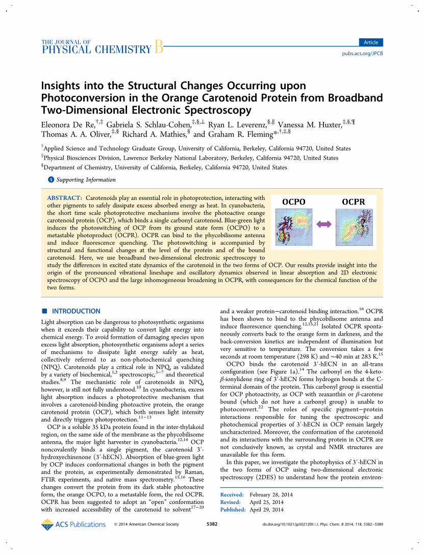
ABSTRACT: Carotenoids play an essential role in photoprotection, interacting withother pigments to safely dissipate excess absorbed energy as heat. In cyanobacteria,the short time scale photoprotective mechanisms involve the photoactive orangecarotenoid protein (OCP), which binds a single carbonyl carotenoid. Blue-green lightinduces the photoswitching of OCP from its ground state form (OCPO) to ametastable photoproduct (OCPR). OCPR can bind to the phycobilisome antennaand induce fluorescence quenching. The photoswitching is accompanied bystructural and functional changes at the level of the protein and of the boundcarotenoid. Here, we use broadband two-dimensional electronic spectroscopy tostudy the differences in excited state dynamics of the carotenoid in the two forms of OCP. Our results provide insight into theorigin of the pronounced vibrational lineshape and oscillatory dynamics observed in linear absorption and 2D electronicspectroscopy of OCPO and the large inhomogeneous broadening in OCPR, with consequences for the chemical function of thetwo forms.
■ INTRODUCTION
Light absorption can be dangerous to photosynthetic organismswhen it exceeds their capability to convert light energy intochemical energy. To avoid formation of damaging species uponexcess light absorption, photosynthetic organisms adopt a seriesof mechanisms to dissipate light energy safely as heat,collectively referred to as non-photochemical quenching(NPQ). Carotenoids play a critical role in NPQ, as validatedby a variety of biochemical,1,2 spectroscopic,3−7 and theoreticalstudies.8,9 The mechanistic role of carotenoids in NPQ,however, is still not fully understood.10 In cyanobacteria, excesslight absorption induces a photoprotective mechanism thatinvolves a carotenoid-binding photoactive protein, the orangecarotenoid protein (OCP), which both senses light intensityand directly triggers photoprotection.11−13
OCP is a soluble 35 kDa protein found in the inter-thylakoidregion, on the same side of the membrane as the phycobilisomeantenna, the major light harvester in cyanobacteria.12,14 OCPnoncovalently binds a single pigment, the carotenoid 3′-hydroxyechinenone (3′-hECN). Absorption of blue-green lightby OCP induces conformational changes in both the pigmentand the protein, as experimentally demonstrated by Raman,FTIR experiments, and native mass spectrometry.15,16 Thesechanges convert the protein from its dark stable photoactiveform, the orange OCPO, to a metastable form, the red OCPR.OCPR has been suggested to adopt an “open” conformationwith increased accessibility of the carotenoid to solvent17−20
and a weaker protein−carotenoid binding interaction.16 OCPRhas been shown to bind to the phycobilisome antenna andinduce fluorescence quenching.12,15,21 Isolated OCPR sponta-neously converts back to the orange form in darkness, and theback-conversion kinetics are independent of illumination butvery sensitive to temperature. The conversion takes a fewseconds at room temperature (298 K) and ∼40 min at 283 K.15
OCPO binds the carotenoid 3′-hECN in an all-transconfiguration (see Figure 1a).14 The carbonyl on the 4-keto-β-ionylidene ring of 3′-hECN forms hydrogen bonds at the C-terminal domain of the protein. This carbonyl group is essentialfor OCP photoactivity, as OCP with zeaxanthin or β-carotenebound (which do not have a carbonyl group) is unable tophotoconvert.22 The roles of specific pigment−proteininteractions responsible for tuning the spectroscopic andphotochemical properties of 3′-hECN in OCP remain largelyuncharacterized. Moreover, the conformation of the carotenoidand its interactions with the surrounding protein in OCPR arenot conclusively known, as crystal and NMR structures areunavailable for this form.In this paper, we investigate the photophysics of 3′-hECN in
the two forms of OCP using two-dimensional electronicspectroscopy (2DES) to understand how the protein environ-
Received: February 28, 2014Revised: April 25, 2014Published: April 29, 2014
Article
pubs.acs.org/JPCB
© 2014 American Chemical Society 5382 dx.doi.org/10.1021/jp502120h | J. Phys. Chem. B 2014, 118, 5382−5389

ment modulates the pigment energetics and dynamics inOCPO and OCPR and relate this to the different biologicalfunction of the two forms (photoactive vs quenching). OCP isan ideal minimal system for the study of protein−pigmentinteractions, as it binds a single carotenoid. In contrast to otherpigment−protein complexes that bind both carotenoids andchlorophyll pigments, the optical response of this minimalsystem is not complicated by inter-chromophore interactions,allowing direct observation of how the protein environmenttunes and alters the properties of the pigment. Carotenoidphotophysics are highly dependent both on the structure of thepigment and on the surrounding environment.23 Therefore,observed differences in the photophysics of the two forms ofOCP must arise from changes in the structure of the carotenoidbound to the protein pocket and in the pigment−proteinbinding interaction.Previous time-resolved fluorescence24 and transient absorp-
tion experiments19,25,26 have investigated the mechanism playedby OCP in energy dissipation. Transient absorption experi-ments in the visible region19,25 suggested that OCPR couldaccept excitation energy from the phycobilisome bilin S1 stateto the carotenoid intramolecular charge transfer (ICT) state.The ICT state can subsequently relax to the ground statedirectly or via the S1 state of 3′-hECN and then decay. Manyopen questions remain about the molecular origin of thedifferent spectroscopic behavior observed for OCPO andOCPR. For example, the origin of the broadness and redshifting of the OCPR linear absorption has not been fullyelucidated, and the origin of the ICT character enhancement inOCPR19,25 is incompletely described.2DES presents numerous advantages over other nonlinear
spectroscopic techniques such as transient absorption (forrecent reviews, see refs 27−30). 2DES affords time and spectralresolution over both excitation and emission processes and hasbeen very successful in unraveling the complicated structure−function relationship of natural photosynthetic systems.31−35
Broadband 2DES probing the visible region allows us toinvestigate and disentangle a spectral region usually congested
in carotenoid photophysics and simultaneously provides thetime resolution to investigate the ultrafast processes associatedwith the excited-state relaxation of carotenoids.36
Our 2DES results for the two forms of OCP are remarkablydifferent. OCPO shows pronounced vibrational dynamics,while OCPR gives a highly inhomogeneously broadened signal.We discuss how the differences in protein environment for thecarotenoid in the two forms might relate to their specificbiological roles (photoactivity and quenching).
■ EXPERIMENTAL METHODS
Sample Preparation. OCP was purified from a frozenpaste of Arthrospira platensis cells (a generous gift of Dr. GeraldCysewski, Cyanotech Corporation) using a procedure similar tothat described by Holt and Krogmann,37 with isoelectricfocusing omitted. Following anion exchange and gel filtrationchromatography, the purified OCP had an A496/A280 ratio of1.8:1. To prepare OCP for spectroscopic measurements,purified OCP in 50 mM Tris-HCl, 100 mM NaCl, pH 8 (24°C) was concentrated to OD 30/cm in a centrifugal spinconcentrator. For the OCPR sample, a 30 μL volume ofconcentrated OCP was initially converted to OCPR by 15 minof illumination with a 505 nm LED (Luxeon Rebel LXML-PE01-0070, Philips Lumileds, 40 nm FWHM) at 273 K. Theilluminated sample was thoroughly mixed with 70 μL of chilledglycerol and illuminated for an additional 5 min. The samplewas then transferred to a 200 μm path length quartz cuvetteand cooled to 77 K in an optical cryostat (Oxford Instruments).The OCPR sample was continuously illuminated with the LEDduring the cooling to cryogenic temperature. The OCPOsample was prepared similarly but in complete darkness. Themaximum OD was 0.28 at 502 nm for the OCPO sample and0.25 at 518 nm for the OCPR sample. Linear absorption traceswere collected before and after every 2DES measurement tocheck for sample stability at 77 K, and no measurable differencewas observed in either sample. We cannot exclude a minorpresence of RCP (red carotenoid protein12,37,38) in our OCPpreparations based on the linear absorption alone. However, its
Figure 1. (a) Structure of the carotenoid 3′-hECN in the protein pocket in OCPO.43 (b) Normalized linear absorption spectra of OCPO (orangeline) and OCPR (red line) at 283 K (dashed line) and at 77 K (solid line). Pictures of the cuvettes containing the samples are shown next to thecorresponding absorption spectra. (c) Energy level scheme of the key transitions within the main molecular pathways for the carotenoid 3′-hECN.The numbered transitions are: (1) S0 → S2 ground-state bleach; (2) S2 → Sm excited-state absorption; (3) S2 emission; (4) S1 excited-stateabsorption. The experimentally observable transitions are described for the two forms of OCP in the text.
The Journal of Physical Chemistry B Article
dx.doi.org/10.1021/jp502120h | J. Phys. Chem. B 2014, 118, 5382−53895383
contribution would be the same in both OCPO and OCPRspectra, and we do not expect it to contribute to any observeddifferences in the dynamics of 3′-hECN in the two forms.Two-Dimensional Electronic Spectroscopy. The 2DES
experimental apparatus has been described in detail pre-viously.36,39,40 A home-built Ti:sapphire regenerative amplifierlaser system pumped a home-built noncollinear opticalparametric amplifier, producing laser pulses centered at 540nm (550 nm for OCPR measurements) with 60 nm FWHM.The combination of a prism compression line followed by adiffraction-based SLM (spatial light modulator) pulse shapercompressed the pulses to a duration of 12 fs, as characterized byTG-FROG (transient grating frequency-resolved opticalgating).41 The beam was split into two replicas by a beamsplitter, delayed with respect to each other via a retroreflectordelay stage; this allowed the control of the waiting time Tbetween pulses 2 and 3. The two beam pairs were further splitinto a total of four beams by a diffractive optic optimized forfirst-order diffraction, allowing for passive phase stabilization.39
The delay between pulses 1 and 2, which corresponds to thecoherence time τ, was implemented by means of movable glasswedges, allowing for interferometric precision in the control ofτ.39
The beams were focused to the sample position in a boxgeometry and had an energy of ∼10 nJ per pulse. Theinteraction with pulses 1, 2, and 3 generates a third-order signalin the phase-matched direction, ks = −k1 + k2 + k3, collinearwith beam 4, the local oscillator (attenuated by 4 orders ofmagnitude as to prevent any strong interaction with thesample). This signal was spectrally dispersed and heterodyne-detected on a CCD camera.The coherence time τ was scanned from −360 to +360 fs in
0.8 fs time steps for fixed values of waiting time T. The resultingarray of interferograms collected for each value of T wasFourier-transformed to produce the final 2DES spectrum.Negative values of the coherence time generated the non-rephasing signal (free induction decay), while positivecoherence times generated the rephasing signal (photon echosignal). The two signals were obtained experimentally byinverting the ordering of pulses 1 and 2. The total 2DESspectrum, the relaxation spectrum, corresponds to thecombined rephasing and non-rephasing components.Dynamics were monitored by varying the waiting time T;
spectra were collected every 10 fs from 0 to 100 fs, then at 120,150, 300, 450, 1000, 2000, 5000, 7000, 10000, 15000, and20000 fs. In order to phase the 2DES data, spectrally resolvedpump−probe experiments were collected separately for thesame values of T under the same experimental conditions.40,42
■ RESULTSLinear Absorption. Figure 1b shows the linear absorption
spectrum of the two forms of OCP at 283 and 77 K. OCPO,represented by an orange line, has resolvable features thatcorrespond to the vibronic structure of the optically bright S0−S2 transition. The 0−0 transition is at 499 nm at 283 K and isred-shifted to 502 nm at 77 K. The OCPO sample appearsorange, as shown in the inset on the left in Figure 1b. Thestructure of 3′-hECN in OCP is shown in Figure 1a.43 Thelinear absorption spectrum at 283 K is asymmetricallybroadened toward longer wavelengths. This broadening isreduced in the 77 K spectrum.Upon blue-green light illumination, the absorption spectrum
broadens and red-shifts. The main peak for the red form OCPR
is at 512 nm at 283 K and red shifts to 522 nm at 77 K. Thevibrational structure that was visible in OCPO is lost, and thespectrum appears nearly Gaussian, as seen in Figure 1b. Noapparent band narrowing or increased resolution of vibronicstructure is observed in the OCPR spectrum at 77 K.
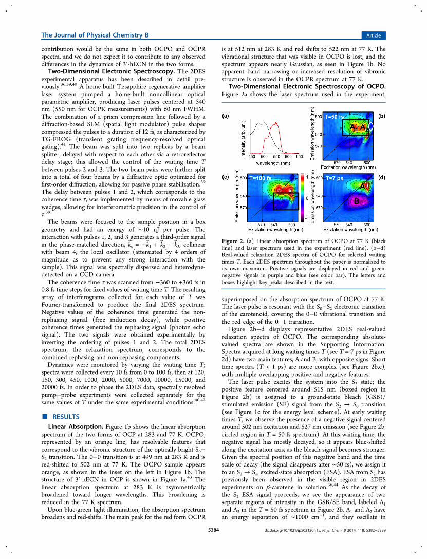
Two-Dimensional Electronic Spectroscopy of OCPO.Figure 2a shows the laser spectrum used in the experiment,
superimposed on the absorption spectrum of OCPO at 77 K.The laser pulse is resonant with the S0−S2 electronic transitionof the carotenoid, covering the 0−0 vibrational transition andthe red edge of the 0−1 transition.Figure 2b−d displays representative 2DES real-valued
relaxation spectra of OCPO. The corresponding absolute-valued spectra are shown in the Supporting Information.Spectra acquired at long waiting times T (see T = 7 ps in Figure2d) have two main features, A and B, with opposite signs. Shorttime spectra (T < 1 ps) are more complex (see Figure 2b,c),with multiple overlapping positive and negative features.The laser pulse excites the system into the S2 state; the
positive feature centered around 515 nm (boxed region inFigure 2b) is assigned to a ground-state bleach (GSB)/stimulated emission (SE) signal from the S2 → S0 transition(see Figure 1c for the energy level scheme). At early waitingtimes T, we observe the presence of a negative signal centeredaround 502 nm excitation and 527 nm emission (see Figure 2b,circled region in T = 50 fs spectrum). At this waiting time, thenegative signal has mostly decayed, so it appears blue-shiftedalong the excitation axis, as the bleach signal becomes stronger.Given the spectral position of this negative band and the timescale of decay (the signal disappears after ∼50 fs), we assign itto an S2 → Sm excited-state absorption (ESA). ESA from S2 haspreviously been observed in the visible region in 2DESexperiments on β-carotene in solution.36,44 As the decay ofthe S2 ESA signal proceeds, we see the appearance of twoseparate regions of intensity in the GSB/SE band, labeled A1and A2 in the T = 50 fs spectrum in Figure 2b. A1 and A2 havean energy separation of ∼1000 cm−1, and they oscillate in
Figure 2. (a) Linear absorption spectrum of OCPO at 77 K (blackline) and laser spectrum used in the experiment (red line). (b−d)Real-valued relaxation 2DES spectra of OCPO for selected waitingtimes T. Each 2DES spectrum throughout the paper is normalized toits own maximum. Positive signals are displayed in red and green,negative signals in purple and blue (see color bar). The letters andboxes highlight key peaks described in the text.
The Journal of Physical Chemistry B Article
dx.doi.org/10.1021/jp502120h | J. Phys. Chem. B 2014, 118, 5382−53895384

intensity with a period of ∼30 fs. The integrated intensity of the2DES relaxation spectra in regions A1 and A2 is plotted as afunction of waiting time T in Figure 3. The oscillation
frequency is close to the frequencies of CC and C−Cstretching of the ground state of the carotenoid 3′-hECN (21and 28 fs, respectively15). We might also expect the intense1008 cm−1 methyl rocking and 980 cm−1 hydrogen-out-of-plane (HOOP) wagging modes observed in prior Ramanstudies15 to contribute to this oscillatory signal, the latter beingunique to 3′-hECN in OCPO. The two peaks A1 and A2 arepart of the same band and decay with the S2 lifetime; therefore,they are most likely due to stimulated emission rather thanground-state bleach processes. They likely oscillate in phase dueto the creation of ground-state wavepackets.Even at early waiting times T, the A band (see Figure 2) is
well rounded, meaning it does not exhibit any diagonalelongation. This can be explained as a very fast randomizationon the S2 potential energy surface of molecules thatunsuccessfully pass through the conical intersection45−47 withthe S1 state and are scattered in a range of trajectories.At later times, the system undergoes ultrafast internal
conversion to the S1 state. A negative feature centered at λt =550 nm (feature B, T = 7 ps spectrum in Figure 2d) appears,corresponding to S1 → SN ESA. The spectra at waiting times Tlarger than ∼2 ps show little evolution of the two main GSB/SE and ESA features. By 20 ps, the signals have entirelydecayed, corresponding to the complete relaxation of thepopulation from S1 to the ground state (spectra not shown).
At early times T, in the region of emission wavelengthsbetween the GSB/SE and the ESA signals (emission wave-length between 535 and 565 nm), we observe the presence ofadditional positive and negative features, overlapping spectrallyand evolving in time. This region is highlighted by a rectangularbox in the T = 100 fs spectrum in Figure 2c. This spectralregion would be difficult to resolve or interpret in a 1Dtransient absorption experiment (which integrates over theexcitation axis), as there are multiple overlapping signals ofopposite sign arising from S2, S1, and possible contributionsfrom other states such as S*.23,48−50 2DES, however, canresolve the evolution of all these features and track them as afunction of waiting time, T.To understand the origin of these peaks, we refer to the
rephasing and non-rephasing components of the early timespectra. Figure 4 shows the rephasing and non-rephasingcomponents of the T = 100 fs spectrum, with a rectangular boxhighlighting the spectral region of interest. The rephasingspectrum shows a similar pattern to the relaxation spectrum inthe boxed region, with alternating positive and negative peaks,while in the non-rephasing component, a single positive peakappears. The peaks are observable in the spectra for waitingtimes shorter than T = 1 ps (data not shown), longer than theS2 lifetime.19 A dispersive lineshape for cross-peaks in therephasing component of 2DES spectra has been previouslyobserved in both theoretical and experimental work.36,44,51 Thisparticular lineshape has been attributed to coupling of anelectronic transition to a high frequency vibration. In 2DESexperiments on β-carotene in solution, an analogous signal hasbeen assigned to the population of a hot ground state viaImpulsive Stimulated Raman Scattering (ISRS).36 Given thesimilarity in dynamics, lineshape and spectral position of thecross-peaks in our spectra, we propose that they arise from theformation of a wavepacket on the hot ground state.
Two-Dimensional Electronic Spectroscopy of OCPR.Figure 5 shows absolute-valued 2DES spectra for selectedwaiting times T for OCPR at 77 K. Figure 6 shows real-valuedspectra for T = 30 fs and 7 ps.52 The laser pulse excites the S0→ S2 transition, and a positive signal appears in the 2D spectra,corresponding to GSB/SE. After excitation, the S2 populationundergoes ultrafast internal conversion to the S1 state, but theS2 lifetime cannot be determined with precision. Due to thespectral position of our laser pulse, we are not able to observethe negative peak corresponding to the S1 → SN excited-stateabsorption signal in our 2D spectra. This ESA signal is in factred-shifted in OCPR and falls outside the bandwidth of ourlaser pulse.19,25 No additional features appear: the GSB/SEsignal decays as the molecule relaxes back to S0, and thisrelaxation is complete by 20 ps. The OCPR 2D spectra are
Figure 3. Evolution in time of the integrated intensity from the 2Dspectra for regions centered at λt = 527 nm, labeled as A1 and A2 inFigure 2b.
Figure 4. 2DES spectra of OCPO for T = 100 fs: relaxation, rephasing, and non-rephasing components.
The Journal of Physical Chemistry B Article
dx.doi.org/10.1021/jp502120h | J. Phys. Chem. B 2014, 118, 5382−53895385
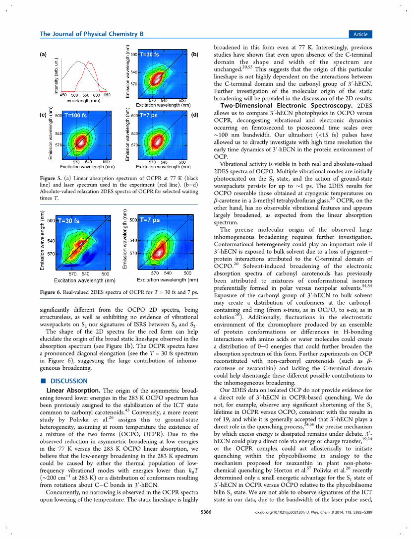
significantly different from the OCPO 2D spectra, beingstructureless, as well as exhibiting no evidence of vibrationalwavepackets on S2 nor signatures of ISRS between S0 and S2.The shape of the 2D spectra for the red form can help
elucidate the origin of the broad static lineshape observed in theabsorption spectrum (see Figure 1b). The OCPR spectra havea pronounced diagonal elongation (see the T = 30 fs spectrumin Figure 6), suggesting the large contribution of inhomo-geneous broadening.
■ DISCUSSIONLinear Absorption. The origin of the asymmetric broad-
ening toward lower energies in the 283 K OCPO spectrum hasbeen previously assigned to the stabilization of the ICT statecommon to carbonyl carotenoids.43 Conversely, a more recentstudy by Polivka et al.26 assigns this to ground-stateheterogeneity, assuming at room temperature the existence ofa mixture of the two forms (OCPO, OCPR). Due to theobserved reduction in asymmetric broadening at low energiesin the 77 K versus the 283 K OCPO linear absorption, webelieve that the low-energy broadening in the 283 K spectrumcould be caused by either the thermal population of low-frequency vibrational modes with energies lower than kBT(∼200 cm−1 at 283 K) or a distribution of conformers resultingfrom rotations about C−C bonds in 3′-hECN.Concurrently, no narrowing is observed in the OCPR spectra
upon lowering of the temperature. The static lineshape is highly
broadened in this form even at 77 K. Interestingly, previousstudies have shown that even upon absence of the C-terminaldomain the shape and width of the spectrum areunchanged.20,53 This suggests that the origin of this particularlineshape is not highly dependent on the interactions betweenthe C-terminal domain and the carbonyl group of 3′-hECN.Further investigation of the molecular origin of the staticbroadening will be provided in the discussion of the 2D results.
Two-Dimensional Electronic Spectroscopy. 2DESallows us to compare 3′-hECN photophysics in OCPO versusOCPR, decongesting vibrational and electronic dynamicsoccurring on femtosecond to picosecond time scales over∼100 nm bandwidth. Our ultrashort (<15 fs) pulses haveallowed us to directly investigate with high time resolution theearly time dynamics of 3′-hECN in the protein environment ofOCP.Vibrational activity is visible in both real and absolute-valued
2DES spectra of OCPO. Multiple vibrational modes are initiallyphotoexcited on the S2 state, and the action of ground-statewavepackets persists for up to ∼1 ps. The 2DES results forOCPO resemble those obtained at cryogenic temperatures onβ-carotene in a 2-methyl tetrahydrofuran glass.36 OCPR, on theother hand, has no observable vibrational features and appearslargely broadened, as expected from the linear absorptionspectrum.The precise molecular origin of the observed large
inhomogeneous broadening requires further investigation.Conformational heterogeneity could play an important role if3′-hECN is exposed to bulk solvent due to a loss of pigment−protein interactions attributed to the C-terminal domain ofOCPO.20 Solvent-induced broadening of the electronicabsorption spectra of carbonyl carotenoids has previouslybeen attributed to mixtures of conformational isomerspreferentially formed in polar versus nonpolar solvents.54,55
Exposure of the carbonyl group of 3′-hECN to bulk solventmay create a distribution of conformers at the carbonyl-containing end ring (from s-trans, as in OCPO, to s-cis, as insolution26). Additionally, fluctuations in the electrostaticenvironment of the chromophore produced by an ensembleof protein conformations or differences in H-bondinginteractions with amino acids or water molecules could createa distribution of 0−0 energies that could further broaden theabsorption spectrum of this form. Further experiments on OCPreconstituted with non-carbonyl carotenoids (such as β-carotene or zeaxanthin) and lacking the C-terminal domaincould help disentangle these different possible contributions tothe inhomogeneous broadening.Our 2DES data on isolated OCP do not provide evidence for
a direct role of 3′-hECN in OCPR-based quenching. We donot, for example, observe any significant shortening of the S1lifetime in OCPR versus OCPO, consistent with the results inref 19, and while it is generally accepted that 3′-hECN plays adirect role in the quenching process,24,56 the precise mechanismby which excess energy is dissipated remains under debate. 3′-hECN could play a direct role via energy or charge transfer,19,24
or the OCPR complex could act allosterically to initiatequenching within the phycobilisome in analogy to themechanism proposed for zeaxanthin in plant non-photo-chemical quenching by Horton et al.57 Polivka et al.26 recentlydetermined only a small energetic advantage for the S1 state of3′-hECN in OCPR versus OCPO relative to the phycobilisomebilin S1 state. We are not able to observe signatures of the ICTstate in our data, due to the bandwidth of the laser pulse used,
Figure 5. (a) Linear absorption spectrum of OCPR at 77 K (blackline) and laser spectrum used in the experiment (red line). (b−d)Absolute-valued relaxation 2DES spectra of OCPR for selected waitingtimes T.
Figure 6. Real-valued 2DES spectra of OCPR for T = 30 fs and 7 ps.
The Journal of Physical Chemistry B Article
dx.doi.org/10.1021/jp502120h | J. Phys. Chem. B 2014, 118, 5382−53895386

and thus we cannot make conclusions on the enhancement ofthe ICT character for 3′-hECN in OCPR nor draw conclusionsregarding its potential involvement in a quenching mechanism.Our data do, however, provide insight into the perturbed localenvironment provided by the protein pocket in OCPO andOCPR.Interestingly, it has not been conclusively demonstrated that
the 4-keto-β-ionylidene ring, nor the ICT state resulting fromthe presence of the conjugated carbonyl group, is necessary forquenching activity in an OCP−phycobilisome complex. Couldthe presence of the 4-keto group be more strictly required forphotoactivity rather than quenching activity? OCP is aphotoswitch, and the unique photochemistry of 3′-hECN inOCPO must be responsible for driving structural changes in theprotein that ultimately lead to formation of the OCPR form,binding of OCP to the phycobilisome, and induction of thequenching mechanism. It is known that interactions betweenthe 4-keto group and absolutely conserved residues in the C-terminal domain (Y203 and W290) are strictly required forOCP photoactivity.22,58 While a relevant modulation of 3′-hECNs conformation(s) and/or photophysical properties inthe OCPR form certainly may occur in the context ofquenching activity, we must also consider the possibility thatthe photophysical properties of OCPR’s 3′-hECN chromo-phore in vitro could simply arise as a secondary consequence ofother mechanistically critical changes in protein structure andchromophore solvent accessibility that occur during thephotochemical mechanism and allow OCP to bind to thephycobilisome. Further studies of the quenching complexformed by OCP bound to the phycobilisome antenna21 areclearly needed to elucidate the specific nature of the quenchingmechanism.
■ CONCLUSIONSHere we have presented 2DES results comparing the excited-state dynamics of 3′-hECN in OCPO and OCPR. The studyinvestigates the photophysics of a carbonyl carotenoid in twodifferent electrostatic environments following photoconversionof the OCP holoprotein. Our results show resolvable and richvibrational dynamics in OCPO, consistent with the carotenoidbeing held in a tightly locked conformation by the proteinenvironment, with consequences for the photoactivity of thisform. OCPR, on the other hand, shows a highly inhomo-geneously broadened behavior. The origin of this largeinhomogeneous broadening can be attributed to conforma-tional heterogeneity due to exposure of the pigment to freesolvent or to variations in the electrostatic environmentexperienced by the carotenoid. Further studies targeted to theinvestigation of the specific pigment−protein interactions in theC-terminal domain of OCP should help elucidate the role ofthe carbonyl group and the protein environment in tuningOCP photochemistry.
■ ASSOCIATED CONTENT*S Supporting InformationAbsolute-valued 2DES spectra for OCPO at selected waitingtimes. This material is available free of charge via the Internet athttp://pubs.acs.org.
■ AUTHOR INFORMATIONCorresponding Author*E-mail: [email protected].
Present Addresses⊥(G.S.S.-C.) Department of Chemistry, Stanford University.∥(R.L.L.) Plant Research Lab, Michigan State University.¶(V.M.H.) Department of Chemistry and Biochemistry,University of Arizona.
NotesThe authors declare no competing financial interest.
■ ACKNOWLEDGMENTS
This work was supported by the Director, Office of Science,Office of Basic Energy Sciences, of the U.S. Department ofEnergy under Contract DE-AC02-05CH11231 and theDivision of Chemical Sciences, Geosciences and BiosciencesDivision, Office of Basic Energy Sciences through Grant DE-AC03-76SF000098 (at LBNL and U.C. Berkeley). The authorsacknowledge J. Zaks for helpful comments on the manuscript.The authors also thank Dr. Gerald Cysewski for the gift of A.platensis cells.
■ REFERENCES(1) Niyogi, K. K.; Bjorkman, O.; Grossman, A. R. The Roles ofSpecific Xanthophylls in Photoprotection. Proc. Natl. Acad. Sci. U.S.A.1997, 94, 14162−14167.(2) Li, Z.; Ahn, T. K.; Avenson, T. J.; Ballottari, M.; Cruz, J. A.;Kramer, D. M.; Bassi, R.; Fleming, G. R.; Keasling, J. D.; Niyogi, K. K.Lutein Accumulation in the Absence of Zeaxanthin RestoresNonphotochemical Quenching in the Arabidopsis thaliana npq1Mutant. Plant Cell 2009, 21, 1798−1812.(3) Ruban, A. V.; Pascal, A. A.; Robert, B.; Horton, P. Activation ofZeaxanthin Is an Obligatory Event in the Regulation of PhotosyntheticLight Harvesting. J. Biol. Chem. 2002, 277, 7785−7789.(4) Ma, Y.-Z.; Holt, N. E.; Li, X.-P.; Niyogi, K. K.; Fleming, G. R.Evidence for Direct Carotenoid Involvement in the Regulation ofPhotosynthetic Light Harvesting. Proc. Natl. Acad. Sci. U.S.A. 2003,100, 4377−4382.(5) Holt, N. E.; Zigmantas, D.; Valkunas, L.; Li, X.-P.; Niyogi, K. K.;Fleming, G. R. Carotenoid Cation Formation and the Regulation ofPhotosynthetic Light Harvesting. Science 2005, 307, 433−436.(6) Ruban, A. V.; Berera, R.; Ilioaia, C.; van Stokkum, I. H. M.;Kennis, J. T. M.; Pascal, A. A.; van Amerongen, H.; Robert, B.; Horton,P.; van Grondelle, R. Identification of a Mechanism of PhotoprotectiveEnergy Dissipation in Higher Plants. Nature 2007, 450, 575−578.(7) Bode, S.; Quentmeier, C. C.; Liao, P.-N.; Hafi, N.; Barros, T.;Wilk, L.; Bittner, F.; Walla, P. J. On the Regulation of Photosynthesisby Excitonic Interactions between Carotenoids and Chlorophylls. Proc.Natl. Acad. Sci. U.S.A. 2009, 106, 12311−12316.(8) Dreuw, A.; Fleming, G. R.; Head-Gordon, M. ChlorophyllFluorescence Quenching by Xanthophylls. Phys. Chem. Chem. Phys.2003, 5, 3247−3256.(9) Dreuw, A.; Fleming, G. R.; Head-Gordon, M. Charge-TransferState as a Possible Signature of a Zeaxanthin-Chlorophyll Dimer in theNon-photochemical Quenching Process in Green Plants. J. Phys.Chem. B 2003, 107, 6500−6503.(10) Ruban, A. V.; Johnson, M. P.; Duffy, C. D. The PhotoprotectiveMolecular Switch in the Photosystem II Antenna. Biochim. Biophys.Acta 2012, 1817, 167−181.(11) Rakhimberdieva, M. G.; Stadnichuk, I. N.; Elanskaya, I. V.;Karapetyan, N. V. Carotenoid-Induced Quenching of the Phycobili-some Fluorescence in Photosystem II-Deficient Mutant of Synecho-cystis sp. FEBS Lett. 2004, 574, 85−88.(12) Wilson, A.; Ajlani, G.; Verbavatz, J.-M.; Vass, I.; Kerfeld, C. A.;Kirilovsky, D. A Soluble Carotenoid Protein Involved in Phyco-bilisome-Related Energy Dissipation in Cyanobacteria. Plant Cell 2006,18, 992−1007.(13) Niyogi, K. K.; Truong, T. B. Evolution of Flexible Non-Photochemical Quenching Mechanisms that Regulate Light Harvest-
The Journal of Physical Chemistry B Article
dx.doi.org/10.1021/jp502120h | J. Phys. Chem. B 2014, 118, 5382−53895387
ing in Oxygenic Photosynthesis. Curr. Opin. Plant Biol. 2013, 16, 307−314.(14) Kerfeld, C. A.; Sawaya, M. R.; Brahmandam, V.; Cascio, D.; Ho,K. K.; Trevithick-Sutton, C. C.; Krogmann, D. W.; Yeates, T. O. TheCrystal Structure of a Cyanobacterial Water-Soluble CarotenoidBinding Protein. Structure 2003, 11, 55−65.(15) Wilson, A.; Punginelli, C.; Gall, A.; Bonetti, C.; Alexandre, M.;Routaboul, J.-M.; Kerfeld, C. A.; van Grondelle, R.; Robert, B.; Kennis,J. T. M.; Kirilovsky, D. A Photoactive Carotenoid Protein Acting asLight Intensity Sensor. Proc. Natl. Acad. Sci. U.S.A. 2008, 105, 12075−12080.(16) Zhang, H.; Liu, H.; Niedzwiedzki, D. M.; Prado, M.; Jiang, J.;Gross, M. L.; Blankenship, R. E. Molecular Mechanism of Photo-activation and Structural Location of the Cyanobacterial OrangeCarotenoid Protein. Biochemistry 2014, 53, 13−19.(17) Wilson, A.; Kinney, J. N.; Zwart, P. H.; Punginelli, C.; D’Haene,S.; Perreau, F.; Klein, M. G.; Kirilovsky, D.; Kerfeld, C. A. StructuralDeterminants Underlying Photoprotection in the Photoactive OrangeCarotenoid Protein of Cyanobacteria. J. Biol. Chem. 2010, 285,18364−18375.(18) Wilson, A.; Gwizdala, M.; Mezzetti, A.; Alexandre, M.; Kerfeld,C. A.; Kirilovsky, D. The Essential Role of the N-Terminal Domain ofthe Orange Carotenoid Protein in Cyanobacterial Photoprotection:Importance of a Positive Charge for Phycobilisome Binding. Plant Cell2012, 24, 1972−1983.(19) Berera, R.; Gwizdala, M.; van Stokkum, I. H. M.; Kirilovsky, D.;van Grondelle, R. Excited States of the Inactive and Active Forms ofthe Orange Carotenoid Protein. J. Phys. Chem. B 2013, 117, 9121−9128.(20) Leverenz, R. L.; Jallet, D.; Mathies, R. A.; Kirilovsky, D.; Kerfeld,C. A. Structural and Functional Modularity of the Orange CarotenoidProtein: Distinct Roles for the N-and C-Terminal Domains inCyanobacterial Photoprotection. Plant Cell 2014, 26, 426−437.(21) Gwizdala, M.; Wilson, A.; Kirilovsky, D. In Vitro Reconstitutionof the Cyanobacterial Photoprotective Mechanism Mediated by theOrange Carotenoid Protein in Synechocystis PCC 6803. Plant Cell2011, 23, 2631−2643.(22) Punginelli, C.; Wilson, A.; Routaboul, J.-M.; Kirilovsky, D.Influence of Zeaxanthin and Echinenone Binding on the Activity of theOrange Carotenoid Protein. Biochim. Biophys. Acta 2009, 1787, 280−288.(23) Polívka, T.; Sundstrom, V. Ultrafast Dynamics of CarotenoidExcited StatesFrom Solution to Natural and Artificial Systems.Chem. Rev. 2004, 104, 2021−2071.(24) Tian, L.; van Stokkum, I. H. M.; Koehorst, R. B. M.; Jongerius,A.; Kirilovsky, D.; van Amerongen, H. Site, Rate, and Mechanism ofPhotoprotective Quenching in Cyanobacteria. J. Am. Chem. Soc. 2011,133, 18304−18311.(25) Berera, R.; van Stokkum, I. H. M.; Gwizdala, M.; Wilson, A.;Kirilovsky, D.; van Grondelle, R. The Photophysics of the OrangeCarotenoid Protein, a Light-Powered Molecular Switch. J. Phys. Chem.B 2012, 116, 2568−2574.(26) Polívka, T.; Chabera, P.; Kerfeld, C. A. Carotenoid−ProteinInteraction Alters the S1 Energy of Hydroxyechinenone in the OrangeCarotenoid Protein. Biochim. Biophys. Acta 2013, 1827, 248−254.(27) Ginsberg, N. S.; Cheng, Y.-C.; Fleming, G. R. Two-DimensionalElectronic Spectroscopy of Molecular Aggregates. Acc. Chem. Res.2009, 42, 1352−1363.(28) Schlau-Cohen, G. S.; Ishizaki, A.; Fleming, G. R. Two-Dimensional Electronic Spectroscopy and Photosynthesis: Fundamen-tals and Applications to Photosynthetic Light-Harvesting. Chem. Phys.2011, 386, 1−22.(29) Lewis, K. L. M.; Ogilvie, J. P. Probing Photosynthetic Energyand Charge Transfer with Two-Dimensional Electronic Spectroscopy.J. Phys. Chem. Lett. 2012, 3, 503−510.(30) Schlau-Cohen, G.; Dawlaty, J.; Fleming, G. Ultrafast Multi-dimensional Spectroscopy: Principles and Applications to Photo-synthetic Systems. IEEE J. Sel. Top. Quantum Electron. 2012, 18, 283−295.
(31) Read, E. L.; Schlau-Cohen, G. S.; Engel, G. S.; Georgiou, T.;Papiz, M. Z.; Fleming, G. R. Pigment Organization and Energy LevelStructure in Light-Harvesting Complex 4: Insights from Two-Dimensional Electronic Spectroscopy. J. Phys. Chem. B 2009, 113,6495−6504.(32) Schlau-Cohen, G. S.; Calhoun, T. R.; Ginsberg, N. S.; Read, E.L.; Ballottari, M.; Bassi, R.; van Grondelle, R.; Fleming, G. R. Pathwaysof Energy Flow in LHCII from Two-Dimensional ElectronicSpectroscopy. J. Phys. Chem. B 2009, 113, 15352−15363.(33) Schlau-Cohen, G. S.; Calhoun, T. R.; Ginsberg, N. S.; Ballottari,M.; Bassi, R.; Fleming, G. R. Spectroscopic Elucidation of UncoupledTransition Energies in the Major Photosynthetic Light-HarvestingComplex, LHCII. Proc. Natl. Acad. Sci. U.S.A. 2010, 107, 13276−13281.(34) Ginsberg, N. S.; Davis, J. A.; Ballottari, M.; Cheng, Y.-C.; Bassi,R.; Fleming, G. R. Solving Structure in the CP29 Light HarvestingComplex with Polarization-Phased 2D Electronic Spectroscopy. Proc.Natl. Acad. Sci. U.S.A. 2011, 108, 3848−3853.(35) Schlau-Cohen, G. S.; De Re, E.; Cogdell, R. J.; Fleming, G. R.Determination of Excited-State Energies and Dynamics in the B Bandof the Bacterial Reaction Center with 2D Electronic Spectroscopy. J.Phys. Chem. Lett. 2012, 3, 2487−2492.(36) Calhoun, T. R.; Davis, J. A.; Graham, M. W.; Fleming, G. R. TheSeparation of Overlapping Transitions in β-Carotene with Broadband2d Electronic Spectroscopy. Chem. Phys. Lett. 2012, 523, 1−5.(37) Holt, T. K.; Krogmann, D. W. A Carotenoid−Protein fromCyanobacteria. Biochim. Biophys. Acta 1981, 637, 408−414.(38) Wu, Y. P.; Krogmann, D. W. The Orange Carotenoid Protein ofSynechocystis PCC 6803. Biochim. Biophys. Acta 1997, 1322, 1−7.(39) Brixner, T.; Stiopkin, I. V.; Fleming, G. R. Tunable Two-Dimensional Femtosecond Spectroscopy. Opt. Lett. 2004, 29, 884−886.(40) Brixner, T.; Mancal, T.; Stiopkin, I. V.; Fleming, G. R. Phase-Stabilized Two-Dimensional Electronic Spectroscopy. J. Chem. Phys.2004, 121, 4221−4236.(41) Trebino, R.; DeLong, K. W.; Fittinghoff, D. N.; Sweetser, J. N.;Krumbugel, M. A.; Richman, B. A.; Kane, D. J. Measuring UltrashortLaser Pulses in The Time-Frequency Domain Using Frequency-Resolved Optical Gating. Rev. Sci. Instrum. 1997, 68, 3277−3295.(42) Hybl, J. D.; Ferro, A. A.; Jonas, D. M. Two-Dimensional FourierTransform Electronic Spectroscopy. J. Chem. Phys. 2001, 115, 6606−6622.(43) Polívka, T.; Kerfeld, C. A.; Pascher, T.; Sundstrom, V.Spectroscopic Properties of the Carotenoid 3′-Hydroxyechinenonein the Orange Carotenoid Protein from the CyanobacteriumArthrospira maxima. Biochemistry 2005, 44, 3994−4003.(44) Christensson, N.; Milota, F.; Nemeth, A.; Sperling, J.;Kauffmann, H. F.; Pullerits, T.; Hauer, J. Two-Dimensional ElectronicSpectroscopy of β-Carotene. J. Phys. Chem. B 2009, 113, 16409−16419.(45) Garavelli, M.; Celani, P.; Bernardi, F.; Robb, M. A.; Olivucci, M.Force Fields for “Ultrafast” Photochemistry: The S2 (1Bu) → S1 (2Ag)→ S0 (1Ag) Reaction Path for all-trans-Hexa-1,3,5-triene. J. Am. Chem.Soc. 1997, 119, 11487−11494.(46) Fuss, W.; Haas, Y.; Zilberg, S. Twin States and ConicalIntersections in Linear Polyenes. Chem. Phys. 2000, 259, 273−295.(47) McCamant, D. W.; Kukura, P.; Mathies, R. A. FemtosecondTime-Resolved Stimulated Raman Spectroscopy: Application to theUltrafast Internal Conversion in β-Carotene. J. Phys. Chem. A 2003,107, 8208−8214.(48) Gradinaru, C. C.; Kennis, J. T.; Papagiannakis, E.; van Stokkum,I. H.; Cogdell, R. J.; Fleming, G. R.; Niederman, R. A.; van Grondelle,R. An Unusual Pathway of Excitation Energy Deactivation inCarotenoids: Singlet-to-Triplet Conversion on an Ultrafast Timescalein a Photosynthetic Antenna. Proc. Natl. Acad. Sci. U.S.A. 2001, 98,2364−2369.(49) Wohlleben, W.; Buckup, T.; Hashimoto, H.; Cogdell, R. J.;Herek, J. L.; Motzkus, M. Pump-Deplete-Probe Spectroscopy and the
The Journal of Physical Chemistry B Article
dx.doi.org/10.1021/jp502120h | J. Phys. Chem. B 2014, 118, 5382−53895388
Puzzle of Carotenoid Dark States. J. Phys. Chem. B 2004, 108, 3320−3325.(50) Polívka, T.; Sundstrom, V. Dark Excited States of Carotenoids:Consensus and Controversy. Chem. Phys. Lett. 2009, 477, 1−11.(51) Mancal, T.; Nemeth, A.; Milota, F.; Lukes, V.; Kauffmann, H. F.;Sperling, J. Vibrational Wave Packet Induced Oscillations in Two-Dimensional Electronic Spectra. II. Theory. J. Chem. Phys. 2010, 132,184515.(52) It was only possible to retrieve the phase factor from pump−probe data for 2DES spectra at select waiting times T, as shown inFigure 6. For comparison, absolute-valued OCPO spectra are shown inthe Supporting Information.(53) Chabera, P.; Durchan, M.; Shih, P. M.; Kerfeld, C. A.; Polívka,T. Excited-State Properties of the 16 kDa Red Carotenoid Proteinfrom Arthrospira maxima. Biochim. Biophys. Acta 2011, 1807, 30−35.(54) Shima, S.; Ilagan, R. P.; Gillespie, N.; Sommer, B. J.; Hiller, R.G.; Sharples, F. P.; Frank, H. A.; Birge, R. R. Two-Photon andFluorescence Spectroscopy and the Effect of Environment on thePhotochemical Properties of Peridinin in Solution and in thePeridinin-Chlorophyll-Protein from Amphidinium carterae. J. Phys.Chem. A 2003, 107, 8052−8066.(55) Enriquez, M. M.; Fuciman, M.; LaFountain, A. M.; Wagner, N.L.; Birge, R. R.; Frank, H. A. The Intramolecular Charge Transfer Statein Carbonyl-Containing Polyenes and Carotenoids. J. Phys. Chem. B2010, 114, 12416−12426.(56) Tian, L.; Gwizdala, M.; van Stokkum, I.; Koehorst, R.;Kirilovsky, D.; van Amerongen, H. Picosecond Kinetics of LightHarvesting and Photoprotective Quenching in Wild-Type and MutantPhycobilisomes Isolated from the Cyanobacterium Synechocystis PCC6803. Biophys. J. 2012, 102, 1692−1700.(57) Horton, P.; Ruban, A. V.; Wentworth, M. Allosteric Regulationof the Light-Harvesting System of Photosystem II. Philos. Trans. R.Soc., B 2000, 355, 1361−1370.(58) Wilson, A.; Punginelli, C.; Couturier, M.; Perreau, F.; Kirilovsky,D. Essential Role of Two Tyrosines and Two Tryptophans on thePhotoprotection Activity of the Orange Carotenoid Protein. Biochim.Biophys. Acta 2011, 1807, 293−301.
The Journal of Physical Chemistry B Article
dx.doi.org/10.1021/jp502120h | J. Phys. Chem. B 2014, 118, 5382−53895389