inorganic scintillators in positron emission...
TRANSCRIPT

INORGANIC SCINTILLATORS IN POSITRON EMISSION
TOMOGRAPHY
CAREL W.E. VAN EIJK*
Delft University of Technology, Mekelweg 15, 2629 JB Delft,
The Netherlands
Abstract. At present large activity is going on in developing positron emission tomography (PET) systems with better specifications. In recent years a number of new gamma-ray scintillators has become commercially available. These new materials were either derived from earlier known scintillators, e.g. Lu1-x YxAlO3:Ce (LuYAP) and Lu2(1-x)Y2xSiO5:Ce(LYSO), or are the result of new discoveries, such as LaCl3:Ce and LaBr3:Ce. The first two materials are primarily of interest for PET because of the relatively high sensitivity for gamma rays and fast response time. The halide scintillators show an energy resolution of ~3% at 662 keV, which is unprecedented for scintillators, a very high light yield and a fast response time. This combination makes LaBr3:Ce an attractive scintillator for time-of-flight (TOF) PET, in spite of the poorer intrinsic sensitivity for annihilation radiation. At the same time the search for and research on new materials is going on. For example LuI3:Ce is a new material with a very high light yield (~90,000 photons per MeV). Both old and new scintillators are considered for application in new PET systems. A review will be presented.
Keywords: gamma-ray scintillators; LuYAP; LYSO; LaCl3:Ce; LaBr3:Ce;LuI3:Ce ; medical diagnostics ; positron emission tomography ; Depth of Interaction ; Time-of-Flight PET
______* To whom correspondence should be addressed. Carel W.E. van Eijk, Radiation Detection & Matter,
R3, Faculty of Applied Sciences, Delft University of Technology, Mekelweg 15, 2629 JB Delft, The Netherlands; e-mail: [email protected]
259
S. Tavernier et al. (eds.), Radiation Detectors for Medical Applications, 259–274.
© 2006 Springer.

C.W.E. VAN EIJK260
1. Introduction
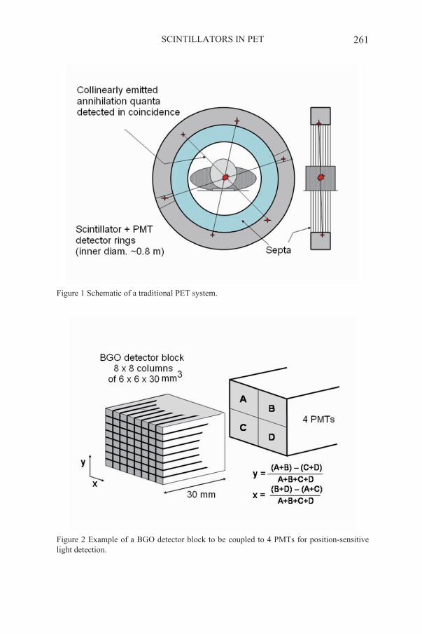
Positron Emission Tomography (PET) is a very powerful medical-diagnostic method for functional imaging.1 Imaging is realized by means of the two 511 keV quanta which are emitted approximately collinearly when a positron, emitted by a radiopharmaceutical introduced into a patient, annihilates in tissue. The two quanta are detected position sensitively in coincidence. See Fig. 1. The point of positron emission is approximately situated on the line of response (LOR) connecting the two positions of detection. Many annihilations give many LORs and from these the radiopharmaceutical distribution can be reconstructed.
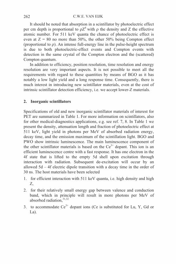
For position-sensitive detection a PET system consists in general of many rings with altogether hundreds of scintillation-detector blocks. See Figures 1 and 2. As the detection efficiency of the 511 keV annihilation quanta plays a very important role, for a long time the BGO scintillator (dense, high atomic number, with large probability of photoelectric effect for 511 keV quanta; see Table 1) was state of the art. In general BGO blocks with saw-cuts are applied (Fig. 2). Depending on the system these cuts provide 36 to 64 crystal columns of e.g. 4 x 4 or 6 x 6 mm2 cross section, coupled at the base in such a way that the scintillation-light distribution allows the determination of the column hit by a radiation quantum using four photomultiplier tubes (PMTs) or two PMTs with a dual structure, and Anger-type logic.2-4 Detector blocks are combined in rings to form typically 24 - 48 planes of scintillator columns. Using coincidences between scintillator columns in the same plane and cross-plane coincidences between columns in adjacent planes, image slices can be obtained at a pitch of half the plane distance.
Originally, planes were separated by septa, i.e. lead collimator plates. In an actual PET scan most of the 511 keV quanta are Compton scattered in the patient. This gives a continuous background. The septa reduce this background significantly. With septa, cross-plane 511 keV - 511 keV coincidences are limited to differences of ~3 planes (two-dimensional imaging; 2D). Modern systems operate with the septa removed and coincidences are accepted between many planes resulting in three-dimensional (3D) imaging.5,6 To some extent the improved coincidence detection efficiency, resulting from the increased solid angle, compensates the increased Compton-scattering background. The latter is reduced also by accepting only photopeak events of the pulse-height spectrum (the better the resolution, the better the reduction will be; the typical energy resolution for BGO is ~20% FWHM). Then, however, events with 511 keV quanta interacting in a scintillator by Compton effect will be eliminated as well. Consequently, depending on the specific use one has to decide whether a narrow photopeak window is used or a more relaxed window.
SCINTILLATORS IN PET 261
Figure 1 Schematic of a traditional PET system.
Figure 2 Example of a BGO detector block to be coupled to 4 PMTs for position-sensitive light detection.
C.W.E. VAN EIJK262
It should be noted that absorption in a scintillator by photoelectric effect per cm depth is proportional to Z4 with the density and Z the effective atomic number. For 511 keV quanta the chance of photoelectric effect is even at Z = 80 no more than 50%, the other 50% being Compton effect (proportional to ). An intense full-energy line in the pulse-height spectrum is due to both photoelectric-effect events and Compton events with detection in the same crystal of the Compton electron and the (scattered) Compton quantum.
In addition to efficiency, position resolution, time resolution and energy resolution are very important aspects. It is not possible to meet all the requirements with regard to these quantities by means of BGO as it has notably a low light yield and a long response time. Consequently, there is much interest in introducing new scintillator materials, even at the cost of intrinsic scintillator detection efficiency, i.e. we accept lower-Z materials.
2. Inorganic scintillators
Specifications of old and new inorganic scintillator materials of interest for PET are summarized in Table 1. For more information on scintillators, also for other medical-diagnostics applications, e.g. see ref. 7, 8. In Table 1 we present the density, attenuation length and fraction of photoelectric effect at 511 keV, light yield in photons per MeV of absorbed radiation energy, decay time, and the emission maximum of the scintillation light. BGO and PWO show intrinsic luminescence. The main luminescence component of the other scintillator materials is based on the Ce3+ dopant. This ion is an efficient luminescence centre with a fast response. It has one electron in the 4f state that is lifted to the empty 5d shell upon excitation through interaction with radiation. Subsequent de-excitation will occur by an allowed 5d – 4f electric dipole transition with a decay time in the order of 30 ns. The host materials have been selected
1. for efficient interaction with 511 keV quanta, i.e. high density and high Z,
2. for their relatively small energy gap between valence and conduction band, which in principle will result in more photons per MeV of absorbed radiation,31,32
3. to accommodate Ce3+ dopant ions (Ce is substituted for Lu, Y, Gd or La).

SCINTILLATORS IN PET 263
Table 1 PET-scintillator candidates
These selection criteria do not guarantee efficient scintillation. On interaction with radiation energetic electrons are produced. These in turn produce more electrons and holes. After thermalization these have to travel to the luminescence centres. The efficiency of the transport is difficult to predict. There can easily be loss due to all kinds of defects in the crystals. The light yields in Table 1 are mainly defined by this loss. In the PWO compounds a very small fraction of the deposited energy results in
density
g/cm3
attenuation
length at
511 keV
mm
photoel
effect%
light
yield
phot/
MeV
dec
time
ns
emission
max
nm
ref
Bi4Ge3O12
(BGO)
7.1 10.4 40 9,000 300 480 9,10
Lu2SiO5:Ce
(LSO)
7.4 11.4 32 26,000 40 420 11-12
LYSO 13
LuAlO3:Ce
(LuAP)
8.3 10.5 30 11,000 18 365 14-18
LuYAP 8,000 21
(65%)
19
Lu2Si2O7:Ce
(LPS)
6.2 14.1 29 20,000 30 380 20
Lu2S3:Ce 6.2 13.8 28,000 32 590 21
Gd2SiO5:Ce
(GSO)
6.7 14.1 25 8,000 60 440 22, 23
PbWO4 (PWO) 8.3 8.7 42 200 15 420 24
PWO: Mo,Y 600 ~15 ~500 25
PWO :Mo,Nb 400 ~10-
103
~500 26
LaCl3:Ce 3.86 28.0 14.7 46,000 25
(65%)
350 27
LaBr3:Ce (5-
30%)
5.07 22.3 13.1 70,000 16
(97%)
380 28
LuI3:Ce (5%) 5.6 18.2 28 90,000 6-140
(72%)
103
(28%)
472, 535 29,30
C.W.E. VAN EIJK264
luminescence. Only the two scintillators at the bottom have a light yield close to the maximum achievable for those materials32.
From the third column we can derive that a 30 mm deep BGO crystal has an interaction probability of 93% for 511 keV quanta. Of this 40% is photoelectric effect and 53% Compton effect. Obviously in a PET system a fraction of the Compton-scattered quanta will escape and be detected in neighbouring crystals. This will lead to deterioration of the position resolution. Compton scattering into the forward direction has the highest probability (angle with direction of incoming 511 keV quantum ~ 32 degrees). Observation of individual crystal responses will help to reduce the deterioration.
LSO/LYSO and LuAP/LuYAP are very interesting candidates to replace BGO in PET. Their attenuation lengths are comparable to that of BGO, though the probability of photoelectric effect is smaller, i.e. that of Compton effect is higher. Furthermore they have a higher light yield and a much faster response.
Originally it appeared to be difficult to grow large stress-free LSO crystals (~ 1,000 cm3) of which small entities can be cut efficiently. These large crystals were reported to be inhomogeneous in light production and the gamma-ray energy resolution is poorer than expected on basis of the light yield.12 An experimental gamma camera was introduced by Siemens/CTI (Knoxville, USA) with a layer of small columns of LSO crystals and it appears that the quality has improved significantly. LSO is now used in Siemens/CTI PET systems.33,34 LSO is available from Siemens/CTI.
LuAP was first proposed as a scintillator in 1994,14 and more detailed papers appeared in 1995.15,16 It is difficult to grow LuAP scintillation crystals. The temperature range in the phase diagram to produce LuAP is very small and one ends up easily with Lu3Al5O12 (LuAG). Yet, several groups, e.g. Crytur Ltd. (Turnov, Czech Republic) and A.G. Petrosyan (Armenian National Academy of Science), were able to supply LuAP:Ce crystals for research.17,18 Another problem of LuAP is the strong scintillation-light absorption. Some groups tried to introduce improvements and to facilitate the crystal-growing process by adding Gd or Y.17 Light yields of ~ 1 - 2 x 10⁴ photons/MeV are reported for these mixed crystals. In case of Gd admixing, in general longer (~ 100 ns) decay time components are introduced. The Crystal Clear Collaboration at CERN has introduced Lu~0.8Y~0.2AlO3:Ce in a small-animal PET system.19,35
Other relatively new scintillators with a high light yield and a fast response time are LPS (lutetium pyro silicate) and Lu2S3:Ce. Although the attenuation lengths of these scintillators are larger than that of BGO they appear to have slightly better properties than GSO, the latter being used in PET by Philips. GSO is commercially available from Hitachi. Of LPS only small samples have been grown so far. R&D of this material is in progress.

SCINTILLATORS IN PET 265
Lu2S3:Ce is in particular of interest because of its emission at 590 nm, which matches the light sensitivity of silicon diodes perfectly. Only small pieces of crystal have been grown of this scintillator.21
It should be noticed that application of scintillators containing lutetium has two disadvantages a) the high price of ~ $ 50/cm3 due to Lu, and b) the presence of the radioactive isotope 176Lu which gives a count rate of ~ 300/s.cm3 (beta decay, end point of 565 keV, some gamma rays). For PET b) is less important.
In the framework of the Crystal Clear Collaboration at CERN, PbWO4
(PWO) crystals have been developed for application in experimental set-ups at the future large hadron collider24. At present the production of ~ 100,000 crystals of ~ 2 x 2 x 20 cm3 is in progress. Obviously it would be attractive to employ such good quality crystals in other fields. From Table 1 we learn that the detection efficiency of PWO at 511 keV is excellent. However, the light yield of PWO is extremely small and this shuts the door to application in PET systems. Several groups have started programmes to answer the question of how to increase the light yield by, say, an order of magnitude. Some results are shown in Table 1. So far by means of doping an increase of fast luminescence appears possible by a factor of ~2 - 3.
Of the three new scintillators listed at the bottom of Table 1, LaCl3:Ceand LaBr3:Ce are unique for their excellent energy resolution of ~3% at 662 keV (see Fig. 3), combined with a high light yield and fast response time.27,28 The efficiency is not as good as that of the other materials and these scintillators are hygroscopic. Yet, as will be discussed in section 4, LaBr3:Ce is of interest for application in PET. LaCl3:Ce and LaBr3:Ce are commercially available from Saint Gobain Crystals and Detectors. LuI3:Ceis still being studied. Also for this material an excellent energy resolution appears feasible. However, it is more hygroscopic than the other two lanthanum halides.
Figure 3 Pulse height spectrum of 137Cs 662 keV gamma rays recorded with diam. 1 x 1 inch LaBr3:Ce crystal showing 3.1% energy resolution FWHM.

C.W.E. VAN EIJK266
3.
On emission a positron travels a distance in the order of 1 mm before it has thermalized and annihilates with an electron. In annihilating the positron and electron have each a small momentum, resulting in a small deviation of the angle between the annihilation quanta from 180 degrees. These two effects result in an intrinsic position resolution for a 0.8 m diameter system of approximately 2 mm FWHM. This value decreases with decreasing system diameter.
Present PET systems have a position resolution of typically ~ 4 mm FWHM in the centre of the system, increasing to ~ 5 - 6 mm if we move in the axial direction to the edge of the system or in the radial direction to, say, 10 cm from the axis.
If not Compton scattered in the patient, annihilation quanta emitted from the central axis of a PET system and in the plane of a detector ring (perpendicular to the axis) will hit a scintillator column in a direction more or less parallel to the length of the column. Quanta emitted off-centre and/or in a tilted plane will hit a column under an angle. They may go through the first encountered column and be detected in a neighbouring column well behind the entrance window. The depth of interaction is not known and consequently a parallax error arises, called radial elongation due to its manifestation upon image reconstruction. This effect decreases with increasing system diameter, i.e. it leads to a requirement opposite to that of the first paragraph of this section. Furthermore, for parallax reduction the length of the scintillator column should be as small as possible. This is in contradiction with the crystal-depth requirement for efficient detection. E.g. for BGO the typical length is 30 mm. This explains efforts to introduce methods that give depth-of-interaction (DOI) information.36-38. This is the more important as the position resolution we are aiming at in newer brain-imaging systems is at the level of 2 - 3 mm and for small-animal PET systems even at ~1.5 mm. Then the cross section of the crystal columns should be similar in size, i.e. about 2 x 2 mm2. As we will discuss below, a detector consisting of a monolithic scintillation crystal of much larger size and position-sensitive light detection may be an option as well.
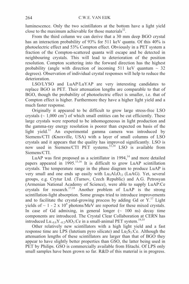
In most DOI studies/applications two or three small columnar crystals of different scintillators, e.g. LSO + GSO38, LSO + GSO + BGO37, LSO + LuAP (see Fig. 4) and LYSO + LuYAP35, are coupled to form one column. The (quantized) DOI information is obtained by using the difference in pulse shape. LSO + LSO with different decay times has been applied in the High Resolution Research Tomograph (HRRT) of Siemens/CTI.34 LSO crystals of 7.5 mm x 2.1 mm x 2.1 mm are used. The spread in decay times from ~36 to ~48 ns in the grown LSO crystals allowed selection of crystals
Position resolution and depth of interaction
SCINTILLATORS IN PET 267
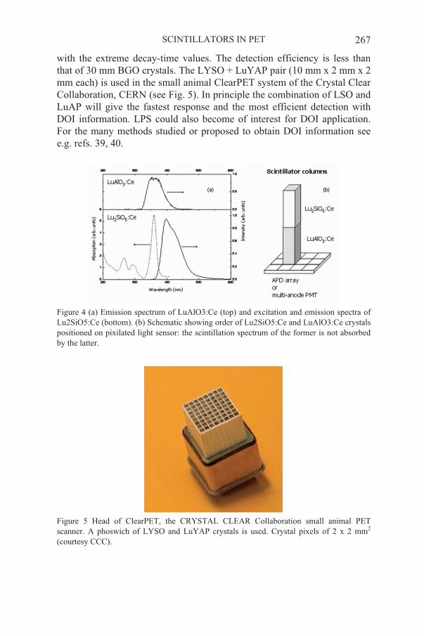
with the extreme decay-time values. The detection efficiency is less than that of 30 mm BGO crystals. The LYSO + LuYAP pair (10 mm x 2 mm x 2 mm each) is used in the small animal ClearPET system of the Crystal Clear Collaboration, CERN (see Fig. 5). In principle the combination of LSO and LuAP will give the fastest response and the most efficient detection with DOI information. LPS could also become of interest for DOI application. For the many methods studied or proposed to obtain DOI information see e.g. refs. 39, 40.
Figure 4 (a) Emission spectrum of LuAlO3:Ce (top) and excitation and emission spectra of Lu2SiO5:Ce (bottom). (b) Schematic showing order of Lu2SiO5:Ce and LuAlO3:Ce crystals positioned on pixilated light sensor: the scintillation spectrum of the former is not absorbed by the latter.
Figure 5 Head of ClearPET, the CRYSTAL CLEAR Collaboration small animal PET scanner. A phoswich of LYSO and LuYAP crystals is used. Crystal pixels of 2 x 2 mm2
(courtesy CCC).
C.W.E. VAN EIJK268
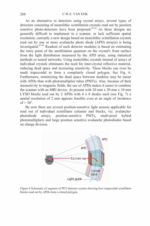
As an alternative to detectors using crystal arrays, several types of detectors consisting of monolithic scintillation crystals read out by position sensitive photo-detectors have been proposed.41,42 As these designs are generally difficult to implement in a scanner, or lack sufficient spatial resolution, currently a new design based on monolithic scintillation crystals read out by one or more avalanche photo diode (APD) array(s) is being investigated.43-45 Readout of such detector modules is based on estimating the entry point of the annihilation quantum on the crystal's front surface from the light distribution measured by the APD array, using statistical methods or neural networks. Using monolithic crystals instead of arrays of individual crystals eliminates the need for inter-crystal reflective material, reducing dead space and increasing sensitivity. These blocks can even be made trapezoidal to form a completely closed polygon. See Fig. 6. Furthermore, minimizing the dead space between modules may be easier with APDs than with photomultiplier tubes (PMTs). Also, because of their insensitivity to magnetic fields, the use of APDs makes it easier to combine the scanner with an MRI device. At present with 20 mm x 20 mm x 10 mm LYSO blocks read out by 2 APDs with 4 x 8 diodes each (see Fig. 7) a spatial resolution of 2 mm appears feasible even at an angle of incidence of > 30o.
By now there are several position-sensitive light sensors applicable for read out of individual scintillator columns and blocks, viz. avalanche-photodiode arrays, position-sensitive PMTs, multi-pixel hybrid photomultipliers and large position sensitive avalanche photodiodes based on charge division.
Figure 6 Schematic of segment of PET-detector system showing how trapezoidal scintillator blocks read out by APDs form a closed polygon.
SCINTILLATORS IN PET 269
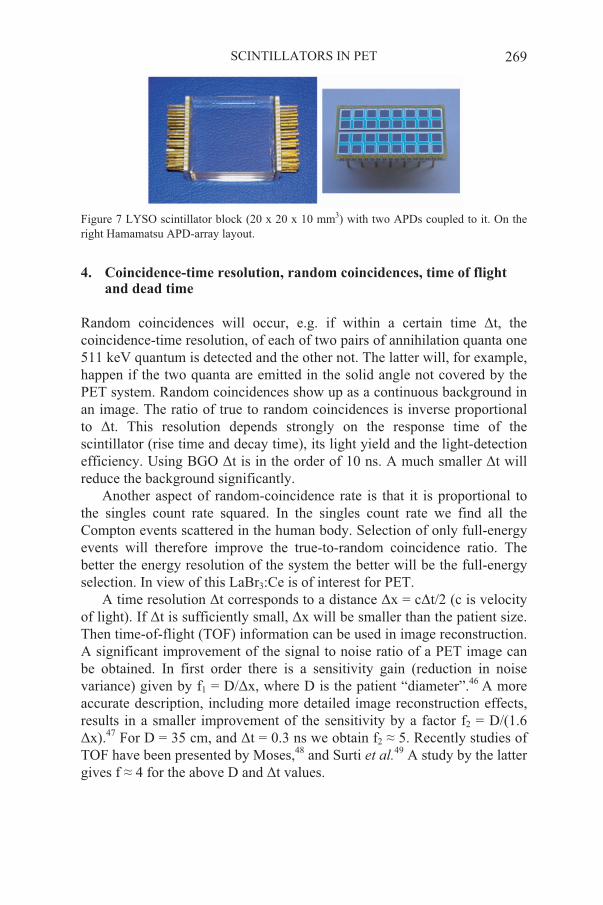
Figure 7 LYSO scintillator block (20 x 20 x 10 mm3) with two APDs coupled to it. On the right Hamamatsu APD-array layout.
4. and dead time
Random coincidences will occur, e.g. if within a certain time t, the coincidence-time resolution, of each of two pairs of annihilation quanta one 511 keV quantum is detected and the other not. The latter will, for example, happen if the two quanta are emitted in the solid angle not covered by the PET system. Random coincidences show up as a continuous background in an image. The ratio of true to random coincidences is inverse proportional to t. This resolution depends strongly on the response time of the scintillator (rise time and decay time), its light yield and the light-detection efficiency. Using BGO t is in the order of 10 ns. A much smaller t will reduce the background significantly.
Another aspect of random-coincidence rate is that it is proportional to the singles count rate squared. In the singles count rate we find all the Compton events scattered in the human body. Selection of only full-energy events will therefore improve the true-to-random coincidence ratio. The better the energy resolution of the system the better will be the full-energy selection. In view of this LaBr3:Ce is of interest for PET.
A time resolution t corresponds to a distance x = c t/2 (c is velocity of light). If t is sufficiently small, x will be smaller than the patient size. Then time-of-flight (TOF) information can be used in image reconstruction. A significant improvement of the signal to noise ratio of a PET image can be obtained. In first order there is a sensitivity gain (reduction in noise variance) given by f1 = D/ x, where D is the patient “diameter”.46 A more accurate description, including more detailed image reconstruction effects, results in a smaller improvement of the sensitivity by a factor f2 = D/(1.6
x).47 For D = 35 cm, and t = 0.3 ns we obtain f2 5. Recently studies of TOF have been presented by Moses,48 and Surti et al.49 A study by the latter gives f 4 for the above D and t values.
Coincidence-time resolution, random coincidences, time of flight
C.W.E. VAN EIJK270
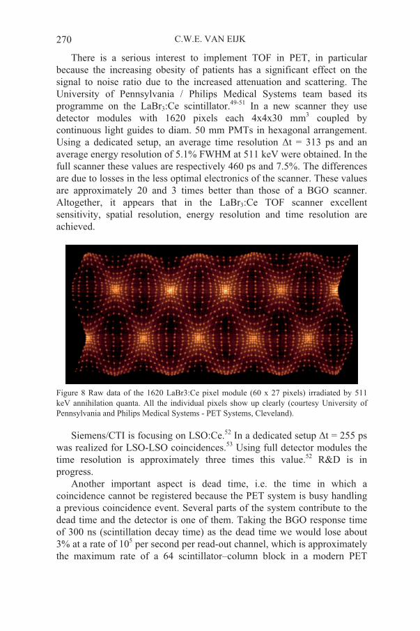
There is a serious interest to implement TOF in PET, in particular because the increasing obesity of patients has a significant effect on the signal to noise ratio due to the increased attenuation and scattering. The University of Pennsylvania / Philips Medical Systems team based its programme on the LaBr3:Ce scintillator.49-51 In a new scanner they use detector modules with 1620 pixels each 4x4x30 mm3 coupled by continuous light guides to diam. 50 mm PMTs in hexagonal arrangement. Using a dedicated setup, an average time resolution t = 313 ps and an average energy resolution of 5.1% FWHM at 511 keV were obtained. In the full scanner these values are respectively 460 ps and 7.5%. The differences are due to losses in the less optimal electronics of the scanner. These values are approximately 20 and 3 times better than those of a BGO scanner. Altogether, it appears that in the LaBr3:Ce TOF scanner excellent sensitivity, spatial resolution, energy resolution and time resolution are achieved.
Figure 8 Raw data of the 1620 LaBr3:Ce pixel module (60 x 27 pixels) irradiated by 511 keV annihilation quanta. All the individual pixels show up clearly (courtesy University of Pennsylvania and Philips Medical Systems - PET Systems, Cleveland).
Siemens/CTI is focusing on LSO:Ce.52 In a dedicated setup t = 255 ps was realized for LSO-LSO coincidences.53 Using full detector modules the time resolution is approximately three times this value.52 R&D is in progress.
Another important aspect is dead time, i.e. the time in which a coincidence cannot be registered because the PET system is busy handling a previous coincidence event. Several parts of the system contribute to the dead time and the detector is one of them. Taking the BGO response time of 300 ns (scintillation decay time) as the dead time we would lose about 3% at a rate of 105 per second per read-out channel, which is approximately the maximum rate of a 64 scintillator–column block in a modern PET

SCINTILLATORS IN PET 271
system. The amount of radioactive material one can efficiently administer to a patient is after all limited. Clearly, the signal processing electronics and data handling will also contribute to the dead time. If the scintillator columns were read out individually the rate would be only ~ 1000 per second and the dead time of the scintillator/detector including electronics would be negligible. The monolithic crystal blocks discussed in section 3 can easily cope with the rates provided that a fast-response scintillator like LYSO or LaBr3:Ce is used and signal processing is fast enough as well.
5. Conclusion
For the improvement of PET the attention is strongly focused on the introduction of depth of interaction (DOI) and time of flight (TOF). In small-animal PET L(Y)SO and LuYAP scintillators are used, for TOF in whole-body PET the new LaBr3:Ce scintillator and L(Y)SO fit into the picture. Furthermore, in addition to smaller geometric cross-sections of crystals and DOI for improvement of spatial resolution, the monolithic block concept is gaining attention. Further development of LuI3:Ce may eventually result in a very interesting PET scintillator with a higher sensitivity for annihilation radiation than LaBr3:Ce and comparable or even better energy resolution, time resolution and light-yield specs.
References
1. S.R. Cherry, J.A. Sorenson, and M.E. Phelps, Positron Emission Tomography, in:
2. M.E. Casey and R. Nutt, A multicrystal two dimensional BGO detector system for
3. T.R. DeGrado, T.G. Turkington, J.J. Williams, C.W. Stearns, J.M. Hoffman, and R.E. Coleman, Performance characteristics of a whole-body PET scanner, J. Nucl. Med. 35,
4. K. Wienhard, M. Dahlbom, L. Eriksson, C. Michel, T. Bruckbauer, U. Pietrzyk, and W.-D. Heiss, The ECAT EXACT HR: performance of a new high resolution positron
5. D.W. Townsend, T.J. Spinks, T. Jones, A. Geissbühler, M. Defrise, M.C. Gilardi, and J. Heather, Three-dimensional reconstruction of PET data from a multi-ring camera, IEEE
6. S.R. Cherry, M. Dahlbom, and E.J. Hoffman, 3D PET using a conventional multislice tomograph without septa, J. Comput. Assist. Tomogr. 15, 655–668 (1991)
7. C.W.E. van Eijk, Inorganic scintillators in medical imaging, Phys. Med. Biol. 47,
8. W.W. Moses and K.S. Shah, Potential for RbGd2Br7:Ce, LaCl3:Ce, LaBr3:Ce, and LuI3:Ce in nuclear medical imaging, Nucl. Instr. Meth. A 537, 317–320 (2005)
9. M.J. Weber and R.R. Monchamp, Luminescence of Bi4Ge3O12: spectral and decay
Physics in Nuclear Medicine, 3rd. ed., Saunders, Philadelphia, pp. 325–360
positron emission tomography, IEEE Trans. Nucl. Sci. 33, 460–463 (1986)
1398–1406 (1994)
scanner, J. Comput. Assist. Tomogr. 18, 110–118 (1994)
Trans. Nucl. Sci. 36, 1056–1065 (1989)
R85–R106 (2002)
properties, J. Appl. Phys. 44, 5495–5499 (1973)
C.W.E. VAN EIJK272
10. I. Holl, E. Lorenz, and G. Mageras, A measurement of the light yield of common inorganic scintillators, IEEE Trans. Nucl. Sci. 35, 105–109 (1988)
11. C.L. Melcher and J.S. Schweitzer, Cerium-doped Lutetium Oxyorthosilicate: A fast, efficient new scintillator, IEEE Trans. Nucl. Sci. 39, 502–505 (1992)
12. C.L. Melcher, M. Schmand, M. Eriksson, L. Eriksson, M. Casey, R. Nutt, J.L. Lefaucheur, and B. Chai, Scintillation properties of LSO:Ce Boules, 1999 IEEE
13. T. Kimble, M. Chou, B.H.T. Chai, Scintillation Properties of LYSO crystals, 2002 IEEE
14. B.I. Minkov, Promising new lutetium based single crystals for fast scintillation,
15. W.W. Moses, S.E. Derenzo, A. Fyodorov, M. Korzhik, A. Gektin, B. Minkov, and V. Aslanov, LuAlO3:Ce – A high density, high speed scintillator for gamma detection, IEEE Trans. Nucl. Sci. 42, 275–279 (1995)
16. A. Lempicki, M.H. Randles, D. Wisniewski, M. Balcerzyk, C. Brecher, and A. Wojtowicz, LuAlO3:Ce and other aluminate scintillators, IEEE Trans. Nucl. Sci. 42,
17. J.A. Mares, M. Nikl, E. Mihokova, J. Kvapil, J. Giba, and K. Blazek, Spectroscopy and transfer processes in LuxGd1-xAlO3 :Ce scintillators, J. of Luminescence 72–74, 737–739
18. C. Dujardin, C. Pedrini, C. Blanc, J.C. Gâcon, J.C. van’t Spijker, O.W.F. Frijns, C.W.E. van Eijk, P. Dorenbos, R. Chen, A. Fremout, F. Tallouf, S. Tavernier, P. Bruyndonckx, and A.G. Petrosyan, Optical and scintillation properties of large LuAlO3:Ce3+ crystals,
19. C. Kuntner, H. Aiginger, E. Auffray, J. Glodo, M. Kapusta, P. Lecoq, M. Moszynski, M. Schneegans, P. Szupryczynski, and A.J. Wojtowicz, Scintillation properties and mechanism in Lu0.8Y0.2AlO3:Ce, Nucl. Instr. Meth. A 486, 176–180 (2002)
20. D. Pauwels, N. Le Masson, B. Viana, A. Kahn-Harari, E.V.D. van Loef, P. Dorenbos, and C.W.E. van Eijk, A novel inorganic scintillator: Lu2Si2O7:Ce3+ (LPS), IEEE Trans.
Nucl. Sci. 47, 1787–1790 (2000) 21. J.C. van’t Spijker, P. Dorenbos, C.P. Allier, C.W.E. van Eijk, A.R.H.F. Ettema, and G.
Huber, Lu2S3:Ce3+, a new red luminescing scintillator, Nucl. Instr. Meth. B 134, 304–309
22. K. Takagi and T. Fukazawa, Cerium-activated Gd2SiO5 single crystal scintillator, Appl.
Phys. Lett. 42, 43–45 (1983) 23. H. Ishibashi, K. Shimizu, K. Susa, and S. Kubota, Cerium doped GSO scintillators and
its application to position sensitive detectors, IEEE Trans. Nucl. Sci. 36, 170–172 (1989) 24. E. Auffrey, F. Cavallari, P. Lecoq, P. Sempere, and M. Schneegans, Status of the PWO
crystal production from Russia for CMS-ECAL, Nucl. Instr. Meth. A 486, 111–115 (2002)
25. M. Nikl, P. Bohacek, E. Mihokova, N. Solovieva, A. Vedda, M. Martini, GP Pazzi, P. Fabeni, and M. Kobayashi, Complete characterization of doubly doped PWO4:Mo,Y
26. M. Kobayashi, Y. Usuki, M. Ishii and M. Nikl, Doping PbWO4 with different ions to increase the light yield, Nucl. Instr. Meth, A 486, 170–175 (2002)
27. E.V.D. van Loef, P. Dorenbos, C.W.E. van Eijk, K. Krämer, H.U. Güdel, High-energy-resolution scintillator: Ce3+ activated LaCl3, Appl. Phys. Lett. 77(10), 1467–1468 (2000)
28. E.V.D. van Loef, P. Dorenbos, C.W.E. van Eijk, K. Krämer, H.U. Güdel, High-energy-resolution scintillator: Ce3+ activated LaBr3, Appl. Phys. Lett. 79(10), 1573–1575 (2001)
NSS/MIC Conference Record, CDROM (1999) N9–3
NSS/MIC Conference Record, CDROM (2002) M10–34
Functional Materials 1, 103–105 (1994)
280–284 (1995)
(1997)
Journal of Physics, Condensed Matter 10, 3061–3073 (1998)
(1998)
scintillators, J. Appl. Phys. 91, 2791–2797 (2002)
SCINTILLATORS IN PET 273
29. K.S. Shah, J. Glodo, M. Klugerman, W. Higgins, T. Gupta, P. Wong, W.W. Moses, S.E. Derenzo, M.J. Weber, and P. Dorenbos, LuI3:5%Ce3+ - A new scintillator gor gamma ray spectroscopy, IEEE Trans. Nucl. Sci. 51(5), 2302–2305 (2004)
30. M. D. Birowosuto, P. Dorenbos, C. W. E. van Eijk, K. W. Krämer, H. U. Güdel, Scintillation Properties of LuI3: Ce3+ - High Light Yield Scintillators, IEEE Trans. Nucl.
Sci. 52(4), 1114–1118 (2005) 31. P.A. Rodnyi, P. Dorenbos, and C.W.E. van Eijk, Energy Loss in Inorganic Scintillators,
Phys. Stat. Sol. (b) 187, 15–29 (1995) 32. C.W.E. van Eijk, Inorganic-scintillator development, Nucl. Instr. Meth. A 460, 1–14
33. D.W. Townsend, presented by C.Morel at the 6th Int. Conf. on Inorganic Scintillators and their use in Scientific and Industrial Applications, SCINT2001, Chamonix, France,
34. K. Wienhard, M. Schmand, M.E. Casey, K. Baker, J. Bao, L. Eriksson, W.F. Jones, C. Knoess, M. Lenox, M. Lercher, P. Luk, C. Michel, J.H. Reed, N. Richerzhagen, J. Treffert, S. Vollmar, J.W. Young, W.D. Heiss, and R. Nutt, The ECAT HRRT: Performance and First Clinincal Application of the New High Resolution Research Tomograph, 2000 IEEE NSS/MIC Conference Record, CDROM 17 (2000) 2–6
35. K. Ziemons, E. Auffray, R. Barbier et al., the Crystal Clear Collaboration, The ClearPET project: development of a 2nd generation high-performance small animal PET scanner, Nucl. Instr. Meth. A 537, 307–311 (2005)
36. J.S. Karp and M.E. Daube-Witherspoon, Depth-of-interaction determination in NaI(Tl) and BGO scintillation crystals using a temperature gradient, Nucl. Instr. Meth. 260, 509–517 (1987)
37. J. Seidel, J.J. Vaquero, S. Siegel, W.R. Gandler, and M.V. Green, Depth Identification Accuracy of a Three Layer Phoswich PET Detector Module, IEEE Trans. Nucl. Sci. 46, 485–490 (1999)
38. A. Saoudi, C.M. Pepin, F. Dion, M. Bentourkia, R. Lecomte, M. Andreaco, M. Casey, R. Nutt, and H. Daudet, Investigation of Depth-of-Interaction by Pulse Shape Discrimination in Multicrystal Detectors Read Out by Avalanche Photodiodes, IEEE
Trans. Nucl. Sci. 46, 462–467 (1999) 39. L.J. Meng and D. Ramsden, Performance Results of a Prototype Depth-encoding PET
Detector, IEEE Trans. Nucl. Sci., 47, 1011–1017 (2000) 40. K.S. Shah et al, presented at the 6th Int. Conf. on Inorganic Scintillators and their use in
Scientific and Industrial Applications SCINT2001, Chamonix, France, 16–21 Sept.
41. C. Morel, S. Delorme, R. Frei, C. Joseph, and J.-F. Loude, Use of a neural network to exploit light division in a triangular scintillating crystal, in: Proceedings of the
International Conference on Inorganic Scintillators and their Applications, SCINT95,Delft University Press, Delft, 1996, pp. 591–595
42. J.W. LeBlanc and R.A.Thompson, A novel PET detector block with three-dimensional hit position encoding, IEEE Trans. Nucl. Sci. 51, 746–751 (2004)
43. P. Bruyndonckx, S. Léonard, J. Liu, S. Tavernier, P. Szupryczynski, and A. Fedorov, Study of Spatial Resolution and Depth of Interaction of APD-Based PET Detector Modules Using Light Sharing Schemes, IEEE Trans. Nucl. Sci. 50(5), 1415–1419 (2003)
44. P. Bruyndonckx, S. Léonard, S. Tavernier, C. Lemaître, O. Devroede, Y. Wu, M. Krieguer, Neural Network-Based Position Estimators for PET Detectors Using Monolithic LSO Blocks, IEEE Trans. Nucl. Sci. 51(5), 2520–2525 (2004)
45. D. J. van der Laan, M. C. Maas, D. R. Schaart, P. Bruyndonckx, S. Léonard, and C. W. E. van Eijk, Performance optimization of continuous scintillator PET detector modules
(2001)
16–21 Sept. 2001
2001
C.W.E. VAN EIJK274
using Cramér-Rao theory combined with Monte Carlo simulations, in: 2004 IEEE NSS-
MIC Conference Record, CDROM M2–85 (2004) 46. T.F. Budinger, Time-of-Flight positron emission tomography – status relative to
conventional PET, J. Nucl. Med. 24, 73–76 (1983) 47. T. Tomitani, Image reconstruction and noise evaluation in photon time-of-flight assisted
positron emission tomography, IEEE Trans. Nucl. Sci. 28, 4582–4589 (1981) 48. W.W. Moses, Time of Flight in PET Revisited, IEEE Trans. Nucl. Sci. 50(5), 1325–1330
(2003)49. S. Surti, J.S. Karp, G. Muehllehner, and P.S. Raby, Investigation of Lanthanum
Scintillators for 3-D PET, 2002 IEEE NSS-MIC Conference Record, CDROM M7–38 (2002)
50. S. Surti, J.S. Karp, and G. Muehllehner, Image quality assessment of LaBr3-based whole-body 3D PET scanners: a Monte Carlo evaluation, Phys. Med. Biol. 49, 4593–4610 (2004)
51. J.S. Karp, A. Kuhn, A.E. Perkins, S. Surti, M.E. Werner, M.E. Daube-Witherspoon, L. Popescu, S. Vandenberghe, and G. Muehllehner, Characterization of a Time-of-Flight PET Scanner based on Lanthanum Bromide, 2005 IEEE NSS-MIC Conference Record,
CDROM M04–8 (2005) 52. C.L. Melcher, presented at this workshop 53. C.L. Melcher, private communication