innovative adhesive systems for dental giomer restorations
TRANSCRIPT

REV.CHIM.(Bucharest)♦ 69♦ No. 10 ♦ 2018 http://www.revistadechimie.ro 2693
* email: [email protected]; Phone: +40-264-580165
Microleakage is associated with marginal micro-infiltration and bacterial penetration through therestoration–tooth interface; reportedly, it is the main reasonfor the replacement of restorative composites and forcomplications associated with restoration failure[1-3].Therefore, a good marginal seal and adhesion of the dentalrestorative material to the tooth structure are majorrequirements to reduce or eliminate the chances ofmicroleakage [4].
The newest restorative composite materials on themarket used in adhesive dentistry are the so-called giomersdeveloped by Shofu, Kyoto, Japan. These materials arehybrid light-cured composites based on pre-reacted glasses(PRG) dispersed in a polymer matrix along with othermicro- and nanofillers. Giomers, like glass-ionomercements, are known as smart materials because of theirproperties of fluoride release and fluoride recharge, whichare clinically desirable properties due to the associatedability to form acid-resistant chemical compounds withthe tooth structures [5-8].
The giomers are used together with an adhesive systemto obtain good adhesion with the dental substrate. Theadhesive systems should provide a seal between the toothsubstrate and the restorative giomer and should create astrong adhesive bond associated with minimal shrinkageduring curing [9].
The adhesive systems developed by Shofu for their owngiomer restorative products are either two-componentsystems (two-step, self-etching, light-cured adhesives),such as Imperva Fluoro Bond (FL-Bond) (1996) and FluoroBond II (2006), or one-component systems (tri-curable all-in-one adhesives), such as Reactmer Bond (2000), FluoroBond Shake - One (2006) and BeautiBond (2006), asalready described [10]. The monomers in commercial
giomer adhesives contain acidic groups in the molecules,i.e., carboxylic groups (4-acryloxyethyltrimellitic acid (4-AET), acryloxyethyltrimellitic anhydride (4-AETA)) andphosphoric or phosphonic acid groups (6-methacryloxy-hexyl phosphonoacetate (6-MHPA))[10].
However there are relatively few in vivo and in vitrostudies concerning the adhesion to the tooth structures ofthe giomer products Beautifil (1995) and Reactmer Paste(2000) (first-generation giomers) and Beautifil II (2006)(second-generation giomer) and their correspondingadhesives, which have produced different results due todifferent tested giomer or adhesive materials, different testprotocols, or different types of restored cavities.
Matis et al. [11] determined that the giomer (Beautifil)and composite (Silux Plus) restorations were notsignificantly different from one another, at 36 months, inany of the eight criteria evaluated: the marginal adaptation,marginal discoloration, retention, anatomical form, caries,staining, surface roughness and sensitivity. Sunico et al.[12]when investigating the two-year clinical performance foundthat the success rate of Beautifil was 80% compared toReactmer 71% in cervical (class V) cavities.
In the most comprehensive clinical study, Gordan et al.evaluated the performance of the Beautifil restorativematerial with the self-etching primer FL-Bond for posteriorrestorations (26 class I and 35 class II in 31 patients). Thereport after the first four years covering 39 restorationsconcluded that marginal adaptation, staining, anatomy,and colour matching constituted the majority of therecorded changes[13]. During the eight-year period, nochanges were detected with respect to surface roughness,postoperative sensitivity or secondary caries. The majorityof changes recorded were for marginal adaptation and
Innovative Adhesive Systems for Dental Giomer Restorations
IOANA HODISAN1,4, CRISTINA PREJMEREAN2*, TINCA BURUIANA3, DOINA PRODAN2, LOREDANA COLCERIU4, IOAN PETEAN1,GABRIEL FURTOS2, VASILE PREJMEREAN5, MARIA TOMOAIA COTISEL1,6
1Babes Bolyai University, Faculty of Chemistry and Chemical Engineering, Physical Chemistry Center, 7-9 Universitatii Str.,400084, Cluj Napoca, Romania2Raluca Ripan Institute of Research in Chemistry, Babes Bolyai University, 5 Fantanele Str., 400000, Cluj Napoca, Romania3Petru Poni Institute of Macromolecular Chemistry, 41A Grigore Ghica Voda Alley, 700487, Iasi, Romania4Iuliu Hatieganu University of Medicine and Pharmacy, Faculty of Dentistry, 8 Victor Babes Str., 400000, Cluj Napoca, Romania5Babes Bolyai University, Faculty of Mathematics and Computer Science, 7-9 Universitatii Str., 400084, Cluj Napoca, Romania6Academy of Romanian Scientists, 54 Splaiul Independentei, 050094, Bucharest, Romania
The aim of this work was to reduce microleakage in giomer restorations by using innovative materials inboth adhesive systems and light-cured dental giomer. Two adhesive systems with different primers wereinvestigated. The innovative compounds in the primers were acrylic acid (AA)/itaconic acid (IA)copolymer modified with methacrylic groups and AA/IA/N-acryloyl-L-leucine copolymer grafted withmethacrylic groups. In addition, the investigated new giomer G contains a pre-reacted glass based on thelatter copolymer. The commercial Beautifil II giomer and the FL-Bond II adhesive system were used forcomparison. Microleakage was evaluated by determining the scores and percentages of dye penetrationlengths after thermocycling of a series of light-cured dental giomer restorations performed on 42 premolarsextracted for orthodontic reasons. A lower microleakage value was recorded for the adhesive systemcontaining the AA/IA/N-acryloyl-L-leucine copolymer grafted with methacrylic groups than for thecommercial adhesive, which was in substantial agreement with SEM and AFM investigations. In this case,remarkable dentin sealing and a strong adhesion at the giomer restoration–tooth interface was observed,and the innovative adhesive was proven to be promising for dental applications.
Keywords: adhesive systems, dental glomers, SEM, AFM
http://www.revistadechimie.ro REV.CHIM.(Bucharest)♦ 69♦ No. 10 ♦ 20182694
marginal staining [14]. After 13 years, no changes wereobserved for 12 of the 25 restorations that remained intact.Changes observed in the other 13 restorations includedcolour match, marginal adaptation and interfacialstaining[15].
Jyothi et al.[16] evaluated the clinical performance ofthe Beautifil II giomer and FL-Bond II (2006) adhesive inclass V non-carious lesions using USPHS criteria andreported a decrease in the Alfa rating (87.5%) for thematerial at the end of one year, with a partial loss ofrestorations along the cervical 1/3 area. Abdel-Karim etal.[17] showed that an evaluation of the marginaladaptation of the Beautifil II giomer and FL-Bond II in classI cavities adhesive yielded 10% Bravo scores at 6 months,which increased to 16.6% Bravo and 16.6% Charlie casesat 30 months and then to 25% Bravo and 16.6% Charliecases at the end of a 36-month period.
In in vitro studies evaluating microleakage as ameasurement of marginal-sealing effectiveness, giomershave been investigated and compared to diacrylic resincomposites, glass-ionomer cements (GICs), and theirhybrids (resin-modified glass-ionomer cements (RMGICs),and compomers), ormocers and silorans [18-20].
Shathi et al.[18] conducted in vivo combined with invitro testing to compare the marginal microleakage ofAdmira/Admira Bond adhesive ormocer restorations withthat of Beautifil/Beauti Bond adhesive giomer restorationsin class I cavities. A higher degree of microleakage wasdetected in giomer than in ormocer restorations. Eldesoukyet al.[19] compared the microleakage found in class IIprimary molars restored with Beautifil II (giomer) andDyract (compomer) materials. Microleakage scores foundat the dentin margin were higher than microleakage scoresfound at the enamel margin in both groups, but giomerrestorations showed less microleakage than did compomerrestorations.
In another in vitro study, Marinova-Takorova et al.[20]compared the microleakage at the interface betweencervical cavity walls and Beautifil II giomer and Filteksilorane-based composite material restorations. None ofthe tested materials was without any microleakage. Theaverage microleakage scores for the giomer were higherthan for the silorane. Bollu et al.[21] evaluated themicroleakage in class V cavities restored with Fuji FillingLC (RMGIC), Beautifil II (giomer) and Ketac N-00 (nano-ionomer). Nano-Ionomer showed less microleakage whichwas statistically significant when compared to giomer. Nostatistical difference was found when comparing the othermaterials.
In studies conducted concerning the adhesion ofgiomers, microleakage was always present at the dentinmargins, with a dye penetration length significantly higherthan that at the enamel margin. If the seal is not perfect,microleakage occurs, compromising the restoration. Thus,there is currently no ideal adhesive system that adheresperfectly to dental tissues, and adhesion at the dentinrequires further improvements.
The aim of the present study is to reduce microleakagein giomer restorations by using innovative materials bothin adhesive systems and in the light-cured dental giomer.The scientific hypothesis to be tested is that the adhesionof light-cured giomers to the tooth substrates will beenhanced by using two-step resin-modified glass-ionomeradhesives having specific composition, as it is done in thiswork.
For this purpose, two acid copolymers (polyalkenoicacids) based on acrylic acid and itaconic acid modifiedwith polymerisable groups and containing or not containing
N-acryloyl–L-leucine were used as the main componentsin the primers of the experimental resin-modified glass-ionomer adhesives. These adhesives were previouslyprepared and tested for monomer conversion and physicalproperties [22,23]. An experimental giomer comprising apre-reacted glass based on the copolymer with L-leucineresidue was used together with the experimentaladhesives.
The evaluation of the microleakage at the tooth/restoration interface was assessed by determining the dyepenetration score and the percentage of dye penetrationlength. The interface morphology was examined byscanning electron microscopy (SEM) and atomic forcemicroscopy (AFM) techniques. The commercial BeautifilII giomer product and FL-Bond II adhesive were used ascontrols for comparison.
Experimental partMaterials and methods
Reagent-grade 2,2-bis[4-(2-hydroxy-3-methacryloxy-propoxy)phenyl]propane (Bis-GMA), triethyleneglycoldimethacrylate (TEGDMA), and 2-hydroxyethylmethacrylate (HEMA) were purchased from AldrichChemical, Milwaukee, WI, USA, and used without furtherpurification. Camphorquinone (CQ), N,N-dimethyl-aminoethyl methacrylate (DMAEMA), and diphenyl-iodonium chloride (CDFI) were supplied by SigmaChemical, St. Louis, MO, USA. Poly(acrylic acid-co-itaconicacid) modified with methacrylic groups (PAlk-1-MA) andpoly(acrylic acid-co-itaconic acid-co-N-acryloyl-L-leucine)modified with methacrylic groups (PAlk-2-MA) weresynthetised as reported elsewhere[24]. Beautifil II giomer- shade A30, batch number 051215 (PN1420), and FL-BondII adhesive system, batch number 041201 (PN 1306), werepurchased from Shofu, Kyoto, Japan.
Preparation of the adhesive systemsTwo innovative two-step adhesive systems (A1 and A2)
comprising a primer and bonding agent were prepared andinvestigated in this study. The A1 and A2 adhesive systemswere prepared according to the method describedelsewhere [23]. The preparation of the primers (primer 1and primer 2, respectively) included the dissolution of PAlk-1-MA- or PAlk-2-MA-modified polyalkenoic acids in waterand the addition of HEMA to the composition in which theinitiating system of polymerisation was previously dissolved(CQ and CDFI). After the mixture was stirred for 30 s,TEGDMA was added under continuous stirring, and at theend, the acetone solvent was incorporated.
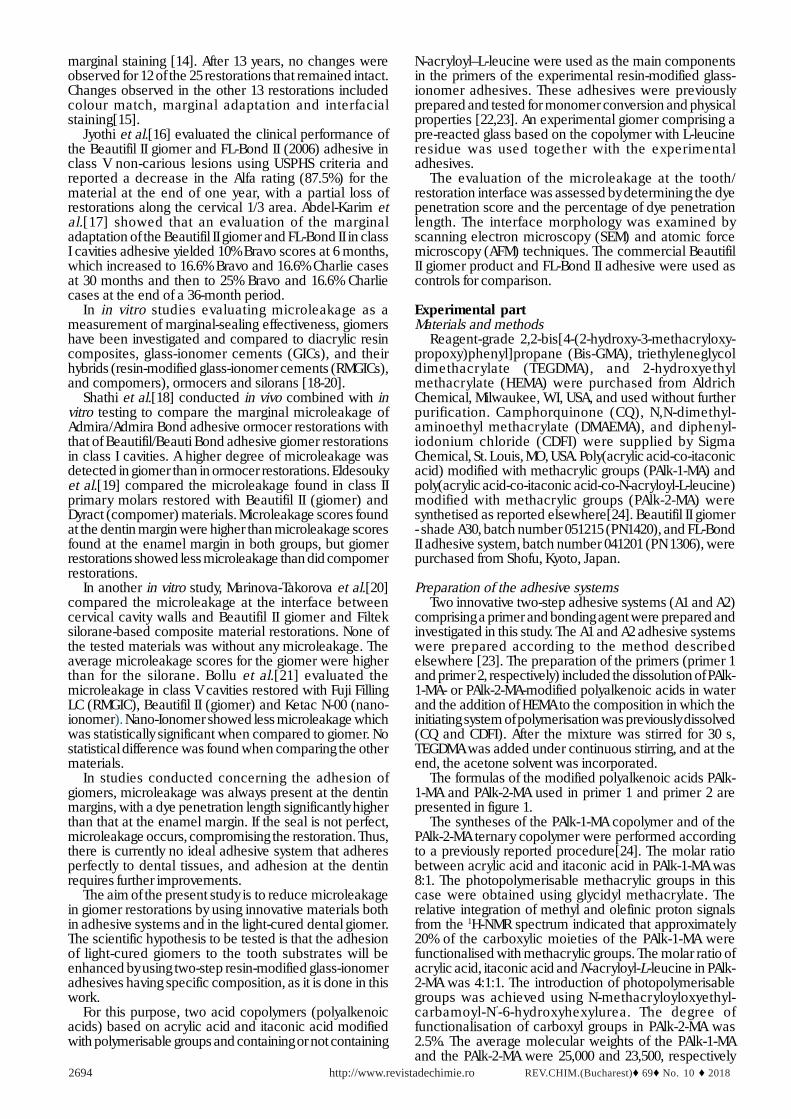
The formulas of the modified polyalkenoic acids PAlk-1-MA and PAlk-2-MA used in primer 1 and primer 2 arepresented in figure 1.
The syntheses of the PAlk-1-MA copolymer and of thePAlk-2-MA ternary copolymer were performed accordingto a previously reported procedure[24]. The molar ratiobetween acrylic acid and itaconic acid in PAlk-1-MA was8:1. The photopolymerisable methacrylic groups in thiscase were obtained using glycidyl methacrylate. Therelative integration of methyl and olefinic proton signalsfrom the 1H-NMR spectrum indicated that approximately20% of the carboxylic moieties of the PAlk-1-MA werefunctionalised with methacrylic groups. The molar ratio ofacrylic acid, itaconic acid and N-acryloyl-L-leucine in PAlk-2-MA was 4:1:1. The introduction of photopolymerisablegroups was achieved using N-methacryloyloxyethyl-carbamoyl-N’-6-hydroxyhexylurea. The degree offunctionalisation of carboxyl groups in PAlk-2-MA was2.5%. The average molecular weights of the PAlk-1-MAand the PAlk-2-MA were 25,000 and 23,500, respectively
REV.CHIM.(Bucharest)♦ 69♦ No. 10 ♦ 2018 http://www.revistadechimie.ro 2695
(measured in dimethylformamide by gel permeationchromatography analysis)[24].
The bonding agent was prepared by mixing a Bis-GMA,HEMA and TEGDMA monomer mixture with apolymerisation initiating system (CQ and DMAEMA).
The commercial adhesive system used as a referencein this study was FL-Bond II, also comprised a primer anda bonding agent. The FL-Bond II primer is an acetone-freeadhesion promoting monomer with no incorporation ofHEMA[25]. This primer contains ethanol, pure water, aphosphonic acid monomer (6-MHPA), and a carboxylic acidmonomer (4-AET)[10,26]. The bonding contains 1,6-bis(methacr yloxy-2-ethoxy-carbonylamino)-2,4,4-trimethylhexane (UDMA), HEMA, TEGDMA and S-PRG filler(surface pre-reacted glass-ionomer)[27]. The compositionof the investigated adhesive systems is presented in table1.Preparation of the giomer G
The light-cured giomer G was prepared as a monopasteby mixing the resin with the hybrid filler. The resin consistedof a mixture of 70% Bis-GMA and 30% TEGDMA in whichthe components of the initiating system were dissolved.To obtain the hybrid filler, the surface pre-reacted glassfiller (SPRGexp), radiopaque glass (barium fluoro-alumino-boro-silicate glass) and fluorohydroxyapatite (FHAP) weremixed and then sifted together. Fluorohydroxyapatite wasadded to the giomer to increase the mechanical propertiesof the material [28-30]. The surface pre-reacted glass (S-PRG) fillers were obtained by an acid–base reactionbetween fluoride containing superficially active glassesand polyalkenoic acids in the presence of water. When thepolyalkenoic acid attacks the particles on the surface ofthe active glass, a polysalt matrix is formed as a result ofthe acid–base reaction in which the non-attacked cores ofthe glass particles surrounded by the hydrogel layer areembedded [22,31].
In the present study, the SPRGexp was prepared by mixinga 50% aqueous solution of PAlk-2-MA polyalkenoic acidwith superficially active glass powder with the compositionSiO2 (49%), Al2O3 (22%), and CaF2 (29%) in a weight ratioof 1:2.4 [22]. The radiopaque glass was synthesised viathe conventional melting method at 1160°C for 30 min
using the corresponding components SiO2 (25%), B2O3(11%), Al2O3 (14%), and BaF2 (50%). Silanation was carriedout with 3-methacryloyloxypropyl-trimethoxy-silane (A-174 silane). The particle size of the SPRGexp and radiopaqueglass was analysed by using the laser diffraction technique(Analysette 22 NanoTec Laser Particle Sizer, Fritsch, Idar-Oberstein,Germany). In the case of SPRGexp, d(0.1)=1.584µm, d(0.5)=10.806 µm and d(0.9)=23.932 µm. Thevolume median diameter, d50, of the radiopaque glass was5 µm, and d(0.1), d(0.5) and d(0.9) were 1.238 µm,7.147µm and 19.122 µm, respectively [22]. The preparationmethod of FHAP was described elsewhere [32]. Thesynthesised nanoparticles in the form of rods had lengthsbetween 15 and 160 nm and a thickness of approximately10 nm. The composition of the investigated restorativegiomers is presented in table 2.
The chemical composition of the Beautifil II productwas presented in several ways by different literaturesources. According to DIONYSOPOULOS et al., it containsS-PRG fillers (68.6% w/v, 83.3% w/w), Bis-GMAcom andTEGDMA [33]. The composition of Beautifil II presented inthe Material Safety Data Sheet is Bis-GMAcom 7.5%,TEGDMA <5%, aluminofluoro-borosilicate glass 70%, Al2O3,DL-camphorquinone, and others [34].
Preparation of dental samplesForty-two recently extracted non-carious human
maxillar y and mandible premolars extracted fororthodontic reasons were selected for this study. Afterextraction, the teeth were cleaned of soft tissue andcalculus and stored in 0.1% thymol solution steam at 9°C.Before use, the teeth were washed under running waterfor 24 h, blot dried and stored in normal saline solution at37°C and 95% humidity until testing [35]. Box-type class Vstandardised cavities were prepared on the facial and oralsurfaces of each tooth, with coronal margins in the enameland apical margins in the cementum, which measured 4mm mesio-distally, 2 mm occluso-gingivally, and 1.5 mmin depth. Diamond fissure burs (D & Z, Hilzingen, Germany)with a diameter of 1 mm were used for cavity preparations.The preparations were divided randomly into 3 equalgroups (n=28/group) and restored with the following
Fig. 1. The modifiedpolyalkenoic acids used for the
obtaining of the primers
http://www.revistadechimie.ro REV.CHIM.(Bucharest)♦ 69♦ No. 10 ♦ 20182696
materials: group I (control): FL-Bond II and Beautifil II;group II: A1 and giomer G; group III: A2 and giomer G.
The clinical protocol used for restoration consisted ofetching with 37% phosphoric acid for 15 s on the enamel,rinsing for 10 s afterwards and then gently drying for 3 swith air spray. The primer was applied with a brush for 20s and then dried for 3 s. The bonding agent was applied onthe enamel and dentin for 15 s, then dried for 3 s and finallylight-cured for 20 s. The giomer was inserted using theoblique layer technique (layers of a maximum 2 mm each)and light-cured after each layer for 20 s using the light-curing device ‘Spectrum 800’ (Dentsply, Germany) (470nm wavelength). Finishing and polishing of the giomer
restorations were performed using the Super Snap finishingand polishing system (Shofu, Kyoto, Japan).
The restored teeth underwent 500 thermocycles in anLTC 100 thermocycling device (LAM TechnologiesElectronic Equipment, Firenze, Italy) that had two waterbaths at 5°C and 55°C. Each cycle lasted for 60s:20s ineach bath and 10 s to complete the transfer between baths.Thermocycling involves subjecting the restoration and thetooth to extreme temperatures compatible with the oralcavity [36]. The International Organization forStandardisation (ISO) recommends 500 thermal cycles inwater between 5 and 55°C as an accelerated ageing test[37]. After thermocycling, the teeth were dried and isolated
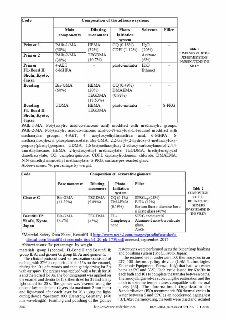
Table 1COMPOSITION OF THE
ADHESIVE SYSTEMSINVESTIGATED IN THE
STUDY
Table 2COMPOSITION
OF THERESTAURATIVE
GIOMERSINVESTIGATED IN
THE STUDY
REV.CHIM.(Bucharest)♦ 69♦ No. 10 ♦ 2018 http://www.revistadechimie.ro 2697
with two consecutive layers of nail varnish on the entiresurface of the tooth, except for an area of approximately 1mm around the restorations. The apices of the teeth wereoccluded with composite resin; then, the teeth wereimmersed in 2% methyl blue solution for 24 h. After the dyewas rinsed off with distilled water, the samples were airdried, and each specimen was embedded in methylmethacrylate and sectioned buccolingually through themiddle of both restorations in slices of 1 mm thicknessusing a diamond saw (Isomet 1000, Buehler, USA).
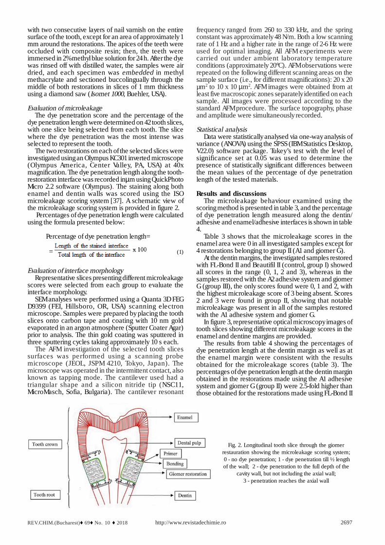
Evaluation of microleakageThe dye penetration score and the percentage of the
dye penetration length were determined on 42 tooth slices,with one slice being selected from each tooth. The slicewhere the dye penetration was the most intense wasselected to represent the tooth.
The two restorations on each of the selected slices wereinvestigated using an Olympus KC301 inverted microscope(Olympus America, Center Valley, PA, USA) at 40xmagnification. The dye penetration length along the tooth-restoration interface was recorded inµm using QuickPhotoMicro 2.2 software (Olympus). The staining along bothenamel and dentin walls was scored using the ISOmicroleakage scoring system [37]. A schematic view ofthe microleakage scoring system is provided in figure 2.
Percentages of dye penetration length were calculatedusing the formula presented below:
Percentage of dye penetration length=
= (1)
Evaluation of interface morphologyRepresentative slices presenting different microleakage
scores were selected from each group to evaluate theinterface morphology.
SEM analyses were performed using a Quanta 3D FEGD9399 (FEI, Hillsboro, OR, USA) scanning electronmicroscope. Samples were prepared by placing the toothslices onto carbon tape and coating with 10 nm goldevaporated in an argon atmosphere (Sputter Coater Agar)prior to analysis. The thin gold coating was sputtered inthree sputtering cycles taking approximately 10 s each.
The AFM investigation of the selected tooth slicessurfaces was performed using a scanning probemicroscope (JEOL, JSPM 4210, Tokyo, Japan). Themicroscope was operated in the intermittent contact, alsoknown as tapping mode. The cantilever used had atriangular shape and a silicon nitride tip (NSC11,MicroMasch, Sofia, Bulgaria). The cantilever resonant
frequency ranged from 260 to 330 kHz, and the springconstant was approximately 48 N/m. Both a low scanningrate of 1 Hz and a higher rate in the range of 2-6 Hz wereused for optimal imaging. All AFM experiments werecarried out under ambient laboratory temperatureconditions (approximately 20°C). AFM observations wererepeated on the following different scanning areas on thesample surface (i.e., for different magnifications): 20 x 20µm2 to 10 x 10 µm2. AFM images were obtained from atleast five macroscopic zones separately identified on eachsample. All images were processed according to thestandard AFM procedure. The surface topography, phaseand amplitude were simultaneously recorded.
Statistical analysisData were statistically analysed via one-way analysis of
variance (ANOVA) using the SPSS (IBM Statistics Desktop,V22.0) software package. Tukey’s test with the level ofsignificance set at 0.05 was used to determine thepresence of statistically significant differences betweenthe mean values of the percentage of dye penetrationlength of the tested materials.
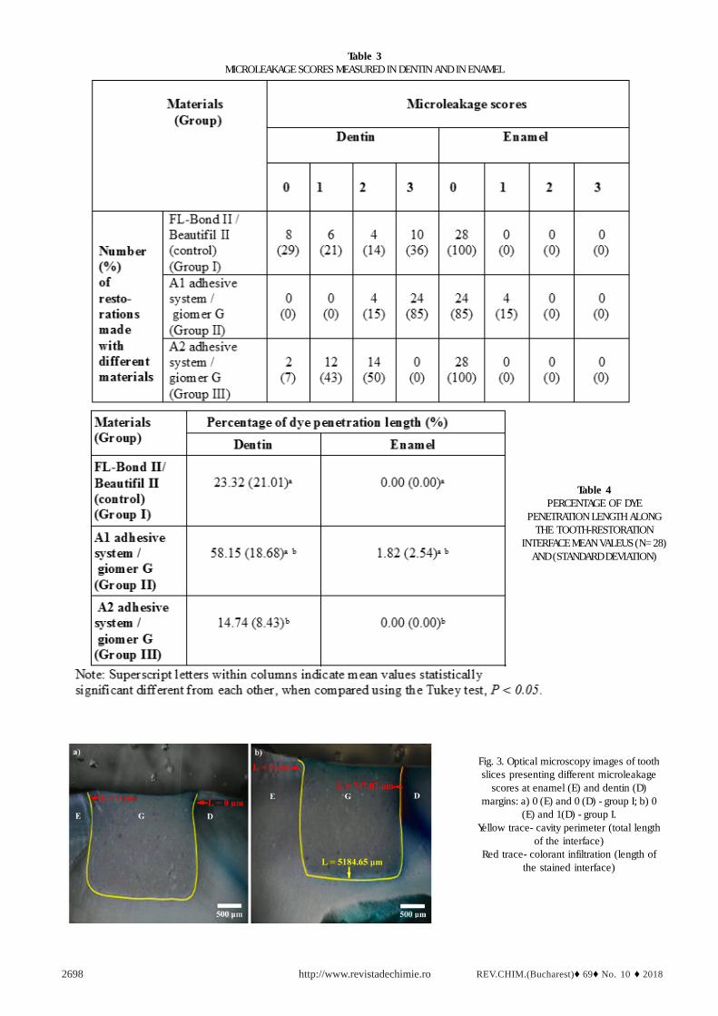
Results and discussionsThe microleakage behaviour examined using the
scoring method is presented in table 3, and the percentageof dye penetration length measured along the dentin/adhesive and enamel/adhesive interfaces is shown in table4.
Table 3 shows that the microleakage scores in theenamel area were 0 in all investigated samples except for4 restorations belonging to group II (A1 and giomer G).
At the dentin margins, the investigated samples restoredwith FL-Bond II and Beautifil II (control, group I) showedall scores in the range (0, 1, 2 and 3), whereas in thesamples restored with the A2 adhesive system and giomerG (group III), the only scores found were 0, 1 and 2, withthe highest microleakage score of 3 being absent. Scores2 and 3 were found in group II, showing that notablemicroleakage was present in all of the samples restoredwith the A1 adhesive system and giomer G.
In figure 3, representative optical microscopy images oftooth slices showing different microleakage scores in theenamel and dentine margins are provided.
The results from table 4 showing the percentages ofdye penetration length at the dentin margin as well as atthe enamel margin were consistent with the resultsobtained for the microleakage scores (table 3). Thepercentages of dye penetration length at the dentin marginobtained in the restorations made using the A1 adhesivesystem and giomer G (group II) were 2.5-fold higher thanthose obtained for the restorations made using FL-Bond II
Fig. 2. Longitudinal tooth slice through the giomerrestauration showing the microleakage scoring system; 0 - no dye penetration; 1 - dye penetration till ½ lengthof the wall; 2 - dye penetration to the full depth of the
cavity wall, but not including the axial wall;3 - penetration reaches the axial wall
http://www.revistadechimie.ro REV.CHIM.(Bucharest)♦ 69♦ No. 10 ♦ 20182698
Table 3MICROLEAKAGE SCORES MEASURED IN DENTIN AND IN ENAMEL
Table 4PERCENTAGE OF DYE
PENETRATION LENGTH ALONGTHE TOOTH-RESTORATION
INTERFACE MEAN VALEUS (N=28)AND (STANDARD DEVIATION)
Fig. 3. Optical microscopy images of toothslices presenting different microleakage
scores at enamel (E) and dentin (D)margins: a) 0 (E) and 0 (D) - group I; b) 0
(E) and 1(D) - group I.Yellow trace- cavity perimeter (total length
of the interface)Red trace- colorant infiltration (length of
the stained interface)
REV.CHIM.(Bucharest)♦ 69♦ No. 10 ♦ 2018 http://www.revistadechimie.ro 2699
Fig. 3. Optical microscopy images of tooth slicespresenting different microleakage scores at
enamel (E) and dentin (D) margins: c) 0 (E) and2 (D) - group III; d) 1(E) and 3 (D) - group II.I.
Yellow trace- cavity perimeter (total length of theinterface) Red trace- colorant infiltration (length
of the stained interface)
Fig. 4. SEM photomicrographs of the interfaces: a), b) dentin / A2adhesive / Giomer G ; c, d) dentin / FL Bond II / Beautifil II. Arrows
show the margins of the ion-exchange layer. Asterisks show theadhesion interface to dentin. (D) - dentin; (AL)- adhesive layer;
(B) - Beautifil II; (G) - Giomer G
Fig. 5. AFM images obtained at adhesion interface between dentin /A2 adhesive /Giomer G: a) topographic image, b) phase image,
c) 3D - view of image (a), and d) cross section on white arrow infigure (a). Scanned area 10µm x 10µm.
Fig. 6. at adhesion interface between dentine / adhesive FL-Bond II/ Beautifil II: a) topographic image, b) phase image, c) 3D - view of
image (a), and d) cross section on white arrow in figure (a).Scanned area 20µm x 20µm
and Beautifil II (Control) and almost four-fold higher thanthose for the restorations made using the A2 adhesivesystem and giomer G (group III). The percentages of dyepenetration length at the dentin margin obtained for therestorations made using the A2 adhesive system andgiomer G (group III) were 1.5 times smaller than thoseobtained for the restorations made using FL-Bond II andBeautifil II (Control).
Statistical analysis indicated significant differences inthe percentages of dye penetration length at the enamelmargin (p=4.626×10⁻⁶) and at the dentin margin(p=3.701×10⁻¹⁵), as determined by one-way ANOVA.
Tukey ’s post-hoc test revealed that there was astatistically significant difference between group I (control)and group II as well as between group II and group III atboth the dentin and enamel margins. At the dentin margin,similar significant differences were observed whencomparing group II with group III (p=5.100×10⁻⁹) andwhen comparing group I with group II (p=5.191×10⁻⁹).The same pattern occurred at the enamel margin, showingsimilar significant differences between the groups.Comparisons of group I with group II and group II withgroup III revealed significant mean differences at the 0.05level (p=3.918×10⁻⁵).
The differences between group I and group III were notstatistically significant at the dentin margin (p=0.148) orthe enamel margin (p=1.000).
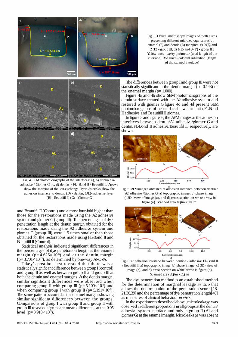
Figure 4a and 4b show SEM photomicrographs of thedentin surface treated with the A2 adhesive system andrestored with giomer G.figure 4c and 4d present SEMphotomicrographs of the interface between dentin, FL BondII adhesive and Beautifill II giomer.
In figure 5 and figure 6, the AFM images at the adhesioninterfaces between dentin/A2 adhesive/giomer G anddentin/FL-Bond II adhesive/Beautifil II, respectively, areshown.
The dye penetration method is an established methodfor the determination of marginal leakage in vitro thatallows the determination of the penetration score [18-21,38,39] and the percentage of dye penetration length[40]as measures of clinical behaviour in vivo.
In the experiments described above, microleakage wasobserved in different proportions in all groups at the dentin/adhesive system interface and only in group II (A1 andgiomer G) at the enamel margin. Microleakage was absent

http://www.revistadechimie.ro REV.CHIM.(Bucharest)♦ 69♦ No. 10 ♦ 20182700
at the enamel margin in groups I (FL-Bond II and BeautifilII) and III (A2 and giomer G).
Our results obtained for microleakage scores at thedentin margin can be considered similar to those presentedby Bollu et al. [21], who used the same materials (BeautifilII and FLBond II) for the restoration of class V cavities. Forinstance, 29% score 0, 21% score 1, 14% score 2, and 36%score 3 obtained in our study are comparable with theirscores, namely 40% score 0, 15% score 1, 5% score 2, and40% score 3 [21]. Even though in their study it is statedthat the microleakage was found to be high at both occlusaland cervical margins [21], they showed only one value forthe percentage of a particular score, not specifying whetherit was for the cervical or occlusal margin, and withoutindicating the mode used to calculate the value of the score.
The absence of score 3 (0%) recorded when using theA2 adhesive system and giomer G indicates a significantlyimproved sealing ability when compared to the score 3 of40% obtained by Bollu [21], or to the score 3 of 36% obtainedby us when using the FL Bond II adhesive and Beautifil IIgiomer.
The differences in the microleakage scores observed inthe enamel compared to the scores obtained in the dentincould be explained by the morphological differences inthe tooth structures dentin and enamel. The enamelstructure is based on hydroxyapatite (Hap) crystals at 92%vol., 2% vol. organic materials and 6% vol. water [41]. Itsstructure allows the creation of microretentions by etchingof the surface. This process will ensure a perfectinterlocking with the components from adhesives, leadingto the realisation of a perfect adhesion at the interface. Incontrast, dentin is more hydrophilic, with a canalicularstructure consisting of 48% vol. Hap, 29% vol. organicmaterials and 23% vol. water [41]. The dentinal tubulestraverse the entire dentine, oriented from the dentin-enameljunction towards the dental pulp. Even if the dentin is driedbefore the adhesive system is applied, dentinal fluidreappears, almost constantly endangering theadhesion[42]. This structure of the dentin makes it morevulnerable to microleakage than enamel [43-46].
The main difference between the experimental groupsof adhesives (groups II and III) and the control one (groupI) is that the primers from the experimental adhesivescontain a relatively high molecular weight acidiccopolymers (polycarboxyl-based copolymers, polyalkenoicacids) grafted with methacrylic groups as the maincomponent, whilst the FL Bond II primer contains lowmolecular weight acidic monomers, namelyacryloxyethyltrimellitic acid (4-AET) and (6-methacryloxy-hexyl phosphonoacetate (6-MHPA). Another difference isthat FL Bond II bonding is filled with SPRG filler, whilst theexperimental bonding is unfilled.
The twofold mechanism of adhesion to dentin isrelatively similar for the experimental adhesives and forthe commercial one mild self-etch approach), theadhesion being realised by micromechanical interlockingand chemical interactions[47]. When an experimentalprimer (PAlk-1-MA or PAlk-2-MA) was applied on the dentin,the polyalkenoic acid dissolved the smear layer, partiallydemineralized the dentin and form strong ionic bonds withthe calcium from the hydroxyapatite[48,49]. Thedemineralization of dentin was minimal in this casebecause the hydroxyapatite buffered the weakpolyalkenoic acid[50]. A zone of chemical interaction(inter-diffusion zone, ion-exchange layer) with a thicknessof a few micrometers was formed in which the calciumpolyalkenoate salt can hardly be dissolved[47]. Afterapplying the bonding and the visible light, due to the grafted
methacrylic groups, the polyalkenoic acids were able topolymerise with the TEGDMA and HEMA from the primerand with the monomers from the bonding, leading to theformation of a unique polymeric network of the adhesive.
When the mild self-etch FL Bond II primer was applied,the infiltration of acidic monomers and the partiallydemineralization of dentin occurred, creating the micro-porosities for micro-mechanical interlocking[51]. Ashallow hybrid layer is formed by the infiltration of themonomers in the hydroxyapatite-coated collagen fibrilnetwork. Additional chemical interactions between theacidic monomers (4-AET and 6-MHPA) and calcium ionsfrom residual hydroxyapatite occurred [10]. However it ispossible that 4-AET calcium salt (Ca-4AET) to have arelatively high solubility as the calcium salt of 4-methacryloyloxyethyl trimellitic acid (Ca-4MET) has, beingtherefore not very stable[52].
The differences concerning microleakage between theexperimental groups (II and III) could be explained bytaking into consideration the chemical composition of theprimers. The two adhesive systems (A1, A2) containedtwo different primers (primer 1 and primer 2) in which themain components were polyalkenoic acids modified withpolymerisable groups (PAlk-1-MA and PAlk-2-MA,respectively) and a bonding agent based on Bis-GMA. Thepolyalkenoic acid with pendant amino acid moieties (PAlk-2-MA) presented more flexibility, allowing more freedomand less steric hindrance when the carboxylic groupsinteracted with the calcium ions from the dentalhydroxyapatite [51, 52]. As a consequence, morecarboxylic groups interacted with the calcium ions fromthe dental tissues in the case of PAlk-2-MA from primer 2compared to PAlk-1-MA in primer 1. In addition, becausePAlk-1-MA exhibited a degree of functionalisation ofcarboxyl groups with methacrylic moieties approximately10-fold greater than that of PAlk-2-MA (20% versus 2.5%),the number of free carboxyl groups that could interact withCa2+ of dental hydroxyapatite was much higher for PAlk-2-MA. This behaviour led to improved sealing and adhesionwhen using PAlk-2-MA (primer 2). Finally, PAlk-2-MAcontained L-leucine residue, respective pendant (- CO -NH-) amide groups which can lead to the formation ofadditional hydrogen bonds with the carboxyl groups ofcollagen[52].
SEM and AFM are powerful tools for the surfaceinvestigation of various sample types (e.g., nanoparticles,bio nanocomposites, biomaterials)[55-61]. Tapping-modeAFM investigation was performed to characterise the tooth-restoration interface by revealing its topography.
In figure 4a and 4b, the good sealing ability of the A2adhesive system to the dentin substrates and to giomer Gcan be observed. The dentin presents a smooth surfacepunctuated by small dots representing the dentin tubules.The microstructure detail in figure 4b reveals the adhesioninterface with dentin at a higher resolution. The ion-exchange layer ranged from 1 to 2µm. The adhesive layerwith a thickness of about 10 µm can be observed betweenthe ion-exchange layer and giomer G. Giomer G has amore heterogeneous microstructure based on a granularmatter having polyhedral shape. A large amount of particlesmeasuring less than 10 microns with sharp or roundededges as well as a few particles having a diameter of about20 microns can be visualized in figure 4b. Base on theparticle size analysis, the first can be attributed to theradiopaque filler particles or small sizes SPRGexp particlesand the second ones can be attributed to the large sizesSPRGexp filler particles[22].

REV.CHIM.(Bucharest)♦ 69♦ No. 10 ♦ 2018 http://www.revistadechimie.ro 2701
The adhesion interface (hybrid layer) between the dentinand Beautifil II appears as an undulated layer closelyfollowing the shape of the dentin and having approximatelythe same width along the interface (fig. 4c, 4d). Theadhesive layer thickness ranged from 18 to 20 µm, and theSPRG filler particles showed irregular polyhedral shapesvarying in size from 5 to15 µm. The morphology of theadhesion zone is better observed at higher magnificationin figure 4d. A good connection was established betweenthe adhesive and the giomer restoration. The surfacemorphology of Beautifil II shows particles of various sizes,mostly below 15 microns having irregular shapes, too. Onecan observe a greater amount of particles embedded inthe polymer matrix than in the experimental giomer. Theblack pores on the samples surface in the SEM images ofthe giomers appeared due to the preparation of the samples(sectioning and sanding) during which particles of differentsizes can be detached from the polymer matrix.
The adhesion morphology of the interface betweendentine / adhesive A2 /
Giomer G is observed in AFM images,(fig. 5), at highmagnification. The dentin is situated at the top of thetopographic image in figure 5a and the adhesive is situatedat the lower side of the image. The ion-exchange layerformed by the reaction between polyalkenoic acid anddentin hydroxyapatite has a thickness of about 2 µm. Thetopography of this layer reveals its nanostructure whichcontains of nano-particles having the average diameteraround 80 nm, as observed in the cross section in figure5d. These nano-structural units can be attributed to thereacted hydroxyapatite with the polyalkenoic acid. A goodsealing of dentinal tubule is observed and an optimalcohesion between dentin and adhesive is achieved. Thefact is sustained by phase image, (fig. 5b), where dentin,ion-exchange layer and adhesive appears as a single solidblock having light brown nuance meanwhile dentinal tubuleis featured in dark brown. The three-dimensional view oftopographic image, figure 5c, presents the bondingachieved by A2 adhesive in a more suggestive manner.The ion-exchange layer in good cohesion with the dentinis better observed.
The SEM and AFM images are in close agreement withthe results obtained by the dye penetration method,showing that the A2 adhesive system provides effectivesealing of the giomer G restorations to the tooth substratewithout gaps or voids.
AFM image from figure 6 shows that FL-Bond II primerrealized the etching of the dentin, which was demineralizedallowing the micro-mechanical interlocking. The dentin issituated at the top and the adhesive at the bottom oftopographic image in figure 6a. A shallow hybrid layer wasformed between dentin and adhesive which have athickness about 1.5 µm. It is well observed in the crossprofile, (fig. 6d), at lateral distance of 4.0 to 5.5 µm.
The mild self-etching manner is also observed in phaseimage, (fig. 6b) where the shallow hybrid layer appears asa mixture of light and dark brown sustaining the micro-mechanical interlocking as an interfacial bondingmechanism to the dentin. Three-dimensional view of theadhesion interface, (fig. 6c), allows a better observation ofFL-Bond II primer interlocking into the partial demineralizeddentin.
ConclusionsThe results obtained in this study and described above
reveal that the composition of the adhesive systemsinfluences the sealing, adhesion and binding of giomer totooth tissue.
Of the two innovative adhesive systems (A1 and A2)developed for dental restorations to repair dentine/enamelcarious injuries by filling directly into the tooth cavity usingthe appropriate dental procedures, the A2 adhesive systemcontaining the PAlk-2-MA-modified polyalkenoic acid ledto improved sealing at the dentin interface and perfectadhesion at the enamel interface in tooth/giomerrestorations.
Our findings suggest that the microleakage in giomerrestorations can be reduced by using a two-step resin-modified glass-ionomer adhesive containing a relativelyhigh molecular weight polyalkenoic acid having a specificchemical formula which includes L-leucine residues andphotopolymerizable methacrylate groups.
The two-step resin-modified glass-ionomer adhesivebased on relatively high molecular weight polyalkenoic acidwith L-leucine residue has proven to be promising forimproving the adhesion to tooth structures in giomerrestorations.
Acknowledgements: This work was supported by two grants of TheRomanian National Authority for Scientific Research and Innovation,UEFISCDI: PN-III-P2-2.1-PED-2016-1936 and PNIII-P2-2.1-PED-2016-1415.
References1.MJOR IA., Oper. Dent., 10, no. 3, 1985, p.88-922.MJOR IA., Acta. Odontol. Scand., 55, no.1,1997, p: 58-633.QVIST V, QVIST J, MJOR IA., Acta. Odontol. Scand., 48, no.5, 1990, p.305-3114.Mc CABE JF, WALLS A. Applied Dental Materials , 9th ed., Singapore:Blackwell publishing, 2008, p.2275.Mc CABE JF, YAN Z, AL NAIMI OT, MAHMOUD G, ROLLAND SL.,Dent. Mater. J., 28, 2009, p. 37-436.ITOTA T, CARRICK TE, YOSHIYAMA M, MC CABE JF., Dent. Mater.,20, no.9, 2004, p.789-7957.FURTOS G, COSMA V, PREJMEREAN C, MOLDOVAN M, BRIE M,COLCERIU A, VEZSENY L, SILAGHI-DUMITRESCU L, SIRBU C., Mater.Sci. Eng. C., 25, 2005, p. 231-2368.WEI J, WANG J, LIU X, MA J, LIU C, FANG J, WEI S., Appl. Surf. Sci.,257, 2011, p.7887-78929.CARVALHO RM, PEREIRA JC, YOSHIYAMA M, PASHLEY DH., Oper.Dent., 21, no.1, 1996, p.17-2410.IKEMURA K, TAY FR, EDO T, PASHLEY DH., Dent. Mater. J., 27, 2008,p.315-33911.MATIS BA, COCHRAN MJ, CARLSON TJ, GUBA C, ECKERT GJ., J.Am. Dent. Assoc., 135, no.4, 2004, p.451-45712.SUNICO MC, SHINKAI K, KATOH Y., Oper. Dent., 30, 2005, p.282-28913.GORDAN VV, SHEN C, WATSON RE, MJOR IA., Am. J. Dent., 18,no.1, 2005, p.45-49.14.GORDAN VV, MONDRAGON E; WATSON RE, GARVAN C, MJORIA., J.A.D.A., 138, no.5, 2007, p.621-627.15.GORDAN VV, BLASER PK, WATSON RE, MJOR IA, McEDWARDS DL,SENSI LG, RILEY III JL., J.A.D.A., 145, no.10, 2014, p.1036-1043.16.JYOTHI KN, ANNAPURNA S, ANIL KUMAR S, VENUGOPAL P,JAYASHANKARA CM., J. Conserv. Dent., 14, no.4, 2011, p.409-41317.ABDEL-KARIM UM, EL-ERAKY M, ETMAN WM.,Tanta. Dent. J., 11,2014, p.213-22218.SHATHI IJ, HOSSAIN M, GAFUR A, RANA S, ALAM S., B.S.M.M.U. J.,10, 2017, p.214-21819.ELDESOUKY HI, HANNO AG, BAKRY NS, AHMED DM., A.D.J., 41,2016, p.188-19320.MARINOVA-TAKOROVA M, KARAJASHEVA D, BOTEVA E., ScriptaScientifica Medicinae Dentalis, 1, no.1, 2015, p.38-4221.BOLLU IP, HARI A, THUMU J, VELAGULA LD, BOLLA N, VARRI S,KASARANENI S, NALLI SVM., J. Clin. Diagn. Res., 10, no.5, 2016, p.ZC66-7022.PREJMEREAN C, PRODAN D, VLASSA M, STREZA M, BURUIANA T,COLCERIU L, PREJMEREAN V, CUC S, MOLDOVAN M., Meas. Sci.Technol., 27, no.12, 2016, p.124008

http://www.revistadechimie.ro REV.CHIM.(Bucharest)♦ 69♦ No. 10 ♦ 20182702
23.SILAGHI-DUMITRESCU L, DADARLAT D, STREZA M, BURUIANA T,PRODAN D, HODISAN I, PREJMEREAN C., J. Therm. Anal. Calorim.,118, no.2, 2014, p.623-63024.BURUIANA T, NECHIFOR M, MELINTE V, PODASCA V, BURUIANAEC., J. Biomater. Sci. Polym. Ed., 25, 2014, p.749-76525. BRESCHI L, MAZZONI A, RUGGERI A, CADENARO M, DI LENARDAR, DE STEFANO DORIGO E., Dent. Mater., 24, 2008, p.90-10126.AKIMOTO N, SAKAMOTO T, KUBOTA Y, KONDO Y,MOMOI Y., Dent.Mater. J., 30, no.4, 2011, p.523-52727.ABO EL NAGA A., J. Am. Sci., 8, no.2, 2012, p.27-3428.LIN J, ZHU J, GU X, WEN W, LI Q, FISCHER-BRANDIES H, WANGH, MEHL C., Acta. Biomater., 7, no.3, 2011, p.1346-135329.MOCANU A, FURTOS G, RAPUNTEAN S, HOROVITZ O, FLORE C,GARBO C, DANISTEANU A, RAPUTEAN G, PREJMEREAN C, TOMOAIA-COTISEL M., Appl. Surf. Sci., 298, 2014, p.225-23530.SANTOS C, LUKLINSKA ZB, CLARKE LR, DAVY KWM., J. Mater. Sci.-Mater. Med., 12, no.7, 2001, p.565-573.31.ZIMEHL, R, HANNING, M., Colloids Surf. A., 163, 2000, p.55-6232.PREJMEREAN, C., MOLDOVAN, M., PETREA, C.M., PRODAN, D.,SILAGHI-DUMITRESCU, L., VASILE, E., FURTOS, G., BOBOAIA, S.,SILAGHI-DUMITRESCU, R., Mat. Plast., 48, no.4, 2011, p.279-28433.DIONYSOPOULOS D, KOLINIOTOU-KOUMPIA E, HELVATZOGLOUA, KOTSANOS N., Dent. Mater. J., 32, no.2, 2013, p.296-30434.MATERIAL SAFETY DATA SHEET, BEAUTIFIL II; http://www.net32.com/images/prodinfo/a/shofu-dental-corp-beautifil-ii-compule-tips-b1-20-pk-1759.pdf accessed February 2018.35.MANICARDI CA, VERSIANI MA, SAQUY PC, PÉCORA JD, DE SOUSA-NETO MD., J. Endod., 37, 2011, p.531-53736.BULLARD RH, LEINFELDER KF, RUSSELL CM., J. Am. Dent. Assoc.,116, no.7, 1988, p.871-87437.*** International Organization for Standardisation (ISO). Dentalmaterials-testing of adhesion to tooth structure. ISO/TS 11405:2003(E)38.ALHABDAN AA., J. Int. Oral Health, 9, 2017, p.141-14539.MAURO SJ, DURAO VCA, BRISO ALF, SUNDEFELD MLMM, RAHALV., Int. J. Adh. Adh., 34, 2012, p.6-1040.VICHI A, MARGVELASHVILI M, GORACCI C, PAPACCHINI F, FERRARIM., Clin. Oral Investig., 17, no.6, 2013, p.1497-150641.OWENS BM, JOHNSON WW, HARRIS EF., Oper. Dent., 31, no.1,2006, p.60-6742.DORFER CE, STACHLE HJ, WURST MW, DUSCHNER H, PIOCH T.,Eur. J. Oral Sci., 108, no.4, 2000, p.346-35143.PRATI C, TAO L, SIMPSON M, PASHLEY DH., J. Dent., 22, no.1, 1994,p.49-56
44.DERHAMI K, COLI P, BRANNSTROM M., Oper. Dent., 20, no.3, 1995,p.100-10545.BEZNOS C., Oper. Dent., 26, no.1, 2001, p.60-69.46.YAZICI AR, CELIK C, OZGÜNALTAY G., Quintessence Int., 35, 2004,p.790-794.47.VAN MEERBEEK B, DE MUNCK J, YOSHIDE Y, INOUE S, VARGAS M,VIJAY P, VAN LANDUYT K, LAMBRECHTS P, VALNERLE G., Oper Dent2003; 28:215-23548.TYAS MJ, BURROW MF., Austr. Dent. J., 49, 2004, p.112-12149.YOSHIDA Y, INOUE S., Japanese Dental Science Review, 48, no.2,2012, p.141-15250.WATSON T. Bonding of glass-ionomer cements to tooth structures.In: Davidson CL, Mjor IA, eds. Advances in glass-ionomer cements.Chicago; Quintessence, 1999, p.121-13651.VAN MEERBEEK B., Espertise magazine, 21, 2012, p.20-2152.VAN LANDUYT KL, SNAUWAERT J, DE MUNK J, PEUMANS M,YOSHIDA Y, POITEVIN A, COUTINHO E, KAZOUMI S, LAMBRECHTS P,VAN MEERBEEK B., Biomater., 28, no.26, 2007, p.3757-378553.GAVIRIA J, GARCIA CG, VÉLEZ E, QUIJANO J., Modeling andNumerical Simulation of Material Science, 3, no.4, 2013, p.149-15454.UPADHYA PN, KISHORE G., Trends Biomater. Artif. Organs, 18,no.2, 2005, p.158-16555.PEDREIRA DE FREITAS AC, ESPEJO LC, BOTTA SB, SA TEIXEIRA F,CERQUEIRA LUZ MA, GARONE-NETTO N, MATOS AB, BARBOSA DASILVEIRA SALVADORI MC., Appl. Surf. Sci., 256, no.9, 2010, p.2915–291956.LOPES MB, SINHORETI MA, GONINI JR. A, COSANI S, Mc CABE JF.,Braz. Dent. J., 20, no.4, 2009, p.279-28357.HABELITZ S, BALOOCH M, MARSHALL SJ, BALOOCH G, MARSHALLGW., Journal of Structural Biology, 138, 2002, p.227–23658.CARRILHO MR, TAY FR, SWORD J, DONNELLY AM, AGEE KA,NISHITANI Y, SADEK FT, CARVALHO RM, PASHLEY DH., Eur. J. OralSci., 115, 2007, p.321–329.59.SAURO S, OSORIO R, WATSON TF, TOLEDANO M., Dent. Mat., 28,2012, p.622 - 63160.TOMOAIA G, SORITAU O, TOMOAIA-COTISEL M, POP L-B, POP A,MOCANU A, HOROVITZ O, BOBOS L-D., Powder Technology, 238,2013, p.99-10761.TOMOAIA G, HOROVITZ O, MOCANU A, NITA A, AVRAM A, RACZ CP, SORITAU O, CENARIU M, TOMOAIA-COTISEL M., Colloids andSurfaces B: Biointerfaces, 135, 2015, p.726-734.
Manuscript received: 26.03.2018