injectable preformed scaffolds with shape-memory properties · injectable preformed scaffolds with...
TRANSCRIPT

Injectable preformed scaffolds withshape-memory propertiesSidi A. Bencherifa,b, R. Warren Sandsa, Deen Bhattaa, Praveen Aranya,b, Catia S. Verbekea, David A. Edwardsa,b,and David J. Mooneya,b,1
aSchool of Engineering and Applied Sciences, Harvard University, Cambridge, MA 02138; and bWyss Institute for Biologically Inspired Engineering at HarvardUniversity, Boston, MA 02115
Edited by David A. Tirrell, California Institute of Technology, Pasadena, CA, and approved October 16, 2012 (received for review July 6, 2012)
Injectable biomaterials are increasingly being explored to minimizerisks and complications associated with surgical implantation. Wedescribe a strategy for delivery via conventional needle–syringe in-jection of large preformedmacroporous scaffolds withwell-definedproperties. Injectable 3D scaffolds, in the form of elastic sponge-likematrices, were prepared by environmentally friendly cryotropic ge-lation of a naturally sourced polymer. Cryogels with shape-memoryproperties may bemolded to a variety of shapes and sizes, and maybe optionally loaded with therapeutic agents or cells. These scaf-folds have the capability to withstand reversible deformations atover 90% strain level, and a rapid volumetric recovery allows thestructurally defined scaffolds to be injected through a small-boreneedle with nearly complete geometric restoration once delivered.These gels demonstrated long-term release of biomolecules in vivo.Furthermore, cryogels impregnated with bioluminescent reportercells provided enhanced survival, higher local retention, and ex-tended engraftment of transplanted cells at the injection site com-paredwith a standard injection technique. These injectable scaffoldsshow great promise for various biomedical applications, includingcell therapies.
syringe-injectable hydrogels | cryogelation | shape retention |controlled delivery | cell therapy
Hydrogels are 3D networks that can absorb a large amount ofwater while maintaining their structural integrity. They are
considered as appropriate scaffolds for tissue engineering applica-tions mainly due to their structural similarities with native soft tis-sue, and are widely used as soft materials in many biomedicalapplications including cell culture, cell encapsulation, controlleddelivery of therapeutic agents, andmedical device fabrication (1–6).Hydrogels typically exhibit a nanoporous network structure, but it
is advantageous to use hydrophilic networks with large inter-connected pores (>10 μm) to allow cell infiltration and deployment,and provide an increased surface area for cell attachment and in-teraction. As a result, macroporous hydrogel scaffolds have beendeveloped using various techniques such as fiber bonding, gasfoaming,microemulsion formation, phase separation, freeze-drying,and porogen leaching (7–15). More recently, gelation at subzerotemperatures, known as cryogelation, has been used to createhydrogels with large interconnected pores (16–18). During cry-ogelation, the reactants are restricted to the unfrozen/semifrozenphases and form a cross-linked network upon polymerization, whilethe ice crystals nucleated from the aqueous phase during freezingfunction as porogens. The melting of these ice crystals at tempera-ture above the freezing temperature gives rise to interconnectedmacroporous networks. Cryogels typically exhibit enhanced me-chanical stability with respect to traditional hydrogels (17). Recentstudies have shown the potential application of cryogels as tissueengineering scaffolds (19-21).Due to the trauma resulting from surgical implantation of 3D
scaffolds, minimally invasive surgical approaches to deliver poly-meric scaffolds have been the focus of recent work (22, 23). Fora biomaterial to be injectable, it has to be able to flow througha small-bore needle or a catheter. Most injectable hydrogels aredelivered in the liquid form (24–26). However, injecting a liquidthat will subsequently polymerize in situ presents a number of
complicating issues, including appropriate gelling time, the need tomaintain proper gelation conditions, formation of gels withappropriate mechanical strength and persistence time, bio-compatibility, and the ability to protect the cargo of proteindrugs or cells in adverse environments (27). Finally, purely liq-uid precursor solutions may leak into the neighboring tissue ordilute with body fluids, which may not only limit hydrogel for-mation but also limit control over the macrostructure of the gel.Recently, hydrogels with shear-thinning behaviors have beendeveloped to address certain of these concerns (27, 28), butthese still do not allow one to precisely control gel location norcreate defined macrostructures. Unlike most in situ gelling sys-tems, injectable cryogels have an interconnected macroporousarchitecture, which appears advantageous over nanoporoushydrogel scaffolds with respect to their ability to facilitate cel-lular infiltration and trafficking.We investigate here the potential of a fully preformed cryogel to
be delivered into the body through a conventional needle–syringeinjection. Alginate, a naturally occurring polysaccharide, which con-sists of residues of α-L-guluronic and β-D-mannuronic acid, was usedto fabricate the scaffolds, as it is considered to be biocompatible (29).Several material properties were investigated to benchmark thesecryogels as an injectable scaffold. Key requirements include the fol-lowing: (i) the injectable scaffold must be compressible and flowunder moderate pressure; (ii) the cryogel should maintain sufficientintegrity and strength during injection for gel recovery; (iii) oncedelivered, the cryogel should rapidly regain its original shape and sizefrom the collapsed state; and finally (iv) the scaffold should retainbiomolecules/cells within the scaffold during the injection processand allow their subsequent release in vivo in a controlled fashion.
ResultsMacroporous Hydrogel Synthesis. To prepare alginate amenable tocryogelation, pendant methacryloyl groups were first introducedinto the alginate main chains (Fig. 1A). 1H NMR spectrum ofmethacrylated (MA)-alginate exhibited peaks of vinyl methyleneand methyl protons at δ5.8 and 5.3, and 1.7, respectively, thatwere newly formed by the reaction of polymer with 2-aminoethylmethacrylate (AEMA) (Fig. S1). The methacrylation efficiencywas calculated based on the ratio of the integrals for alginateprotons to the methylene protons of methacryloyl groups, andMA-alginate macromonomer was found to have an approximatedegree of methacrylation of 50%.Next, alginate hydrogels were prepared both at room tempera-
ture (RT) and subzero temperature, which were designated asconventional nanoporous hydrogels and macroporous cryogels,respectively. Macroporous 3D alginate cryogels were preparedusing MA-alginate by the process of cryogelation at −20 °C using
Author contributions: S.A.B., R.W.S., D.A.E., and D.J.M. designed research; S.A.B., R.W.S.,D.B., P.A., and C.S.V. performed research; S.A.B., R.W.S., and D.J.M. analyzed data; andS.A.B. and D.J.M. wrote the paper.
The authors declare no conflict of interest.
This article is a PNAS Direct Submission.1To whom correspondence should be addressed. E-mail: [email protected].
This article contains supporting information online at www.pnas.org/lookup/suppl/doi:10.1073/pnas.1211516109/-/DCSupplemental.
19590–19595 | PNAS | November 27, 2012 | vol. 109 | no. 48 www.pnas.org/cgi/doi/10.1073/pnas.1211516109
Dow
nloa
ded
by g
uest
on
May
10,
202
0
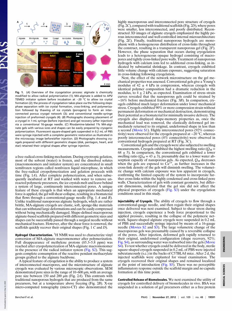
a free-radical cross-linkingmechanism.During cryotropic gelation,most of the solvent (water) is frozen, and the dissolved solutes(macromonomers and initiator system) are concentrated in smallsemifrozen regions called nonfrozen liquid microphase, in whichthe free-radical cryopolymerization and gelation proceeds withtime (Fig. 1A). After complete polymerization, and when subse-quently incubated at RT and washed with water to remove un-reacted polymeric precursors, the ice crystalsmelt and leave behinda system of large, continuously interconnected pores. A uniquefeature of these cryogels is that when an appropriate mechanicalforce is applied, the gel will shear-collapse, resulting in a biomaterialthat flows through a conventional-gauge needle (Fig. 1 A and B).Unlike traditional nanoporous alginate hydrogels, which are ratherbrittle, MA-alginate cryogels are elastic, soft, sponge-like materialsthat can withstand large deformations and can be easily compressedwithout being mechanically damaged. Shape-defined macroporousalginate-based scaffolds preparedwith different geometric sizes andshapes can be successfully passed through a surgical needle withoutmechanical fracture. However, after the shear force is removed, thescaffolds quickly recover their original shapes (Fig. 1 C and D).
Hydrogel Characterization. 1H NMR was used to characterize vinylconversion of MA-alginate macromonomer after polymerization.Full disappearance of methylene protons (δ5.3–5.8 ppm) wasreached after cryopolymerization of MA-alginate macromonomerin the presence of the radical initiator system (Fig. S2). This sug-gests complete consumption of the reactive pendant methacrylategroups grafted to the alginate backbone.A typical feature of cryogelation is the ability to produce a system
of interconnected macropores, and the microstructure of alginatecryogels was evaluated by various microscopic observations. SEMdemonstrated pore sizes in the range of 10–600 μm, with an averagepore size between 150 and 200 μm (Fig. 2A). This contrasts withtraditional nanoporous hydrogels that are produced from the sameprecursors, but at a temperature above freezing (Fig. 2B). X-raymicro-computed tomography (micro-CT) also demonstrated the
highly macroporous and interconnected pore structure of cryogels(Fig. 2C), comparedwith traditional scaffolds (Fig. 2D), where poreswere smaller, less interconnected, and poorly distributed. Recon-structed 3D images of alginate cryogels emphasized the highly po-rous interconnected and well-controlled internal microarchitecture(Fig. 2E). Finally, traditional nanoporous hydrogels are mono-phasic with a homogeneous distribution of cross-links throughoutthe construct, resulting in a transparent nanoporous gel (Fig. 2F).However, the phase separation that occurs during cryogelationleads to a heterogeneous opaque hydrogel consisting of macro-pores and tightly cross-linked porewalls. Treatment of nanoporoushydrogels with calcium ions led to additional cross-linking, as in-dicated by substantial shrinkage. In contrast, cryogels exhibitedlittle volume change with calcium exposure, suggesting saturationin cross-linking following cryogelation.Next, the effect of the network microstructure on the gel me-
chanical properties was assessed. Conventional gels give a Young’smodulus of 42 ± 4 kPa in compression, whereas cryogels withidentical polymer composition had a dramatic reduction in themodulus, to 4 ± 2 kPa, as expected. Examination of stress–straincurves revealed that the nanoporous gels could be strained by∼16% before mechanical fracture (Fig. 3B). In comparison, cry-ogels exhibited much larger deformation under lower mechanicalstress. Cryogels exhibited 90%ormore compression strain withoutpermanent deformation ormechanical destruction, demonstratingtheir potential as a biomaterial forminimally invasive delivery. Thecryogels also displayed shape-memory properties as, once themechanical load was removed, the elastically deformed gel im-mediately returned to its original, undeformed shape in less thana second (Movie S1). Highly interconnected pores (92% connec-tivity) were observed for the cryogels prepared at −20 °C, whereaspoorly interconnected pores (4% connectivity) were observed inconventional hydrogels prepared at RT (Fig. 3C).Conventional gels and the cryogelswere also subjected to swelling
measurements. Cryogels exhibited the highest swelling ratio (QM =68 ± 5). In comparison, the conventional gels exhibited a lowerswelling ratio (QM = 43 ± 3), presumably due to a lower water ab-sorption capacity of nanoporous gels. As expected, QM decreaseswhen the gels are exposed to Ca2+, as further increases in thecross-link density of the gels will diminish swelling. The volumet-ric change with calcium exposure was less apparent in cryogels,confirming the limited capacity of the system to incorporate fur-ther cross-links within the highly condensed and cross-linked porewalls. Characterization of gels with constant geometry, but differ-ent dimensions, indicated that the gel size did not affect thephysical properties of cryogels (Fig. S3) under the cryogelationconditions used in this study.
Injectability of Cryogels. The ability of cryogels to flow through aconventional-gauge needle, and then regain their original shapesonce delivered was next examined. Subject to shear stress duringinjection, cryogels experience a body force proportional to theapplied pressure, resulting in the collapse of the polymeric net-work. Square-shaped alginate cryogels were suspended in 0.2 mLof PBS and successfully syringe injected by means of a 16-gaugeneedle (Movies S2 and S3). The large volumetric change of themacroporous gels was presumably caused by a reversible collapseof the pores. After injection, deformed gels rapidly returned totheir original, undeformed configuration (shape recovery, 92%;Fig. S4), as surrounding water was reabsorbed into the gels (MovieS4). To test whether cryogels could be delivered in the body, sterilesquare-shaped cryogels suspended in 0.2 mL of PBS were injectedsubcutaneously (s.c.) in the backs of C57BL/6J mice. After 2 d, theinjected scaffolds were explanted for visual examination. Thecryogels recovered their original shapes and remained localizedat the point of introduction (Fig. S5). There was no perceptibleinflammatory response outside the scaffold margin and no capsuleformation at this time point.
Controlled Release of Biomolecules.We next examined the utility ofcryogels for controlled delivery of biomolecules in vivo. BSA wassuspended in a solution of gel precursors either as a free protein
Fig. 1. (A) Overview of the cryogelation process: alginate is chemicallymodified to allow radical polymerization (1); MA-alginate is added to APS/TEMED initiator system before incubation at −20 °C to allow ice crystalformation (2); the process of cryogelation takes place via the following steps:phase separation with ice crystal formation, cross-linking, and polymeriza-tion followed by thawing of ice crystals (porogens) to form an inter-connected porous cryogel network (3); and conventional needle–syringeinjection of preformed cryogels (4). (B) Photographs showing placement ofa cryogel in 1-mL syringe (before injection) and gel recovery (after injection)via a conventional 16-gauge needle. (C) Rhodamine-labeled 1% MA-algi-nate gels with various sizes and shapes can be easily prepared by cryogenicpolymerization. Fluorescent square-shaped gels suspended in 0.2 mL of PBSwere syringe injected with a complete geometric restoration as illustrated inthe microscopy image before/after injection. (D) Photographs showing cry-ogels prepared with different geometric shapes (disk, pentagon, heart, andstar) retained their original shapes after syringe injection.
Bencherif et al. PNAS | November 27, 2012 | vol. 109 | no. 48 | 19591
ENGINEE
RING
MED
ICALSC
IENCE
S
Dow
nloa
ded
by g
uest
on
May
10,
202
0

[rhodamine-labeled BSA (Rhod-BSA)] or as a reactive substance[methacrylated rhodamine-labeled BSA (Rhod-BSA-MA)] beforecryogenic treatment to allow their incorporation in the pore wallsof the cryogel. This gives rise to a macroporous matrix immobiliz-ing BSA (encapsulation efficiency: EE = 90%; Fig. S4). BSA-con-taining alginate cryogels were then syringe delivered into the s.c.space of C57BL/6J mice. Each mouse received a total of two cry-ogel injections, cross-linked Rhod-BSA-MA (right flank) and en-capsulated Rhod-BSA (left flank). The dye-labeled cryogels couldbe readily located in tissue at the site of injection (Fig. 4A). At 3 dpostinjection, one set of the square-shaped scaffolds located un-derneath the skin (Fig. 4B) was surgically removed and analyzed.The explanted cryogels demonstrate no alteration in size, andminimal inflammation was observed by histology (Fig. 4C). Thescaffolds were encased within host tissue in vivo (Fig. 4D), andfluorescent microscopy revealed the original geometry of the gels,indicating that the structural integrity and the geometric squareshape was retained within the s.c. pocket (Fig. 4E). Sustained re-lease of BSA was achieved from the injected square-defined scaf-folds as indicated by the spread of Rhod-BSA into the surroundingtissue over time (Fig. 4E), and diminishing fluorescence of the
cryogels (Fig. 4F). The release profiles for physically entrapped andchemically anchored BSA were similar, suggesting that proteinrelease may be triggered by matrix degradation rather than bydiffusion. The delayed release profiles suggest a very slow degra-dation rate of the matrix.The release of granulocyte–macrophage colony-stimulating fac-
tor (GM-CSF), an important hematopoietic growth factor and im-mune modulator, was also studied to examine the ability of thecryogels to provide a sustained release of potentially useful proteindrugs. Approximately 80% of encapsulated GM-CSF was releasedfrom the cryogels within 3 d, and release was nearly complete overa period of 4 wk (Fig. S6).
Loading Cryogels with Cells for Transplantation. The ability of cry-ogels to be loaded with cells for transplantation was examined inthe next series of studies. A cell-adhesive peptide, containing theRGD sequence, was coupled to alginate via covalent coupling ofthe peptide to polymerizable acryloyl-PEG-N-hydroxysuccinimide(ACRL-PEG-NHS) to promote cell adhesion. Cells were initiallyentrapped in cryogels (Fig. S7), resulting in cells being homoge-neously distributed in gel pores. Adhesion of cells to the cryogelmatrix occurswhen the accessibleRGDpeptides immobilizedon thematrix interact with integrin receptors on cell surfaces. RGD mod-ification of alginate cryogels enhanced attachment (Fig. 5A) andspreading of cells (Fig. 5B), as contrasted to many fewer adherentcells in cryogels that were not modified with a peptide (Fig. 5C) orwere modified with a non–cell-adhesive control peptide (Fig. 5D).One important aspect of the alginate cryogel technology is its
potential to be used as an injectable cell delivery vehicle. The effectof fluid velocity, pressure, and shear stress resulting from injection oncell retention and viability was also analyzed. Injection had aminimaleffect on the release of bound cells (cell retention efficiency: CRE =80%; Fig. S8), and the viability of cells in injected cryogels was 92 ±3% (Fig. 5E), comparable with the control group. The proliferativecapacity of cells was not lost, as the cells grew to confluencywithin thecryogels after incubation postinjection (Fig. 5F).The ability of cryogels to transplant and subsequently localize
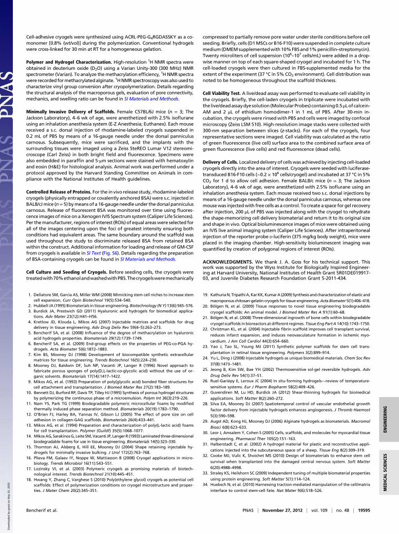
cells was next examined. Square-shaped rhodamine-labeled RGD-containing alginate cryogels (4× 4× 1mm)were prepared (Fig. 6A),purified, sterilized, and subsequently seeded with bioluminescentreporter cells, and maintained in culture for 1 d to promote celladhesion (Fig. 6 B and C). Healthy C57BL/6J mice received a s.c.injection on their backs of a series of bioluminescent reporter cellsseeded on rhodamine-labeled cryogels with and without adhesionpeptide. As a control, a bolus of free cells was also injected. Im-mediately after injection, detection of red-emitting rhodaminedyes indicated successful injection and in vivo localization of flu-orescently-labeled scaffolds (Fig. 6D). Both types of cryogelsmaintained cells at the point of introduction area after 2 d (Fig.6E) as the bioluminescence was particularly bright, whereas sim-ple cell injections did not. RGD appeared to lead to more cellretention. By day 15, RGD had a major effect in supporting cell
Fig. 2. SEM cross-sectional image of 1% (wt/vol) MA-alginatecryogel showing interconnected macroporous network (A) andnanoporous structure of conventional hydrogels (B). Two-di-mensional micro-CT images of alginate cryogel (C) and con-ventional hydrogel (D), and reconstructed three-dimensionalmicro-CT image of alginate cryogel (E). Photographs of cylin-drical MA-alginate hydrogels, demonstrating the effect of Ca2+
treatment on cryogels and conventional hydrogels (F).
Fig. 3. Comparison of physical properties of conventional nanoporous gelsversus cryogels. Both gel types were fabricated using 1% (wt/vol) MA-alginate.Young’s moduli for alginate cryogels and conventional hydrogels (A). Stress vs.strain curves for macroporous and nanoporous rhodamine-labeled alginatehydrogels subjected to compression tests (B). Evaluation of pore connectivity(C) and weight swelling ratio (D) of macroporous and conventional alginatehydrogels. Swelling ratios were determined for gels in the absence (−) andpresence (+) of CaCl2. Values in A, C, and D represent mean and SD (n = 4).
19592 | www.pnas.org/cgi/doi/10.1073/pnas.1211516109 Bencherif et al.
Dow
nloa
ded
by g
uest
on
May
10,
202
0

engraftment, as many more transplanted cells were presentcompared with cryogels without RGD peptides (Fig. 6F).
DiscussionThe results of this study suggest that a minimally invasive deliveryapproach can be used to introduce preformed shape-memorymacroporous cryogels via needle–syringe injection into the body
without mechanical fracture. These cryogels could be readilyinjected into the s.c. layer of the skin, used to fill in vivo tissuedefects and cavities, or used as patches on internal organs. Thesematerials have an interconnectedmacroporous architecture, whichis advantageous with respect to their ability to facilitate cellularinfiltration, and incorporation of payloads (e.g., cells) after de-vices are manufactured. The most important requirement of thesematerials for minimally invasive therapies is the ability to collapse,and faithfully and rapidly resume their original structure onceplaced in the body. Shape memory allows for this function. Fur-thermore, the ability of these materials to reassume specific, pre-defined shapes after injection is likely to be useful in applicationssuch as tissue patches (e.g., cardiac patches) where one desiresa patch of a specific size and shape, or when one desires to fill alarge defect site with multiple smaller objects that pack in such amanner to leave voids that enhance diffusional transport to/fromthe objects and the host, and promote vascularization around eachobject (30, 31). The polymeric system can be used as a deliverydevice for biomolecules, including proteins, as long-term sus-tained release of encapsulated rhodamine-labeled BSA and GM-CSF demonstrate. Also, the feasibility of this approach to trans-plant cells was examined with bioluminescent reporter cells. Thebioactive cryogels provided a favorable microenvironment to fa-cilitate cell survival and engraftment of transplanted cells.Cryogels have tissue-like compliance and are mechanically robust
due to a highly cross-linked polymer phase that results from stericallyentangled polymer chains, a high degree of chemical modification ofalginate, a high polymer concentration, and nearly complete re-activity of methacryoyl groups during cryogelation. Unlike conven-tional gels, a unique characteristic of these scaffolds is that when anappropriate shear stress is applied, the cryogels can be reversiblycompressed at over 90% strain levels, resulting in injectable mac-roporous preformed scaffolds. The large volumetric change of thecryogels is caused by reversible collapse of the interconnected poresforcing water contained in themacropores to flow out of the gel. Thecryogels have the ability to memorize a macroscopic and permanentshape, to be manipulated and temporarily collapsed, and then laterto be relaxed to their original structure without residual permanentdeformation. The relaxation is associated with the energy storedduring the elastic gel collapse. The gels in this study were fabricatedwith high–molecular-weight alginate but can be formedwith a varietyof materials, including biodegradable polymers. One could furthertune their performance by altering their composition, formulation,and degradation profiles. The ability of these gels to be syringe
Fig. 4. Minimally invasive s.c. injection of alginatecryogels into thebacks of C57BL/6Jmice (A). Hydrogellocalization after s.c. injection of preformed rhoda-mine-labeled 1%MA-alginate cryogels (4× 4× 1mm)in the subcutis of a mouse after 3 d (B). Histologicalanalysis (H&E stain) of explanted alginate cryogel atday 3. The arrows indicate the interface between thes.c. connective tissue (Lower Left) and the cryogelmatrix (Upper Right). (Magnification: 10×.) (C). Pho-tographs showing merged phase-contrast and fluo-rescence of s.c. injected rhodamine-labeled alginatemacroporous scaffold with restoration of geometryafter placement (D). Three days postinjection, thedashed lines indicate original square-shaped geom-etry of recovered scaffolds and the arrows show thespread of released BSA into the surrounding tissue(E). In vivo release profiles of cross-linked (chemicallyanchored) or encapsulated (physically entrapped)rhodamine-labeled BSA containing injected cryogels(F). Values represent mean and SD (n = 4).
Fig. 5. Adhesion of MSC cells in MA-alginate cryogels. Phase-contrast im-age showing elongated cells in RGD-containing cryogels (A). Confocalmicrographs of seeded cells after 5 d of culture are shown in a typical RGD-modified cryogel (B), unmodified cryogel (C), and RGE-modified cryogel (D).Live/dead cell assay of MSCs (E) (1 d incubation postinjection) and confocalimage showing cells (F) (6 d incubation postinjection) in RGD-modified MA-alginate cryogels. Cryogels were injected through a 16-gauge needle beforeimaging (E and F). (Scale bars: A–C, E, F, 20 μm; D, 10 μm.)
Bencherif et al. PNAS | November 27, 2012 | vol. 109 | no. 48 | 19593
ENGINEE
RING
MED
ICALSC
IENCE
S
Dow
nloa
ded
by g
uest
on
May
10,
202
0
delivered at a specific location without the need of an invasive sur-gery may decrease scarring, lessen the risk of infection, and reducerecovery times comparedwith traditional procedures. Themaximuminjectable gel size investigated to date is 8× 8× 1mm for the square-shaped cryogels. However, the technique used to fabricate gels isamenable to scaling up for larger injectable structures.These cryogels can act as a delivery vehicle for therapeutics
and cells, with sustained release over time. By providing a pro-tein drug depot at the site of injection, the cryogels can poten-tially achieve high local protein concentrations without systemicexposure to the bioactive proteins. Biomacromolecules, such asBSA used here as a model protein and GM-CSF, can be physi-cally entrapped during polymerization. Due to the large size ofthe protein paired with a dense polymer microstructure, theseproteins were retained in the cryogel during injection and re-leased in vitro and in vivo in a sustained fashion.The open pore structure of the cryogels with bioactive surfaces
and tissue-like elasticity provide an appropriate microenviron-ment for cells. Absorption of a cell suspension occurs throughthe pores by convection, which was used to uniformly seed cellsthroughout the gel. Cell entrapment results in a very efficientimmobilization system, in which the cells are initially caged in gelpores. To prevent anoikis and to retain cells, adhesion peptideswere grafted to the cryogels (32). During the syringe injection ofcell-seeded cryogels, over 80% of cells initially in cryogels were
retained, with high viability. These cell-seeded injectable porousscaffolds are potentially of wide utility for tissue regeneration; onecould apply this technology to develop biologically active scaffoldsthat controllably deliver growth factors while providing cellularbuilding blocks to enhance tissue formation. In an attempt to im-prove cell transplantation, biocompatible macroporous alginatecryogels have been used to create a niche that provides the ap-propriate microenvironment to promote survival of transplantedcells. This microenvironment includes chemical and physical cuesdesigned to guide cell growth and integration with the host tissue.The uniformdistribution and viability of seeded cells are unaffectedby the shear thinning during injection and the gel constructs remainat the point of introduction, suggesting that the localized deliveryof gels may be useful for the delivery of cells in close proximity toa lesion site in tissue regeneration efforts. More broadly, this mayprovide a core technology useful to solve some of the fundamentalproblems associated with current cell-based therapies—the rapidloss of cell viability, low engraftment efficiency, and absence ofcontrol over cell fate after introduction into the body (32). Theindependent tuning of cell-adhesive properties is desirable in cre-ating a cellular microenvironment suitable for survival, consistentwith previous studies (33). Biomimetic cryogels seeded with bio-luminescent reporter cells and introduced s.c. in mice showed en-hanced bioluminescence signals, indicating that cell delivery usingcryogels decreases cell loss from the injection site, enhances sur-vival, and promotes engraftment of cells in vivo.
Materials and MethodsMaterials. Sodium alginate with high guluronate content (LF 20/60) was pur-chased from ProNova Biomedical; and 2-morpholinoethanesulfonic acid (Mes),sodium chloride (NaCl), calcium chloride (CaCl2) sodium hydroxide (NaOH), N-hydroxysuccinimide (NHS), 1-ethyl-3-(3-dimethylaminopropyl)-carbodiimidehydrochloride (EDC), AEMA, methacrylic anhydride (MAH), BSA, 5(6)-carboxy-X-rhodamine N-succinimidyl ester (rhodamine-NHS), MA, and acetone werepurchased from Sigma-Aldrich. ACRL-PEG-NHS (3.5 kDa) was purchased fromJenKen Technology. The integrin binding peptide (Gly)4-Arg-Gly-Asp-Ala-Ser-Ser-Lys-Tyr was purchased from Commonwealth Biotech. Clonally derived mu-rine mesenchymal stem cells (D1 MSCs) (ATCC) and luciferase-transduced B16-F10 cells (ATCC) were cultured in Dulbecco’s modified Eagle medium (DMEM)supplemented with 10% (vol/vol) FBS and 1% (vol/vol) penicillin–streptomycin,all obtained from Invitrogen. Live/Dead Viability/Cytotoxicity Kit was pur-chased from Invitrogen. D-Luciferin was obtained from Gold Biotechnology.
Chemical Modification. MA-alginate was prepared by reacting alginate withAEMA. Sodium alginate (1 g) was dissolved in a buffer solution [0.6% (wt/vol),pH ∼6.5] of 100 mM Mes. NHS (1.3 g) and EDC (2.8 g) were added to themixture to activate the carboxylic acid groups of alginate. After 5 min,AEMA (2.24 g; molar ratio of NHS:EDC:AEMA = 1:1.3:1.1) was added to theproduct and the solution was stirred at RT for 24 h. The mixture was pre-cipitated in acetone, filtered, and dried in a vacuum oven overnight at RT. 1HNMR was used to characterize chemical modification of alginate and degreeof functionalization of MA-alginate. Rhod-BSA-MA was prepared by react-ing Rhod-BSA with MAH. Rhod-BSA was synthesized according to ref. 34.Rhod-BSA (100 mg) was dissolved in deionized water, and MAH (30 mg) wasadded to the mixture. After 30 min, the mixture was precipitated in an ex-cess of acetone, filtered, and dried in a vacuum oven overnight at RT.
Hydrogel Fabrication. Macroporous and conventional (nanoporous) matricesweresynthesizedbyredox-inducedfree-radicalpolymerizationofMA-alginateinwater. ACRL-PEG-G4RGDASSKY and ACRL-PEG-G4RGEASSKY were synthesizedaccording to ref. 5.Alginate cryogelswere synthesizedbymixing 10mg [1% (wt/vol)] of MA-alginate macromonomer in deionized water with tetramethyle-thylenediamine (TEMED) [0.5% (wt/vol)] and ammonium persulfate (APS)[0.25% (wt/vol)]. Fabrication conditions were chosen to allow the solution tofreeze before the gelation takes place and thus eliminate much of the de-pendence of the resulting structure on freezing rate, object size, and geometry.More specifically, a very small amount of initiator was used, the precursor so-lution was precooled to 4 °C to decrease the rate of polymerization beforefreezing, and once the initiator was added to the prepolymer solution, the so-lution was quickly poured into a precooled (−20 °C) Teflon mold. The solutionfroze within a minute while a witness solution stored at 4 °C remained unpo-lymerized for at least 20 min. After a complete incubation period of 17 h, thegels were heated to RT to remove ice crystals andwashedwith deionizedwater.
Fig. 6. Injectable preseeded scaffolds promote in situ localization and reten-tion of bioluminescent reporter cells. Photographs showing alginate cryogelscaffolds (white) and rhodamine-labeled alginate scaffolds (pink) (A). Biolu-minescent cells were seeded on 1% RGD-modified MA-alginate cryogels at aconcentration of 200 × 103 cells/scaffold, and cultured for 1 d. Optical imagingdepicts distribution of bioluminescent cells (B). SEM image shows cells (pseu-docolored blue) homogeneously adherent to the gel (C). Real-time fluores-cence in vivo imaging showing injected Rhod-labeled cryogels. Cells injecteds.c. via Rhod-labeled cryogels (1), Rhod-labeled RGD-cryogels (2), or as a free-floating cell suspension (B) in BALB/c mice at day 0 (D). Noninvasive bio-luminescence in vivo imaging of transplanted cells in BALB/c mice at day 2 (E)and day 15 (F) postinjection. The samemice were used in each of these imagesand were arranged in the same relative positions before imaging (D–F).
19594 | www.pnas.org/cgi/doi/10.1073/pnas.1211516109 Bencherif et al.
Dow
nloa
ded
by g
uest
on
May
10,
202
0
Cell-adhesive cryogels were synthesized using ACRL-PEG-G4RGDASSKY as a co-monomer [0.8% (wt/vol)] during the polymerization. Conventional hydrogelswere cross-linked for 30 min at RT for a homogeneous gelation.
Polymer and Hydrogel Characterization. High-resolution 1H NMR spectra wereobtained in deuterium oxide (D2O) using a Varian Unity-300 (300 MHz) NMRspectrometer (Varian). To analyze themethacrylation efficiency, 1HNMR spectrawere recorded formethacrylatedalginate. 1HNMRspectroscopywasalsousedtocharacterize vinyl group conversion after cryopolymerization. Details regardingthe structural analysis of the macroporous gels, evaluation of pore connectivity,mechanics, and swelling ratio can be found in SI Materials and Methods.
Minimally Invasive Delivery of Scaffolds. Female C57BL/6J mice (n = 3; TheJackson Laboratory), 4–6 wk of age, were anesthetized with 2.5% isofluraneusing an inhalation anesthesia system (E-Z Anesthesia; Euthanex). Each mousereceived a s.c. dorsal injection of rhodamine-labeled cryogels suspended in0.2 mL of PBS by means of a 16-gauge needle under the dorsal panniculuscarnosus. Subsequently, mice were sacrificed, and the implants with thesurrounding tissues were imaged using a Zeiss SteREO Lumar V12 stereomi-croscope (Carl Zeiss) in both bright field and fluorescence. Specimens werealso embedded in paraffin and 5-μm sections were stained with hematoxylinand eosin (H&E) for histological analysis. Animal work was performed under aprotocol approved by the Harvard Standing Committee on Animals in com-pliance with the National Institutes of Health guidelines.
Controlled Release of Proteins. For the in vivo release study, rhodamine-labeledcryogels (physically entrapped or covalently anchored BSA) were s.c. injected inBALB/cJmice (n= 5) bymeansof a 16-gaugeneedleunder the dorsal panniculuscarnosus. Release of fluorescent BSA was monitored over time using fluores-cence imagesofmiceonaXenogen IVIS Spectrum system (Caliper Life Sciences).Per themanufacturer, regions of interest (ROIs) of equal areaswere selected forall of the images centering upon the foci of greatest intensity ensuring bothconditions had equivalent areas. The same boundary around the scaffold wasused throughout the study to discriminate released BSA from retained BSAwithin the construct. Additional information for loading and release ofGM-CSFfrom cryogels is available in SI Text (Fig. S6). Details regarding the preparationof BSA-containing cryogels can be found in SI Materials and Methods.
Cell Culture and Seeding of Cryogels. Before seeding cells, the cryogels weretreatedwith70%ethanolandwashedwithPBS.Thecryogelsweremechanically
compressed to partially remove pore water under sterile conditions before cellseeding. Briefly, cells (D1MSCs or B16-F10)were suspended in complete culturemedium(DMEMsupplementedwith10%FBSand1%penicillin–streptomycin).Twenty microliters of cell suspension (106–107 cells/mL) were added in a drop-wise manner on top of each square-shaped cryogel and incubated for 1 h. Thecell-loaded cryogels were then cultured in FBS-supplemented media for theextent of the experiment (37 °C in 5% CO2 environment). Cell distribution wasnoted to be homogeneous throughout the scaffold thickness.
Cell Viability Test. A live/dead assay was performed to evaluate cell viability inthe cryogels. Briefly, the cell-laden cryogels in triplicate were incubated withthe live/deadassaydye solution (MolecularProbes) containing0.5μLofcalcein-AM and 2 μL of ethidium homodimer-1 in 1 mL of PBS. After 30-min in-cubation, the cryogels were rinsedwith PBS and cells were imaged by confocalmicroscopy (Zeiss LSM 510). High-resolution image stacks were collected with300-nm separation between slices (z-stacks). For each of the cryogels, fourrepresentative sections were imaged. Cell viability was calculated as the ratioof green fluorescence (live cell) surface area to the combined surface area ofgreen fluorescence (live cells) and red fluorescence (dead cells).
Delivery of Cells. Localizeddeliveryof cellswas achievedby injecting cell-loadedcryogels directly into the area of interest. Cryogels were seededwith luciferase-transduced B16-F10 cells (∼0.2 × 10⁶ cells/cryogel) and incubated at 37 °C in 5%CO2 for 1 d to allow cell adhesion. Female BALB/c mice (n = 3; The JacksonLaboratory), 4–6 wk of age, were anesthetized with 2.5% isoflurane using aninhalation anesthesia system. Each mouse received two s.c. dorsal injections bymeans of a 16-gauge needle under the dorsal panniculus carnosus, whereas onemousewas injectedwith free cells as a control. To create a space for gel recoveryafter injection, 200 μL of PBS was injected along with the cryogel to rehydratethe shape-memorizing cell delivery biomaterial and return it to its original sizeand shape in vivo. Optical bioluminescence images ofmicewere obtained usingan IVIS live animal imaging system (Caliper Life Sciences). After intraperitonealinjection of the reporter probe D-luciferin (375 mg/kg body weight), mice wereplaced in the imaging chamber. High-sensitivity bioluminescent imaging wasquantified by creation of polygonal regions of interest (ROIs).
ACKNOWLEDGMENTS. We thank J. A. Goss for his technical support. Thiswork was supported by the Wyss Institute for Biologically Inspired Engineer-ing at Harvard University, National Institutes of Health Grant 5R01DE019917-03, and Juvenile Diabetes Research Foundation Grant 5-2011-434.
1. Dellatore SM, Garcia AS, Miller WM (2008) Mimicking stem cell niches to increase stemcell expansion. Curr Opin Biotechnol 19(5):534–540.
2. Hubbell JA (1995) Biomaterials in tissue engineering. Biotechnology (NY) 13(6):565–576.3. Burdick JA, Prestwich GD (2011) Hyaluronic acid hydrogels for biomedical applica-
tions. Adv Mater 23(12):H41–H56.4. Kretlow JD, Klouda L, Mikos AG (2007) Injectable matrices and scaffolds for drug
delivery in tissue engineering. Adv Drug Deliv Rev 59(4–5):263–273.5. Bencherif SA, et al. (2008) Influence of the degree of methacrylation on hyaluronic
acid hydrogels properties. Biomaterials 29(12):1739–1749.6. Bencherif SA, et al. (2009) End-group effects on the properties of PEG-co-PGA hy-
drogels. Acta Biomater 5(6):1872–1883.7. Kim BS, Mooney DJ (1998) Development of biocompatible synthetic extracellular
matrices for tissue engineering. Trends Biotechnol 16(5):224–230.8. Mooney DJ, Baldwin DF, Suh NP, Vacanti JP, Langer R (1996) Novel approach to
fabricate porous sponges of poly(D,L-lactic-co-glycolic acid) without the use of or-ganic solvents. Biomaterials 17(14):1417–1422.
9. Mikos AG, et al. (1993) Preparation of poly(glycolic acid) bonded fiber structures forcell attachment and transplantation. J Biomed Mater Res 27(2):183–189.
10. Bennett DJ, Burford RP, Davis TP, Tilley HJ (1995) Synthesis of porous hydrogel structuresby polymerizing the continuous phase of a microemulsion. Polym Int 36(3):219–226.
11. Nam YS, Park TG (1999) Biodegradable polymeric microcellular foams by modifiedthermally induced phase separation method. Biomaterials 20(19):1783–1790.
12. O’Brien FJ, Harley BA, Yannas IV, Gibson LJ (2005) The effect of pore size on celladhesion in collagen-GAG scaffolds. Biomaterials 26(4):433–441.
13. Mikos AG, et al. (1994) Preparation and characterization of poly(L-lactic acid) foamsfor cell transplantation. Polymer (Guildf) 35(5):1068–1077.
14. MikosAG, SarakinosG, LeiteSM,Vacanti JP, Langer R (1993) Laminated three-dimensionalbiodegradable foams for use in tissue engineering. Biomaterials 14(5):323–330.
15. Thornton AJ, Alsberg E, Hill EE, Mooney DJ (2004) Shape retaining injectable hy-drogels for minimally invasive bulking. J Urol 172(2):763–768.
16. Plieva FM, Galaev IY, Noppe W, Mattiasson B (2008) Cryogel applications in micro-biology. Trends Microbiol 16(11):543–551.
17. Lozinsky VI, et al. (2003) Polymeric cryogels as promising materials of biotech-nological interest. Trends Biotechnol 21(10):445–451.
18. Hwang Y, Zhang C, Varghese S (2010) Poly(ethylene glycol) cryogels as potential cellscaffolds: Effect of polymerization conditions on cryogel microstructure and proper-ties. J Mater Chem 20(2):345–351.
19. KathuriaN, TripathiA,KarKK,KumarA (2009) Synthesis and characterizationof elastic and
macroporous chitosan-gelatin cryogels for tissue engineering.Acta Biomater 5(1):406–418.20. Bölgen N, et al. (2009) Tissue responses to novel tissue engineering biodegradable
cryogel scaffolds: An animal model. J Biomed Mater Res A 91(1):60–68.21. Bölgen N, et al. (2008) Three-dimensional ingrowth of bone cells within biodegradable
cryogel scaffolds in bioreactors at different regimes. Tissue EngPartA 14(10):1743–1750.22. Christman KL, et al. (2004) Injectable fibrin scaffold improves cell transplant survival,
reduces infarct expansion, and induces neovasculature formation in ischemic myo-
cardium. J Am Coll Cardiol 44(3):654–660.23. Yao J, Tao SL, Young MJ (2011) Synthetic polymer scaffolds for stem cell trans-
plantation in retinal tissue engineering. Polymers 3(2):899–914.24. Yu L, Ding J (2008) Injectable hydrogels as unique biomedical materials. Chem Soc Rev
37(8):1473–1481.25. Jeong B, Kim SW, Bae YH (2002) Thermosensitive sol-gel reversible hydrogels. Adv
Drug Deliv Rev 54(1):37–51.26. Ruel-Gariépy E, Leroux JC (2004) In situ-forming hydrogels—review of temperature-
sensitive systems. Eur J Pharm Biopharm 58(2):409–426.27. Guvendiren M, Lu HD, Burdick JA (2012) Shear-thinning hydrogels for biomedical
applications. Soft Matter 8(2):260–272.28. Silva EA, Mooney DJ (2007) Spatiotemporal control of vascular endothelial growth
factor delivery from injectable hydrogels enhances angiogenesis. J Thromb Haemost
5(3):590–598.29. Augst AD, Kong HJ, Mooney DJ (2006) Alginate hydrogels as biomaterials. Macromol
Biosci 6(8):623–633.30. Leor J, Amsalem Y, Cohen S (2005) Cells, scaffolds, and molecules for myocardial tissue
engineering. Pharmacol Ther 105(2):151–163.31. Halberstadt C, et al. (2002) A hydrogel material for plastic and reconstructive appli-
cations injected into the subcutaneous space of a sheep. Tissue Eng 8(2):309–319.32. Cooke MJ, Vulic K, Shoichet MS (2010) Design of biomaterials to enhance stem cell
survival when transplanted into the damaged central nervous system. Soft Matter
6(20):4988–4998.33. Straley KS, Heilshorn SC (2009) Independent tuning of multiple biomaterial properties
using protein engineering. Soft Matter 5(1):114–124.34. Huebsch N, et al. (2010) Harnessing traction-mediated manipulation of the cell/matrix
interface to control stem-cell fate. Nat Mater 9(6):518–526.
Bencherif et al. PNAS | November 27, 2012 | vol. 109 | no. 48 | 19595
ENGINEE
RING
MED
ICALSC
IENCE
S
Dow
nloa
ded
by g
uest
on
May
10,
202
0