inhibition of staphyloxanthin virulence factor biosynthesis in
TRANSCRIPT
Inhibition of Staphyloxanthin Virulence Factor Biosynthesis in Staphylococcus aureus: In Vitro,in Vivo, and Crystallographic Results†
Yongcheng Song,‡ Chia-I Liu,§,¶,⊥ Fu-Yang Lin,[ Joo Hwan No,[ Mary Hensler,# Yi-Liang Liu,[ Wen-Yih Jeng,§,¶,⊥
Jennifer Low,| George Y. Liu,| Victor Nizet,# Andrew H.-J. Wang,§,¶,⊥ and Eric Oldfield*,‡,[
Department of Chemistry and Center for Biophysics and Computational Biology, UniVersity of Illinois at UrbanasChampaign, 600 S. MathewsAVenue, Urbana, Illinois 61801, Institute of Biological Chemistry, Academia Sinica, Nankang, Taipei 11529, Taiwan, National Core Facility ofHigh-Throughput Protein Crystallography, Academia Sinica, Nankang, Taipei 11529, Taiwan, Institute of Biochemical Sciences, College of LifeScience, National Taiwan UniVersity, Taipei 10098, Taiwan, Department of Pediatrics and Skaggs School of Pharmacy and PharmaceuticalSciences, UniVersity of California, San Diego, 9500 Gilman DriVe, La Jolla, CA, and DiVision of Pediatric Infectious Diseases andImmunobiology Research Institute, Cedars-Sinai Medical Center, Los Angeles, California 90048
ReceiVed February 11, 2009
The gold color of Staphylococcus aureus is derived from the carotenoid staphyloxanthin, a virulence factorfor the organism. Here, we report the synthesis and activity of a broad variety of staphyloxanthin biosynthesisinhibitors that inhibit the first committed step in its biosynthesis, condensation of two farnesyl diphosphate(FPP) molecules to dehydrosqualene, catalyzed by the enzyme dehydrosqualene synthase (CrtM). The mostactive compounds are phosphonoacetamides that have low nanomolar Ki values for CrtM inhibition and areactive in whole bacterial cells and in mice, where they inhibit S. aureus disease progression. We also reportthe X-ray crystallographic structure of the most active compound, N-3-(3-phenoxyphenyl)propylphospho-noacetamide (IC50 ) 8 nM, in cells), bound to CrtM. The structure exhibits a complex network of hydrogenbonds between the polar headgroup and the protein, while the 3-phenoxyphenyl side chain is located in ahydrophobic pocket previously reported to bind farnesyl thiodiphosphate (FsPP), as well as biphenylphosphonosulfonate inhibitors. Given the good enzymatic, whole cell, and in vivo pharmacologic activities,these results should help guide the further development of novel antivirulence factor-based therapies for S.aureus infections.
Introduction
Infections caused by Staphylococcus aureus are a growingcause of concern1,2 because of the widespread development ofantibiotic resistance and the shortfall in the introduction of newtypes of anti-infective agents. An alternative strategy that is nowgaining interest is targeting of bacterial virulence factors,3-5
molecules that are essential for bacterial growth and/or inva-siveness in vivo. Since these factors are by definition notessential for survival in vitro, screening for virulence factorinhibitors can be challenging. However, at least in the case ofS. aureus,6,7 one important virulence factor is a brightly coloredcarotenoid pigment, staphyloxanthin (STXa), whose biosynthesiscan be readily monitored spectrophotometrically. The carotenoidis produced by the condensation of the C15 isoprenoid farnesyldiphosphate (FPP) to form presqualene diphosphate and thendehydrosqualene, followed by a series of oxidations andglycosylations, in a series of reactions catalyzed by the enzymesCrtM, N, O, P,...,8 and inhibition of these enzymes (e.g., CrtNby diphenylamine) has been known for many years to result in
colorless bacteria.9,10 In later work,6 it was shown that loss ofthe STX pigment made S. aureus susceptible to killing byreactive oxygen species (ROS, such as H2O2, ClO-, OH)produced by neutrophils, blocking infectivity. Consequently,inhibition of staphyloxanthin biosynthesis is a potential noveltarget of anti-infective therapy against pigmented S. aureusstrains.11-13
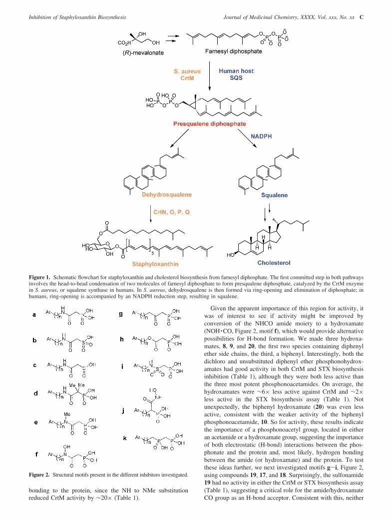
The first committed step in staphyloxanthin biosynthesis(Figure 1) involves the condensation of two FPP molecules toform dehydrosqualene. This reaction is thought to occur via apresqualene diphosphate intermediate and is very similar (oridentical) to that catalyzed by squalene synthase (SQS) in plants,animals, and some protozoa, where the squalene so producedis then converted into sterols such as cholesterol, ergosterol,and the plant sterols. It was thus of interest to see if any of themany known SQS inhibitors previously developed as cholesterollowering drug leads might also have activity in blockingstaphyloxanthin biosynthesis and hence S. aureus virulence. Werecently reported that one class of inhibitor, phosphonosul-fonates, did exhibit such activity.14 The phosphonosulfonates(and related bisphosphonates) were developed earlier by Magninet al.15,16 using FPP as a “template”. The bisphosphonateanalogues of FPP tended to bind to bone and also causedelevation of liver enzyme function, and the farnesyl side chainwas metabolically reactive. However, the phosphonosulfonatesdid not have these drawbacks, and replacement of the farnesylside chain by a diphenyl ether removed the metabolic instability.Using 1, we found good CrtM inhibition restored ROS (H2O2,neutrophil) sensitivity and, importantly, observed a majordecrease in bacterial burden following S. aureus challenge inmice.14 There are, however, many other conceivable backbones(as well as side chains) that might also have good or even better
† Crystal structure coordinates have been deposited in the Protein DataBank and will be released upon publication (2ZY1).
* To whom correspondence should be addressed. Phone: 217-333-3374.Fax: 217-244-0997. E-mail: [email protected].
‡ Department of Chemistry, University of Illinois at UrbanasChampaign.§ Institute of Biological Chemistry.¶ National Core Facility of High-Throughput Protein Crystallography.⊥ National Taiwan University.[ Center for Biophysics and Computational Biology, University of
Illinois at UrbanasChampaign.# University of California, San Diego.| Cedars-Sinai Medical Center.a Abbreviations: CrtM, dehydrosqualene synthase; FPP, farnesyl diphos-
phate; QSAR, quantitative structure-activity relationship; SQS, squalenesynthase; STX, staphyloxanthin.
J. Med. Chem. XXXX, xxx, 000 A
10.1021/jm9001764 CCC: $40.75 XXXX American Chemical Society
activity. To explore some of these possibilities, we wereparticularly interested to see if it might be possible to reducebackbone charge/acidity/polarity while still retaining CrtMactivity, since this might improve inhibitor uptake into bacterialand host cells, as well as further reducing bone affinity. Thepossibility that less polar analogues might still have good activityis supported by the observation that, unlike bisphosphonateinhibitors of farnesyl diphosphate synthase (FPPS), our pub-lished CrtM results indicated that Mg2+ binding (which usuallyinvolves binding to two anionic groups) is not essential forpotent phosphonosulfonate inhibition of CrtM.14 For example,while 1 binds to CrtM with two Mg2+ (PDB file 2ZCQ), 2 bindswith only one Mg2+ (PDB file 2ZCR), and 3 has no Mg2+ at allin its X-ray crystallographic structure (PDB file 2ZCS). So,unlike the situation found with FPPS, it seemed likely that abroad range of backbone structures having fewer anionic groupsmight be developed, since the number of metal ions involvedin binding to CrtM is quite variable.
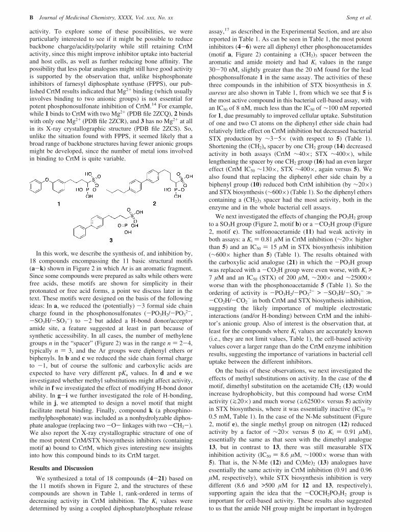
In this work, we describe the synthesis of, and inhibition by,18 compounds encompassing the 11 basic structural motifs(a-k) shown in Figure 2 in which Ar is an aromatic fragment.Since some compounds were prepared as salts while others werefree acids, these motifs are shown for simplicity in theirprotonated or free acid forms, a point we discuss later in thetext. These motifs were designed on the basis of the followingideas: In a, we reduced the (potentially) -3 formal side chaincharge found in the phosphonosulfonates (-PO3H2/-PO3
2-,-SO3H/-SO3
-) to -2 but added a H-bond donor/acceptoramide site, a feature suggested at least in part because ofsynthetic accessibility. In all cases, the number of methylenegroups n in the “spacer” (Figure 2) was in the range n ) 2-4,typically n ) 3, and the Ar groups were diphenyl ethers orbiphenyls. In b and c we reduced the side chain formal chargeto -1, but of course the sulfonic and carboxylic acids areexpected to have very different pKa values. In d and e weinvestigated whether methyl substitutions might affect activity,while in f we investigated the effect of modifying H-bond donorability. In g-i we further investigated the role of H-bonding,while in j, we attempted to design a novel motif that mightfacilitate metal binding. Finally, compound k (a phosphino-methylphosphonate) was included as a nonhydrolyzable diphos-phate analogue (replacing two -O- linkages with two -CH2-).We also report the X-ray crystallographic structure of one ofthe most potent CrtM/STX biosynthesis inhibitors (containingmotif a) bound to CrtM, which gives interesting new insightsinto how this compound binds to its CrtM target.
Results and Discussion
We synthesized a total of 18 compounds (4-21) based onthe 11 motifs shown in Figure 2, and the structures of thesecompounds are shown in Table 1, rank-ordered in terms ofdecreasing activity in CrtM inhibition. The Ki values weredetermined by using a coupled diphosphate/phosphate release
assay,17 as described in the Experimental Section, and are alsoreported in Table 1. As can be seen in Table 1, the most potentinhibitors (4-6) were all diphenyl ether phosphonoacetamides(motif a, Figure 2) containing a (CH2)3 spacer between thearomatic and amide moiety and had Ki values in the range30-70 nM, slightly greater than the 20 nM found for the leadphosphonsulfonate 1 in the same assay. The activities of thesethree compounds in the inhibition of STX biosynthesis in S.aureus are also shown in Table 1, from which we see that 5 isthe most active compound in this bacterial cell-based assay, withan IC50 of 8 nM, much less than the IC50 of ∼100 nM reportedfor 1, due presumably to improved cellular uptake. Substitutionof one and two Cl atoms on the diphenyl ether side chain hadrelatively little effect on CrtM inhibition but decreased bacterialSTX production by ∼3-5× (with respect to 5) (Table 1).Shortening the (CH2)n spacer by one CH2 group (14) decreasedactivity in both assays (CrtM ∼40×; STX ∼400×), whilelengthening the spacer by one CH2 group (16) had an even largereffect (CrtM IC50 ∼130×, STX ∼400×, again versus 5). Wealso found that replacing the diphenyl ether side chain by abiphenyl group (10) reduced both CrtM inhibition (by ∼20×)and STX biosynthesis (∼600×) (Table 1). So the diphenyl etherscontaining a (CH2)3 spacer had the most activity, both in theenzyme and in the whole bacterial cell assays.
We next investigated the effects of changing the PO3H2 groupto a SO3H group (Figure 2, motif b) or a -CO2H group (Figure2, motif c). The sulfonoacetamide (11) had weak activity inboth assays: a Ki ) 0.81 µM in CrtM inhibition (∼20× higherthan 5) and an IC50 ) 15 µM in STX biosynthesis inhibition(∼600× higher than 5) (Table 1). The results obtained withthe carboxylic acid analogue (21) in which the -PO3H groupwas replaced with a -CO2H group were even worse, with Ki >7 µM and an IC50 (STX) of 200 µM, ∼200× and ∼25000×worse than with the phosphonoacetamide 5 (Table 1). So theordering of activity is -PO3H2/-PO3
2- > -SO3H/-SO3- .
-CO2H/-CO2- in both CrtM and STX biosynthesis inhibition,
suggesting the likely importance of multiple electrostaticinteractions (and/or H-bonding) between CrtM and the inhibi-tor’s anionic group. Also of interest is the observation that, atleast for the compounds where Ki values are accurately known(i.e., they are not limit values, Table 1), the cell-based activityvalues cover a larger range than do the CrtM enzyme inhibitionresults, suggesting the importance of variations in bacterial celluptake between the different inhibitors.
On the basis of these observations, we next investigated theeffects of methyl substitutions on activity. In the case of the dmotif, dimethyl substitution on the acetamide CH2 (13) wouldincrease hydrophobicity, but this compound had worse CrtMactivity (J20×) and much worse (J62500× versus 5) activityin STX biosynthesis, where it was essentially inactive (IC50 ≈0.5 mM, Table 1). In the case of the N-Me substituent (Figure2, motif e), the single methyl group on nitrogen (12) reducedactivity by a factor of ∼20× versus 5 (to Ki ) 0.91 µM),essentially the same as that seen with the dimethyl analogue13, but in contrast to 13, there was still measurable STXinhibition activity (IC50 ) 8.6 µM, ∼1000× worse than with5). That is, the N-Me (12) and C(Me)2 (13) analogues haveessentially the same activity in CrtM inhibition (0.91 and 0.96µM, respectively), while STX biosynthesis inhibition is verydifferent (8.6 and >500 µM for 12 and 13, respectively),supporting again the idea that the -COCH2PO3H2 group isimportant for cell-based activity. These results also suggestedto us that the amide NH group might be important in hydrogen
B Journal of Medicinal Chemistry, XXXX, Vol. xxx, No. xx Song et al.
bonding to the protein, since the NH to NMe substitutionreduced CrtM activity by ∼20× (Table 1).
Given the apparent importance of this region for activity, itwas of interest to see if activity might be improved byconversion of the NHCO amide moiety to a hydroxamate(NOH ·CO, Figure 2, motif f), which would provide alternativepossibilities for H-bond formation. We made three hydroxa-mates, 8, 9, and 20, the first two species containing diphenylether side chains, the third, a biphenyl. Interestingly, both thedichloro and unsubstituted diphenyl ether phosphonohydrox-amates had good activity in both CrtM and STX biosynthesisinhibition (Table 1), although they were both less active thanthe three most potent phosphonoacetamides. On average, thehydroxamates were ∼6× less active against CrtM and ∼2×less active in the STX biosynthesis assay (Table 1). Notunexpectedly, the biphenyl hydroxamate (20) was even lessactive, consistent with the weaker activity of the biphenylphosphonoacetamide, 10. So for activity, these results indicatethe importance of a phosphonoacetyl group, located in eitheran acetamide or a hydroxamate group, suggesting the importanceof both electrostatic (H-bond) interactions between the phos-phonate and the protein and, most likely, hydrogen bondingbetween the amide (or hydroxamate) and the protein. To testthese ideas further, we next investigated motifs g-i, Figure 2,using compounds 19, 17, and 18. Surprisingly, the sulfonamide19 had no activity in either the CrtM or STX biosynthesis assay(Table 1), suggesting a critical role for the amide/hydroxamateCO group as an H-bond acceptor. Consistent with this, neither
Figure 1. Schematic flowchart for staphyloxanthin and cholesterol biosynthesis from farnesyl diphosphate. The first committed step in both pathwaysinvolves the head-to-head condensation of two molecules of farnesyl diphosphate to form presqualene diphosphate, catalyzed by the CrtM enzymein S. aureus, or squalene synthase in humans. In S. aureus, dehydrosqualene is then formed via ring-opening and elimination of diphosphate; inhumans, ring-opening is accompanied by an NADPH reduction step, resulting in squalene.
Figure 2. Structural motifs present in the different inhibitors investigated.
Inhibition of Staphyloxanthin Biosynthesis Journal of Medicinal Chemistry, XXXX, Vol. xxx, No. xx C

Table 1. Enzyme (CrtM, Ki), Pigment (STX, S. aureus, IC50), and Cell Growth (IC50) Inhibition Results for 4-21
a The value given are the Ki values for CrtM inhibition, in µM. b The values given are the IC50 values for STX (staphyloxanthin) virulence factor inhibitionin S. aureus and are in µM. c The values given are the Ki values for human squalene synthase inhibition (in vitro) and are in µM. d The values given are theIC50 values for MCF-7 cell growth inhibition, in µM. e The values given are the IC50 values for NCI-H460 cell growth inhibition, in µM. f The values givenare the IC50 values for SF-268 cell growth inhibition, in µM.
D Journal of Medicinal Chemistry, XXXX, Vol. xxx, No. xx Song et al.
the ester (17) nor the ketone (18) had significant activity in eitherassay (Table 1).
We then investigated two additional motifs, j and k in Figure2. The hydroxamate (j, 15) had modest activity in both assays(Ki, IC50 ∼4 µM) but was ∼100× (CrtM) to ∼500× (STX)less active than was 5. The phosphinomethylphosphonate 7(motif k) was a potent CrtM inhibitor (Ki ) 220 nM), but againwe believe, due to its increased polarity, it was ∼50× lesseffective in STX biosynthesis inhibition in whole cells than wasthe most potent phosphonoacetamide (5).
Finally, we investigated the effects of all 18 compounds onhuman SQS inhibition, together with their activity in growthinhibition of three human tumor cell lines (MCF-7, NCI-460,and SF-268) (Table 1). Although inhibition of human SQS maynot be a particularly important toxicologic consideration, givena choice, it would seem to be preferable to have a STXbiosynthesis inhibitor with poor activity against SQS, as opposedto one with potent SQS activity, and as can be seen in Table 1,several potent CrtM inhibitors do have relatively little activityin the SQS assay. More important, in essentially all cases wefind no inhibition of human cell growth (in three human celllines, IC50 > 300 µM), supporting the idea that these compoundswill have low toxicity. In fact, only the ester 17 had significantactivity (IC50 ≈ 70-90 µM) on human cell growth, but sincethis compound is inactive in STX biosynthesis inhibition, it isnot likely to be of interest.
Structure and Structure-Function Aspects. The resultsdescribed above are of interest, since they represent thedevelopment of several new, chemically diverse virulenceinhibitors for S. aureus. However, the relationships betweenstructure and activity need to be explored in more detail inorder to obtain a better understanding of how, in particular,the most active compounds act on their CrtM target. Whatis interesting about the most active compounds, the phospho-noacetamides, is that while they contain the same 3-(3-phenoxyphenyl)propyl (PhOC6H4(CH2)3-) side chain asfound in the most active phosphonosulfonates,18 the “back-bone” is clearly about two bond lengths greater in the newcompounds. So we next examined the effects of this commonfeature on binding.
In the CrtM enzyme, there are two binding sites for FPP,14
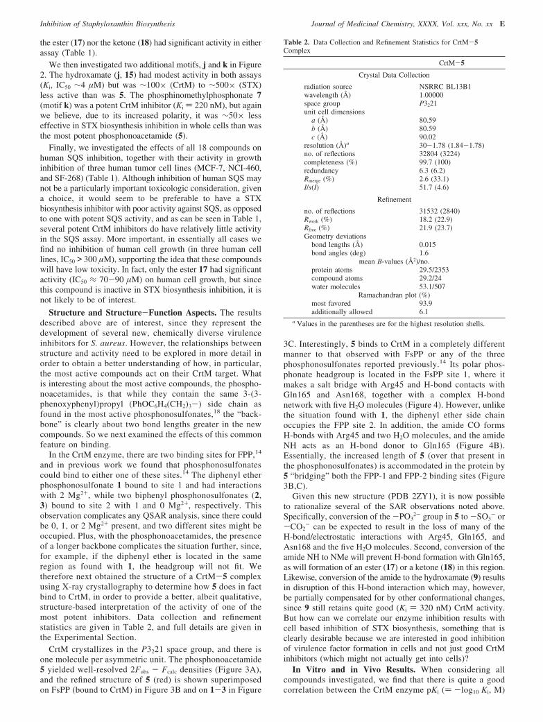
and in previous work we found that phosphonosulfonatescould bind to either one of these sites.14 The diphenyl etherphosphonosulfonate 1 bound to site 1 and had interactionswith 2 Mg2+, while two biphenyl phosphonosulfonates (2,3) bound to site 2 with 1 and 0 Mg2+, respectively. Thisobservation complicates any QSAR analysis, since there couldbe 0, 1, or 2 Mg2+ present, and two different sites might beoccupied. Plus, with the phosphonoacetamides, the presenceof a longer backbone complicates the situation further, since,for example, if the diphenyl ether is located in the sameregion as found with 1, the headgroup will not fit. Wetherefore next obtained the structure of a CrtM-5 complexusing X-ray crystallography to determine how 5 does in factbind to CrtM, in order to provide a better, albeit qualitative,structure-based interpretation of the activity of one of themost potent inhibitors. Data collection and refinementstatistics are given in Table 2, and full details are given inthe Experimental Section.
CrtM crystallizes in the P3221 space group, and there isone molecule per asymmetric unit. The phosphonoacetamide5 yielded well-resolved 2Fobs - Fcalc densities (Figure 3A),and the refined structure of 5 (red) is shown superimposedon FsPP (bound to CrtM) in Figure 3B and on 1-3 in Figure
3C. Interestingly, 5 binds to CrtM in a completely differentmanner to that observed with FsPP or any of the threephosphonosulfonates reported previously.14 Its polar phos-phonate headgroup is located in the FsPP site 1, where itmakes a salt bridge with Arg45 and H-bond contacts withGln165 and Asn168, together with a complex H-bondnetwork with five H2O molecules (Figure 4). However, unlikethe situation found with 1, the diphenyl ether side chainoccupies the FPP site 2. In addition, the amide CO formsH-bonds with Arg45 and two H2O molecules, and the amideNH acts as an H-bond donor to Gln165 (Figure 4B).Essentially, the increased length of 5 (over that present inthe phosphonosulfonates) is accommodated in the protein by5 “bridging” both the FPP-1 and FPP-2 binding sites (Figure3B,C).
Given this new structure (PDB 2ZY1), it is now possibleto rationalize several of the SAR observations noted above.Specifically, conversion of the -PO3
2- group in 5 to -SO3- or
-CO2- can be expected to result in the loss of many of the
H-bond/electrostatic interactions with Arg45, Gln165, andAsn168 and the five H2O molecules. Second, conversion of theamide NH to NMe will prevent H-bond formation with Gln165,as will formation of an ester (17) or a ketone (18) in this region.Likewise, conversion of the amide to the hydroxamate (9) resultsin disruption of this H-bond interaction which may, however,be partially compensated for by other conformational changes,since 9 still retains quite good (Ki ) 320 nM) CrtM activity.But how can we correlate our enzyme inhibition results withcell based inhibition of STX biosynthesis, something that isclearly desirable because we are interested in good inhibitionof virulence factor formation in cells and not just good CrtMinhibitors (which might not actually get into cells)?
In Vitro and in Vivo Results. When considering allcompounds investigated, we find that there is quite a goodcorrelation between the CrtM enzyme pKi () -log10 Ki, M)
Table 2. Data Collection and Refinement Statistics for CrtM-5Complex
CrtM-5
Crystal Data Collection
radiation source NSRRC BL13B1wavelength (Å) 1.00000space group P3221unit cell dimensions
a (Å) 80.59b (Å) 80.59c (Å) 90.02
resolution (Å)a 30-1.78 (1.84-1.78)no. of reflections 32804 (3224)completeness (%) 99.7 (100)redundancy 6.3 (6.2)Rmerge (%) 2.6 (33.1)I/s(I) 51.7 (4.6)
Refinement
no. of reflections 31532 (2840)Rwork (%) 18.2 (22.9)Rfree (%) 21.9 (23.7)Geometry deviations
bond lengths (Å) 0.015bond angles (deg) 1.6
mean B-values (Å2)/no.protein atoms 29.5/2353compound atoms 29.2/24water molecules 53.1/507
Ramachandran plot (%)most favored 93.9additionally allowed 6.1
a Values in the parentheses are for the highest resolution shells.
Inhibition of Staphyloxanthin Biosynthesis Journal of Medicinal Chemistry, XXXX, Vol. xxx, No. xx E

and cell STX biosynthesis inhibition pIC50 () -log10 IC50,M) values, as can be seen in Figure 5A where the R2 valueis 0.73 (for the 14 compounds having nonlimit Ki/IC50 values).However, when we add results for the 36 compounds reported
Figure 3. X-ray crystallographic results: (A) electron density for 5 in CrtM; (B) superposition of 5 (red) in the CrtM active site (2ZY1) with thatof two molecules (green, yellow) of S-thiolofarnesyl diphosphate (2ZCP); (C) superposition of 5 (red) with 1 (blue), 2 (yellow), 3 (cyan) in theCrtM active site (2ZCQ, 2ZCR, 2ZCS).
Figure 4. Interactions between 5 and different residues in the CrtMactive site: (A) Pymol26 view; (B) Ligplot27 interactions.
Figure 5. Figure showing correlations between CrtM inhibition andSTX biosynthesis inhibition: (A) data for the 14 compounds reportedin this work; (B) combination of 36 phosphonosulfonate inhibitor results(ref 18) with the 14 compounds reported here; (C) combinatorialdescriptor search result for all 50 compounds tested (here and in ref18) in CrtM and STX biosynthesis inhibition. The lower R2 value inpart B is likely due the high diversity of the large data set; the R2
improves to 0.68 by using the combinatorial descriptor approach.19
F Journal of Medicinal Chemistry, XXXX, Vol. xxx, No. xx Song et al.
previously to the correlation, the n ) 50 compound data setexhibits a much worse correlation (Figure 5B) with an R2 )0.42. This is similar to the results we reported previouslywhere we found for 10 different enzyme/cell assays that onaverage the R2 value for the pKi/pIC50 correlation was only0.32 (ref 19), making any predictions of cell-based activity,based on enzyme activity, in some cases, impossible. Thelarge discrepancies found were, we proposed, likely to bedue to the neglect of factors that affect inhibitor uptake intocells, and we described a general method in which this aspectmight be taken into consideration, by using a “combinatorialdescriptor approach”.19 That is, we described cell activityby using the following equation:
pIC50(cell) ) apKi(enzyme) + bB + cC + d
where a-d are linear regression coefficients and where B andC are mathematical descriptors (such as SlogP) chosen in acombinatorial manner from a large series of potential descriptors(such as the 230 descriptors in the program MOE20). Applyingthis same method to the combined data set (50 compounds),we now obtain (Figure 5C) R2 ) 0.68, a significant improvement.
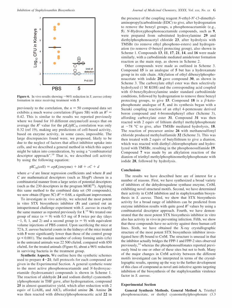
To investigate in vivo activity, we selected the most potentin vitro STX biosynthesis inhibitor (5) and carried out anintraperitoneal challenge experiment with S. aureus in exactlythe same manner as reported previously for 1.14 We treated onegroup of mice (n ) 9) with 0.5 mg of 5 twice per day (days-1, 0, 1, and 2) and a second group (n ) 9) with equivalentvolume injections of PBS control. Upon sacrificing the mice at72 h, S. aureus bacterial counts in the kidneys of the mice treatedwith 5 were significantly lower than those of the control group(p < 0.001). The median number of colony forming units (cfu)in the untreated animals was 22 500 cfu/mL compared with 850cfu/mL for the treated animals (Figure 6), about a 96% reductionin surviving bacteria in the treatment group.
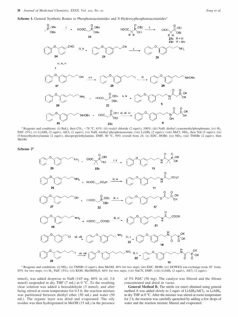
Synthetic Aspects. We outline here the synthetic schemesused to prepare 4-21; full protocols for each compound aregiven in the Experimental Section. A general synthetic routeto the most active phosphonoacetamide and N-hydroxyac-etamide (hydroxamate) compounds is shown in Scheme 1.The reaction of aldehyde 24 and sodium diethyl cyanometh-ylphosphonate in THF gave, after hydrogenation, compound25 in almost quantitative yield, which after reduction with 2equiv of LiAlH4 and AlCl3 afforded amine 26. Amine 26was then reacted with dibenzylphosphonoacetic acid 22 in
the presence of the coupling reagent N-ethyl-N′-(3-dimethyl-aminopropyl)carbodiimide (EDC) to give, after hydrogenationto remove the benzyl groups, a phosphonoacetamide (e.g.,5). N-Hydroxyphosphonoacetamide compounds, such as 9,were prepared from substituted hydroxylamine 29 anddiethylphosphonoacetyl chloride 23, after hydrolysis withTMSBr (to remove ethyl phosphono-esters) and hydrogen-ation (to remove O-benzyl protecting group), also shown inScheme 1. Compounds 13, 11, 17, 21, 14, and 16 were madesimilarly, with a carbodiimide mediated amide/ester formationreaction as the main step, as shown in Scheme 2.
Other compounds were made as outlined in Scheme 3.Compound 15 is an analogue of 5 but has a hydroxamategroup in its side chain. Alkylation of ethyl dibenzylphospho-noacetate with iodide 28 gave compound 30, as shown inScheme 3. The carboxylate ethyl ester was then selectivelyhydrolyzed (1 M KOH) and the corresponding acid coupledwith O-benzylhydroxylamine under standard carbodiimideconditions, followed by hydrogenation to remove three benzylprotecting groups, to give 15. Compound 18 is a �-keto-phosphonate analogue of 5, and its synthesis began with aSuzuki coupling reaction of an ethyl 4-pentenoate derivedboron compound and 4-bromodiphenyl ether (Scheme 3),affording carboxylate ester 31. Compound 31 was thenreacted with 2 equiv of lithium diethyl methylphosphonateat -78 °C to give, after TMSBr mediated hydrolysis, 18.The reaction of precursor amine 26 with methanesulfonylchloride produced methylsulfamide 32 (Scheme 3). This wasthen treated with 2 equiv of butyllithium to give a dianion,which was reacted with diethyl chlorophosphate and hydro-lyzed with TMSBr, resulting in the phosphonosulfamide 19.Compound 7 was made by an alkylation reaction of thedianion of triethyl methylphosphinomethylphosphonate withiodide 28, followed by hydrolysis.
Conclusions
The results we have described here are of interest for anumber of reasons. First, we have synthesized a broad varietyof inhibitors of the dehydrosqualene synthase enzyme, CrtM,exhibiting novel structural motifs. Second, we have determinedtheir activity in CrtM inhibition and in staphyloxanthin biosyn-thesis in S. aureus. Third, we show that STX biosynthesisactivity for a broad range of inhibitors can be predicted fromenzyme inhibition results with quite good R2 values by using acombinatorial descriptor approach. Fourth, we have demon-strated that the most potent STX biosynthesis inhibitor in vitroalso has activity in vivo in preventing infection. Fifth, we showthat these compounds have no activity against three human celllines. Sixth, we have obtained the X-ray crystallographicstructure of the most potent STX biosynthesis inhibitor inves-tigated here (5) bound to CrtM. The structure is unusual in thatthe inhibitor actually bridges the FPP-1 and FPP-2 sites observedpreviously,14 whereas the phosphonosulfonates reported previ-ously bind to one or other of these sites but not to both. Manyof the major changes in CrtM activity between the differentmotifs investigated can be interpreted in terms of the crystal-lographic results, opening up the way to the further developmentof this class of compound as novel anti-infective agents targetinginhibition of the biosynthesis of the staphyloxanthin virulencefactor in S. aureus.
Experimental Section
General Synthesis Methods. General Method A. Triethylphosphonoacetate, or diethyl cyanomethylphosphonate (3.3
Figure 6. In vivo results showing ∼96% reduction in S. aureus colonyformation in mice receiving treatment with 5.
Inhibition of Staphyloxanthin Biosynthesis Journal of Medicinal Chemistry, XXXX, Vol. xxx, No. xx G
mmol), was added dropwise to NaH (145 mg, 60% in oil, 3.6mmol) suspended in dry THF (7 mL) at 0 °C. To the resultingclear solution was added a benzaldehyde (3 mmol), and afterbeing stirred at room temperature for 0.5 h, the reaction mixturewas partitioned between diethyl ether (50 mL) and water (50mL). The organic layer was dried and evaporated. The oilyresidue was then hydrogenated in MeOH (15 mL) in the presence
of 5% Pd/C (50 mg). The catalyst was filtered and the filtrateconcentrated and dried in vacuo.
General Method B. The nitrile (or ester) obtained using generalmethod A was added slowly to 2 equiv of LiAlH4/AlCl3, or LiAlH4,in dry THF at 0 °C. After the mixture was stirred at room temperaturefor 2 h, the reaction was carefully quenched by adding a few drops ofwater and the reaction mixture filtered and evaporated.
Scheme 1. General Synthetic Routes to Phosphonoacetamides and N-Hydroxyphosphonoacetamidesa
a Reagents and conditions: (i) BuLi, then CO2, -78 °C, 63%; (ii) oxalyl chloride (2 equiv), 100%; (iii) NaH, diethyl cyanomethylphosphonate; (iv) H2,Pd/C (5%); (v) LiAlH4 (2 equiv), AlCl3 (2 equiv); (vi) NaH, triethyl phosphonoacetate; (vii) LiAlH4 (2 equiv); (viii) MsCl, NEt3, then NaI (5 equiv); (ix)O-benzylhydroxylamine (2 equiv), diisopropylethylamine, DMF, 80 °C, 50% overall from 24; (x) EDC, HOBt; (xi) NEt3; (xii) TMSBr (2 equiv), thenMeOH.
Scheme 2a
a Reagents and conditions: (i) NEt3; (ii) TMSBr (2 equiv), then MeOH, 48% for two steps; (iii) EDC, HOBt; (iv) DOWEX ion-exchange resin, H+ form,85% for two steps; (v) H2, Pd/C (5%); (vi) KOH, MeOH/H2O, 66% for two steps; (vii) NaCN, DMF; (viii) LiAlH4 (2 equiv), AlCl3 (2 equiv).
H Journal of Medicinal Chemistry, XXXX, Vol. xxx, No. xx Song et al.

General Method C. To a solution of a carboxylic acid (1 mmol)and an amine (1 mmol) in CH2Cl2 (5 mL) were added N-ethyl-N′-(3-dimethylaminopropyl)carbodiimide (EDC) (1.5 mmol) and 1-hydroxy-benzotriazole (1 mmol). After the mixture was stirred for 2 h at roomtemperature, 50 mL of ethyl acetate was added and the reaction mixturewashed successively with 1 N HCl (5 mL), water (5 mL), and saturatedNaHCO3 (5 mL), dried, and evaporated. The amide was purified usingflash chromatography (silica gel, ethyl acetate).
General Method D. A DMF solution (3 mL) containing a halide(3 mmol), O-benzylhydroxylamine (6 mmol) and diisopropylethyl-amine (3 mmol) was heated at 90 °C for 24 h. After the mixturewas cooled, diethyl ether (50 mL) was added and the mixture waswashed with H2O (20 mL), dried, and evaporated. The alkylatedhydroxylamine, such as 29, was purified by using column chro-matography (silica gel; hexane/ethyl acetate, 6/1).
General Method E. To a diethyl phosphonate (1 mmol) in dryCH3CN (3 mL) was added TMSBr (2 mmol) at room temperature.After 6 h, the solution was evaporated and methanol (5 mL) added.Neutralization with 1 N KOH to pH 8, followed by evaporation todryness and triturating with acetone, gave a white powder.
All reagents used were purchased from Aldrich (Milwaukee, WI).The purities of all compounds were routinely monitored by using1H and 31P NMR spectroscopy at 400 or 500 MHz on Varian (PaloAlto, CA) Unity spectrometers. All compounds were of g95%purity, as determined by combustion analysis. The details of thesesyntheses are as follows.
Diethylphosphonoacetyl Chloride (23a). Compound 23a wasprepared by mixing diethylphosphonoacetic acid (1.5 mmol) withoxalyl chloride (3 mmol) in benzene (5 mL) in the presence of onedrop of DMF for 1 h, followed by evaporation. The oily residuewas used immediately for the next reaction.
3-(3-Phenoxyphenyl)propyl Iodide (28). Alcohol 27, obtainedfrom 3-phenoxybenzaldehyde (3 mmol) following general methodsA and B, in CH2Cl2 (10 mL) containing NEt3 (0.5 mL, 3.6 mmol)was reacted with methanesulfonyl chloride (230 µL, 3 mmol) at 0°C. After 1 h, diethyl ether (50 mL) and water (50 mL) were addedand the organic layer was collected, washed with 1 N HCl andsaturated NaHCO3, dried, and evaporated to dryness. The oilyresidue was treated with NaI (1.35 g, 9 mmol) in acetone (7 mL)at 60 °C for 1 h. The reaction mixture was then partitioned betweendiethyl ether (50 mL) and water (50 mL) and the organic layerwashed with 5% Na2S2O3, dried, and evaporated to dryness to giveiodide 28. The iodide thus obtained is quite pure, according to 1Hand 13C NMR spectra, and may be used in the next step withoutfurther purification.
N-[3-(3-(3,4-Dichlorophenoxy)phenyl)propyl]phosphonoacetam-ide Dipotassium Salt (4). 3-(3-(3,4-Dichlorophenoxy)phenyl)-propylamine was prepared from 3-(3,4-dichlorophenoxy)benzalde-
hyde (1 mmol), using general method A, and was then coupledwith dibenzylphosphonoacetic acid according to general methodC to give the dibenzyl ester of 4. The benzyl groups wereremoved by catalytic hydrogenation (5% Pd/C in methanol for 1 h)followed by neutralization with KOH to give compound 4 as a whitepowder (245 mg, 48% overall yield). Anal. (C17H16Cl2-K2NO5P ·0.5CH3OH) C, H, N. 1H NMR (400 MHz, D2O): δ1.60-1.70 (m, 2H, CH2); 2.35 (d, J ) 20 Hz, 2H, CH2P); 2.46 (t,J ) 7.6 Hz, 2H, PhCH2); 2.98 (t, J ) 7.2 Hz, 2H, CH2N);6.70-7.30 (m, 7H, aromatic). 31P NMR (D2O): δ 13.6.
N-[3-(3-Phenoxyphenyl)propyl]phosphonoacetamide Dipotassi-um Salt (5). Amine 26 was prepared from 3-phenoxybenzaldehyde(1 mmol) using general method A and was then coupled withdibenzylphosphonoacetic acid according to general method C to givethe dibenzyl ester of 5. The benzyl groups were removed byhydrogenation for 1 h, catalyzed with 5% Pd/C in methanol, followedby neutralization with KOH to give compound 5 as a white powder(307 mg, 62% overall yield). Anal. (C17H18K2NO5P ·1.5H2O) C, H,N. 1H NMR (400 MHz, D2O): δ 1.60-1.70 (m, 2H, CH2); 2.35 (d, J) 20 Hz, 2H, CH2P); 2.46 (t, J ) 7.6 Hz, 2H, PhCH2); 2.98 (t, J )7.2 Hz, 2H, CH2N); 6.70-7.30 (m, 9H, aromatic). 31P NMR (D2O):δ 13.7.
N-[3-(3-(4-Chlorophenoxy)phenyl)propyl]phosphonoacetamideDipotassium Salt (6). 6 was prepared in the same way as 5, butusing 3-(4-chlorophenoxy)benzaldehyde (1 mmol) as starting mate-rial, as a white powder (267 mg, 58% overall yield). Anal.(C17H17ClK2NO5P ·0.3KCl ·1.5H2O) C, H, N. 1H NMR (400 MHz,D2O): δ 1.60-1.70 (m, 2H, CH2); 2.37 (d, J ) 20 Hz, 2H, CH2P);2.49 (t, J ) 7.6 Hz, 2H, PhCH2); 3.01 (t, J ) 7.2 Hz, 2H, CH2N);6.70-7.30 (m, 8H, aromatic). 31P NMR (D2O): δ 13.5.
3-(3-Phenoxyphenyl)propylphosphinylmethylphosphonic AcidTripotassium Salt (7). Triethyl methylphosphinylmethylphospho-nate (1 mmol) was treated with BuLi (2.2 mmol) in THF at -78°C for 1 h, followed by addition of iodide 28 (1.1 mmol). Thereaction mixture was allowed to warm to room temperature over3 h and was then quenched with saturated NH4Cl. The productwas purified with column chromatography (silica gel; ethyl acetate/methanol, 20/1) and deprotected using general method E to give 7as a white powder (320 mg, 62% overall yield). Anal.(C17H19K3O6P2 ·H2O) C, H. 1H NMR (400 MHz, D2O): δ 1.30-1.60(m, 6H, 3CH2); 1.75-1.85 (m, 2H, CH2P); 2.43 (t, J ) 7.6 Hz,2H, PhCH2); 6.70-7.30 (m, 9H, aromatic). 31P NMR (D2O): δ 16.3(s, 1P); 39.9 (s, 1P).
N-Hydroxy-N-[3-(3-(3,4-dichlorophenoxy)phenyl)propyl]phos-phonoacetamide Dipotassium Salt (8). 8 was prepared in the sameway as 9, but using 3-(3,4-dichlorophenoxy)benzaldehyde (3 mmol)as starting material, as a white powder (428 mg, 28% overall yield).
Scheme 3a
a Reagents and conditions: (i) NaH, ethyl dibenzylphosphonoacetate, 68%; (ii) KOH, MeOH/H2O; (iii) O-benzylhydroxylamine, EDC, HOBt; (iv) H2,Pd/C (5%), 31% from 30; (v) 9-BBN, then Pd(PPh3)4, K3PO4, 80 °C, 48%; (vi) diethyl methylphosphonate (2 equiv), BuLi, -78 C, 65%; (vii) TMSBr, thenMeOH; (viii) MsCl, NEt3; (ix) BuLi (2 equiv), -78 °C, then diethyl chlorophosphate, 76%; (x) BuLi (2 equiv), -78 °C, then 28, 58%.
Inhibition of Staphyloxanthin Biosynthesis Journal of Medicinal Chemistry, XXXX, Vol. xxx, No. xx I
Anal. (C17H16Cl2K2NO6P ·KBr) C, H, N. 1H NMR (400 MHz, D2O):δ 1.75-1.85 (m, 2H, CH2); 2.48 (t, J ) 7.6 Hz, 2H, PhCH2); 2.63(d, J ) 20 Hz, 2H, CH2P); 3.46 (t, J ) 7.2 Hz, 2H, CH2N);6.70-7.35 (m, 7H, aromatic). 31P NMR (D2O): δ 15.6.
N-Hydroxy-N-[3-(3-phenoxyphenyl)propyl]phosphonoacetam-ide Dipotassium Salt (9). General method D with iodide 28 gavesubstituted hydroxylamine 29 (1 mmol), which was reacted withthe acid chloride in the presence of NEt3 (2 mmol) in CH2Cl2 (5mL) at 0 °C. After the mixture was stirred for 1 h, the couplingproduct was purified by using column chromatography (silica gel,ethyl acetate) and was then deprotected following general methodE. Hydrogenation (5% Pd/C, MeOH) gave compound 9 as a whitepowder (312 mg, 56% overall yield). 1H NMR (400 MHz, D2O):δ 1.65-1.75 (m, 2H, CH2); 2.42 (t, J ) 7.6 Hz, 2H, PhCH2); 2.66(d, J ) 20 Hz, 2H, CH2P); 3.39 (t, J ) 7.2 Hz, 2H, CH2N);6.70-7.20 (m, 9H, aromatic). 31P NMR (D2O): δ 15.8.
N-[3-(4-Biphenyl)propyl]phosphonoacetamide (10). 3-(4-Biphe-nyl)propylamine was prepared from 4-phenylbenzaldehyde (1mmol), using general method A, and was then coupled withdibenzylphosphonoacetic acid according to general method C togive the dibenzyl ester of 10. The benzyl groups were removed byhydrogenation (catalyzed with 5% Pd/C in methanol) for 1 h,followed by neutralization with KOH, to give compound 10 as awhite powder (222 mg, 65% overall yield). Anal.(C17H20K2NO4P ·0.25CH3OH) C, H, N. 1H NMR (400 MHz, D2O):δ 1.65-1.75 (m, 2H, CH2); 2.31 (d, J ) 20 Hz, 2H, CH2P); 2.55(t, J ) 7.6 Hz, 2H, PhCH2); 3.03 (t, J ) 7.2 Hz, 2H, CH2N);7.20-7.55 (m, 9H, aromatic). 31P NMR (D2O): δ 12.8.
N-[3-(3-Phenoxyphenyl)propyl]sulfoacetamide (11). Amine 26(1 mmol) was coupled with sulfoacetic acid (1 mmol) accordingto general method C (without addition of 1-hydroxybenzotriazole)to give 11. The product was purified by using column chromatog-raphy (DOWEX ion-exchange resin, H+ form, methanol as eluent)as an off-white powder (315 mg, 85% overall yield). Anal.(C17H19NO5S) C, H, N. 1H NMR (400 MHz, D2O): δ 1.60-1.70(m, 2H, CH2); 2.44 (m, 2H, PhCH2); 3.02 (m, 2H, CH2N); 3.59 (s,2H, CH2S); 6.70-7.30 (m, 9H, aromatic).
N-Methyl-N-[3-(3-phenoxyphenyl)propyl]phosphonoacetamideDipotassium Salt (12). Amine 26 (1 mmol) was reacted with benzylchloroformate (ZCl, 1 mmol) in the presence of NEt3 to giveZ-protected amine 26 which was then methylated in THF with MeI(1.5 equiv) and NaH (1.2 equiv) overnight. After hydrogenation(5% Pd/C in MeOH) to remove the Z-protecting group, theN-methylated amine 5 was coupled with dibenzylphosphonoaceticacid, according to general method B, to give the dibenzyl ester of12. The benzyl groups were removed by hydrogenation (5% Pd/Cin methanol) for 1 h, followed by neutralization with KOH to givecompound 12 as a white powder (220 mg, 50% overall yield). Anal.(C18H20K2NO5P) C, H, N. The NMR spectrum of 12 showed thattwo rotamers (with respect to the amide bond) exist with ratio of∼45:55. 1H NMR (400 MHz, D2O): δ 1.60-1.80 (m, 2H, CH2);2.35-2.45 (m, 2H, CH2P); 2.45-2.95 (m, 5H, Me and PhCH2);3.10-3.40 (m, 2H, CH2N); 6.80-7.30 (m, 9H, aromatic). 31P NMR(D2O): δ 13.6.
N-[3-(3-Phenoxyphenyl)propyl]phosphonodimethylacetamide Di-potassium Salt (13). Diethyl phosphonodimethylacetate (3 mmol)was treated with 3 N KOH (5 mL) in ethanol (8 mL) for 24 h,followed by acidifiction with HCl to give the correspondingcarboxylic acid. As with compound 23a, the acid was thenconverted to the acid chloride 23b, which was reacted with 1 equivof amine 26 in the presence of NEt3 in CH2Cl2 (5 mL) at 0 °C.After the mixture was stirred for 1 h, the coupling product waspurified by using column chromatography (silica gel, ethyl acetate)and was then deprotected following general method E to give 13as a white powder (335 mg, 21% overall yield). Anal.(C19H22K2NO5P ·0.5KBr ·H2O) C, H, N. 1H NMR (400 MHz, D2O):δ 1.09 (d, J ) 13.6 Hz, 6H, 2CH3); 1.60-1.70 (m, 2H, CH2); 2.46(m, 2H, PhCH2); 2.99 (m, 2H, CH2N); 6.85-7.25 (m, 9H, aromatic).31P NMR (D2O): δ 22.9.
N-[2-(3-Phenoxyphenyl)ethyl]phosphonoacetamide DipotassiumSalt (14). 3-Phenoxybenzyl chloride (2 mmol) and NaCN (2.2mmol) were stirred in DMF (2 mL) overnight at 60 °C. After themixture was cooled, diethyl ether (50 mL) was added and themixture was washed with water and the organic layer dried andevaporated. 2-(3-Phenoxyphenyl)ethylamine was prepared from thenitrile so obtained, using general method B, and was then coupledwith dibenzylphosphonoacetic acid according to general methodC to give the dibenzyl ester of 14. The benzyl groups were re-moved by catalytic hydrogenation (5% Pd/C in methanol for 1 h)followed by neutralization with KOH to give compound 14 as awhite powder (387 mg, 45% overall yield). Anal. (C16H16-K2NO5P ·H2O) C, H, N. 1H NMR (400 MHz, D2O): δ 2.39 (d, J )20 Hz, 2H, CH2P); 2.58 (t, J ) 7.6 Hz, 2H, PhCH2); 3.05 (t, J )7.2 Hz, 2H, CH2N); 6.70-7.30 (m, 9H, aromatic). 31P NMR (D2O):δ 13.8.
N-Hydroxy-2-phosphono-5-(3-phenoxyphenyl)pentamide Dipo-tassium Salt (15). Iodide 28 was added to a cold DMF solutioncontaining ethyl dibenzylphosphonoacetate (1 equiv) and NaH (1.1equiv). After the mixture was stirred for 3 h at room temperature,the product 30 was purified by using column chromatography (silicagel; hexane/ethyl acetate, 1/1). 30 was then treated with 3 N KOHin EtOH/H2O (3:1) for 24 h and the resulting solution was reducedin volume and then acidified with 3 N HCl, to give the correspond-ing carboxylic acid. The acid so obtained was reacted withO-benzylhydroxylamine, according to general method C, to giveprotected 15, which was then hydrogenated in the presence of 5%Pd/C in MeOH for 1 h to afford, after neutralization with KOH,15 as a white powder (293 mg, 21% overall yield). Anal.(C17H18K2NO6P ·0.5C2H5OH) C, H, N. 1H NMR (400 MHz, D2O):δ 1.25-1.75 (m, 4H, CH2); 2.20-2.50 (m, 3H, CH + PhCH2);6.70-7.25 (m, 9H, aromatic). 31P NMR (D2O): δ 17.5.
N-[4-(3-Phenoxyphenyl)butyl]phosphonoacetamide DipotassiumSalt (16). Iodide 28 (2 mmol) and NaCN (2.2 mmol) were stirredin DMF (2 mL) overnight at 60 °C. After the mixture was cooled,diethyl ether (50 mL) was added and the mixture washed with waterand the organic layer evaporated. 4-(3-Phenoxyphenyl)butylaminewas prepared from the nitrile so obtained, using general methodB, and was then coupled with dibenzylphosphonoacetic acid,according to general method C, to give the dibenzyl ester of 16.The benzyl groups were removed by catalytic hydrogenation (5%Pd/C in methanol for 1 h) followed by neutralization with KOH togive compound 16 as a white powder (475 mg, 51% overall yield).Anal. (C18H20K2NO5P ·1.5H2O) C, H, N. 1H NMR (400 MHz, D2O):δ 1.55-1.70 (m, 4H, CH2); 2.39 (d, J ) 20 Hz, 2H, CH2P); 2.48(t, J ) 7.6 Hz, 2H, PhCH2); 3.01 (t, J ) 7.2 Hz, 2H, CH2N);6.70-7.30 (m, 9H, aromatic). 31P NMR (D2O): δ 13.5.
3-(3-Phenoxyphenyl)propyl Phosphonoacetate Dipotassium Salt(17). Alcohol 27 was coupled with dibenzylphosphonoacetic acidaccording to general method C to give dibenzyl ester of 17. Thebenzyl groups were removed by catalytic hydrogenation (5% Pd/Cin methanol for 1 h) followed by neutralization with KOH to givecompound 17 as a white powder (180 mg, 42% overall yield). Anal.(C17H17K2O6P) C, H. 1H NMR (400 MHz, D2O): δ 1.60-1.70 (m,2H, CH2); 2.38 (d, J ) 20 Hz, 2H, CH2P); 2.44 (t, J ) 7.6 Hz, 2H,PhCH2); 3.47 (t, J ) 7.2 Hz, 2H, CH2O); 6.70-7.30 (m, 9H,aromatic). 31P NMR (D2O): δ 13.9.
2-Oxo-6-(4-phenoxyphenyl)hexylphosphonic Acid DipotassiumSalt (18). 9-BBN (0.5 M in THF, 9 mL) was added to ethyl4-pentenoate (3 mmol) at 0 °C and the reaction mixture stirred atroom temperature for 2 h. 4-Bromodiphenylether (3 mmol),Pd(PPh3)4 (0.15 mmol), K3PO4 (6 mmol), and H2O (2 mL) werethen added, and the reaction mixture was refluxed overnight. Theorganic layer was evaporated and purified by using columnchromatography (silica gel; hexane/ether, 6/1) to afford ester 31,which was then reacted with 2 equiv of the lithium salt of diethylmethylphosphonate at -78 °C, for 2 h. The reaction was quenchedwith saturated NH4Cl, diethyl ether added to extract the product,and the organic solvent removed. The oily residue was purified byusing column chromatography (silica gel, ethyl acetate) anddeprotected according to general method E to give compound 18
J Journal of Medicinal Chemistry, XXXX, Vol. xxx, No. xx Song et al.
as a white powder (312 mg, 20% overall yield). Anal.(C18H19K2O5P ·0.5KBr ·2H2O) C, H. 1H NMR (400 MHz, D2O): δ1.30-1.50 (m, 4H, CH2); 2.44 (t, J ) 7.6 Hz, 2H, PhCH2); 2.54 (t,J ) 7.6 Hz, 2H, CH2CO); 2.70 (d, J ) 20 Hz, 2H, CH2P);6.80-7.25 (m, 9H, aromatic). 31P NMR (D2O): δ 11.0.
N-[3-(3-Phenoxyphenyl)propyl]phosphonomethylsufamide Di-potassium Salt (19). Amine 26 prepared from 3-phenoxybenzal-dehyde (3 mmol) using general method A was reacted with 1 equivof methanesulfonyl chloride in CH2Cl2 in the presence of 1.2 equivof NEt3 at 0 °C. After 1 h, 50 mL of ethyl acetate was added andthe reaction mixture was washed successively with 1 N HCl, water,NaHCO3, then dried and evaporated. The oily residue was treatedwith 2.2 equiv of BuLi at -78 °C for 1 h followed by addition of0.6 equiv of diethyl chlorophosphate. The reaction mixture waswarmed to 0 °C over 1 h and then quenched with saturated NH4Cl.Column chromatography (silica gel, ethyl acetate) followed byhydrolysis using general method E gave 19 as a white powder (366mg, 42% overall yield). Anal. (C16H18K2NO6P ·KBr) C, H, N. 1HNMR (400 MHz, D2O): δ 1.60-1.75 (m, 2H, CH2); 2.50 (t, J )7.6 Hz, 2H, PhCH2); 2.87 (t, J ) 7.2 Hz, 2H, CH2N); 1.18 (d, J )20 Hz, 2H, CH2P); 6.70-7.30 (m, 9H, aromatic). 31P NMR (D2O):δ 4.4.
N-Hydroxy-N-[3-(4-methylbiphenyl)propyl]phosphonoacetam-ide (20). Compound 20 was prepared in the same manner as 9, using4-methylphenylbenzaldehyde as starting material, as a white powder(175 mg, 48% overall yield). Anal. (C18H22NO5P ·0.2HBr) C, H, N.1H NMR (400 MHz, D2O): δ 1.70-1.80 (m, 2H, CH2); 2.21 (s, 3H,Me), 2.46 (t, J ) 7.6 Hz, 2H, PhCH2); 2.66 (d, J ) 20 Hz, 2H, CH2P);3.42 (t, J ) 7.2 Hz, 2H, CH2N); 7.0-7.30 (m, 8H, aromatic). 31P NMR(D2O): δ 15.9.
N-[3-(3-Phenoxyphenyl)propyl]phosphonomalonamide Potassi-um Salt (21). Amine 26 (1 mmol) was coupled with malonic acidmonoethyl ester according to general method C to give the ethylester of 21, which was then hydrolyzed with 3 equiv of KOH inMeOH/H2O for 1 h. The reaction mixture was acidified andextracted with ethyl acetate, and the organic layer was evaporated.The oily residue was dissolved in methanol, neutralized with KOH,and evaporated to give 21 as a white powder (250 mg mg, 66%overall yield). Anal. (C18H18KNO4 ·0.25KCl ·0.5H2O) C, H, N. 1HNMR (400 MHz, D2O): δ 1.80-1.90 (m, 2H, CH2); 2.62 (t, J )7.6 Hz, 2H, PhCH2); 3.32 (s, 2H, CH2COO); 3.33 (m, 2H, CH2N);6.70-7.40 (m, 9H, aromatic).
Enzyme and Biological Assays. CrtM Expression and Purifica-tion. CrtM with a histidine tag was overexpressed in E. coliBL21(DE3) cells and CrtM protein purified as described previ-ously.14 A 50 mL overnight culture was transferred into 1 L LBmedium supplemented with 100 µg/mL ampicillin. Induction wascarried out with 1 mM IPTG for 4 h at 37 °C, when the cell culturereached an OD of 0.6 at 600 nm. The cell extract was loaded ontoa Ni-NTA column and CrtM eluted by using a 100 mL lineargradient of 0-0.5 M imidazole in 50 mM Tris-HCl buffer, pH 7.4.
CrtM Inhibition Assay. The condensation of farnesyl diphos-phate was monitored by a continuous spectrophotometric assay forphosphate releasing enzymes.1 The reaction buffer contained 50mM Tris-HCl, 1 mM MgCl2, 450 µM FPP, pH 7.4. The compoundsinvestigated were preincubated with 2 µg of CrtM for 30 min at20 °C. Reactions were carried out by using 96-well plates with200 µL of reaction mixture in each well. The IC50 values wereobtained by fitting the inhibition data to a normal dose-responsecurve in Origin 6.1 (OriginLab Corporation, Northampton, MA).The Ki values reported are calculated on the basis of the substrateconcentrations used, the IC50 values found, and the kinetic constantfor CrtM, as described previously.14
Staphyloxanthin Biosynthesis Inhibition Assay. The S. aureusstrain used was the WT clinical isolate (Pig1). S. aureus waspropagated in Todd-Hewitt broth (THB) or on THB agar (TBA;Difco, Detroit, MI). For in vitro pigment inhibition studies, S. aureuswas cultured in THB (1 mL) in the presence of inhibitor compoundsfor 72 h, in duplicate. Prior to assay, the bacteria were centrifugedand washed twice in PBS. Staphyloxanthin was extracted withMeOH, and the OD was determined at 450 nm using a Perkin-
Elmer MBA 2000 (Norwalk, CT) spectrophotometer. The IC50
values were obtained by fitting the OD data to a normaldose-response curve, using GraphPad PRISM.
Human SQS Enzyme Expression, Purification, and InhibitionAssay. E. coli expressing a human SQS construct were cultured inLuria-Bertani medium supplemented with kanamycin (30 µg/mL)and chloramphenicol (34 µg/mL) at 37 °C, until the cells reachedan OD of 0.4 at 600 nm, and were then induced at 37 °C for 4 hby incubation with 1 mM isopropyl-1-thio-�-D-galactopyranoside.Cells were harvested by centrifugation (10 min, 4000 rpm) andresuspended in 10 mL of lysis/elution buffer (20 mM NaH2PO3,pH 7.4, 10 mM CHAPS, 2 mM MgCl2, 10% glycerol, 10 mMmercaptoethanol, 500 mM NaCl, 10 mM imidazole, and a proteaseinhibitor cocktail), disrupted by sonication, and centrifuged at16 000 rpm for 30 min. The supernatant (40 mL) was then appliedto a HiTrap nickel-chelating HP column (Amersham Biosciences).Enzyme purification was performed according to the manufacturer’sinstructions using a Pharmacia FPLC system. Unbound protein waswashed off with 50 mM imidazole, and then the His6-HsSQS waseluted with 1 M imidazole. Purity was confirmed by SDS-PAGEelectrophoresis. Fractions containing the enzyme were pooled anddialyzed against buffer A (25 mM sodium phosphate, pH 7.4, 20mM NaCl, 2 mM dithiothreitol, 1 mM EDTA, 10% glycerol, 10%methanol), concentrated, then stored at -80 °C.
SQS activity was based on measuring the conversion of [3H]FPPto [3H]squalene. Final assay concentrations were 50 mM MOPS(pH 7.4), 20 mM MgCl2, 5 mM CHAPS, 1% Tween-80, 10 mMDTT, 0.025 mg/mL BSA, 0.25 mM NADPH, and 7.5 ng of purifiedrecombinant human SQS. The reaction was started with the additionof substrate (3HFPP, 0.1 nmol, 2.22 × 10⁶ dpm), and the finalvolume of the reaction was 200 µL. After incubation at 37 °C for5 min, an amount of 40 µL of 10 M NaOH was added to stop thereaction, followed by 10 µL of a (100:1) mixture of 98% EtOHand squalene. The resulting mixtures were mixed vigorously byvortexing. Then 10 µL aliquots were applied to 2.5 cm × 10 cmchannels of a silica gel thin layer chromatogram, and newly formedsqualene was separated from unreacted substrate by chromatographyin toluene-EtOAc (9:1). The region of the squalene band wasscraped and immersed in Hydrofluor liquid scintillation fluid andassayed for radioactivity. IC50 values were calculated from thehyperbolic plot of percent of inhibition versus inhibitor concentra-tion, using GraphPad PRISM.
Human Cell Growth Inhibition Assay. Three human cell linesMCF-7, NCI-H460, and SF-268 were obtained from the NationalCancer Institute. Cells were cultured in RPMI-1640 mediumsupplemented with 10% fetal bovine serum and 2 mM L-glutamineat 37 °C in a 5% CO2 atmosphere with 100% humidity. A brothmicrodilution method was used to calculate IC50 values for growthinhibition by each compound. Cells were inoculated at a densityof 5000 cells/well into 96-well flat-bottom culture plates containing10 µL of the test compound, previously half-log serial diluted (from0.316 mM to 0.1 pM) for a final volume of 100 µL. Plates werethen incubated for 4 days at 37 °C in a 5% CO2 atmosphere at100% humidity, after which an MTT ((3-(4,5-dimethylthiazole-2-yl)-2,5-diphenyltetrazolium bromide) cell proliferation assay (ATCC,Manassas, VA) was used to quantify cell viability. The IC50 valueswere obtained by fitting the OD data to a normal dose-responsecurve, using GraphPad PRISM.
Murine Model of Kidney Infection. The 10-12 week old CD1male mice (Charles River Laboratory) were randomized into twogroups at the start of the experiment and administered either 0.5mg of BPH-652 or PBS control, ip, twice a day, starting on day 1to day 2 (a total of eight doses). All mice were injected intraperi-toneally (ip) with 108 early stationary phase S. aureus on day 0.After 3 days, animals were euthanized, kidneys homogenized inPBS, and plated on THA for quantitative bacterial culture.
Statistics. The significance of experimental differences in themouse in ViVo challenge studies were evaluated by use of the two-tailed Student’s t test.
X-ray Crystallography. Native CrtM was eluted from Ni-NTAbeads by incubation with factor Xa (Novagen) to cleave it from
Inhibition of Staphyloxanthin Biosynthesis Journal of Medicinal Chemistry, XXXX, Vol. xxx, No. xx K

the polyhistidine-containing N-terminal thioredoxin fusion tag. Thecleaved product was equilibrated with buffer containing 150 mMNaCl, 5 mM DTT, 1 mM �-mercaptoethanol, 5% glycerol, and 20mM Tris, pH 7.5 and then concentrated to 15 mg/mL. Native CrtMcrystals (space group P3221) were grown using the hanging-dropmethod by mixing equal amounts of reservoir with 0.12-0.58 Mpotassium sodium tartrate at room temperature. BPH-830 wasincorporated by soaking crystals with a solution of 5 (10 mM inDMSO) for 3 h at room temperature. X-ray diffraction data werecollected at SPXF beamline BL13B1 at the National SynchrotronRadiation Research Center (NSRRC), Hsinchu, Taiwan. All dif-fraction images were recorded using an ADSD Q210 CCD detector,and the data were indexed, integrated, and scaled by using theHKL2000 package.21 The structure of the CrtM-5 complex wasdetermined by molecular replacement using CNS22 using the refinednative CrtM (PDB 2ZCO) as a search model. Iterative cycles ofmodel building with Xtalview23 and computational refinement withCNS were performed, in which 5% reflections were set aside forRfree calculation.24 The stereochemical quality was assessed withthe program PROCHECK.25 Figures were obtained by usingPymol.26
Acknowledgment. This work was supported by the UnitedStates Public Health Service (Grant AI074832 to G.Y.L., GrantHD051796 to V.N., and Grants AI074233, GM073216, andGM65307 to E.O.). Y.S. was supported by a Leukemia andLymphoma Society Special Fellowship. Diffraction data wereobtained at the National Synchrotron Radiation Research Centerof Taiwan and was supported by grants from Academia Sinicaand the National Core Facility of High-Throughput ProteinCrystallography (Grant SC95-3112-B-001-015-Y to A.H.-J.W.).
References(1) Bancroft, E. A. Antimicrobial resistance: it’s not just for hospitals.
JAMA, J. Am. Med. Assoc. 2007, 298 (15), 1803–1804.(2) Klevens, R. M.; Morrison, M. A.; Nadle, J.; Petit, S.; Gershman, K.;
Ray, S.; Harrison, L. H.; Lynfield, R.; Dumyati, G.; Townes, J. M.;Craig, A. S.; Zell, E. R.; Fosheim, G. E.; McDougal, L. K.; Carey,R. B.; Fridkin, S. K. Invasive methicillin-resistant Staphylococcusaureus infections in the United States. JAMA, J. Am. Med. Assoc. 2007,298 (15), 1763–1771.
(3) Treating Infectious Diseases in a Microbial World: Report of TwoWorkshops on NoVel Antibacterial Therapeutics; National ResearchCouncil: Washington, DC, 2006; pp 21-22.
(4) Wang, R.; Braughton, K. R.; Kretschmer, D.; Bach, T. H.; Queck,S. Y.; Li, M.; Kennedy, A. D.; Dorward, D. W.; Klebanoff, S. J.;Peschel, A.; DeLeo, F. R.; Otto, M. Identification of novel cytolyticpeptides as key virulence determinants for community-associatedMRSA. Nat. Med. 2007, 13 (12), 1510–1514.
(5) Escaich, S. Antivirulence as a new antibacterial approach forchemotherapy. Curr. Opin. Chem. Biol. 2008, 12 (4), 400–408.
(6) Liu, G. Y.; Essex, A.; Buchanan, J. T.; Datta, V.; Hoffman, H. M.;Bastian, J. F.; Fierer, J.; Nizet, V. Staphylococcus aureus goldenpigment impairs neutrophil killing and promotes virulence throughits antioxidant activity. J. Exp. Med. 2005, 202 (2), 209–215.
(7) Clauditz, A.; Resch, A.; Wieland, K. P.; Peschel, A.; Gotz, F.Staphyloxanthin plays a role in the fitness of Staphylococcus aureusand its ability to cope with oxidative stress. Infect. Immun. 2006, 74(8), 4950–4953.
(8) Pelz, A.; Wieland, K. P.; Putzbach, K.; Hentschel, P.; Albert, K.; Gotz,F. Structure and biosynthesis of staphyloxanthin from Staphylococcusaureus. J. Biol. Chem. 2005, 280 (37), 32493–32498.
(9) Hammond, R. K.; White, D. C. Inhibition of vitamin K2 and carotenoidsynthesis in Staphylococcus aureus by diphenylamine. J. Bacteriol.1970, 103 (3), 611–615.
(10) Hammond, R. K.; White, D. C. Inhibition of carotenoid hydroxylationin Staphylococcus aureus by mixed-function oxidase inhibitors. J.Bacteriol. 1970, 103 (3), 607–610.
(11) Daum, R. S. Removing the golden coat of Staphylococcus aureus.N. Engl. J. Med. 2008, 359 (1), 85–87.
(12) Walsh, C. T.; Fischbach, M. A. Inhibitors of sterol biosynthesis asStaphylococcus aureus antibiotics. Angew. Chem., Int. Ed. Engl. 2008,47 (31), 5700–5702.
(13) Haebich, D.; von Nussbaum, F. “Superbugs bunny” outsmarts ourimmune defense. ChemMedChem 2008, 3 (8), 1173–1177.
(14) Liu, C. I.; Liu, G. Y.; Song, Y.; Yin, F.; Hensler, M. E.; Jeng, W. Y.;Nizet, V.; Wang, A. H.; Oldfield, E. A cholesterol biosynthesisinhibitor blocks Staphylococcus aureus virulence. Science 2008, 319(5868), 1391–1394.
(15) Magnin, D. R.; Biller, S. A.; Dickson, J. K., Jr.; Logan, J. V.;Lawrence, R. M.; Chen, Y.; Sulsky, R. B.; Ciosek, C. P., Jr.; Harrity,T. W.; Jolibois, K. G.; et al. 1,1-Bisphosphonate squalene synthaseinhibitors: interplay between the isoprenoid subunit and the diphosphatesurrogate. J. Med. Chem. 1995, 38 (14), 2596–2605.
(16) Magnin, D. R.; Biller, S. A.; Chen, Y.; Dickson, J. K., Jr.; Fryszman,O. M.; Lawrence, R. M.; Logan, J. V.; Sieber-McMaster, E. S.; Sulsky,R. B.; Traeger, S. C.; Hsieh, D. C.; Lan, S. J.; Rinehart, J. K.; Harrity,T. W.; Jolibois, K. G.; Kunselman, L. K.; Rich, L. C.; Slusarchyk,D. A.; Ciosek, C. P., Jr. R-Phosphonosulfonic acids: potent andselective inhibitors of squalene synthase. J. Med. Chem. 1996, 39 (3),657–660.
(17) Rieger, C. E.; Lee, J.; Turnbull, J. L. A continuous spectrophotometricassay for aspartate transcarbamylase and ATPases. Anal. Biochem.1997, 246 (1), 86–95.
(18) Song, Y.; Lin, F.-Y.; Yin, F.; Hensler, M.; Poveda, C. A. R.;Mukkamala, D.; Cao, R.; Wang, H.; Morita, C. T.; Pacanowska, D. G.;Nizet, V.; Oldfield, E. Phosphonosulfonate are potent, selectiveinhibitors of dehydrosqualene synthase and staphyloxanthin biosyn-thesis in Staphylococcus aureus. J. Med. Chem. 2009, 52 (4), 976–988.
(19) Mukkamala, D.; No, J. H.; Cass, L. M.; Chang, T. K.; Oldfield, E.Bisphosphonate inhibition of a Plasmodium farnesyl diphosphatesynthase and a general method for predicting cell-based activity fromenzyme data. J. Med. Chem. 2008, 51 (24), 7827–7833.
(20) Molecular Operating EnVironment (MOE); Chemical ComputingGroup, Inc.: Montreal, Quebec, 2006.
(21) Otwinowski, Z.; Minor, W. ; Processing of X-ray diffraction datacollected in oscillation mode. Macromol. Crystallogr. 1997, 276, 307–326.
(22) Brunger, A. T.; Adams, P. D.; Clore, G. M.; DeLano, W. L.; Gros,P.; Grosse-Kunstleve, R. W.; Jiang, J. S.; Kuszewski, J.; Nilges, M.;Pannu, N. S.; Read, R. J.; Rice, L. M.; Simonson, T.; Warren, G. L.Crystallography & NMR system: a new software suite for macromo-lecular structure determination. Acta Crystallogr., Sect. D: Biol.Crystallogr. 1998, 54 (Part 5), 905–921.
(23) McRee, D. E. XtalView Xfit. A versatile program for manipulatingatomic coordinates and electron density. J. Struct. Biol. 1999, 125(2V3), 156–165.
(24) Brunger, A. T. Assessment of phase accuracy by cross validation: thefree R value. Methods and applications. Acta Crystallogr. D 1993, 49(1), 24–36.
(25) Laskowski, R. A.; MacArthur, M. W.; Moss, D. S.; Thornton, J. M.PROCHECK: a program to check the stereochemical quality of proteinstructures. J. Appl. Crystallogr. 1993, 26, 283–291.
(26) DeLano, W. L. The PyMOL Molecular Graphics System; DeLanoScientific: Palo Alto, CA, 2008; http://www.pymol.org.
(27) Wallace, A. C.; Laskowski, R. A.; Thornton, J. M. Ligplot: a programto generate schematic diagrams of protein-ligand interactions. ProteinEng. 1995, 8, 127–134.
JM9001764
L Journal of Medicinal Chemistry, XXXX, Vol. xxx, No. xx Song et al.