inhibition of mammalian brain acetylcholinesterase by ketamine
TRANSCRIPT
Biochemxal Pharmacology, Vol. 23. pp. 1647-1652 Pergamon Press 1974. Printed in Great Bntain
INHIBITION OF MAMMALIAN BRAIN ACETYLCHOLINESTERASE BY KETAMINE”
M. L. COHEN,? S. L. CHAN, H. N. BHARGAVA and ANTHONY J. TREVOR
Department of Pharmacology, University of California at San Francisco, San Francisco, Calif. 94143, U.S.A.
(Received 24 August 1973; accepted 9 November 1973)
Abstract-Ketamine caused a reversible inhibition of both membrane-bound and purified forms ofacctylcholinesterase (AChE; EC 3.1.1.7.) prepared from beef brain caudate nucleus tissue. Apparent Ki values (x 10m4 M) ranged between 4.9 and 6.9 for the different enzyme forms. Inhibition was of the mixed kinetic type, which suggests interactions of the drug with both active site(s) and other anionic sites on the enzyme. Rat brain AChE was inhibited by ketamine in vitro at concentrations commensurate with brain levels of the drug deter- mined after i.v. administration to rats. Gas chromatographic analyses demonstrated a 26 per cent increase in rat brain acetylcholine (ACh) 30 set after the administration of ketamine (20 mg/kg, i.v.). ACh accumulation via inhibition of AChE may underlie certain pharmaco- logical effects of ketamine which resemble cholinergic stimulation.
WHEN USED clinically as an anesthetic agent, ketamine causes certain pharmacologi- cal effects which resemble cholinergic stimulation. These include lacrimation and salivation, ‘s2 increased skeletal muscle tone2s3 and increased uterine tone.4 The sia- logogic actions, as well as certain central manifestations of disorientation on emer- gence from anesthesia, are antagonized by anticholinergic drugs.s-7 Since the amine group of ketamine is partly charged at physiological pH, it is possible that the drug could cause acetycholine (ACh) accumulation via inhibition of acetylcholinesterase (AChE; EC 3.1.1.7.) In this study, the effects of ketamine on membrane-bound and purified forms of mammalian brain AChE were examined. Inhibition of the enzyme was related to the levels of ketamine achieved in brain tissue after administration of the drug in vivo, and to observed changes in the levels of ACh in the brain.
MATERIALS AND METHODS
Brain acetylcholinesterase preparations. Purified forms of AChE were prepared from bovine brain caudate nucleus tissue as previously described.8,9 The procedure resulted in separation of three forms of the enzyme with estimated molecular weights of 390,000 (A), 270,000 (B) and 130,000 (C). These forms had average specific activities of 480 (A), 400 (B) and 575 (C) m-moles acetylthiocholine (ATC) hydrolyzed/mg of protein/hr.
Synaptosomal membrane fractions containing AChE activity were prepared from homogenates of bovine caudate nucleus tissue in 0.32 M sucrose using the method
* This work was supported in part by United States Public Health Service Grant N.S. 10913. t Present address: Roche Institute of Molecular Biology, Nutley, N.J.
1647
I548 M. L. COHL~. S. L. CIIA~, H. N. BHAKGAVA and A. .I. TKEVOK
of DeRobertis et u/.‘~ Such preparations had an average AChE specific activity of 45 pmoles ATC hydrolyzed/mg of protein/hr.
In some experiments, AChE was prepared from homogenates of whole brain tissue from male Sprague-Dawley rats (1 O@ 120 g) following the procedure of DeRobertis Et al.‘o to the separation of an Ml pellet. This was resuspended in 0.32 M sucrose (pH 7.0) for studies on a membrane-bound form of AChE. Soluble forms of rat brain
AChE were obtained by mixing the M 1 pellet in sucrose with added EDTA (0.5 mM) and imidazole buffer (5 mM) at O-4’ for 36 hr, followed by centrifugation at 100,000 g for 60 min to remove particulate material.
Estimation qf brain ACh urzd choline. Male SpragueeDawley rats (SU-100 g) were injected via tail vein with either saline or ketamine (20 mg/kg). At selected time inter- vals, the rats were guillotined and the brains were removed rapidly (within 1520 set) and placed in liquid nitrogen. Brain levels of ACh and choline were determined simultaneously using the gas chromatographic procedure of Jenden et al.’ “12 with the following slight modifications. The dichloromethane extract (1 ~1) was injected into a Varian 1700 gas chromatograph equipped with flame ionization detector and a 10 ft by 2 mm (id.) column containing Gas-Chrom Q coated with a 5% mixture of OV-101 and dodecyldimethylenetriamine succinamide. Operating conditions were: column temperature, 120”; injector and detector temperatures, 165” and 275” respectively; nitrogen, air and hydrogen flow rates, 17. 300 and 30 ml/min respect- ively. Under these conditions, the retention times of the demethylated derivatives of ACh, propionylcholine and pivaloylcholine were 145. 210 and 267 set respectively. Concentrations of ACh and choline were calculated by peak height ratio analyses.
Other analyticuI methods. AChE activity was determined by the spectrophoto- metric method of Ellman et ~1.‘~ at 25” using acetylthiocholine as substrate. Inhibi- tion of AChE activity was plotted as LineweaverBurk plots with lines calculated by the least squares method of linear regression analysis. Apparent dissociation con- stants (Ki) were calculated from the slopes of the inhibited reactions at two substrate concentrations and confirmed by direct observation of Hunter and Downs plots.i4 Protein was estimated by the procedure of Lowry et ~1.‘~ using bovine serum albu- min standards. Brain levels of ketamine after intravenous administration to rats were determined by a modification of the gas chromatographic procedure of Chang and Glazko16 as reported previously.”
Chemicals. All reagents used were of analytical grade. Acetylthiocholine (ATC) was obtained from Sigma Chemical Co. (St. Louis, MO.). 5,5’-Dithio-his-(2-nitrobenzoic acid), propionyl chloride and pivaloyl chloride were obtained from Aldrich Chemical Co. (Milwaukee, Wis.). Crystalline ketamine hydrochloride and its metabolites were donated by Parke Davis & Co. (Ann Arbor, Mich.) through the courtesy of Dr. A. J. Glazko. Acetonitrile (chromatoquality), dichloromethane (spectroquality), Pen- tane (spectroquality) and anhydrous ether (purified by passage over neutral Alumina, Biorad Lab., Richmond, Calif.) were obtained from Matheson, Coleman & Bell (New Jersey). p-Toluenesulfonic acid, silver salt, was obtained from Eastman Kodak Co. (Rochester, N.Y.). OV-101 was obtained from Applied Science Labs. (Pa.). Acetylcho- line perchlorate (AChClO,). pivaloylcholine perchlorate (PivChClO,), choline sul- fonate and succinamide polymer were synthesized according to Jenden et al.’ 1,12 A solution of sodium thiophenoxide was prepared in freshly distilled anhydrous butanone (J. T. Baker, AR) and stored frozen, sealed in ampules under nitrogen.
Ketamine inhibition of brain acetylcholinesterase 1649
OA 00 OC n Membrane bound
I I I 10-5 10-4 to-3 IO-2
Ketamine, molar
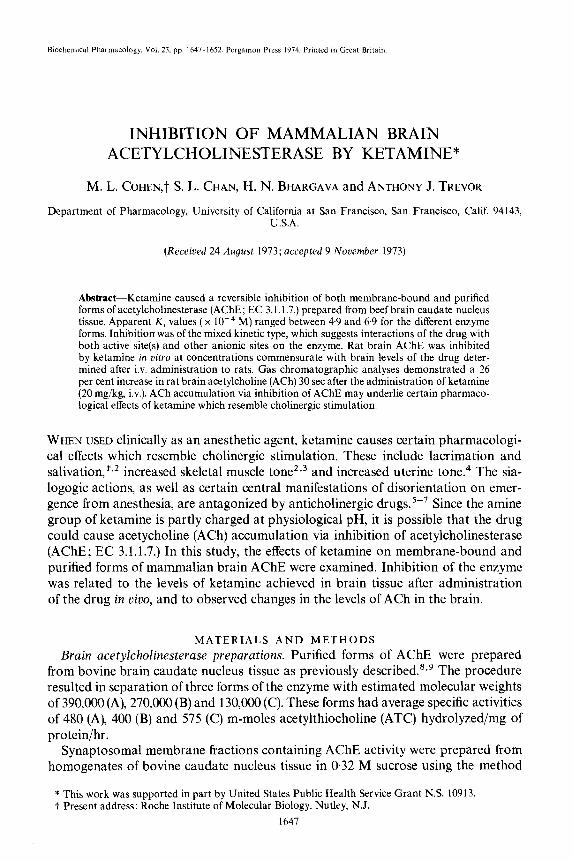
FIG. 1. Ketamine inhibition of bovine brain AChE. AChE activity was determined in the presence of keta- mine by the spectrophotometric procedure of Ellman et al.” at pH 7.9 and substrate concentration of 1 mM ATC. Control activity, expressed as pmoles ATC hydrolyzed/mg of protein/hr, for the enzyme forms was: 480 x lo3 (form A); 400 x lo3 (form B); 575 x lo3 (form C) and 45 (membrane-bound). Points represent mean values from three determinations (which differed by less than 4 per cent) at each
inhibitor concentration.
RESULTS
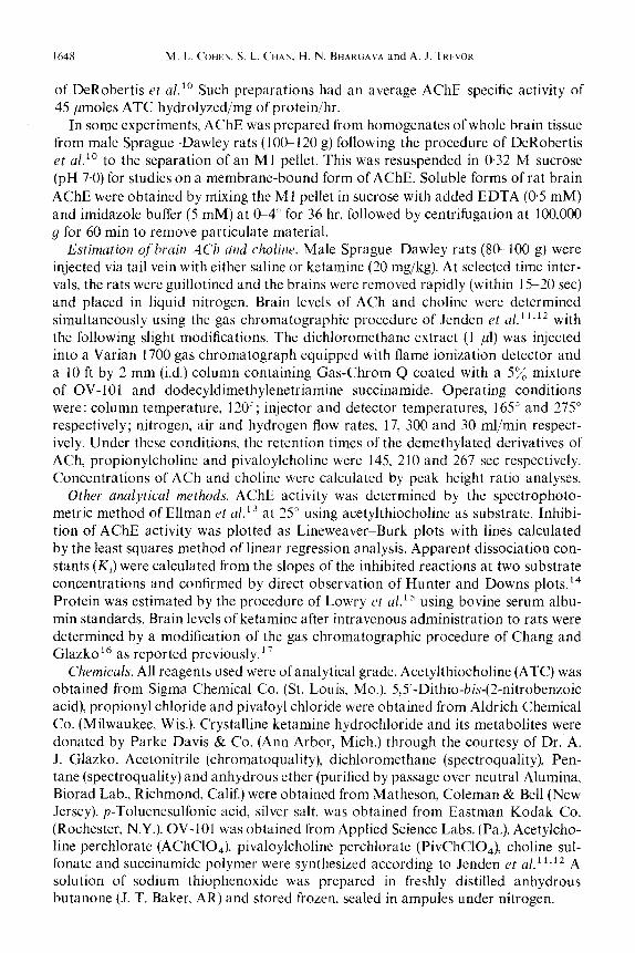
Ketamine inhibition of bovine brain acetylcholinesterase. Membrane-bound (synap- tosomal) and three purified forms of AChE from bovine brain tissue were inhibited by ketamine in a concentration-dependent manner (Fig. 1). The forms showed no marked differences in sensitivity to ketamine, apparent Ki values (x 1O-4 M) being 4.9 for membrane-bound AChE and 6.9, 66 and 5.1 for purified forms C, B and A respectively. Inhibition was 90 per cent reversible after dialysis against 0.1 M phos- phate buffer for 60 min to remove ketamine. Figure 2 is a Lineweaver-Burk plot for
FIG. 2. Lineweaver-Burk plot of ketamine inhibition of purified AChE (form C). Enzyme activity was measured by the method of Ellman et ~1.‘~ at substrate (ATC) concentrations from @1 to 5 mM at pH 7.9. Control activity at optimum substrate concentration (1 mM) was 575 m-moles ATC hydrolyzed/mg of protein/hr. Lines were plotted from calculations employing the least squares method of linear regression
analysis.

I650 M. L. COHEY, S. L. CHAN. H. N. BHAIIGAVA and A.J. TRDOR
IXIO-3 M IXIO-p M
Ketamine, molar
FIG. 3. Ketamine inhibition of rat brain AChE. Enzyme activity was estimated by the method of Ellman et a1.‘3 at pH 7.9 and optimal substrate concentration (1 mM ATC). Control activity was 9.4 and 143 pmoles ATC hydrolyzed/mg ofproteimhr for the particulate and soluble form of the enzyme respectively. Points represent mean values from three determinations (which differed by less than 4 per cent) at each
inhibitor concentration.
ketamine inhibition of AChE form C (mol. wt, 130,000), suggesting mixed kinetics of inhibition with changes in I&,, and apparent K, value. Similar data were obtained for the other forms of the enzyme. The demethylated metabolite of ketamine (meta- bolite I) also inhibited bovine brain AChE (data not shown), but at concentrations approximately 10 times that required for inhibition by ketamine.
Ketamine inhibition of rat brain acetylcholinesterase. Ketamine inhibited mem- brane-bound and solubilized forms of rat brain AChE in a concentration-dependent manner (Fig. 3). Approximately 20 and 40 per cent inhibition of both forms of the enzyme occurred at concentrations of 5 x 10e4 and lop3 M ketamine respectively. The levels of ketamine achieved in rat brain were estimated after intravenous administration of the drug at a dose (20 mg/kg) that produced a loss of righting reflex of c-8 min.i7 Table 1 shows brain levels of ketamine at three time intervals after administration of the drug with an estimation of “apparent molarity.” The latter
data are approximations which assume a specific gravity of 1.0 for brain tissue; they do not take into account possible regional differences in ketamine level or differences in the intra-extracellular distribution of the drug. Peak levels of ketamine achieved in rat brain tissue after administration in vivo were approximately 4 x lop4 M.
TABLE I. RATBRAIN LEVELSOF KETAMINEAFTER INTRAVENOUS INJECTION*
Time after injection (min)
Ketamine
@g/g tissue)
“Apparent molarity”
(M)
0.5 95.7 k 12.6 4.1 x 1om4 I ,o 63.0 f 2.X 2.7 x 10-a
10~0 24.6 k 3.9 I.0 x 1om4
* Ketamine (20 mg/kg) was injected into the tail vein of rats. Brain tissue was assayed for ketamine at indicated times by the gas chromatographic pro- cedure noted in Methods. Ketamine levels shown are mean values from four separate animals + S.E.M.
Ketamine inhibition of brain acetylcholinesterase 1651
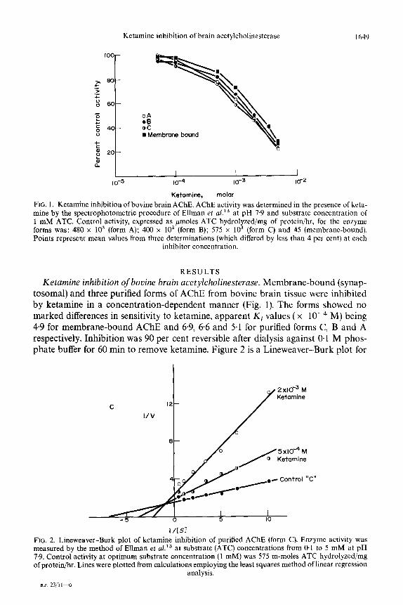
TABLE 2. EFFECT OF KETAMINE ON RAT BRAIN ACh LEVELS*
Time after Increase injection ACh Choline in ACh
Condition (min) (nmoles/g) (nmoles/g) (%)
Saline 0.5 Ketamine 0.5
Saline 1.0 Ketamine 1.0
Saline 10.0 Ketamine 10.0
17.75 + 0.65 50.36 k 0.58 2242 + 0.41 47.33 f 3.57 26.3 (P < 0001) 18.69 + 0.36 56.61 k 6.27 22.74 f 1.20 60.03 k 1.12 21.7 (P < 0.02)
20.39 f 0.77 46.04 + 7.65 19.67 + 0.99 51.89 k 2.1 (P > 005)
* Ketamine, 20 mg/kg, was injected into the tail vein of rats. Brain tissue was assayed for ACh and choline at indicated times by the gas chroma- tographic procedure noted in Methods. Levels shown are mean values from four to six separate animals &- S.E.M. P values were calculated by the paired Student’s t-test.
Effect of ketamine on rat brain ACh levels. The whole brain levels of ACh and cho- line determined at three time periods after the intravenous administration of keta- mine to rats are shown in Table 2. Control (saline-treated) levels of brain ACh are comparable to those reported previously.” Ketamine (20 mg/kg, iv.) caused signifi- cant increases in brain ACh at 30 (P < 0.001) and 60 (P < 0.02) set, but not at 10 min after administration. Brain choline levels exhibited some variability and were not changed significantly after ketamine treatment.
DISCUSSION
The anesthetic agent ketamine caused reversible inhibition of both membrane- bound and purified forms of mammalian brain AChE. The competitive component of inhibition may reflect an interaction of the positively charged nitrogen moiety of ketamine (pKa, 7.5) with the anionic region of the enzyme active site. The possibility of ketamine interactions with AChE at secondary anionic sites is suggested by the noncompetitive component of inhibition. Such interactions have been suggested for other inhibitors of AChE which possess charged nitrogen groups, including deca- methonium and tubocurarine.‘g*20
Ketamine caused approximately 40 per cent inhibition of rat brain AChE in vitro at a concentration only 25 times that estimated to occur in brain tissue after intra- venous administration of the drug (20 mg/kg) to the intact animal. The same dose of ketamine caused approximately 26 and 22 per cent increases in brain ACh levels at 30 and 60 set after administration respectively. These data suggest that cholinergic stimulation via AChE inhibition could underlie certain of the pharmacological effects of ketamine. Other anesthetic agents, including barbiturates, chloral hydrate and ether, have been shown to elevate brain ACh levels when used under conditions of deep anesthesia. 21,22 In preliminary studies23,24 ether and cyclopropane, both of which exert effects resembling cholinergic stimulation, were reported to cause inhibi- tion of brain AChE in vitro. Since there have been no reports of AChE inhibition by barbiturates at concentrations similar to those occurring in vivo in the brain, it is possible that their effects on brain ACh levels result from mechanisms other than enzyme inhibition.

1652 M. L. COHEN. S. L. CHAN. H. N. BHAKGAVA and A. J. TMVOR
Similarities have been noted between AChE and cholinergic receptors in terms of ligand binding properties,‘” which suggest the possibility of a direct interaction of ketamine with certain cholinergic receptors. This may be the basis for the reported interactions between ketamine and certain neuromuscular blocking agents at the myoneural junction.26
REFERENCES
1. G. CORSSEN, M. MIYASAKA and E. F. DOMINO, Anesth. A&g. 47, 746 (1968). 2. J. W. PENDER, J. Am. med. Ass. 215, 1126(1971). 3. G. CORSSEN and E. F. DOMINO, Anesth. Analg. 45, 29 (1966). 4. B. LITTLE, T. CHANG, L. CHUCOT, W. A. DILL, L. L. ENRITE. A. J. GLAZKO, M. JASSANI, H. KRETCHMER
and A. Y. SWEET, Am. J. Obstet. Gynec. 113, 247 (1972). 5. M. MORGAN, L. LOH, L. SINGER and P. H. MOORE, Anuesthesia 26, 158 (1971). 6. J. F. BOVIL, J. W. DUND~E. D. L. COPPEL and J. MOORE.. Luncrt 1. 1285 (1971) 7. P. P. BOSOMWORTH, Anesth. A&g. 50, 471 (1971). 8. S. L. CHAN, D. Y. SHIRACHI and A. J. TREVOR, J. Neurochcm. 19, 437 (1972). 9. S. L. CHAN, D. Y. SHIRACHI, H. N. BHARGAVA, E. GARIIN~R and A. J. TREVOR. J. Neurochem. 19. 2747
(1972). 10. E. DEROBERTIS. M. ALBERICI, G. RODKIGUEZ and J. M. AZCUKKA, L[fi Sci. 5, 577 (1966). 1 I. D. J. JENDEN, R. A. B~~ITH and M. ROCH, Analvt. Chem. 44. 1879 (1972). 12. D. J. JENDEN, M. ROCH and R. A. BOOTH, J. chiomat. Sci. 10, 151 (1972). 13. G. L. ELLMAN. D. COURTNEY, V. ANURES and R. M. FEATHERSTONE, Biochem. Pharmac. 7, 88 (1961). 14. A. HUNTER and C. E. DOWNS, J. biol. Chem. 157, 427 (1945). 15. 0. H. LOWRY, W. J. ROSEBROUGH, A. L. FARR and R. J. RANIIALL, J. biol. Chem. 193, 265 (1957). 16. T. CHANG and A. J. GLAZKO, Anesthesiology 36, 401 (1972). 17. M. COHEN, S. L. CHAN, W. WAY and A. J. TREVOR. Anesthesiology 39, 370 (1973). 18. L. B. CAMPBELL and D. J. JENDEN. J. Nemochrm. 17, 1697 (1970). 19. R. J. KITZ. L. M. BRASWELL and S. GINSBERG, Molrc. Phurmac. 6, 108 (1970). 20. B. D. ROUFOGALIS and E. E. QUIST, MO&. Pharmac. 8, 41 (1972). 21. J. CROSSLAND and A. J. MEKRICK. J. Physiol., Land. 125, 56 (1954). 22. J. CROSSLAND and P. SLATER. Br. J. Ph&mac. Chemother. 33, 42 (1968). 23. U. R. MAHESHWAKI. S. L. CHAN and A. J. TREVOR. Proc. west. Pharmac. Sot. 16. 175 (1973) 24. U. R. MAHESHWARI, S. L. CHAN and A. J. TRFVOR. Pharmucologi~t 15, 183 (197<). ~ ’ 25. B. BELLEAU and V. DITULLIO, Can. J. Biochrm. Physiol. 49, I 131 (1971). 26. R. CRONELLY, K. L. DRETCHEN, M. D. SOKOLL and J. P. LONG, Eur. J. Pharmac. 22, 17 (1973).