inhibition of embryonic development by microcystin-lr in zebrafish, danio rerio
TRANSCRIPT
Inhibition of embryonic development by microcystin-LR
in zebrafish, Danio Rerio
Pei-Jen Wanga, Ming-Shan Chienb, Fong-June Wua,Hong-Nong Choua,c, Shyh-Jye Leeb,c,*
aInstitute of Fisheries Science, National Taiwan University, Taipei, Taiwan, ROCbInstitute of Zoology, National Taiwan University, Taipei, Taiwan, ROC
cDepartment of Life Sciences, National Taiwan University, Taipei, Taiwan, ROC
Received 7 September 2004; revised 22 October 2004; accepted 25 October 2004
Available online 10 December 2004
Abstract
Microcystin-LR (MC-LR), a cyanobacterial toxin, is a potent inhibitor of protein phosphatase 1 (PP1) and protein
phosphatase 2A (PP2A). PP1 and PP2A are critical regulators in embryonic development. However, the effects of MC-LR in
embryonic development have been controversial. MC-LR has been demonstrated to be highly toxic in medaka, but not in
zebrafish or rabbit embryos. The causes of difference may be due to membrane impermeability that impaired the delivery of
MC-LR into cytoplasm of zebrafish and rabbit embryos. Therefore, we microinjected MC-LR directly into developing zebrafish
embryos and investigated the effects of MC-LR on embryonic development. We demonstrated that MC-LR induced the
lethality of zebrafish embryos in a dose- and time-dependent manner. MC-LR also induced the loss of blastomere coherence via
the interference of b-catenin and cadherins distributions. Furthermore, the MC-LR treated fry revealed various developmental
defects. These results suggested that MC-LR might affect the phosphorylation equilibrium of signaling molecules, including
b-catenin and cadherins, required early in zebrafish embryonic development.
q 2004 Elsevier Ltd. All rights reserved.
Keywords: Microcystin; PP1; PP2A; Zebrafish; Embryos; b-catenin and cadherin
1. Introduction
The toxicity of MCs has been associated with fish killed
worldwide (Watanabe et al., 1996). Among the fish species
killed by MCs, cyprinids are most affected, and to a less
extent to the others (Fischer and Dietrich, 2000). By
analyzing the diets of affected species, it appears that the
toxicity of MCs depends on the amount of toxin ingested.
However, by gavaging a bolus dose of MC-LR containing
0041-0101/$ - see front matter q 2004 Elsevier Ltd. All rights reserved.
doi:10.1016/j.toxicon.2004.10.016
* Corresponding author. Address: 1 Roosevelt Road, Section 4,
Institute of Zoology, National Taiwan University, 209 Fisheries
Science Building, Taipei, Taiwan 106, ROC. Tel.: C886 1 2
33665902; fax: C886 1 2 33662457.
E-mail address: [email protected] (S.-J. Lee).
lyophilized cyanobacteria to carp or rainbow trout, it
appears that carp is more susceptible to MC-LR compared
to rainbow trout (Tencalla et al., 1994; Tencalla and
Dietrich, 1997). Therefore, the MC-LR gavaging exper-
iments appeared not to be consistent to serve as a bioassay
system in fish.
MC-LR is a potent inhibitor of serine/threonine protein
phosphatase 1 and 2A (PP1 and PP2A) with an IC50 of
about 0.1–1.0 nM (Honkanen et al., 1990). The inhibition
of PP1 and PP2A results in hyperphosphorylation of
proteins, which may be responsible for the toxicity of
microcystins. During embryonic development, the equili-
brium of phosphorylation and dephosphorylation status of
signaling molecules is tightly regulated for the control of
rapid cleavages and cellular differentiation. Dynamic
changes in cytoskeleton play a major role in embryo
Toxicon 45 (2005) 303–308
www.elsevier.com/locate/toxicon

P.-J. Wang et al. / Toxicon 45 (2005) 303–308304
reorganization. Microcystins have been demonstrated to
impair cytosleleton by hyperphosphorylation of the major
intermediate proteins keratin 8 and 18 in a dose- and time-
dependent manner (Ohta et al., 1992; Toivola et al.,
1997). Microcystins have also been reported to perturb the
functions of motor protein dynein (Runnegar et al., 1997)
and tubulin (Ding et al., 2000). It is therefore reasonable
to hypothesize that MC-LR should disrupt early embryo-
nic development by interfering with the cytoskeletal
dynamic within the embryo. A couple of attempts have
been reported to evaluate the effects of MC-LR on early
embryonic development, including rabbit (Frangez et al.,
2003) and zebrafish (Oberemm et al., 1997, 1999) studies.
However, both studies revealed no significant defects of
early embryos treated with MC-LR. These results are
clearly against our hypothesis and the well established
PP1 and PP2A blocking ability by MC-LR. In both
investigations, the membrane-intact embryos were used. It
was very likely that the membrane barrier precluded MC-
LR from entering cytoplasm. However, Wiegand et al.
(1999), have shown that there was a detectable uptake of
C-14 labeled MC-LR by early zebrafish embryos and the
lack of toxicity may due to the activities of detoxication
enzymes like glutathione S-transferase (GST). C-14
labeled MC-LR uptake reached 0.44 ng per egg, which
seems to be high enough to exert MC-LR’s functions.
However, since the C-14 labeled MC-LR uptake was
measured using the whole egg (the embryo itself, the
chorion, and the perivitelline fluid), one could not exclude
the possibility that MC-LR was trapped mainly in the
chorion and perivitelline fluid. Therefore, instead of
relying on cellular uptake of MC-LR, we took a more
direct approach by microinjecting MC-LR into zebrafish
embryos to bypass membrane barriers. We found that
(1) MC-LR dose- and time-dependently induced embryo-
nic malformation and death. (2) MC-LR caused the loss of
blastomere coherence by disrupting the distributions of
b-catenin and cadherin.
2. Materials and methods
2.1. Fish Husbandry and embryo collection
Zebrafish, Danio rerio, were raised on a 14-h day/10-h
night cycle at 26 8C. Eggs were collected at 15–20 min
intervals after spawning, washed and incubated in Ringer’s
solution (116 mM NaCl, 2.9 mM KCl, 1.8 mM CaCl2 and
5 mM HEPES) at 28.5 8C until use. All chemicals were from
Sigma (St Louis, MO) unless otherwise stated.
2.2. Purification of MC-LR
MC-LR was extracted and purified from Microcystin
aeruginosa as described in detail by Lee et al. (1998).
2.3. Microinjection procedures
Glass capillaries of 1.14 mm O.D. and 0.5 mm I.D.
(World Precision Instrument Inc., Sarasota, FL) were pulled
using a horizontal puller (P-97, Sutter Instrument, Navato,
CA). Embryos at desired stages as judged according to
Kimmel et al. (1995) were immobilized at an injection
trough on a 100 mm 2% agar plate. MC-LR was dissolved in
injection buffer (68.5 mM NaCl, 1.35 mM KCl, 5 mM
Na2HPO4 and 1 mM KH2PO4 and 0.25% (w/v) phenol red)
with different concentrations of MC-LR as indicated. An
injection pipette was forced into the chorion and the yolk
cell to reach the junction between yolk cell and blastodisc
where 2.3 nL of solution was ejected by using a nanoliter
injector (World Precision Instrument Inc., Sarasota, FL).
After injection, embryos were recovered from injection
troughs and cultured in Ringer’s solution at 28.5 8C until
examined.
2.4. Whole-mount immunocytochemistry and confocal
microscopy
Zebrafish embryos at designated stages were fixed in 4%
paraformaldehyde in PBS (137 mM NaCl, 2.7 mM KCl,
10 mM Na2HPO4 and 2 mM KH2PO4) overnight at 4 8C,
washed in PBS and manually dechorionated. Twenty
embryos were incubated with a polyclonal anti-b-catenin
antibody (Sigma C2206, diluted at 1:400) or a polyclonal
anti-pan cadherin antibody (Sigma C3678, diluted at 1:100)
with continuous rotation at 4 8C overnight. After four
washes in PBS containing 0.5% Triton X-100 (PBT),
embryos were incubated in a 1:250 or 1:500 dilution of a
FITC-conjugated goat anti-rabbit antibody (#111-095-144,
Jackson ImmunoResearch Laboratories, West Grove, PA)
for anti-b-catenin or anti-pan cadherin antibody-labeled
embryos, respectively, at 4 8C overnight. Embryos were
transferred to 30% glycerol first and then 50% glycerol for
confocal microscopic observations at the Center of Two-
Photon Laser Confocal Microscope, National Taiwan
University. To reveal the nuclei, embryos were washed in
PBT and then incubated in 10 mg/mL Hoechst dye for 30
mins at room temperature before examination. Unbound
Hoechst dye was removed by washing in PBT and examined
under an epifluorecent microscope.
2.5. Embryo observations and photography
Observation of embryonic development was made at
designated time under a Leica Mz75 stereomicroscope, a
Leica DMIRB fluorescent microscope, or a Leica TCS SP2
confocal microscope equipped with an Ar–Kr two photon
laser system located. All photographs were taken by a Nikon
digital camera and analyzed using Adobe Photoshop 7.0
except for the confocal images, which were photographed
and edited utilizing the Leica Confocal Software.

P.-J. Wang et al. / Toxicon 45 (2005) 303–308 305
2.6. Statistical analysis
Experimental values are expressed as meansGstandard
error of means and analyzed in Excel using Two-Factor
ANOVA with Replication.
Fig. 2. MC-LR causes gradual death of zebrafish embryos in a dose-
dependent manner. An hour post injection with MC-LR at indicated
amounts in nM gradual death occurred and the embryos survival
rate continued to decrease until 24 h post injection. Fertilized eggs
were injected at the 2–4 cell stage and cultured in Ringer’s solution
at 28.5 8C. Embryos were examined at the designated time and the
white/opaque embryos were scored as dead. These results were
cumulated data of five independent experiments.
3. Results
To study the toxicity of MC-LR in zebrafish, we
microinjected MC-LR into 2–4 cell stage embryos. Within
half an hour after injection of microcystin-LR (MC-LR),
embryos showed signs of malformation. The most notable
change in the MC-LR-injected embryos was the loss of
coherence of blastomeres. A normal zebrafish embryo has a
tightly adhered blastomeres arranged in tiers as shown in
Fig. 1A. In contrast, MC-LR induced the deterioration of
embryos by blocking the adherence of blastomeres (Fig. 1C).
We were also interested to know if MC-LR affected nuclear
division, since cell cycle progression is regulated by
phosphorylation and dephosphorylation. In the control-
injected embryo all nuclei were clearly visible by Hoechst
dye staining except one nucleus was out of focus as shown in
Fig. 1B. In contrast, we observed only two nuclei in an 8-cell
stage embryo treated with 750 nM MC-LR in one focal plane
Fig. 1. MC-LR causes the loss of adherence of blastomeres in
zebrafish embryos. After the injection of 900 nM MC-LR, a
zebrafish embryo could still divide, but the blastomeres lost
compact adherence to each other (C) as compared to the control-
injected embryos (A). Blastomeres of the control-injected 8-cell
embryos were nicely arranged into two tiers with nuclei clearly
stained by Hoechst dye in each blastomere (B). One nucleus is out
of focus in panel B. The nuclear cleavage appears not normal in
MC-LR injected embryo at the first glance, since only 2 nuclei are
visible on the focal plan shown in panel D. However, it was due to
the irregular arrangement of blastomeres that we were able to locate
all 8 nuclei in the 8-cell stage embryo by scanning through different
focal planes.
shown in Fig. 1D. However, by examining through different
focal planes, we were able to find all 8 nuclei in the embryo
treated with MC-LR (data not shown). Therefore, MC-LR
appeared not interfering with the karyokinesis. Although, MC-
LR-treated embryos could still develop, but they were
gradually dying and embryos turned opaque and disintegrat-
ing. To analyze the dose-dependent lethality of MC-LR, we
injected embryos with different amounts of MC-LR. Injecting
embryos after the 1-cell stage allowed us to remove easily
unfertilized eggs from our statistics. We then examined
survival rates of embryos at designated time points (Fig. 2). As
shown in Fig. 2, 93.2G4.8% (NZ5) of 311 control-injected
embryos survived at 48 h post injection. As MC-LR was
injected, the survival rates of embryos decreased with the
increase in time of culture post fertilization and the elevation of
intracellular MC-CR concentration from 300 to 900 nM. At
the end of examination (48 h post fertilization), embryonic
survival rate decreased significantly to 82.5G3.9% of 319
embryos, 51.5G12.1% of 353 embryos and 18.5G10.3% of
343 embryos injected with 300, 750 and 900 nM MC-LR,
respectively, in 5 independent trials. Using the Two-Factor
Analysis of Variance, the effects of intracellular MC-LR
concentration and the time of culture post fertilization on
embryonic survival were analyzed. Both factors and their
interaction significantly (p%4.65E-23) affected the survival
rate of MC-LR-injected embryos. Thus, MC-LR induced
embryonic death in a dose- and time-dependent manner.
For those MC-LR-treated fry survived at 48 h post injection,
various defects were shown in Fig. 3. The most frequent
observed defects appeared in tails. Tails were curving
(Fig. 3A), bent or twisting (Fig. 3B). In some cases, cyclopia
occurred while a residual eye was still visible (arrow) as shown
in Fig. 3C. Fig. 3D shows a fry with edema in pericardial sac
(PS) and hatching gland (HG). Some of those fry with edema
could survive up to 6–7 days post fertilization, but the edema in

Fig. 3. MC-LR induces different developmental defects in zebrafish fry. MC-LR-treated embryos were cultured in Ringer’s solution for 48 h and
photographed except (E), which was taken at 6 days post fertilization. Various defects were detected in MC-LR-treated embryos, including
trunk and tail curving (A), bent or twisting tails (B), cyclopia whose residual eye was indicated by an arrow (C), edema in pericardial sac (PS)
and hatching gland (HG) (D), curving, rounding, deteriorated and eventually dying (F). Edema in pericardial sac and hatching gland could be
worse where the curving of the trunk occurred if they survived in this case 6 days post fertilization (E).
P.-J. Wang et al. / Toxicon 45 (2005) 303–308306
pericardial sac and hatching gland was worse that a curvature
of trunk occurred (Fig. 3E). In some severe cases, the fry was
curving, rounding, deteriorating and eventually dying (Fig.
3F). Due to the broad spectrum of defect patterns, we assume
MC-LR perturbed the phosphorylation status of some key
enzymes, which might interfere with downstream molecules
required for proper embryonic development and subsequent
organogenesis.
Blocking of blastomeres coherence by MC-LR (Fig. 1) is
an intriguing observation. Since E-cadherin has been shown to
mediate the blastomere adherence via b-catenin (Gotz et al.,
2000), we investigated whether MC-LR inhibits blastomeres
coherence by disturbing b-catenin/cadherin signaling by
examining the distributions of b-catenin and cadherins in
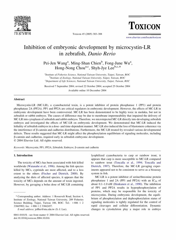
embryos treated with or without MC-LR. In Fig. 4, we show
confocal photographs of a single section of 3 mm (Fig. 4A, B,
E, F) or 3D stacking graphs of 20 sections (Fig. 4C, D, G, H) for
embryos treated in the presence (Fig. 4B, D, F, H) or absence
(Fig. 4A, C, E, G) of 900 nM MC-LR. Both b-catenin (Fig. 4A,
C) and cadherins (Fig. 4E, G) were clearly localized at the
cleavage furrows between adjacent blastomeres in the absence
of MC-LR. In contrast, b-catenin (Fig. 4B, D) and cadherins
(Fig. 4F, H) were present only in the barely touched boundary
between blastomeres. These results suggested that MC-LR
perturbs the blastomere coherence by interfering with the
localization of b-catenin and cadherin.
4. Discussion
The sensitivity to MC-LR exposure is variable in
developmental stages and among fish species. The main
cause of variability is the MC-LR uptake that is presumably
mediated by bile acid transporters (Eriksson et al., 1990;
Runnegar et al., 1991, 1995). To assay the toxicity of
MC-LR during early embryonic development in zebrafish,
we microinjected MC-LR directly into embryos and we
observed that (1) MC-LR dose- and time-dependently
increased the lethality of embryos. (2) MC-LR caused
various embryo abnormalities, which suggested that MC-LR
affected the phosphorylation status of upstream factors
mastering the embryonic development and subsequent
organogenesis. (3) MC-LR induced the loss of blastomere
coherence by interfering the distributions of b-catenin and
cadherins.
Zebrafish embryos and larvae were found to be
insensitive to the toxicity of MC-LR or crude extracts of
cyanobacteria. Although no acute effects were observed
during embryonic development, a reduced survival rate and
a retarded growth were reported in MC-LR and MC-RR pre-
exposed larvae at 21 days of age (Oberemm et al., 1997,
1999). The lack of MC-LR toxicity in embryos is
questionable, since the activities of MC-LR targets, PP1
and PP2A, have been shown to be critical for regulating cell
cycle progression during embryonic development (Gotz
et al., 2000). The most plausible explanation is the poor
membrane permeability of microcystins in the absence of
bile acid transporters. Jacquet et al. (2004) have nicely
demonstrated the lethality of MC-LR on development of
medaka fish embryos by microinjection. Using the similar
approach and MC-LR dosages, we were able to demonstrate
that MC-LR caused the death of embryos in a dose- and
time-dependent manner (Fig. 2). We also observed a high
rate in malformation in MC-LR-treated zebrafish embryos
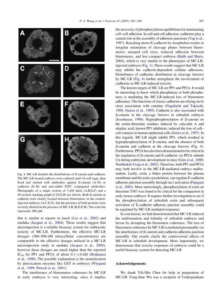
Fig. 4. MC-LR disturbs the distributions of b-catenin and cadherin.
The MC-LR treated embryos were cultured until 16-cell stage, then
fixed and stained with antibodies against b-catenin (A–D) or
cadherin (E–H) and anti-rabbit FITC conjugated antibodies.
Photographs of a single section of 3 mM thick (A,B,E,F) and a
20-section stacking graph (C,D,G,H) are shown. Both b-catenin or
cadherin were clearly located between blastomeres in the control-
injected embryos (A,C,E,G), but the presence of both proteins were
severely aborted in the presence of MC-LR (B,D,F,H). The scale bar
represents 200 mM.
P.-J. Wang et al. / Toxicon 45 (2005) 303–308 307
that is similar to reports in loach (Liu et al., 2002) and
medaka (Jacquet et al., 2004). These results suggest that
microinjection is a reliable bioassay system for embryonic
toxicity of MC-LR. Furthermore, the effective MC-LR
dosages (300–900 nM intracellular concentration) are
comparable to the effective dosages utilized in a MC-LR
microinjection study in medaka (Jacquet et al., 2004),
however those dosages are much higher than the reported
IC50 for PP1 and PP2A of about 0.1–1.0 nM (Honkanen
et al., 1990). The possible explanation is the neutralization
by detoxication enzymes like GST in embryos (Wiegand
et al., 1999; Pietsch et al., 2001).
The interference of blastomeres coherence by MC-LR
in early embryos is very interesting, since it implies
the necessity of phosphorylation equilibrium for maintaining
cell–cell adhesion. In cell and cell adhesion, cadherins play a
central role in the assembly of adherens junctions (Yap et al.,
1997). Knocking down E-cadherin by morpholino results in
irregular orientation of cleavage planes between blasto-
meres, unequal cell sizes, reduced adhesion between
blastomeres, and less compact embryos (Babb and Marrs,
2004), which is very similar to the phenotypes of MC-LR-
injected embryos (Fig. 1). These results suggest that MC-LR
may inhibit the cadherin-dependent cellular adhesions.
Disturbance of cadherins distribution in cleavage furrows
by MC-LR (Fig. 4) further strengthens the involvement of
cadherins in MC-LR-induced toxicity.
The known targets of MC-LR are PP1 and PP2A. It would
be interesting to know which phosphatase or both phospha-
tases is mediating the MC-LR-induced loss of blastomere
adherence. The functions of classic cadherins are relying on its
close association with catenins (Nagafuchi and Takeichi,
1988; Ozawa et al., 1989). Cadherin is also associated with
b-catenin at the cleavage furrows in zebrafish embryos
(Jesuthasan, 1998). Hyperphosphorylation of b-catenin on
the serine-threonine residues induced by calyculin A and
okadaic acid, known PP1 inhibitors, induced the loss of cell–
cell contacts in human epidermal cells (Serres et al., 1997). In
this regards, MC-LR might inhibit PP1, which resulted in
hyperphosphorylation of b-catenin, and the absence of both
b-catenin and cadherin at the cleavage furrows (Fig. 4).
Furthermore, PP2A has also been demonstrated to be critical in
the regulation of b-catenin and E-cadherin via PP2A subunit
Ca during embryonic development in mice (Gotz et al., 2000;
Nunbhakdi-Craig et al., 2002). Therefore, both PP1 and PP2A
may both involve in the MC-LR-mediated embryo malfor-
mation. Lastly, ezrin, a linker protein between the plasma
membrane and the actin cytoskeleton, can regulate E-cadherin
adherens junction assembly through rac1 activation (Pujuguet
et al., 2003). More interestingly, phosphorylation of ezrin on
threonine T567 was found to be critical for the compaction in
early mouse embryos. It requires further investigation to see if
the phosphorylation of zebrafish ezrin and subsequent
activation of E-cadherin adherens junction assembly could
be regulated by MC-LR mediated responses.
In conclusion, we had demonstrated that MC-LR induced
the malformation and lethality of zebrafish embryos and
larvae by disrupting the blastomere coherence. The loss of
blastomere coherence by MC-LR is mediated presumably via
the interference of b-catenin and cadherin adherens junction
assembly. Our results clarify the controversial effects of
MC-LR in zebrafish development. More importantly, we
demonstrate that toxicity responses of embryos could be a
useful bioassay system for detecting MC-LR.
Acknowledgements
We thank Yih-Min Chen for help in preparation of
MC-LR. Fong-June Wu was a recipient of Undergraduate

P.-J. Wang et al. / Toxicon 45 (2005) 303–308308
Research Grants (NSC91-2815-C-002-059-B) supported by
National Science Council of Taiwan, R.O.C.
References
Babb, S.G., Marrs, J.A., 2004. E-cadherin regulates cell movements
and tissue formation in early zebrafish embryos. Developmental
Dynamics 230, 263–277.
Ding, W.X., Shen, H.M., Ong, C.N., 2000. Microcystic cyanobac-
teria extract induces cytoskeletal disruption and intracellular
glutathione alteration in hepatocytes. Environmental Health
Perspectives 108, 605–609.
Eriksson, J.E., Gronberg, L., Nygard, S., Slotte, J.P.,
Meriluoto, J.A., 1990. Hepatocellular uptake of 3H-dihydromi-
crocystin-LR, a cyclic peptide toxin. Biochemica et Biophysica
Acta 1025, 60–66.
Fischer, W.J., Dietrich, D.R., 2000. Pathological and biochemical
characterization of microcystin-induced hepatopancreas and
kidney damage in carp (Cyprinus carpio). Toxicology and
Applied Pharmacology 164, 73–81.
Frangez, R., Zuzek, M.C., Mrkun, J., Suput, D., Sedmak, B.,
Kosec, M., 2003. Microcystin-LR affects cytoskeleton and
morphology of rabbit primary whole embryo cultured cells in
vitro. Toxicon 41, 999–1005.
Gotz, J., Probst, A., Mistl, C., Nitsch, R.M., Ehler, E., 2000. Distinct
role of protein phosphatase 2A subunit C alpha in the regulation
of E-cadherin and beta-catenin during development. Mechan-
isms of Development 93, 83–93.
Honkanen, R.E., Zwiller, J., Moore, R.E., Daily, S.L., Khatra, B.S.,
Dukelow, M., Boynton, A.L., 1990. Characterization of
Microcystin-Lr, A Potent Inhibitor of Type-1 and Type-2A
Protein Phosphatases. Journal of Biological Chemistry 265,
19401–19404.
Jacquet, C., Thermes, V., de Luze, A., Puiseux-Dao, S., Bernard, C.,
Joly, J.S., Bourrat, F., Edery, M., 2004. Effects of microcystin-
LR on development of medaka fish embryos (Oryzias latipes).
Toxicon 43, 141–147.
Jesuthasan, S., 1998. Furrow-associated microtubule arrays are
required for the cohesion of zebrafish blastomeres following
cytokinesis. Journal of Cell Biology 111, 3695–3703.
Kimmel, C.B., Ballard, W.W., Kimmel, S.R., Ullmann, B.,
Schilling, T.F., 1995. Stages of embryonic-development of the
zebrafish. Developmental Dynamics 203, 253–310.
Lee, T.H., Chen, Y.M., Chou, H.N., 1998. First report of
microcystins in Taiwan. Toxicon 36, 247–255.
Liu, Y.D., Song, L.R., Li, X.Y., Liu, T.M., 2002. The toxic effects
of microcystin-LR on embryo-larval and juvenile development
of loach, Misguruns mizolepis Gunthe. Toxicon 40, 395–399.
Nagafuchi, A., Takeichi, M., 1988. Cell binding function of E-
cadherin is regulated by the cytoplasmic domain. EMBO
Journal 7, 3679–3684.
Nunbhakdi-Craig, V., Machleidt, T., Ogris, E., Bellotto, D.,
White, C.L., Sontag, E., 2002. Protein phosphatase 2A
associates with and regulates atypical PKC and the epithelial
tight junction complex. Journal of Cell Biology 158, 967–978.
Oberemm, A., Fastner, J., Steinberg, C.E.W., 1997. Effects of
microcystin-LR and cyanobacterial crude extracts on embryo-
larval development of zebrafish (Danio rerio). Water Research
31, 2918–2921.
Oberemm, A., Becker, J., Codd, G.A., Steinberg, C., 1999. Effects
of cyanobacterial toxins and aqueous crude extracts of
cyanobacteria on the development of fish and amphibians.
Environmental Toxicology 14, 77–88.
Ohta, T., Nishiwaki, R., Komori, A., Suganuma, M., Fujiki, H.,
1992. Hyperphosphorylation of Cytokeratins 8 and 18 by
Microcystin-LR, a new liver-tumor promoter, in primary
cultured rat hepatocytes. Carcinogenesis 13, 2443–2447.
Ozawa, M., Baribault, H., Kemler, R., 1989. The cytoplasmic
domain of the cell adhesion molecule uvomorulin associates
with three independent proteins structurally related in different
species. EMBO Journal 8, 1711–1717.
Pietsch, C., Wiegand, C., Ame, M.V., Nicklisch, A., Wunderlin, D.,
Pflugmacher, S., 2001. The effects of a cyanobacterial crude
extract on different aquatic organisms: evidence for cyanobac-
terial toxin modulating factors. Environmental Toxicology 16,
535–542.
Pujuguet, P., del Maestro, L., Gautreau, A., Louvard, D., Arpin, M.,
2003. Ezrin regulates E-cadherin-dependent adherens junction
assembly through Rac1 activation. Molecular Biology of the
Cell 14, 2181–2191.
Runnegar, M.T., Gerdes, R.G., Falconer, I.R., 1991. The uptake of
the cyanobacterial hepatotoxin microcystin by isolated rat
hepatocytes. Toxicon 29, 43–51.
Runnegar, M., Berndt, N., Kaplowitz, N., 1995. Microcystin uptake
and inhibition of protein phosphatases: effects of chemoprotec-
tants and self-inhibition in relation to known hepatic transpor-
ters. Toxicology and Applied Pharmacology 134, 264–272.
Runnegar, M., Wei, X., Zhang, L., HammAlvarez, S.F., 1997.
Inhibition of protein phosphatase 2A leads to enhanced
phosphorylation of kinesin and cytoplasmic dynein and their
associated proteins and to decreased microtubule-based vesicle
motility in intact hepatocytes. Hepatology 26, 537.
Serres, M., Grangeasse, C., Haftek, M., Durocher, Y., Duclos, B.,
Schmitt, D., 1997. Hyperphosphorylation of beta-catenin on
serine-threonine residues and loss of cell–cell contacts induced
by calyculin A and okadaic acid in human epidermal cells.
Experimental Cell Research 231, 163–172.
Tencalla, F., Dietrich, D., 1997. Biochemical characterization of
microcystin toxicity in rainbow trout (Oncorhynchus mykiss).
Toxicon 35, 583–595.
Tencalla, F.G., Dietrich, D.R., Schlatter, C., 1994. Toxicity of
microcystis-aeruginosa peptide toxin to yearling rainbow-trout
(Oncorhynchus-Mykiss). Aquatic Toxicology 30, 215–224.
Toivola, D.M., Goldman, R.D., Garrod, D.R., Eriksson, J.E., 1997.
Protein phosphatases maintain the organization and structural
interactions of hepatic keratin intermediate filaments. Journal of
Cell Science 110, 23–33.
Watanabe, M.F., Harada, K.I., Carmicael, W.W., Fujiki, H., 1996.
Toxic Microcystis. CRC press, Boca Raton, FL p. 262.
Wiegand, C., Pflugmacher, S., Oberemm, A., Meems, N.,
Beattie, K.A., Steinberg, C.E.W., Codd, G.A., 1999. Uptake
and effects of microcystin-LR on detoxication enzymes of early
life stages of the zebra fish (Danio rerio). Environmental
Toxicology 14, 89–95.
Yap, A.S., Brieher, W.M., Gumbiner, B.M., 1997. Molecular
and functional analysis of cadherin-based adherens
junctions. Annual Review of Cell and Developmental Biology
13, 119–146.