inhibition of angiogenesis and endothelial cell functions - molecular
TRANSCRIPT
Inhibition of angiogenesis and endothelial cellfunctions are novel sulforaphane-mediatedmechanisms in chemoprevention
Elisabeth Bertl, Helmut Bartsch,and Clarissa Gerhauser
Division of Toxicology and Cancer Risk Factors, German CancerResearch Center, Heidelberg, Germany
AbstractSulforaphane, an aliphatic isothiocyanate, is a knowncancer chemopreventive agent. Aiming to investigateantiangiogenic potential of sulforaphane, we here reporta potent decrease of newly formed microcapillaries in ahuman in vitro antiangiogenesis model, with an IC50 of0.08 Mmol/L. The effects of sulforaphane on endothelialcell functions essential for angiogenesis were investigatedin HMEC-1, an immortalized human microvascular endo-thelial cell line. Molecular signaling pathways leading toactivation of endothelial cell proliferation and degradationof the basement membrane were analyzed by reversetranscription-PCR. Sulforaphane showed time- and con-centration-dependent inhibitory effects on hypoxia-induced mRNA expression of vascular endothelial growthfactor and two angiogenesis-associated transcriptionfactors, hypoxia-inducible factor-1A and c-Myc, in a con-centration range of 0.8 to 25 Mmol/L. In addition, theexpression of the vascular endothelial growth factorreceptor KDR/flk-1 was inhibited by sulforaphane at thetranscriptional level. Sulforaphane could also affect base-ment membrane integrity, as it suppressed transcriptionof the predominant endothelial collagenase matrix metal-loproteinase-2 and its tissue inhibitor of metalloproteinase-2. Migration of HMEC-1 cells in a wound healing assaywas effectively prevented by sulforaphane at submicro-molar concentrations, and we determined an IC50 of 0.69Mmol/L. In addition, within 6 hours of incubation,sulforaphane inhibited tube formation of HMEC-1 cellson basement membrane matrix at 0.1, 1, and 10 Mmol/L
concentrations. These effects were not due to inhibition ofHMEC-1 cell proliferation; however, after 72 hours ofincubation, sulforaphane nonselectively reduced HMEC-1cell growth with an IC50 of 11.3 Mmol/L. In conclusion, wehave shown that sulforaphane interferes with all essentialsteps of neovascularization from proangiogenic signalingand basement membrane integrity to endothelial cellproliferation, migration, and tube formation. These novelantiangiogenic activities of sulforaphane are likely tocontribute to its cancer chemopreventive and therapeuticpotential. [Mol Cancer Ther 2006;5(3):575–85]
IntroductionSulforaphane [1-isothiocyanato-(4R )-(methylsulfinyl)-butane: CH3S(O)(CH2)4-N = C = S] is a naturally occurringcancer chemopreventive isothiocyanate found as a gluco-sinolate precursor in cruciferous vegetables (Brassicaceae;ref. 1). In animal models, sulforaphane prevented 7,12-dimethylbenz[a]anthracene-induced preneoplastic lesionsin mouse mammary glands (2) and rat mammary tumor-igenesis (3). In addition, sulforaphane treatment inhibitedazoxymethane-induced aberrant crypt foci in rat colon (4).Lately, sulforaphane was shown to retard the growth ofPC-3 human prostate cancer xenografts in nude mice (5).Sulforaphane acts through various chemopreventive
mechanisms. (a) Sulforaphane modulates carcinogen me-tabolism by inhibition of phase 1 cytochrome P450enzymes and benzo[a]pyrene-DNA binding (6). (b) Sulfor-aphane potently induces enzymes of phase 2 metabolism,including glutathione S-transferases, NAD(P)H:quinoneoxidoreductase, UDP-glucuronosyl transferase, and thio-redoxin reductase, in various cancer cell lines and in vivo (2,7–12). These gene products are regulated by the antioxi-dant response element and mediate detoxification and/orantioxidant function, thereby protecting cells from geno-toxic damage. The transcription of antioxidant responseelement–driven genes is regulated, at least in part, bynuclear transcription factor Nrf2, which is sequestered incytoplasm by Kelch-like ECH-associated protein 1. Expo-sure of cells to antioxidant response element inducers,including sulforaphane, results in the dissociation of Nrf2from Kelch-like ECH-associated protein 1 and facilitatestranslocation of Nrf2 to the nucleus, where it binds to anti-oxidant response element, eventually resulting in the trans-criptional regulation of target genes (reviewed in ref. 13).Apparently, this association is not so clear for humanKelch-like ECH-associated protein 1 (14). (c) Sulforaphanealso exerts anti-inflammatory properties. Using lipopoly-saccharide (LPS)–stimulatedmurinemacrophages, we havedescribed the down-regulation of LPS-mediated expressionof inducible nitric oxide synthase (NOS), cyclooxygenase-2,
Received 8/15/05; revised 12/15/05; accepted 1/11/06.
The costs of publication of this article were defrayed in part by thepayment of page charges. This article must therefore be hereby markedadvertisement in accordance with 18 U.S.C. Section 1734 solely toindicate this fact.
Note: The current address for E. Bertl is RCC Ltd., Zelgliweg 1, CH-4452Itingen, Switzerland.
Requests for reprints: Clarissa Gerhauser, Division of Toxicology andCancer Risk Factors, German Cancer Research Center, C010-2Chemoprevention, Im Neuenheimer Feld 280, 69120 Heidelberg,Germany. Phone: 49-6221-42-33-06; Fax: 49-6221-42-33-59.E-mail: [email protected]
Copyright C 2006 American Association for Cancer Research.
doi:10.1158/1535-7163.MCT-05-0324
575
Mol Cancer Ther 2006;5(3). March 2006
on April 15, 2019. © 2006 American Association for Cancer Research. mct.aacrjournals.org Downloaded from
and tumor necrosis factor-a (15). The major mechanism ofsulforaphane action was inhibition of nuclear factor-nB(NF-nB) binding to DNA presumably through modulationof intracellular redox conditions via dithiocarbamoylationof essential thiol groups involved in the activation ofNF-nB. Sulforaphane-mediated inhibition of LPS-inducedNF-nB-mediated transactivation was recently confirmed inHT-29 cells stably transfected with NF-nB luciferaseconstructs (16). (d) In addition, sulforaphane possessesantiproliferative and apoptosis-inducing properties asshown in various cancer cell lines and in vivo (17, 18).Various chemopreventive agents have been shown to
inhibit angiogenesis (i.e., the formation of new bloodvessels from preexisting microvasculature; ref. 19). Asinflammation may play a key role in angiogenesis (20), wewere interested whether sulforaphane might possessantiangiogenic properties. Angiogenesis is a physiologicprocess relevant for tissue growth, remodeling, and woundhealing but is also a prerequisite for tumor growth andmetastasis (21). A microtumor requires an intact bloodvessel system for supply with oxygen and nutrients and thepossibility to shed its metabolites to grow beyond a criticalsize of 1 to 2 mm2. Expression of persisting angiogenicactivity was described as one of the earliest events in thetransformation of a normal to a neoplastic cell (22). In 1971,Folkman first postulated inhibition of angiogenesis as ananticancer strategy (23); nowadays, antiangiogenic strate-gies are also regarded as a feasible mechanism in chemo-prevention, turning cancer into a manageable chronicdisease (24, 25).The initiation of blood vessel formation is the result of a
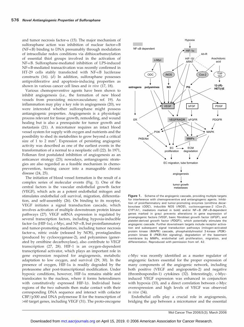
complex series of molecular events (Fig. 1). One of thecentral factors is the vascular endothelial growth factor(VEGF), which acts as a potent endothelial mitogen andstimulates endothelial cell survival, migration, differentia-tion, and self-assembly (26). On binding to its receptor,VEGF initiates a signal transduction cascade, whichinvolves activation of multiple downstream protein kinasepathways (27). VEGF mRNA expression is regulated byseveral transcription factors, including hypoxia-induciblefactor-1a (HIF-1a), c-Myc, and NF-nB, but proinflammatoryand tumor-promoting mediators, including tumor necrosisfactor-a, nitric oxide (released by NOS), prostaglandins(produced by cyclooxygenase-2), and polyamines (gener-ated by ornithine decarboxylase), also contribute to VEGFtranscription (27, 28). HIF-1 is an oxygen-dependenttranscriptional activator, which plays an important role ingene expression required for angiogenesis, metabolicadaptation to low oxygen, and survival (29, 30). In thepresence of oxygen, HIF-1a is readily degraded by theproteasome after post-transcriptional modification. Underhypoxic conditions, however, HIF-1a remains stable andtranslocates to the nucleus, where it forms heterodimerswith constitutively expressed HIF-1h. Individual basicregions of the two subunits then make contact with theircorresponding DNA sequence and interact with cofactorCBP/p300 and DNA polymerase II for the transcription of>60 target genes, including VEGF (31). The proto-oncogene
c-Myc was recently identified as a master regulator ofangiogenic factors essential for the proper expression ofmany components of the angiogenic network, includingboth positive (VEGF and angiopoietin-2) and negative(thrombospondin-1) cytokines (32). Interestingly, c-Myc-induced VEGF expression was enhanced in conjunctionwith hypoxia (33), and a direct correlation between c-Mycoverexpression and high levels of VEGF was observedin vivo (34).Endothelial cells play a crucial role in angiogenesis,
bridging the gap between a microtumor and the essential
Figure 1. Scheme of the angiogenic cascade, providing multiple targetsfor interference with chemopreventive and antiangiogenic agents. Inhibi-tion of proinflammatory and tumor-promoting enzymes [ornithine decar-boxylase (ODC ), inducible NOS (iNOS ), cyclooxygenase-2 (Cox-2 ),CYP1A1, mediators marked in bold] and/or NF-nB (NF-nB-dependentgenes marked in gray) prevents alterations in gene expression ofproangiogenic factors [VEGF, basic fibroblast growth factor (bFGF ), andplatelet-derived growth factor (PDGF)], which potentially stimulate theangiogenic cascade. Further downstream targets include receptor activa-tion and subsequent signal transduction pathways [mitogen-activatedprotein kinase (MAPK ) cascade, phosphatidylinositol 3-kinase (PI3K )-protein kinase B (PKB )/Akt signaling], degradation of the basementmembrane by MMPs, endothelial cell proliferation, migration, anddifferentiation. Reproduced with permission from ref. 42.
Novel Antiangiogenic Properties of Sulforaphane576
Mol Cancer Ther 2006;5(3). March 2006
on April 15, 2019. © 2006 American Association for Cancer Research. mct.aacrjournals.org Downloaded from
factors for tumor expansion. After mitogenic stimulationby a proangiogenic signal, they proliferate and migrateinto the perivascular stroma, initiate capillary sprouting byforming capillary-like tubes, and thus supply a microtumorwith essential nutrients and oxygen (35). Endothelial cellmigration is controlled by the surrounding extracellularmatrix. Therefore, endothelial cells produce type IV colla-genase as well as other members of the matrix metal-loproteinase (MMP) and serine protease family, which areessentially required for angiogenesis, tumor cell invasion,and metastasis (36, 37). Especially, MMP-2 (gelatinase A)is mostly expressed by microvascular endothelial cells ofblood vessels within and surrounding the tumor. Produc-tion of MMP precursor enzymes is regulated at thetranscriptional level, whereas activation of the proenzymesis tightly controlled by post-transcriptional mechanisms(38). An additional level of control is the interactionwith endogenous inhibitory proteins, the tissue inhibitorsof metalloproteinases (TIMP), which bind MMPs in a 1:1stoichiometric fashion and reversibly inhibit MMP enzy-matic activity (39).In this report, we describe novel antiangiogenic proper-
ties of sulforaphane. These effects were detected in ahuman in vitro antiangiogenic assay. In addition, weinvestigated the influence of sulforaphane on hypoxia-stimulated mRNA expression of VEGF and its receptorKDR/flk-1, HIF-1a, c-Myc, MMP-2, and TIMP-2 in culturedhuman microvascular endothelial cells (HMEC-1). Wefurther analyzed the effect of sulforaphane on essentialendothelial cell functions of HMEC-1 cells, includingmigration, differentiation, and proliferation. We concludethat these novel antiangiogenic activities of sulforaphaneare based on multiple interactions with the angiogeniccascade and might contribute to its chemopreventive andtherapeutic potential.
Materials andMethodsChemicalsAll cell culture materials were obtained from Invitrogen
(Eggenstein, Germany). Fetal bovine serum was providedby Pan (Aidenbach, Germany). q-Amino-caproic acid,aprotinin, sulforhodamine B, fibrinogen, thrombin, andbovine type B gelatin were purchased from Sigma(Taufkirchen, Germany). Matrigel was obtained from BDBiosciences (Heidelberg, Germany). RNeasy Mini kit,RNase-free DNase set, and all designed primers werefrom Qiagen (Hilden, Germany). Moloney murine leuke-mia virus reverse transcriptase and random primers forthe generation of cDNA were provided by Promega(Mannheim, Germany). Euro Taq polymerase was fromBioCat (Heidelberg, Germany). RNase inhibitor anddeoxynucleotide triphosphates were provided by Eppen-dorf (Hamburg, Germany). All materials and equipmentfor gel electrophoresis were purchased from Bio-Rad(Munich, Germany). All other chemicals were obtainedfrom Sigma. Sulforaphane was synthesized as describedearlier (15).
Cell CultureHuman microvascular endothelial cells (HMEC-1),
estrogen receptor–negative mammary tumor cells (SK-BR3), human colon adenocarcinoma cells (HCT-116), andmurine fibroblasts (NIH-3T3) were cultured as describedpreviously (40).
Human In vitro Antiangiogenesis AssayThe assay is based on the culture of human placental
blood vessels in fibrin gels described by Brown et al. (41). Inbrief, superficial vessels of human placentas were cut tofragments of 1 to 2 mm long and embedded in a fibrin gel(1 mL) containing 0.5 unit thrombin, 0.3% fibrinogen, and5 Ag/mL aprotinin in 24-well plates. The gel was overlaidwith 1 mL medium mix consisting of 1 part endothelialbasal medium MCDB 131 supplemented with 10 mmol/LL-glutamine and 1 part Medium 199 containing 100 units/mL penicillin G sodium, 100 units/mL streptomycinsulfate, and 250 ng/mL amphotericin B and supplementedwith 0.1% q-amino-caproic acid and 20% heat-inactivatedfetal bovine serum, which was changed twice weekly.The vessels were cultured at 37jC in a humidified 5%CO2 environment for 3 weeks. Resveratrol (1 Amol/L) wasused as a positive control. Sulforaphane was dissolved anddiluted in 100% DMSO to a final concentration of 0.01 to20 mmol/L and added to the medium (1 AL/mL, 0.1% finalDMSO concentration). Each experiment was repeated atleast thrice with placentas from different donors. Foranalysis of microvessel density (MVD), standardizeddigital images were acquired with a color digital micro-scopic camera system (Leitz Diavert microscope, Leica,Bensheim, Germany; AxioCam, Carl Zeiss, Gottingen,Germany) with a resolution of 1,300 � 1,030 pixel at �32magnification and processed with AxioVision Release3.1 software package (Carl Zeiss). The measurement ofMVD (mm2) was carried out using Adobe Photoshop 7.0with histogram function to obtain the pixel area of newlyformed capillaries in relation to the overall number ofpixels in the taken picture, which was then converted tomm2. Results are mean F SD of data originated from threeindependent experiments (42).
Inhibition of Cell ProliferationInhibition of cell proliferation of HMEC-1, SK-BR3, HCT-
116, and NIH-3T3 cells as well as flow cytometric analysesof sulforaphane-treated HMEC-1 cells were tested asdescribed previously (40). The influence of sulforaphaneon HMEC-1 cell proliferation in assays for endothelial cellfunctions was also assessed as described before (40).
Endothelial Cell Migration andTube FormationHMEC-1 cell migration as well as the formation of
capillary-like structures on a basement membrane prepa-ration were measured as described previously (40).
ReverseTranscription-PCRTotal RNA from 3 � 105 HMEC-1 cells (treated as
indicated in figure legends) was isolated using QiagenRNeasy Mini kits for total RNA extraction according to themanufacturer’s manual and treated with DNase I beforeuse. Experiments under hypoxic conditions were doneusing chambers for anaerobe bacterial culture (Merck,
Molecular Cancer Therapeutics 577
Mol Cancer Ther 2006;5(3). March 2006
on April 15, 2019. © 2006 American Association for Cancer Research. mct.aacrjournals.org Downloaded from
Darmstadt, Germany). Hypoxia was monitored by indica-tor sticks and determined as pO2 V 3 mm Hg. RNA (0.5 Ag)was transcribed into cDNA using Moloney murine leuke-mia virus reverse transcriptase and random hexamerprimers. Specific primers were designed using the Heidel-berg Unix Sequence Analysis Resources computer systemat the German Cancer Research Center (Heidelberg,Germany; Table 1). For amplification of cDNA fragments,PCR conditions were 94jC for 5 minutes followed by theindicated number of cycles. Cycling conditions were 94jCfor 1 minute followed by 1 minute at the indicatedannealing temperature and 72jC for 1 minute. The programwas terminated with a 7-minute extension interval at 72jC.Reaction conditions were optimized for each primer pair.PCR products were separated on 1.8% agarose gels andvisualized by ethidium bromide staining. For quantifica-tion of mRNA expression, densitometric scans of ethidiumbromide–stained gels were acquired using a Herolab EASYRH-3 densitometer (Herolab GmbH, Wiesloch, Germany)with EasyWin 32 software and semiquantitatively evaluat-ed using TINA software version 2.09a (Raytest Isotopen-messgerate GmbH, Staubenhardt, Germany). Stainingintensities were normalized to glyceraldehyde-3-phosphatedehydrogenase mRNA expression, background stainingwas subtracted, and values were expressed as percentageof induced expression in comparison with maximumcontrol values.
Gelatin ZymographyThe presence of secreted MMP-2 activity in conditioned
medium of HMEC-1 cells was analyzed by gelatinzymography (43). HMEC-1 cells were cultured in serum-free MCDB 131 endothelial basal medium supplementedas described above at 1.8 � 106 cells/mL/well in 24-wellplates. Aliquots from cell culture supernatants werecollected in a time- and concentration-dependent manneras indicated in the figure legends, centrifuged for10 minutes at 2,000 rpm, and sterile filtered. Proteinseparation was done by electrophoresis on a 7.5% SDS-polyacrylamide gel containing 0.1% gelatin under nonre-
ducing conditions. For molecular weight estimations,individual bands of a prestained protein standard mix(Bio-Rad) were detected and identified by their uniquecolor. After electrophoresis, gels were soaked in 2.5%Triton X-100 for 1 hour and then incubated in renaturationbuffer composed of 50 mmol/L Tris-HCl (pH 7.6), 15mmol/L CaCl2, 150 mmol/L NaCl, and 0.2% Brij 35 for48 hours. After staining with 0.1% Coomassie brilliantblue R250 in water, ethanol, acetic acid (55 + 45 + 10) for20 minutes, gels were destained in water, methanol, aceticacid (80 + 10 + 10) to reveal clear areas corresponding toprotein bands with gelatinolytic (i.e., metalloproteinases)activity. Densitometric scans were acquired as describedabove.
Statistical AnalysisResults are mean F SD of data originated from three
independent experiments unless stated otherwise. Forstatistical evaluation Student’s t test was applied. For theendothelial cell migration assay, paired Student’s t test wasdone comparing the migration area after 18 hours to time 0.P < 0.05 was considered as statistically significant and P <0.005 was considered as highly significant.
ResultsAngiogenesis is a multistep process that offers varioustargets for intervention: (a ) inhibition of release ofangiogenic factors or neutralization of released angiogenicmediators; (b) inhibition of synthesis and turnover of vesselbasement membrane; and (c) inhibition of vascular endo-thelial cell proliferation, migration, and differentiation (44).
Antiangiogenic Activity in the Human In vitro Anti-angiogenesis ModelBased on these strategies, we have established a human
in vitro antiangiogenesis assay, which covers multiple stepsof the angiogenic process and is sensitive to knownantiangiogenic compounds as well as selected chemo-preventive agents (42). Placental vessel fragments werecultured on fibrin gels in the presence or absence of various
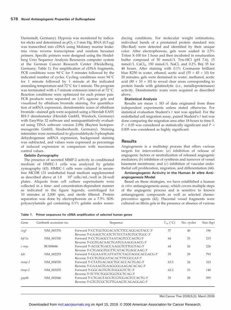
Table 1. Primer sequences for cDNA amplification of selected human genes
Gene Genbank accession no. Sequence Tm (jC) No. cycles Size (bp)
vegf NM_003376 Forward 5V-CCTGGTGGACATCTTCCAGGAGTACC-3V 57 40 196Reverse 5V-GAAGCTCATCTCTCCTATGTGCTGGC-3V
hif-1a NM_001530 Forward 5V-CCTGAGCCTAATAGTCCCAGTG-3V 64 33 215Reverse 5V-GGTGACAACTGATCGAAGGAACG-3V
c-myc BC008686 Forward 5V-ACGCTGACCAAGGTGTTGGTAG-3V 64 26 226Reverse 5V-CTGAGGTGGTTCATACTGAGCAAG-3V
kdr NM_002253 Forward 5V-GGAAATCATTATTCTAGTAGGCACGACG-3V 55 29 793Reverse 5V-CCTGTGGATACACTTTCGCGAT-3V
mmp-2 NM_004530 Forward 5V-CTATGACAGCTGCACCACTGAG-3V 63.5 24 163Reverse 5V-GAAAGTGAAGGGGAAGACACAG-3V
timp-2 NM_003255 Forward 5V-GGCAGTGTGTGGGGTCTC-3V 62.2 33 140Reverse 5V-TCTTCTGGGTGGTGCTCAG-3V
gapdh NM_002046 Forward 5V-CTGAGTACGTCGTGGAGTCCACTG-3V 55 28 595Reverse 5V-GTGTCGCTGTTGAAGTCAGAGGAG-3V
Novel Antiangiogenic Properties of Sulforaphane578
Mol Cancer Ther 2006;5(3). March 2006
on April 15, 2019. © 2006 American Association for Cancer Research. mct.aacrjournals.org Downloaded from
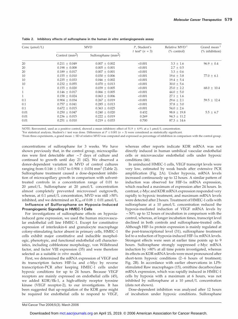
concentrations of sulforaphane for 3 weeks. We haveshown previously that, in the control group, microcapilla-ries were first detectable after f7 days of culture andcontinued to growth until day 21 (42). We observed adonor-dependent variation in MVD of control culturesranging from 0.146 F 0.017 to 0.904 F 0.034 mm2 (Table 2).Sulforaphane treatment caused a dose-dependent inhibi-tion of microcapillary growth in comparison with solvent-treated controls in a concentration range of 0.01 to20 Amol/L. Sulforaphane at 20 Amol/L concentrationalmost completely prevented microvessel outgrowth,whereas, at 0.1 Amol/L concentration, MVD was still >50%inhibited, and we determined an IC50 of 0.08F 0.01 Amol/L.
Influence of Sulforaphane on Hypoxia-InducedProangiogenic Signaling in HMEC-1CellsFor investigations of sulforaphane effects on hypoxia-
induced gene expression, we used the human microvascu-lar endothelial cell line HMEC-1. Except for constitutiveexpression of interleukin-6 and granulocyte macrophagecolony-stimulating factor absent in primary cells, HMEC-1cells exhibit major constitutive and inducible morphol-ogic, phenotypic, and functional endothelial cell character-istics, including cobblestone morphology, von Willebrandfactor, and factor VIII expression (35) and were thereforeselected as a suitable in vitro model.First, we determined the mRNA expression of VEGF and
its transcription factors HIF-1a and c-Myc by reversetranscription-PCR after keeping HMEC-1 cells underhypoxic conditions for up to 24 hours. Because VEGFreceptors are mainly expressed on endothelial cells (45),we added KDR/flk-1, a high-affinity receptor tyrosinekinase (VEGF receptor-2), to our investigations. It hasbeen suggested that up-regulation of the KDR gene mightbe required for endothelial cells to respond to VEGF,
whereas other reports indicate KDR mRNA was notdirectly induced in human umbilical vascular endothelialcells or microvascular endothelial cells under hypoxicconditions (46).In uninduced HMEC-1 cells, VEGF transcript levels were
very low, estimated by weak bands after extensive PCRamplification (Fig. 2A). Under hypoxia, mRNA levelsincreased continuously up to 12 hours. A similar pattern ofinduction was observed for HIF-1a mRNA expression,which reached a maximum of expression after 24 hours. Incontrast, c-Myc andKDRmRNAexpression responded veryrapidly to hypoxic treatment, and maximum mRNA levelswere detected after 2 hours. Treatment of HMEC-1 cells withsulforaphane at a 10 Amol/L concentration reduced thehypoxia-mediated induction of VEGF mRNA levels byf50% up to 12 hours of incubation in comparison with thecontrol, whereas, at longer incubation times, transcript leveldeclined in both controls and sulforaphane-treated cells.Although HIF-1a protein expression is mainly regulated atthe post-transcriptional level (31), sulforaphane treatmentled to a reduction of hypoxia-induced HIF-1amRNA levels.Strongest effects were seen at earlier time points up to 9hours. Sulforaphane strongly suppressed c-Myc mRNAinduction by >80% at all time points investigated, whereasits effects on KDRmRNA levels weremost pronounced aftershort-term hypoxic conditions (2–6 hours of treatment;Fig. 2B). In accordance with earlier observations in LPS-stimulated Raw macrophages (15), ornithine decarboxylasemRNA expression, which was rapidly induced in HMEC-1cells by hypoxia with a maximum at 6 hours, was notinhibited by sulforaphane at a 10 Amol/L concentration(data not shown).Dose-dependent inhibition was analyzed after 12 hours
of incubation under hypoxic conditions. Sulforaphane
Table 2. Inhibitory effects of sulforaphane in the human in vitro antiangiogenesis assay
Conc (Amol/L) MVD P , Student’st test* (n = 3)
Relative MVDc
(% control)Grand meanc
(% inhibition)
Control (mm2) Sulforaphane (mm2)
20 0.211 F 0.049 0.007 F 0.002 <0.001 3.3 F 1.6 96.9 F 0.420 0.198 F 0.008 0.005 F 0.001 <0.001 2.7 F 0.520 0.189 F 0.017 0.007 F 0.001 <0.001 3.3 F 0.610 0.155 F 0.010 0.030 F 0.006 <0.001 19.6 F 3.8 77.0 F 6.110 0.235 F 0.033 0.046 F 0.002 <0.001 19.4 F 5.410 0.232 F 0.055 0.070 F 0.013 <0.001 30.0 F 5.61 0.155 F 0.020 0.039 F 0.005 <0.001 25.0 F 2.2 68.0 F 10.41 0.146 F 0.017 0.066 F 0.005 <0.001 44.0 F 5.01 0.158 F 0.024 0.063 F 0.006 <0.001 27.1 F 1.60.1 0.904 F 0.034 0.267 F 0.019 <0.001 29.6 F 2.1 59.5 F 12.40.1 0.757 F 0.041 0.285 F 0.013 <0.001 37.8 F 3.00.1 0.672 F 0.015 0.363 F 0.025 <0.001 54.0 F 2.60.01 0.250 F 0.047 0.240 F 0.020 0.432 98.8 F 19.8 5.5 F 6.70.01 0.236 F 0.015 0.222 F 0.019 0.269 94.3 F 11.20.01 0.251 F 0.010 0.219 F 0.033 0.700 87.3 F 14.6
NOTE: Resveratrol, used as a positive control, showed a mean inhibitory effect of 51.9 F 4.9% at a 1 Amol/L concentration.*For statistical analysis, Student’s t test was done. Differences at P V 0.001 (n = 3) were considered as statistically significant.cFrom three experiments, a grand mean F SD of relative MVD was computed and expressed as percentage of inhibition in comparison with the control group.
Molecular Cancer Therapeutics 579
Mol Cancer Ther 2006;5(3). March 2006
on April 15, 2019. © 2006 American Association for Cancer Research. mct.aacrjournals.org Downloaded from
potently reduced hypoxia-induced levels of c-Myc, KDR,and VEGF mRNA by >50% at concentrations above 1.56Amol/L, whereas its effect on HIF-1amRNA induction wasslightly less pronounced, with f50% reduction at 6.25Amol/L concentration (Fig. 2C and D). Sulforaphane alsovery potently reduced the hypoxia-mediated induction ofinducible NOS mRNA levels by >50% at a concentration of0.4 Amol/L (data not shown).
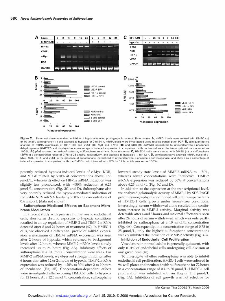
Sulforaphane-Mediated Effects on Basement Mem-braneModulatorsIn a recent study with primary human aortic endothelial
cells, short-term chronic exposure to hypoxic conditionsresulted in an up-regulation of MMP-2 and TIMP-2 mRNAdetected after 8 and 24 hours of treatment (47). In HMEC-1cells, we observed a differential profile of mRNA expres-sion: a maximum of TIMP-2 mRNA expression was seenafter 2 hours of hypoxia, which returned to backgroundlevels after 12 hours, whereas MMP-2 mRNA levels slowlyincreased up to 24 hours (Fig. 3A). Inhibitory effects ofsulforaphane at a 10 Amol/L concentration were weak. ForMMP-2 mRNA levels, we observed stronger inhibition after6 hours than after 12 or 24 hours of hypoxia. TIMP-2 mRNAexpression was reduced by f20% to 30% after 2 to 9 hoursof incubation (Fig. 3B). Concentration-dependent effectswere investigated after exposing HMEC-1 cells to hypoxiafor 12 hours. At a 12.5 Amol/L concentration, sulforaphane
lowered steady-state levels of MMP-2 mRNA to f50%,whereas lower concentrations were ineffective. TIMP-2mRNA expression was reduced by 50% at concentrationsabove 6.25 Amol/L (Fig. 3C and D).In addition to the expression at the transcriptional level,
we analyzed gelatinolytic activity of MMP-2 by SDS-PAGEgelatin zymography in conditioned cell culture supernatantsof HMEC-1 cells grown under serum-free conditions.Interestingly, serum withdrawal alone resulted in a contin-uous increase in MMP-2 activity. Marginal activity wasdetectable after 4 and 8 hours, andmaximal effectswere seenafter 24 hours of serum withdrawal, which was only partlyinhibited by sulforaphane at a 10 Amol/L concentration(Fig. 4A). Consequently, in a concentration range of 0.78 to25 Amol/L, only the highest sulforaphane concentrationsweakly inhibited the induction of MMP-2 activity (Fig. 4B).
Inhibition of Endothelial Cell ProliferationVasculature in normal adults is generally quiescent, with
only 0.01% of endothelial cells undergoing cell division atany given time (48).To investigate whether sulforaphane was able to inhibit
endothelial cell proliferation, HMEC-1 cells were cultured in96-well plates and incubated with sulforaphane for 72 hoursin a concentration range of 0.4 to 50 Amol/L. HMEC-1 cellproliferation was inhibited with an IC50 of 11.3 Amol/L(Fig. 5A). Inhibition of cell growth was not selective for
Figure 2. Time- and dose-dependent inhibition of hypoxia-induced proangiogenic factors. Time course: A, HMEC-1 cells were treated with DMSO (�)or 10 Amol/L sulforaphane (+) and exposed to hypoxia for 2 to 24 h. mRNA levels were investigated using reverse transcription-PCR. B, semiquantitativeanalysis of mRNA expression of HIF-1 ( ) and VEGF ( ; top ) and c-Myc ( ) and KDR ( ; bottom ) normalized to glyceraldehyde-3-phosphatedehydrogenase (GAPDH ) and displayed as a percentage of induced expression in comparison with control values at the transcriptional maximum set as100%. Stippled, crossed , or striped columns, sulforaphane treatment. Dose response: C, HMEC-1 cells were treated with DMSO (�) or sulforaphane(SFN ) in a concentration range of 0.78 to 25 Amol/L, respectively, and exposed to hypoxia (+) for 12 h. D, semiquantitative analysis mRNA levels of c-Myc, KDR, HIF-1, and VEGF in the presence of sulforaphane, normalized to glyceraldehyde-3-phosphate dehydrogenase, and shown as a percentage ofinduced expression in comparison with the DMSO control treated with LPS for 12 h, which was set as 100%.
Novel Antiangiogenic Properties of Sulforaphane580
Mol Cancer Ther 2006;5(3). March 2006
on April 15, 2019. © 2006 American Association for Cancer Research. mct.aacrjournals.org Downloaded from
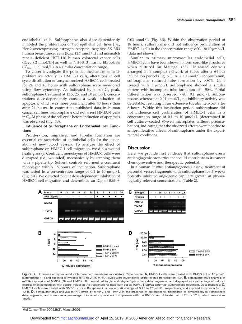
endothelial cells. Sulforaphane also dose-dependentlyinhibited the proliferation of two epithelial cell lines [i.e.,Her-2-overexpressing estrogen receptor–negative SK-BR3human breast cancer cells (IC50, 12.7 Amol/L) and mismatchrepair–deficient HCT-116 human colorectal cancer cells(IC50, 8.2 Amol/L)] as well as NIH-3T3 murine fibroblasts(IC50, 11.9 Amol/L) in a similar concentration range.To closer investigate the potential mechanism of anti-
proliferative activity in HMEC-1 cells, alterations in cellcycle distribution of unsynchronized HMEC-1 cells treatedfor 24 and 48 hours with sulforaphane were monitoredusing flow cytometry. As indicated by a sub-G1 peak,sulforaphane treatment at 12.5, 25, and 50 Amol/L concen-trations dose-dependently caused a weak induction ofapoptosis, which was more prominent after 48 hours thanafter 24 hours. In contrast to published data in humancancer cell lines, sulforaphane did not arrest HMEC-1 cellsin G2-M phase of the cell cycle before induction of apoptosiswas observed (Fig. 5B).
Influence of Sulforaphane on Endothelial Cell Func-tionsProliferation, migration, and tubular formation are
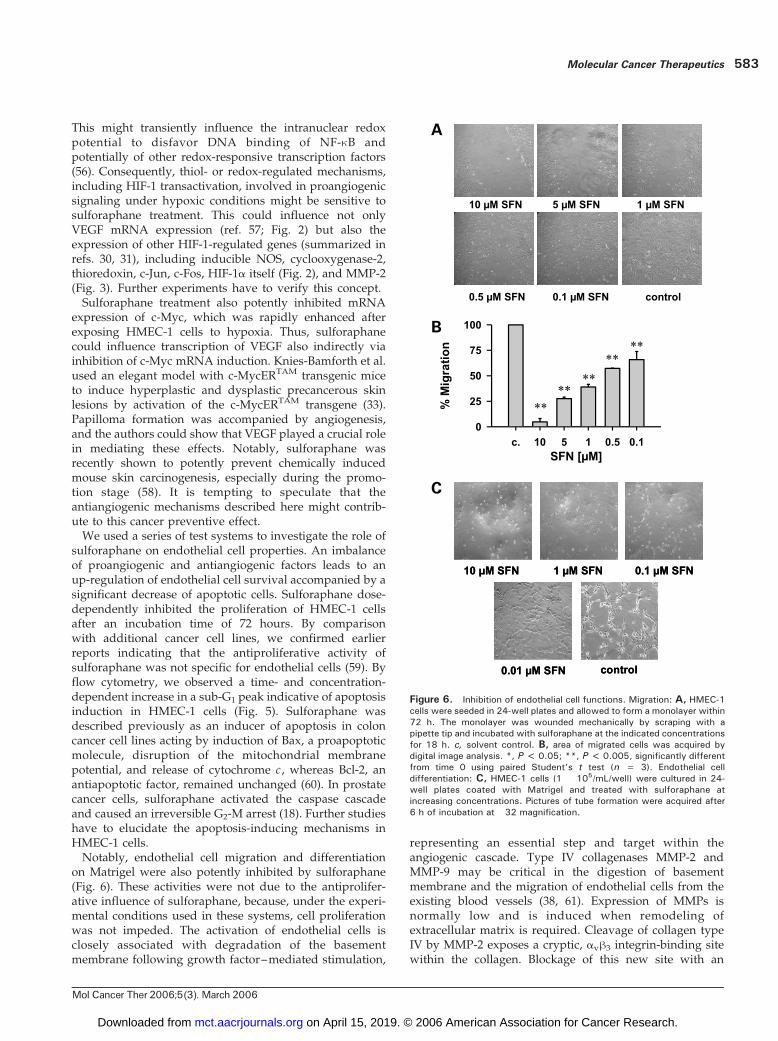
essential characteristics of endothelial cells for the gener-ation of new blood vessels. To analyze the effect ofsulforaphane on HMEC-1 cell migration, we did a woundhealing assay. Confluent monolayers of HMEC-1 cells weredisrupted (i.e., wounded) mechanically by scraping themwith a pipette tip. Solvent controls reformed a confluentmonolayer within 18 hours of incubation. Sulforaphanewas tested in a concentration range of 0.1 to 10 Amol/L(Fig. 6A). We detected potent dose-dependent inhibition ofHMEC-1 cell migration and determined an IC50 of 0.69 F
0.03 Amol/L (Fig. 6B). Within the observation period of18 hours, sulforaphane did not influence proliferation ofHMEC-1 cells in the concentration range of 0.1 to 10 Amol/L(data not shown).Similar to primary microvascular endothelial cells,
HMEC-1 cells have been shown to form cord-like structureswhen cultured on Matrigel (35). Untreated controlsarranged in a complex network of tubes after a 6-hourincubation period (Fig. 6C). At a 10 Amol/L concentration,sulforaphane reduced tube formation by >80%. Cellstreated with 1 Amol/L sulforaphane showed a similarpattern with incomplete tube formation of f50%. Partialdifferentiation was observed with 0.1 Amol/L sulfora-phane, whereas, at 0.01 Amol/L, no inhibitory activity wasdetectable, resulting in an extensive tubular network after6 hours. Within this incubation period, sulforaphane didnot influence cell proliferation of HMEC-1 cells in aconcentration range of 0.1 to 10 Amol/L (determined incell culture–coated 96-well microplates without preincu-bation), indicating that the observed effects were not due toantiproliferative effects of sulforaphane under the experi-mental conditions.
DiscussionHere, we provide first evidence that sulforaphane exertsantiangiogenic properties that could contribute to its cancerchemopreventive and therapeutic potential.In a human in vitro antiangiogenesis assay, treatment of
placental vessel fragments with sulforaphane for 3 weekspotently inhibited angiogenic capillary growth at physio-logically relevant concentrations (Table 2).
Figure 3. Influence on hypoxia-inducible basement membrane modulators. Time course: A, HMEC-1 cells were treated with DMSO (�) or 10 Amol/Lsulforaphane (+) and exposed to hypoxia for 2 to 24 h. mRNA levels were investigated using reverse transcription-PCR. B, semiquantitative analysis ofmRNA expression of MMP-2 ( ) and TIMP-2 ( ), normalized to glyceraldehyde-3-phosphate dehydrogenase, and displayed as a percentage of inducedexpression in comparison with control values at the transcriptional maximum set as 100%. Stippled columns, sulforaphane treatment. Dose response: C,HMEC-1 cells were treated with DMSO (�) or sulforaphane in a concentration range of 0.78 to 25 Amol/L, respectively, and exposed to hypoxia (+) for12 h. D, semiquantitative analysis mRNA levels of MMP-2 and TIMP-2 in the presence of sulforaphane, normalized to glyceraldehyde-3-phosphatedehydrogenase, and shown as a percentage of induced expression in comparison with the DMSO control treated with LPS for 12 h, which was set as100%.
Molecular Cancer Therapeutics 581
Mol Cancer Ther 2006;5(3). March 2006
on April 15, 2019. © 2006 American Association for Cancer Research. mct.aacrjournals.org Downloaded from
The model covers multiple steps relevant of angiogen-esis, including (a) production of growth factors; (b)activation of endothelial cells; (c) production of lyticenzymes to digest the basement membrane and extra-cellular matrix; and (d) endothelial cell migration, prolif-eration, and tube formation. Angiogenesis is induced bymechanical damage to the vessels. We have previouslyidentified sulforaphane as a very potent inhibitor of NF-nB-mediated expression of inducible NOS and cyclo-oxygenase-2 (15). Because nitric oxide and prostaglandinscontribute to the expression of VEGF (27), we assume thatpart of the sulforaphane-mediated inhibitory potential inthis model is based on the inhibition of the proangiogenicstimulus triggered by the mechanical stress caused by thepreparation of the vessel fragments. In addition, Xu et al.have recently reported that sulforaphane inhibited NF-nB-mediated VEGF expression in human prostate cancer PC-3C4 cells (49).Beside the expression of proinflammatory mediators (50),
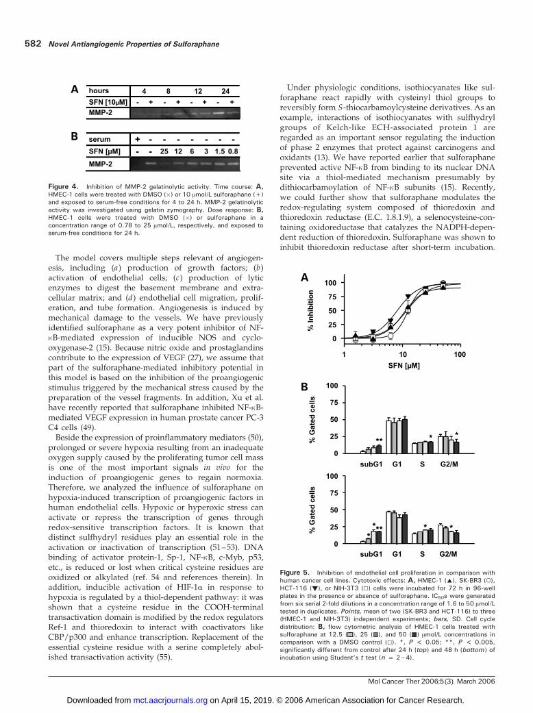
prolonged or severe hypoxia resulting from an inadequateoxygen supply caused by the proliferating tumor cell massis one of the most important signals in vivo for theinduction of proangiogenic genes to regain normoxia.Therefore, we analyzed the influence of sulforaphane onhypoxia-induced transcription of proangiogenic factors inhuman endothelial cells. Hypoxic or hyperoxic stress canactivate or repress the transcription of genes throughredox-sensitive transcription factors. It is known thatdistinct sulfhydryl residues play an essential role in theactivation or inactivation of transcription (51–53). DNAbinding of activator protein-1, Sp-1, NF-nB, c-Myb, p53,etc., is reduced or lost when critical cysteine residues areoxidized or alkylated (ref. 54 and references therein). Inaddition, inducible activation of HIF-1a in response tohypoxia is regulated by a thiol-dependent pathway: it wasshown that a cysteine residue in the COOH-terminaltransactivation domain is modified by the redox regulatorsRef-1 and thioredoxin to interact with coactivators likeCBP/p300 and enhance transcription. Replacement of theessential cysteine residue with a serine completely abol-ished transactivation activity (55).
Under physiologic conditions, isothiocyanates like sul-foraphane react rapidly with cysteinyl thiol groups toreversibly form S-thiocarbamoylcysteine derivatives. As anexample, interactions of isothiocyanates with sulfhydrylgroups of Kelch-like ECH-associated protein 1 areregarded as an important sensor regulating the inductionof phase 2 enzymes that protect against carcinogens andoxidants (13). We have reported earlier that sulforaphaneprevented active NF-nB from binding to its nuclear DNAsite via a thiol-mediated mechanism presumably bydithiocarbamoylation of NF-nB subunits (15). Recently,we could further show that sulforaphane modulates theredox-regulating system composed of thioredoxin andthioredoxin reductase (E.C. 1.8.1.9), a selenocysteine-con-taining oxidoreductase that catalyzes the NADPH-depen-dent reduction of thioredoxin. Sulforaphane was shown toinhibit thioredoxin reductase after short-term incubation.
Figure 4. Inhibition of MMP-2 gelatinolytic activity. Time course: A,HMEC-1 cells were treated with DMSO (�) or 10 Amol/L sulforaphane (+)and exposed to serum-free conditions for 4 to 24 h. MMP-2 gelatinolyticactivity was investigated using gelatin zymography. Dose response: B,HMEC-1 cells were treated with DMSO (�) or sulforaphane in aconcentration range of 0.78 to 25 Amol/L, respectively, and exposed toserum-free conditions for 24 h.
Figure 5. Inhibition of endothelial cell proliferation in comparison withhuman cancer cell lines. Cytotoxic effects: A, HMEC-1 (E), SK-BR3 (o),HCT-116 (!), or NIH-3T3 (5) cells were incubated for 72 h in 96-wellplates in the presence or absence of sulforaphane. IC50s were generatedfrom six serial 2-fold dilutions in a concentration range of 1.6 to 50 Amol/Ltested in duplicates. Points, mean of two (SK-BR3 and HCT-116) to three(HMEC-1 and NIH-3T3) independent experiments; bars, SD. Cell cycledistribution: B, flow cytometric analysis of HMEC-1 cells treated withsulforaphane at 12.5 ( ), 25 ( ), and 50 ( ) Amol/L concentrations incomparison with a DMSO control (5). *, P < 0.05; **, P < 0.005,significantly different from control after 24 h (top ) and 48 h (bottom ) ofincubation using Student’s t test (n = 2–4).
Novel Antiangiogenic Properties of Sulforaphane582
Mol Cancer Ther 2006;5(3). March 2006
on April 15, 2019. © 2006 American Association for Cancer Research. mct.aacrjournals.org Downloaded from
This might transiently influence the intranuclear redoxpotential to disfavor DNA binding of NF-nB andpotentially of other redox-responsive transcription factors(56). Consequently, thiol- or redox-regulated mechanisms,including HIF-1 transactivation, involved in proangiogenicsignaling under hypoxic conditions might be sensitive tosulforaphane treatment. This could influence not onlyVEGF mRNA expression (ref. 57; Fig. 2) but also theexpression of other HIF-1-regulated genes (summarized inrefs. 30, 31), including inducible NOS, cyclooxygenase-2,thioredoxin, c-Jun, c-Fos, HIF-1a itself (Fig. 2), and MMP-2(Fig. 3). Further experiments have to verify this concept.Sulforaphane treatment also potently inhibited mRNA
expression of c-Myc, which was rapidly enhanced afterexposing HMEC-1 cells to hypoxia. Thus, sulforaphanecould influence transcription of VEGF also indirectly viainhibition of c-Myc mRNA induction. Knies-Bamforth et al.used an elegant model with c-MycERTAM transgenic miceto induce hyperplastic and dysplastic precancerous skinlesions by activation of the c-MycERTAM transgene (33).Papilloma formation was accompanied by angiogenesis,and the authors could show that VEGF played a crucial rolein mediating these effects. Notably, sulforaphane wasrecently shown to potently prevent chemically inducedmouse skin carcinogenesis, especially during the promo-tion stage (58). It is tempting to speculate that theantiangiogenic mechanisms described here might contrib-ute to this cancer preventive effect.We used a series of test systems to investigate the role of
sulforaphane on endothelial cell properties. An imbalanceof proangiogenic and antiangiogenic factors leads to anup-regulation of endothelial cell survival accompanied by asignificant decrease of apoptotic cells. Sulforaphane dose-dependently inhibited the proliferation of HMEC-1 cellsafter an incubation time of 72 hours. By comparisonwith additional cancer cell lines, we confirmed earlierreports indicating that the antiproliferative activity ofsulforaphane was not specific for endothelial cells (59). Byflow cytometry, we observed a time- and concentration-dependent increase in a sub-G1 peak indicative of apoptosisinduction in HMEC-1 cells (Fig. 5). Sulforaphane wasdescribed previously as an inducer of apoptosis in coloncancer cell lines acting by induction of Bax, a proapoptoticmolecule, disruption of the mitochondrial membranepotential, and release of cytochrome c , whereas Bcl-2, anantiapoptotic factor, remained unchanged (60). In prostatecancer cells, sulforaphane activated the caspase cascadeand caused an irreversible G2-M arrest (18). Further studieshave to elucidate the apoptosis-inducing mechanisms inHMEC-1 cells.Notably, endothelial cell migration and differentiation
on Matrigel were also potently inhibited by sulforaphane(Fig. 6). These activities were not due to the antiprolifer-ative influence of sulforaphane, because, under the experi-mental conditions used in these systems, cell proliferationwas not impeded. The activation of endothelial cells isclosely associated with degradation of the basementmembrane following growth factor–mediated stimulation,
representing an essential step and target within theangiogenic cascade. Type IV collagenases MMP-2 andMMP-9 may be critical in the digestion of basementmembrane and the migration of endothelial cells from theexisting blood vessels (38, 61). Expression of MMPs isnormally low and is induced when remodeling ofextracellular matrix is required. Cleavage of collagen typeIV by MMP-2 exposes a cryptic, avh3 integrin-binding sitewithin the collagen. Blockage of this new site with an
Figure 6. Inhibition of endothelial cell functions. Migration: A, HMEC-1cells were seeded in 24-well plates and allowed to form a monolayer within72 h. The monolayer was wounded mechanically by scraping with apipette tip and incubated with sulforaphane at the indicated concentrationsfor 18 h. c, solvent control. B, area of migrated cells was acquired bydigital image analysis. *, P < 0.05; **, P < 0.005, significantly differentfrom time 0 using paired Student’s t test (n = 3). Endothelial celldifferentiation: C, HMEC-1 cells (1 � 105/mL/well) were cultured in 24-well plates coated with Matrigel and treated with sulforaphane atincreasing concentrations. Pictures of tube formation were acquired after6 h of incubation at �32 magnification.
Molecular Cancer Therapeutics 583
Mol Cancer Ther 2006;5(3). March 2006
on April 15, 2019. © 2006 American Association for Cancer Research. mct.aacrjournals.org Downloaded from
antibody decreased migration of endothelial cells andin vitro angiogenesis and reduced tumor growth in animalmodels (36). We could show a weak inhibition of MMP-2activity after serum withdrawal (Fig. 4). This inhibition ismost likely mediated at the transcriptional level (Fig. 3),but we cannot exclude a direct inhibition of zinc-dependentgelatinolytic activity. Inhibition or down-regulation ofMMP-2 activity might contribute to the observed inhibitoryeffects of sulforaphane on endothelial cell differentiation.In conclusion, our investigations have revealed novel
antiangiogenic properties of sulforaphane based onmultipleinteractions with critical steps in the angiogenic cascade.VEGF expression stimulated by HIF-1 and c-Myc, respec-tively, as well as endothelial cell migration and differen-tiation represent important targets of sulforaphane action.These antiangiogenic properties not only might be relevantfor the effects of sulforaphane in cancer prevention butalso might contribute to its cancer therapeutic efficacy thatis presently emerging (5, 62).
Acknowledgments
We thank G. Bastert, G. Fanderich, the nurses of the Department ofGynecology and Obstetrics, University of Heidelberg, and all donormothers for their assistance in supplying the starting material for theantiangiogenesis assay; C. Ittrich (German Cancer Research Center) forsupport with statistical evaluation of the result; our former colleague C.Herhaus for syntheses of sulforaphane; P. Huber (Radiologic UniversityHospital, Heidelberg, Germany) for providing the hypoxic chambers; F.J.Candal (Centers for Disease Control and Prevention, Atlanta, GA) for theHMEC-1 cell line; B. Vogelstein (Johns Hopkins University, Baltimore, MD)for providing the HCT-116 cell line; and N. Hay (University of Chicago,Chicago, IL) for the NIH-3T3 cell line.
References
1. Zhang Y, Talalay P, Cho CG, Posner GH. A major inducer ofanticarcinogenic protective enzymes from broccoli: isolation and elucida-tion of structure. Proc Natl Acad Sci U S A 1992;89:2399–403.
2. Gerhauser C, You M, Liu J, et al. Cancer chemopreventive potential ofsulforamate, a novel analogue of sulforaphane that induces phase 2 drug-metabolizing enzymes. Cancer Res 1997;57:272–8.
3. Zhang Y, Kensler TW, Cho CG, Posner GH, Talalay P. Anticarcinogenicactivities of sulforaphane and structurally related synthetic norbornylisothiocyanates. Proc Natl Acad Sci U S A 1994;91:3147–50.
4. Chung FL, Conaway CC, Rao CV, Reddy BS. Chemoprevention ofcolonic aberrant crypt foci in Fischer rats by sulforaphane and phenethylisothiocyanate. Carcinogenesis 2000;21:2287–91.
5. Singh AV, Xiao D, Lew KL, Dhir R, Singh SV. Sulforaphane inducescaspase-mediated apoptosis in cultured PC-3 human prostate cancer cellsand retards growth of PC-3 xenografts in vivo . Carcinogenesis 2004;25:83–90.
6. Fahey JW, Talalay P. Antioxidant functions of sulforaphane: a potentinducer of phase II detoxication enzymes. Food Chem Toxicol 1999;37:973–9.
7. Basten GP, Bao Y, Williamson G. Sulforaphane and its glutathioneconjugate but not sulforaphane nitrile induce UDP-glucuronosyl transfer-ase (UGT1A1) and glutathione transferase (GSTA1) in cultured cells.Carcinogenesis 2002;23:1399–404.
8. Hintze KJ, Keck AS, Finley JW, Jeffery EH. Induction of hepaticthioredoxin reductase activity by sulforaphane, both in Hepa1c1c7 cellsand in male Fisher 344 rats. J Nutr Biochem 2003;14:173–9.
9. Jowsey IR, Jiang Q, Itoh K, Yamamoto M, Hayes JD. Expressionof the aflatoxin B1-8,9-epoxide-metabolizing murine glutathione S -transferase A3 subunit is regulated by the Nrf2 transcription factorthrough an antioxidant response element. Mol Pharmacol 2003;64:1018–28.
10. Munday R, Munday CM. Induction of phase II detoxification enzymesin rats by plant-derived isothiocyanates: comparison of allyl isothiocyanate
with sulforaphane and related compounds. J Agric Food Chem 2004;52:1867–71.
11. Svehlikova V, Wang S, Jakubikova J, Williamson G, Mithen R,Bao Y. Interactions between sulforaphane and apigenin in the inductionof UGT1A1 and GSTA1 in CaCo-2 cells. Carcinogenesis 2004;25:1629–37.
12. Hu R, Hebbar V, Kim BR, et al. In vivo pharmacokinetics andregulation of gene expression profiles by isothiocyanate sulforaphane inthe rat. J Pharmacol Exp Ther 2004;310:263–71.
13. Lee JS, Surh YJ. Nrf2 as a novel molecular target for chemo-prevention. Cancer Lett 2005;224:171–84.
14. Eggler AL, Liu G, Pezzuto JM, van Breemen RB, Mesecar AD.Modifying specific cysteines of the electrophile-sensing human Keap1protein is insufficient to disrupt binding to the Nrf2 domain Neh2. Proc NatlAcad Sci U S A 2005;102:10070–5.
15. Heiss E, Herhaus C, Klimo K, Bartsch H, Gerhauser C. Nuclear factornB is a molecular target for sulforaphane-mediated anti-inflammatorymechanisms. J Biol Chem 2001;276:32008–15.
16. Jeong WS, Kim IW, Hu R, Kong AN. Modulatory properties of variousnatural chemopreventive agents on the activation of NF-nB signalingpathway. Pharm Res 2004;21:661–70.
17. Kim BR, Hu R, Keum YS, et al. Effects of glutathione on antioxidantresponse element-mediated gene expression and apoptosis elicited bysulforaphane. Cancer Res 2003;63:7520–5.
18. Singh SV, Herman-Antosiewicz A, Singh AV, et al. Sulforaphane-induced G2-M phase cell cycle arrest involves checkpoint kinase 2-mediated phosphorylation of cell division cycle 25C. J Biol Chem 2004;279:25813–22.
19. Losso JN. Targeting excessive angiogenesis with functional foodsand nutraceuticals. Trends Food Sci Technol 2003;14:455–68.
20. Szekanecz Z, Koch AE. Vascular endothelium and immune responses:implications for inflammation and angiogenesis. Rheum Dis Clin North Am2004;30:97–114.
21. Carmeliet P, Jain RK. Angiogenesis in cancer and other diseases.Nature 2000;407:249–57.
22. Ziche M, Gullino PM. Angiogenesis and neoplastic progressionin vitro . J Natl Cancer Inst 1982;69:483–7.
23. Folkman J. Tumor angiogenesis: therapeutic implications. N Engl JMed 1971;285:1182–6.
24. Sharma RA, Harris AL, Dalgleish AG, Steward WP, O’Byrne KJ.Angiogenesis as a biomarker and target in cancer chemoprevention.Lancet Oncol 2001;2:726–32.
25. Tosetti F, Ferrari N, De Flora S, Albini A. ‘‘Angioprevention’’:angiogenesis is a common and key target for cancer chemopreventiveagents. FASEB J 2002;16:2–14.
26. Affara NI, Robertson FM. Vascular endothelial growth factoras a survival factor in tumor-associated angiogenesis. In vivo 2004;18:525–42.
27. Josko J, Mazurek M. Transcription factors having impact on vascularendothelial growth factor (VEGF) gene expression in angiogenesis. MedSci Monit 2004;10:RA89–98.
28. Xie K, Wei D, Shi Q, Huang S. Constitutive and inducible expressionand regulation of vascular endothelial growth factor. Cytokine GrowthFactor Rev 2004;15:297–324.
29. Shi YH, Fang WG. Hypoxia-inducible factor-1 in tumor angiogenesis.World J Gastroenterol 2004;10:1082–7.
30. Harris AL. Hypoxia—a key regulator in angiogenesis. Nat Rev Cancer2002;2:38–47.
31. Semenza GL. Targeting HIF-1 for cancer therapy. Nat Rev Cancer2003;3:721–32.
32. Baudino TA, McKay C, Pendeville-Samain H, et al. c-Myc is essentialfor vasculogenesis and angiogenesis during development and tumorprogression. Genes Dev 2002;16:2530–43.
33. Knies-Bamforth UE, Fox SB, Poulsom R, Evan GI, Harris AL. c-Mycinteracts with hypoxia to induce angiogenesis in vivo by a vascularendothelial growth factor-dependent mechanism. Cancer Res 2004;64:6563–70.
34. Aref S, Mabed M, Zalata K, Sakrana M, El Askalany H. The interplaybetween c-Myc oncogene expression and circulating vascular endothelialgrowth factor (sVEGF), its antagonist receptor, soluble Flt-1 in diffuse
Novel Antiangiogenic Properties of Sulforaphane584
Mol Cancer Ther 2006;5(3). March 2006
on April 15, 2019. © 2006 American Association for Cancer Research. mct.aacrjournals.org Downloaded from
large B cell lymphoma (DLBCL): relationship to patient outcome. LeukLymphoma 2004;45:499–506.
35. Unger RE, Krump-Konvalinkova V, Peters K, Kirkpatrick CJ. In vitroexpression of the endothelial phenotype: comparative study of primaryisolated cells and cell lines, including the novel cell line HPMEC-ST1.6R.Microvasc Res 2002;64:384–97.
36. Egeblad M, Werb Z. New functions for the matrix metalloproteinasesin cancer progression. Nat Rev Cancer 2002;2:161–74.
37. Rundhaug JE. Matrix metalloproteinases, angiogenesis, and cancer.Clin Cancer Res 2003;9:551–4.
38. Nagase H, Woessner JF, Jr. Matrix metalloproteinases. J Biol Chem1999;274:21491–4.
39. Lambert E, Dasse E, Haye B, Petitfrere E. TIMPs as multifacialproteins. Crit Rev Oncol Hematol 2004;49:187–98.
40. Bertl E, Becker H, Eicher T, et al. Inhibition of endothelial cell functionsby novel potential cancer chemopreventive agents. Biochem Biophys ResCommun 2004;325:287–95.
41. Brown KJ, Maynes SF, Bezos A, Maguire DJ, Ford MD, Parish CR. Anovel in vitro assay for human angiogenesis. Lab Invest 1996;75:539–55.
42. Bertl E, Klimo K, Heiss E, et al. Identification of novel inhibitors ofangiogenesis using a human in vitro anti-angiogenic assay. Int J CancerPrev 2004;1:47–61.
43. Tremblay P, Houde M, Arbour N, et al. Differential effects of PKCinhibitors on gelatinase B and interleukin 6 production in the mousemacrophage. Cytokine 1995;7:130–6.
44. Gastl G, Hermann T, Steurer M, et al. Angiogenesis as a target fortumor treatment. Oncology 1997;54:177–84.
45. Terman BI, Carrion ME, Kovacs E, Rasmussen BA, Eddy RL, ShowsTB. Identification of a new endothelial cell growth factor receptor tyrosinekinase. Oncogene 1991;6:1677–83.
46. Gerber HP, Condorelli F, Park J, Ferrara N. Differential transcriptionalregulation of the two vascular endothelial growth factor receptor genes.Flt-1, but not Flk-1/KDR, is up-regulated by hypoxia. J Biol Chem 1997;272:23659–67.
47. Ning W, Chu TJ, Li CJ, Choi AM, Peters DG. Genome-wide analysis ofthe endothelial transcriptome under short-term chronic hypoxia. PhysiolGenomics 2004;18:70–8.
48. Keshet E, Ben-Sasson SA. Anticancer drug targets: approachingangiogenesis. J Clin Invest 1999;104:1497–501.
49. Xu C, Shen G, Chen C, Gelinas C, Kong AN. Suppression of NF-nBand NF-nB-regulated gene expression by sulforaphane and PEITC throughInBa, IKK pathway in human prostate cancer PC-3 cells. Oncogene 2005;24:4486–95.
50. Naldini A, Carraro F. Role of inflammatory mediators in angiogenesis.Curr Drug Targets Inflamm Allergy 2005;4:3–8.
51. Rupec RA, Baeuerle PA. The genomic response of tumor cells tohypoxia and reoxygenation. Differential activation of transcription factorsAP-1 and NF-nB. Eur J Biochem 1995;234:632–40.
52. Michiels C, Minet E, Mottet D, Raes M. Regulation of gene expressionby oxygen: NF-nB and HIF-1, two extremes. Free Radic Biol Med 2002;33:1231–42.
53. Maulik N. Redox signaling of angiogenesis. Antioxid Redox Signal2002;4:805–15.
54. Wu X, Bishopric NH, Discher DJ, Murphy BJ, Webster KA. Physicaland functional sensitivity of zinc finger transcription factors to redoxchange. Mol Cell Biol 1996;16:1035–46.
55. Ema M, Hirota K, Mimura J, et al. Molecular mechanisms oftranscription activation by HLF and HIF1a in response to hypoxia: theirstabilization and redox signal-induced interaction with CBP/p300. EMBO J1999;18:1905–14.
56. Heiss E, Gerhauser C. Time-dependent modulation of thioredoxinreductase activity might contribute to sulforaphane-mediated inhibition ofNF-nB binding to DNA. Antioxid Redox Signal. Antioxid Redox Signal2005;7:1601–11.
57. Forsythe JA, Jiang BH, Iyer NV, et al. Activation of vascularendothelial growth factor gene transcription by hypoxia-inducible factor1. Mol Cell Biol 1996;16:4604–13.
58. Gills JJ, Jeffery EH, Matusheski NV, Moon RC, Lantvit DD,Pezzuto JM. Sulforaphane prevents mouse skin tumorigenesis duringthe stage of promotion. Cancer Lett 2005 (doi:10.1016/j.canlet.2005.05.007).
59. Wu X, Kassie F, Mersch-Sundermann V. Induction of apoptosis intumor cells by naturally occurring sulfur-containing compounds. Mutat Res2005;589:81–102.
60. Gamet-Payrastre L, Li P, Lumeau S, et al. Sulforaphane, anaturally occurring isothiocyanate, induces cell cycle arrest andapoptosis in HT29 human colon cancer cells. Cancer Res 2000;60:1426–33.
61. Schnaper HW, Grant DS, Stetler-Stevenson WG, et al. Type IVcollagenase(s) and TIMPs modulate endothelial cell morphogenesisin vitro . J Cell Physiol 1993;156:235–46.
62. Pham NA, Jacobberger JW, Schimmer AD, Cao P, Gronda M, HedleyDW. The dietary isothiocyanate sulforaphane targets pathways ofapoptosis, cell cycle arrest, and oxidative stress in human pancreaticcancer cells and inhibits tumor growth in severe combined immunodefi-cient mice. Mol Cancer Ther 2004;3:1239–48.
Molecular Cancer Therapeutics 585
Mol Cancer Ther 2006;5(3). March 2006
on April 15, 2019. © 2006 American Association for Cancer Research. mct.aacrjournals.org Downloaded from
2006;5:575-585. Mol Cancer Ther Elisabeth Bertl, Helmut Bartsch and Clarissa Gerhäuser chemopreventionnovel sulforaphane-mediated mechanisms in Inhibition of angiogenesis and endothelial cell functions are
Updated version
http://mct.aacrjournals.org/content/5/3/575
Access the most recent version of this article at:
Cited articles
http://mct.aacrjournals.org/content/5/3/575.full#ref-list-1
This article cites 61 articles, 20 of which you can access for free at:
Citing articles
http://mct.aacrjournals.org/content/5/3/575.full#related-urls
This article has been cited by 10 HighWire-hosted articles. Access the articles at:
E-mail alerts related to this article or journal.Sign up to receive free email-alerts
Subscriptions
Reprints and
To order reprints of this article or to subscribe to the journal, contact the AACR Publications
Permissions
Rightslink site. (CCC)Click on "Request Permissions" which will take you to the Copyright Clearance Center's
.http://mct.aacrjournals.org/content/5/3/575To request permission to re-use all or part of this article, use this link
on April 15, 2019. © 2006 American Association for Cancer Research. mct.aacrjournals.org Downloaded from