inhaled corticosteroid treatment for 6 months was not sufficient to normalize phagocytosis in
TRANSCRIPT

da Silva-Martins et al. Clinical and Translational Allergy 2013, 3:28http://www.ctajournal.com/content/3/1/28
RESEARCH Open Access
Inhaled corticosteroid treatment for 6 monthswas not sufficient to normalize phagocytosis inasthmatic childrenCarmen Lívia Faria da Silva-Martins1,2,3, Shirley Claudino Couto1 and Maria Imaculada Muniz-Junqueira1*
Abstract
Background: Corticosteroids are the first-line therapy for asthma; however, the effect of corticosteroids on theinnate immune system remains unclear. This study’s objective was to evaluate the effect of inhaled corticosteroidtherapy (ICT) on phagocytic functions.
Methods: To evaluate the impact of ICT, the phagocytosis of Saccharomyces cerevisiae by blood monocytes andneutrophils and the production of superoxide anions were assessed before and after three and six months of ICTtreatment in 58 children with persistent asthma and 21 healthy controls.
Results: We showed that the phagocytic capacity of monocytes and neutrophils that occurred via patternrecognition receptors or was mediated by complement and immunoglobulin receptors in asthmatic children beforetreatment was significantly lower than in healthy controls (p<0.05, Mann–Whitney test) and was not influenced bythe severity of the clinical form of the disease. Although there was clinical improvement with treatment, ICT for6 months was not sufficient to normalize phagocytosis by the phagocytes. Superoxide anion production was alsodecreased in the asthmatic children before treatment, and ICT normalized the O- production only for children withmild persistent asthma when assessed at baseline but caused this function to decrease after stimulation (p<0.05,Kruskal-Wallis test).
Conclusions: Our data suggest that an immunodeficiency in phagocytes remained even after treatment. However,this immunodeficiency does not appear to correspond with the clinical evolution of asthma because animprovement in clinical parameters occurred.
Keywords: Asthma, Immunodeficiency, Neutrophils, Monocytes, Oxygen radical production, Phagocytosis
IntroductionAsthma is a serious global health problem throughout theworld. An estimated 300 million individuals are affectedby this disease [1], which is the most common chronicdisease of childhood [2].Corticosteroids are the first-line drugs for asthma ther-
apy and are by far the most effective anti-inflammatorytreatment [3], resulting in marked reductions in asthmamorbidity and mortality [4,5].The goals of asthma treatment are to control the clin-
ical symptoms for extended periods, prevent asthma
* Correspondence: [email protected] of Cellular Immunology, Pathology, Faculty of Medicine, CampusDarcy Ribeiro, Asa Norte, University of Brasilia, Brasilia, DF 70.910-900, BrazilFull list of author information is available at the end of the article
© 2013 da Silva-Martins et al.; licensee BioMedCreative Commons Attribution License (http:/distribution, and reproduction in any medium
exacerbations, maintain pulmonary function, and controlairway inflammation [6]. Inhaled corticosteroids (ICS) arevery effective in controlling asthma symptoms in patientsof all ages and severities and may prevent irreversible air-way changes [4,5,7]. However, corticosteroid treatmentalso has several side effects [8] and may influence thefunctions of immune system cells [9,10].Children with asthma have an increased frequency of pul-
monary infections, and it is possible that alterations in thefunction of phagocytes, which are engaged in the first lineof defense against pathogens, may play a role in these infec-tions [11]. Monocyte chemotaxis in vitro is inhibited byhigh concentrations of steroids [12]. In addition, corticoste-roids affect nitric oxide production, total free radical pro-duction, and nitric oxide synthase activity in the monocytes
Central Ltd. This is an Open Access article distributed under the terms of the/creativecommons.org/licenses/by/2.0), which permits unrestricted use,, provided the original work is properly cited.

da Silva-Martins et al. Clinical and Translational Allergy 2013, 3:28 Page 2 of 12http://www.ctajournal.com/content/3/1/28
of asthmatic patients [10]. Furthermore, there are reportsof defective phagocytosis of pathogens in asthma patients[13]. Phagocytosis by alveolar macrophages is impaired inchildren with poorly controlled asthma [11], and oxidativestress is emerging as a common mechanism that may alterboth macrophage and neutrophil functions [13].The influence of ICS treatment on phagocyte functions
in asthmatic children remains unclear. Either the treatmentor the disease may influence phagocyte functions directlyor through several cytokines and substances produced aspart of the altered immune response in affected individuals.Therefore, the objective of this study was to evaluate theundetermined influence of ICS treatment on the phago-cytic function of monocytes and neutrophils from asth-matic children after 3 and 6 months of therapy and tocompare phagocytosis and the production of toxic oxygenradicals in asthmatic children and healthy control subjects.This study may shed light on the effects of corticosteroidson cells of the innate immune system in asthmatic childrenand may improve the understanding of the mechanism ofaction of this drug. It was showed that the phagocytic cap-acity of monocytes and neutrophils that occurred via pat-tern recognition receptors or was mediated by complementand immunoglobulin receptors in asthmatic children beforetreatment was significantly lower than in healthy controlsand inhaled corticosteroid treatment for 6 months was notsufficient to normalize phagocytosis in asthmatic children.
MethodsStudy groupsOne author (CLFSM), a pediatric pneumologist, conductedthe clinical evaluation of all control and asthmatic childrenand sequentially selected and screened 58 children whowere seen in the pediatric asthma outpatient clinic at BrasiliaUniversity Hospital for enrolment in this study. The childrenwere placed in the following groups: a group of 58 out-patient asthmatic children with variable disease severity (27girls and 31 boys; 7.6 ± 3.4 years) from whom blood wascollected before, at 3 months, and at 6 months of inhaledcorticosteroid therapy. Asthma was classified as mild, mod-erate, or severe persistent disease by the frequency of thesymptoms, presence of nocturnal asthma, frequency of acuteexacerbations, medications required for control, physical ac-tivity limitations, or altered pulmonary functioning [14].Twenty-four (41.3%) asthmatic children had mild asthma,20 (34.5%) children had moderate asthma and 14 (24.1%)had severe persistent asthma. A group of 21 healthy children(12 girls and 9 boys; 10.7 ± 2.10 years) without the diseaseand without a personal or familial history of allergy com-prised the control group. At the time of clinical examination,peripheral blood was collected to assess phagocytosis andsuperoxide anion production.The exclusion criteria were as follows: children < 2 years
or > 18 years, any other clinically significant pulmonary
disease or other disease, those who presented with any con-dition that might alter the function of the immune system,any children taking medication other than asthma treat-ment, and any children that had previously used inhaledcorticosteroids, or that used oral corticosteroids in the last6 months.During the first evaluation, the group of asthmatic
children was assessed for the severity of asthma, asthmacontrol test (ACT), peak expiratory flow (PEF), andbody mass index (BMI). The ACT was administered tochildren aged 4–11 years (7 answers) and >12 years (5answers). The test was adapted for children < 4 years,with the mother responding to the questions. The chil-dren were considered clinically controlled (ACT ≥ 25),partially controlled (ACT = 20–24) or uncontrolled(ACT < 20) [15].The PEF value was recorded as the best of three forced
expirations by means of a peak flow meter and expressed asthe percentage of predicted of normal values for height andsex values for children ≥ 100 cm. In seven children withmild persistent asthma (mPA), one child with moderatepersistent asthma (MPA) and three with severe persistentasthma (SPA) the PEF were not assessed. All these childrenyounger than 5 years had familial history of asthma. Thethresholds of PEF used to severity evaluation were: mPA≥80% predicted value, MPA= 60-80% predicted value, andSPA ≤ 60% predicted value.BMI was assessed to exclude any possible influence of
child nutritional status on phagocyte functions.During the second (3 months) and third (6 months)
clinical follow-up visits, the children were assessed withthe ACT, PFE and BMI.The children were treated with ICS. A long-acting
β2-agonist (LABA) was added when necessary to con-trol symptoms. All 24 children with mild persistentasthma (mPA), 10 children <4 years with moderate per-sistent asthma (MPA) and 5 children < 4 years with se-vere persistent asthma (SPA) received monotherapywith 250 μg beclomethasone dipropionate twice day.Eight children with MPA and three with SPA receivedbudesonide + formoterol 6/200 μg twice day and 1children with SPA received 12/400 μg twice day. Twochildren with MPA and five children with SPA receivedfluticasone propionate + salmeterol 25/125 μg twiceday (Table 1).Peripheral blood was collected from control and asth-
matic children who had fasted for > 12 hours prior to theirblood being drawn. Hemogram (Cell-dyn 3.700) was auto-matically assessed.The Human Research Ethical Committee of the School
of Medicine of the University of Brasilia approved the ex-perimental protocol (process no. 02/2007).All parents provided formal informed consent for their
child’s participation in this study.

Table 1 Clinical characteristics of healthy controls and asthmatic children before inhaled corticosteroid therapyshowing mild, moderate and severe persistent asthma
Parameters Normalcontrol
Mild persistentasthma
Moderate persistentasthma
Severe persistentasthma
Test p
Number 21/78 24/58 (41.3%) 20/58 (34.5%) 14/58 (24.1%)
Gender (boys/girls) 9/12 13/11 12/8 6/8 Chi-square p=0.64
Age (years) (mean±SD) 10.7±2.1 6.32±2.99 9.22±3.33 7.24±3.53 ANOVA* p<0.05
Total leukocytes 7390 9720 8120 8305 Kruskal-Wallisp=0.05
Neutrophils 3440 4150 3640 4260 Kruskal-Wallisp=0.66
Monocytes 568 568 536 643 Kruskal-Wallisp=0.57
Eosinophils 245 460 635 487 ANOVA p=0.003
BMI (Kg/m2) (mean±SD) 43.31±32.1 51.9±34.2 47.4±31.2 63.7±32.5 Kruskal-Wallisp=0.29
PEF (%) (mean±SD) 86.9±18.6 81.2±17.3 75.6±15.3 65.6±12.0 ANOVA** p<0.05
Treatment***
ICS (%) 23/23 (100%) 10/20 (50%) 05/14 (35.7%)
ICS + LABA (%)
Budesonide + formoterol 0/23 (0%) 08/20 (40%) 04/14 (28.6%)
Fluticasone propionate +salmeterol
0/23 (0%) 02/20 (10%) 05/14 (35.7%)
BMI Body Mass Index (percentile), PEF Peak Expiratory Flow, mPA mild persistent asthma, MPA moderate persistent asthma, SPA severe persistent asthma,mPA, MPA, SPA data before treatment. Beclomethasone dipropionate = 250 μg to 500 μg/day; Budesonide + formoterol 6/200 μg or 12/400 μg twice day;Fluticasone propionate + salmeterol = 25/125 μg to 50/250 μg twice day K-W= Kruskal-Wallis + Dunn’s method; * mPA and SPA < C; ** SPA < C;*** Treatment followed GINA.
da Silva-Martins et al. Clinical and Translational Allergy 2013, 3:28 Page 3 of 12http://www.ctajournal.com/content/3/1/28
Phagocytosis testPhagocytosis of Saccharomyces cerevisiae was adaptedfrom a previously described technique [16]. Briefly, 40 μLsamples per marked area of heparinized whole peripheralblood obtained from each subject were placed on dupli-cate slides containing 8 areas that were each 7-mm indiameter and incubated in a humidified chamber for45 min at 37°C. The slides were then rinsed with 0.15 Mphosphate-buffered saline (PBS) (pH 7.2) at 37°C to re-move non-adherent cells. Adherent cells (12,534±5,050cells/marked area; 5.63±0.85% monocytes and 93.5±1.08%neutrophils) (viability > 98%) were incubated with a sus-pension of 2.5×10⁵ S. cerevisiae in 20 μL Hanks-Tris solu-tion (Sigma Co., St Louis, MO, USA) (pH 7.2) containing10% heat-inactivated fetal calf serum (FCS) (Gibco/Invitrogen, Grand Island, NY, USA) for 30 min in a hu-midified chamber at 37°C. The slides were then rinsedwith 0.15 M PBS at 37°C to eliminate non-phagocytosedS. cerevisiae, and the final wash step was performed with30% FCS in Hanks-Tris solution. The slides were fixedwith methanol and stained with 10% Giemsa stain. Thenumber of S. cerevisiae that were phagocytosed by either200 monocytes or 200 neutrophils in individual prepara-tions was assessed by light microscopy. The phagocyticindex was calculated as the mean number of phagocytosedS. cerevisiae per phagocytosing monocyte or neutrophil
multiplied by the percentage of these cells engaged inphagocytosis.The internalization of particles by phagocytes occurs
via receptors. When phagocytosis occurs via pattern-recognition receptors, the phagocyte recognizes directlyconserved pattern molecular in the surface of the par-ticle to be phagocytosed. When phagocytosis is facili-tated by opsonins, the ingestion occurs via receptors tocomponents of complement or via receptors to FcIgG.Saccharomices cerevisiae (Baker’s yeasts) suspensionswere prepared according to a previously described tech-nique [16] to assess phagocytosis via pattern-recognitionreceptors and facilitated by opsonins. Yeasts were usedwith or without previous incubation with fresh serumfrom the donor. In the former case, yeast cells were con-sidered sensitized, because they were opsonized by com-plement molecules and antibodies in serum. Thesemolecules adhere on yeast surface and will be recog-nized by their neutrophil and monocyte receptors (CR1,CR3 and FcR) during the process respective of phagocyt-osis [16]. Yeast cells that were not pre-incubated withfresh serum from the donor, but were incubated withfetal calf serum, were considered as non-sensitized, be-cause they were non-opsonized and their phagocytosisoccurs via the pattern-recognition receptors (PRRs) ofneutrophils and monocyte [17]. For opsonization, the

da Silva-Martins et al. Clinical and Translational Allergy 2013, 3:28 Page 4 of 12http://www.ctajournal.com/content/3/1/28
S. cerevisiae were sensitized by incubation at 37°C for30 min with 10% fresh serum from the donor in Hanks-Trissolution. The yeast cells that were pre-incubated withinactivated fetal calf serum were considered non-sensitized,and their phagocytosis occurred via the pattern-recognitionreceptors of phagocytes [17].
Nitro blue tetrazolium slide testThe nitro blue tetrazolium (NBT) test evaluated theability to generate toxic oxygen radicals (superoxideanion/O2
−) that are capable of reducing the compoundNBT to an insoluble form, formazan, which is identi-fied via optical microscopy as a blue color in the cy-toplasm of the cell [18]. The amount of reduced NBTis directly proportional to the amount of oxygen ra-dicals (O2
−) produced by the phagocytes. After thephagocytes were adhered, cells from the control andasthmatic children were incubated with 0.05% NBTsolution in Hanks-Tris solution (Sigma, St Louis, MO,USA) for 20 min at 37°C in a humidified chamber.The slides were then washed, fixed with methanoland stained with a 1.4% safranin and 28.6% glycerol.The percentage of phagocytes that contained reducedcytoplasmic NBT was assessed by optical microscopy,
Table 2 Clinical characteristics of asthmatic children before, 3
Parameters Before 3 mo
PEF (%) (mean±SD)
mPA 81.2±17.3 90.6±
MPA 75.6±15.3 79.3±
SPA 65.6±12.0 72.8±
BMI (Kg/m2) (mean±SD)
mPA 51.9±34.3 45.2±
MPA 47.4±31.2 50.5±
SPA 63.7±32.5 61.8±
ACT n (%)
mPA
≥25 0/23 (0%) 1/23 (
20-24** 11/23 (47.8%) 19/23 (
<20*** 12/23 (52.2%) 3/23 (1
MPA
≥25 0/20 0%) 1/20
20-24** 2/20 (10%) 7/20
<20*** 18/20 (90%) 12/20
SPA
≥25 0/14 (0%) 0/14
20-24 2/14 (14.3%) 3/14 (2
<20 12/14 (85.7%) 11/14 (7
PEF Peak Expiratory Flow, BMI Body Mass Index (percentile), ACT Asthma Control TeSPA severe persistent asthma, * 6 m > before ** 3 m and 6 m > before *** 3 m and
and the source of the individual preparations was re-vealed only at the end of the evaluation [18].
Statistical analysisThe data were previously tested with Bartlett’s test for equalvariances and the Kolmogorov–Smirnov test for normalityof distribution. The Mann–Whitney test was used to com-pare two non-normal samples. The Kruskal-Wallis test,followed by Dunn’s method, was used to compare multiplenon-normal samples. Spearman’s test was used to evaluatethe correlation between samples. The chi-square test wasused to compare proportions. For homogeneity of datapresentation, all values were expressed as the median, quar-tiles and extremes, and outlier values were indicated. Differ-ences and correlations with a two-tailed value of p<0.05were considered statistically significant. The Prism 5 soft-ware package (GraphPad, San Diego, CA, USA) was usedfor statistical tests and graphical presentation of the data.
ResultsClinical and demographic characteristicsThe characteristics of the children enrolled in the studyand the clinical outcomes during 3 and 6 months of cor-ticosteroid treatment are summarized in Tables 1 and 2.
and 6 months after inhaled corticosteroid therapy
nths 6 months Test p
ANOVA
23.6 99.2±18.4 p < 0.05*
16.8 86.3±18.3 p < 0.05*
17.0 77.9±19.8 p > 0.05
32.2 43.4±37.5 Kruskal-Wallis p>0.05
31.5 50.0±30.5
29.6 63.2±29.5
4.3%) 4/22 (18.2%) Chi-square
82.6%) 16/22 (72.7%) p<0.05
3.0%) 2/22 (9.1%)
(5%) 3/20 (15%) Chi-square
(35%) 10/20 (50.0%) p<0.05
(60%) 7/20 (35.0%)
(0%) 0/14 (0%) Chi-square
1.4%) 4/14 (28.6%) p>0.05
8.57%) 10/14 (71.4%)
st, mPA mild persistent asthma, MPA moderate persistent asthma,6 m < before.

da Silva-Martins et al. Clinical and Translational Allergy 2013, 3:28 Page 5 of 12http://www.ctajournal.com/content/3/1/28
Children from the control group had the mean age 3 yearsolder than the asthmatic children (10.7 ± 2.10 years versus7.6 ± 3.4 years; p=0.001, t test), but no difference was ob-served based on gender between the children with or with-out asthma (p=0.6, Chi-square test). To exclude a possibleinfluence of age as an independent confounding variable onthe results of phagocyte functions, the univariate correlationbetween age and monocyte and neutrophil phagocytic indi-ces and % NBT reduction for healthy control and asthmaticchildren was tested. No correlation was observed betweenage and these phagocyte functions (Spearman’s test, p>0.05).Therefore, no adjustment of results of phagocyte functionsfor age was done. Furthermore, previous observations hadshown that there are not differences among preschool chil-dren, schoolchildren and adolescent phagocyte functions.No statistically significant difference was observed be-
tween groups for leukocytes, except for eosinophils. Themedian of the number of eosinophils in peripheral bloodwas significantly higher in asthmatic children than healthycontrol (245 for the control group and 460, 635, and 487for mPA, MPA and SPA groups, respectively, before ICT;p=0.003, ANOVA) (Table 1).ACT and PEF showed the clinical improve after 3 and
6 months follow up (Tables 1 and 2).In the 3 months evaluation, the phagocyte tests was done
in 50 children (n = 50), and in the 6 months evaluation,the phagocyte tests of was done in 47 children (n = 47).
Figure 1 In vitro evaluation of the phagocytic indices of monocytes (lasthma (mPA), moderate persistent asthma (MPA) and severe persisteusing 2.5×10⁵ yeast cells per well. For the top panels, sensitized yeasts weranalyses showed that the median value of all groups of asthmatic children was loobserved between the groups with mild, moderate and severe persistent asthmaas the median, quartile and extreme, and outlier values are marked.
No difference was observed between the phagocyticindices in children with different clinical severities ofpersistent asthmaTo determine whether the severity of persistent asthmainfluenced the phagocytic capacity of monocytes and neu-trophils, the phagocytic indices of monocytes and neutro-phils from children with different severities of asthma werecompared. All groups of asthmatic children showed alower phagocytic index than the healthy control children;however, no difference was observed between the childrenwith mild, moderate or severe persistent asthma (Kruskal-Wallis test followed by Dunn’s method, p<0.01) (Figure 1).
Monocytes and neutrophils from children with asthmashowed less phagocytosis through pattern molecularreceptors, and treatment with ICS for 6 months was notsufficient to normalize phagocytic functionTo assess the influence of inhaled corticosteroids on phago-cytosis, the phagocytic capacity of monocytes and neutro-phils from children with different severities of asthma wascompared before and after corticosteroid treatment.Using non-sensitized S. cerevisiae, the monocytes of chil-
dren with mild, moderate and severe persistent asthmashowed a lower phagocytic index than healthy children.The median phagocytic index of the monocytes from chil-dren with asthma from all groups prior to treatment wassignificantly lower than that in the control healthy children
eft) and neutrophils (right) in individuals with mild persistentnt asthma (SPA) before treatment and in normal control childrene used. For the bottom panels, non-sensitized yeasts were used. Statisticalwer than that of the healthy control children; however, no difference was(Kruskal-Wallis test followed by the Dunn’s method). The data are expressed

da Silva-Martins et al. Clinical and Translational Allergy 2013, 3:28 Page 6 of 12http://www.ctajournal.com/content/3/1/28
(p<0.05, Mann–Whitney test) (Figure 2C, F, I), and this de-ficiency was caused by lower quantitative involvement ofphagocytes in phagocytosis when compared to the controlhealthy children (p<0.05, Mann–Whitney test) (Figure 2B,E, H), because there was no difference in the median num-ber of phagocytosed S. cerevisiae per monocyte betweenthe control and asthmatic children (p>0.05, Mann–Whitneytest) (Figure 2A, D, G). Subsequent to the inhaled cortico-steroid treatment of children with asthma, there were nodifferences among the groups for phagocytic index, percent-age of monocytes engaged in phagocytosis and the numberof yeast cells phagocytosed over six months of treatment(Figure 2).Similar results were observed for the neutrophils. Prior
to the inhaled corticosteroid treatment, the neutrophilsfrom children with mild, moderate and severe asthma also
Figure 2 Influence of inhaled corticosteroid treatment on the phagocchildren with mild, moderate and severe persistent asthma before anhealthy controls using non-sensitized Saccharomyces cerevisiae. The doutlier data are indicated. Top: average number of S. cerevisiae yeast cells inmonocytes engaged in phagocytosis (B, E, H); Bottom: monocyte phagocyand in text.
exhibited significantly lower phagocytic indices (p < 0.05,Mann–Whitney test; Figure 3C, F, I) and lower propor-tions of cells involved in phagocytosis (p < 0.05, Mann-Whiney test; Figure 3B, E, H) than the neutrophilsfrom control healthy children for non-sensitized yeastcells (Figure 3). ICS therapy for 3 and 6 months did notmodify phagocytosis by the neutrophils (Figure 3).
Monocytes and neutrophils from children with asthmashowed less phagocytosis of opsonized yeast cells viacomplement and immunoglobulin receptors, andtreatment with inhaled corticosteroids (ICS) for 6 monthswas not sufficient to normalize phagocyte functionUsing sensitized S. cerevisiae, the monocytes and neutrophilsfrom children with mild, moderate and severe persistentasthma also showed lower phagocytic indices than the
ytic capacity of monocytes via pattern-recognition receptors ind during the 3rd and 6th months of treatment compared toata are expressed as the median, quartile and extreme values. Thegested by phagocytosing monocytes (A, D, G); Middle: percentage oftic index (C, F, I). The statistical differences are marked in the graphics
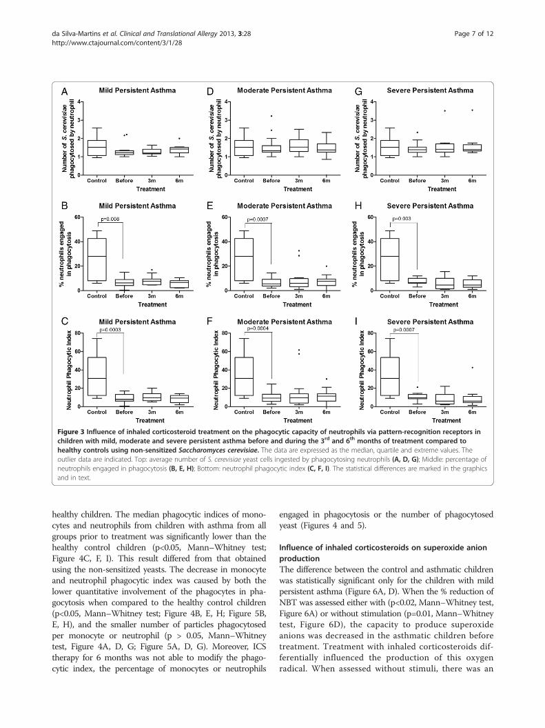
Figure 3 Influence of inhaled corticosteroid treatment on the phagocytic capacity of neutrophils via pattern-recognition receptors inchildren with mild, moderate and severe persistent asthma before and during the 3rd and 6th months of treatment compared tohealthy controls using non-sensitized Saccharomyces cerevisiae. The data are expressed as the median, quartile and extreme values. Theoutlier data are indicated. Top: average number of S. cerevisiae yeast cells ingested by phagocytosing neutrophils (A, D, G); Middle: percentage ofneutrophils engaged in phagocytosis (B, E, H); Bottom: neutrophil phagocytic index (C, F, I). The statistical differences are marked in the graphicsand in text.
da Silva-Martins et al. Clinical and Translational Allergy 2013, 3:28 Page 7 of 12http://www.ctajournal.com/content/3/1/28
healthy children. The median phagocytic indices of mono-cytes and neutrophils from children with asthma from allgroups prior to treatment was significantly lower than thehealthy control children (p<0.05, Mann–Whitney test;Figure 4C, F, I). This result differed from that obtainedusing the non-sensitized yeasts. The decrease in monocyteand neutrophil phagocytic index was caused by both thelower quantitative involvement of the phagocytes in pha-gocytosis when compared to the healthy control children(p<0.05, Mann–Whitney test; Figure 4B, E, H; Figure 5B,E, H), and the smaller number of particles phagocytosedper monocyte or neutrophil (p > 0.05, Mann–Whitneytest, Figure 4A, D, G; Figure 5A, D, G). Moreover, ICStherapy for 6 months was not able to modify the phago-cytic index, the percentage of monocytes or neutrophils
engaged in phagocytosis or the number of phagocytosedyeast (Figures 4 and 5).
Influence of inhaled corticosteroids on superoxide anionproductionThe difference between the control and asthmatic childrenwas statistically significant only for the children with mildpersistent asthma (Figure 6A, D). When the % reduction ofNBT was assessed either with (p<0.02, Mann–Whitney test,Figure 6A) or without stimulation (p=0.01, Mann–Whitneytest, Figure 6D), the capacity to produce superoxideanions was decreased in the asthmatic children beforetreatment. Treatment with inhaled corticosteroids dif-ferentially influenced the production of this oxygenradical. When assessed without stimuli, there was an

Figure 4 Influence of inhaled corticosteroid treatment on the phagocytic capacity of monocytes via opsonin receptors in children withmild, moderate and severe persistent asthma before and during the 3rd and 6th months of treatment compared to healthy controlsusing sensitized Saccharomyces cerevisiae. The data are expressed as the median, quartile and extreme values. The outlier data are indicated.Top: average number of S. cerevisiae yeast cells ingested by phagocytosing monocytes (A, D, G); Middle: percentage of monocytes engaged inphagocytosis (B, E, H); Bottom: monocyte phagocytic index (C, F, I). The statistical differences are marked in the graphics and in text.
da Silva-Martins et al. Clinical and Translational Allergy 2013, 3:28 Page 8 of 12http://www.ctajournal.com/content/3/1/28
increase in the % reduction of NBT after 6 m of treat-ment (p<0.05, Kruskal-Wallis test followed by Dunn’smethod, Figure 6D). However, when the % NBT reduc-tion was assessed after phagocytosis stimulation, therewas decreased production of superoxide anions after 3and 6 m of follow-up (p<0.05, Kruskal-Wallis testfollowed by Dunn’s method) (Figure 6A).
DiscussionThis prospective study evaluated for the first time the in-fluence of 3 and 6 months of treatment with inhaled corti-costeroids on the neutrophil and monocyte functions ofasthmatic children. Our data showed that 6 months of ICSwas not sufficient to return the decreased phagocyte func-tions to normal in the asthmatic children. Although6 months of treatment caused some improvement in theclinical parameters, such as an increase in peak expiratory
flow, and a significant increase in percentage of childrenwith mild and moderate persistent asthma showing ACT >25 and ACT = 20–24, (Table 2), no concomitant recoveryof phagocyte function was observed.Our data showed that the phagocytic capacity of mono-
cytes and neutrophils in children with mild, moderate andsevere persistent asthma was lower than in the healthycontrol children. A significant decrease in phagocytosis bymonocytes and neutrophils [19,20], bronchial macro-phages [21] and alveolar macrophages [11] has also beenshown in asthmatic individuals.Our data demonstrated that the severity of asthma did
not influence the immunodeficiency in phagocytes. Thephagocytic indices of the neutrophils and monocytes fromthe children with mild, moderate or severe persistentasthma were decreased, and there were no differencesamong these groups (Figure 1). Our data differed from those

Figure 5 Influence of inhaled corticosteroid treatment on the phagocytic capacity of neutrophils via opsonin receptors in childrenwith mild, moderate and severe persistent asthma before and during the 3rd and 6th months of treatment compared to healthycontrols using sensitized Saccharomyces cerevisiae. The data are expressed as the median, quartile and extreme values. The outlier data areindicated. Top: average number of S. cerevisiae yeast cells ingested by phagocytosing neutrophils (A, D, G); Middle: percentage of neutrophilsengaged in phagocytosis (B, E, H); Bottom: neutrophil phagocytic index (C, F, I). The statistical differences are marked in the graphics and in text.
da Silva-Martins et al. Clinical and Translational Allergy 2013, 3:28 Page 9 of 12http://www.ctajournal.com/content/3/1/28
of Fitzpatrick et al. [11], who observed greater deficiencies insevere asthma patients. However, Alexis et al. [21] alsoshowed decreased phagocytosis in mild asthma patients,whereas Lay et al. [22] observed increased phagocytosis bymacrophages obtained from the sputum of asthma patients.The differences between our observations and those of otherresearchers who have assessed phagocytosis in asthma aremost likely the result of the following factors: differences inthe clinical form of the disease, the severity of the disease,the age of individuals, the treatment used, the stimuli used,the source of phagocytes tested, the cell type assessed, andgenetic differences between individuals [11,19-22].The deficiency in phagocytosis was evident when phago-
cytosis was assessed either through pathogen associatedmolecular pattern receptors (non-sensitized) or throughcomplement and antibody receptors (sensitized). To beable to phagocytose, the phagocyte needs to move towardthe S. cerevisiae and ingest the particle. Because the
reduction in phagocytosis was attributable to the de-creased quantitative involvement of the phagocytes inphagocytosis, it is possible that phagocytes in asthmaticchildren had decreased capacity to move toward the yeast.In fact, it was previously shown that β2-agonists and glu-cocorticoids, which are commonly used for the treatmentof obstructive lung diseases, influence chemokine releaseand receptor sensitivity and, consequently, the chemotaxisof these cells [23].When sensitized phagocytosis through FcγR and comple-
ment receptors was assessed, the deficiency was evidencedas both to the decreased quantitative involvement of thephagocytes in phagocytosis and the decreased number ofyeast cells phagocytosed. The reasons for this effect are notclear; however, one possible explanation is a decrease in theexpression of receptors for complement and/or FcγR in themembranes of the monocytes and neutrophils from asthmapatients. Alexis et al. [19] found a significant correlation

Figure 6 Comparison of phagocyte production of toxic oxygen radical molecules assessed as the per cent reduction of nitro bluetetrazolium among the groups with mild, moderate and severe persistent asthma and healthy control children. In A, B, C: understimulation. In D, E, F: baseline. The data are expressed as the median, quartile and extreme values. The outlier data are indicated. The statisticaldifferences are marked in the graphics and in text.
da Silva-Martins et al. Clinical and Translational Allergy 2013, 3:28 Page 10 of 12http://www.ctajournal.com/content/3/1/28
between the expression of CD64 (FcγR) and phagocytosisby bronchial macrophages from asthmatic individuals andthe reduced expression of CD11b, a component of the CR3receptor, and phagocytosis in sputum and in neutrophilsand monocytes from asthmatics.Although there was clinical improvement in asthma
with corticosteroid therapy, we can hypothesize that theeffect of the drug on phagocytes decreasing the phago-cytosis may have contributed to the insufficient recoveryof phagocytosis after six months of clinical follow-up. Infact, corticosteroids decrease phagocytosis by monocytesand the production of inflammatory cytokines [3,13].Corticosteroids also increase the production of IL-10, acytokine that deactivates monocytes [3,5]. Therefore, itis possible that phagocytosis did not return to normalbecause of the action exerted by the corticosteroids onthe phagocytes. Another possibility to explain this lackof response of phagocytes to ICT by us observed mightbe that the insufficient recovery of immune alterationsremained downmodulating phagocyte function.Superoxide anion production, which was assessed as
the per cent reduction of nitro blue tetrazolium, byphagocytes from asthmatic and control children was sta-tistically decreased only for children with mild persistentasthma. The response to ICS after 6 months of treat-ment was different when assessed at baseline or afterstimulation. Inhaled corticosteroids increased the per
cent NBT reduction only when assessed without sti-mulation; however, these values were to the values ofhealthy, normal children, they did not exceed the valuesobserved in normal children. However, when superoxideanion production was evaluated after stimulation withsensitized S. cerevisiae, there was a decrease in super-oxide anion production. It is possible that decreasedphagocytosis may have played a role in the decreasedsuperoxide anion production by these cells.Generation of radical oxygen species occurs through
several enzymatic pathways or chemical process that areessential in many physiological reactions such as killinginvading pathogens, and takes place in every cell. How-ever, increased levels of ROS can produce harmful pa-thophysiological disorders that damage DNA, lipids,proteins, and carbohydrates, leading to enhanced inflam-matory response [24]. Oxidative stress has been proven toaffect smooth muscle contraction, induce airway hyper-responsiveness, and increase mucus secretion, and exces-sive ROS production can trigger key alterations, leading toan antioxidant-oxidant imbalance that has been shown inpatients with asthma and differ significantly according toseverity of the disease [24-26]. It was also observed thatresting or stimulated phagocytes may produce differentlyreactive oxygen species that depends on the disease form[27]. Predominance of enhancement of ROS has been ob-served in asthma [25-27]. The differences between our

da Silva-Martins et al. Clinical and Translational Allergy 2013, 3:28 Page 11 of 12http://www.ctajournal.com/content/3/1/28
observations and other researchers who have assessedROS in asthma are most likely the result of the type ofROS evaluated, the cell type assessed and differences inthe clinical form and severity of the disease.Influence of treatment with corticoid has been also evalu-
ated. It was observed that budesonide reduced the oxidativestress in the in guinea pig [28] and ICT was able to influ-ence the production of NADP oxidase in a dose dependentmanner [29]. Treatment with glucocorticosteroids as anti-oxidants has been suggested, based in its antioxidants prop-erties [30]. We only observed influence of ICT in mildpersistent asthma. A possible explanation by the differentresponse observed by us is the type of ROS analyzed andthe cell tested.Oxidative injury leads to increased lipid peroxidation, in-
creased airway reactivity and secretions, production ofchemoattractant molecules, and increased vascular perme-ability [31-33], which collectively lead to the augmentationof the existing inflammation that is a hallmark of asthma.Therefore, lower oxygen radical production in asthma maycontribute to the decreased immunopathogenesis by thesemolecules in asthma. On the other hand, a lower produc-tion of superoxide anions can lead to immunodeficiencyand may hinder the immune defense against infectiousagents.Although corticosteroids are considered the first-line
drugs for asthma therapy, the number of children thatmeet the clinical control evaluated by ACT (>25) in chil-dren that were followed-up for 6 m in this study wassmall. Significant variability in the response to inhaledcorticosteroids for persistent asthma has been shown[34], and uncontrolled asthma occurs in more than 50%of children who receive treatment with low-dose inhaledcorticosteroids [35]. It is possible that this lower clinicalresponse occurred because there was a high percentageof moderate and severe persistent asthma among theassessed children. Furthermore, the children were trea-ted at home, and although adherence to treatment wasencouraged in all clinical evaluation, there is no assur-ance regarding the regular use of corticosteroids duringthe follow-up period. This uncertainty is a limitation ofour study. The lower age of asthmatic children thanhealthy control children was also a limitation, however,we had already showed that there are not differences inphagocytosis between children higher 2 years old [16].Some children received different ICS (budesonide orfluticasone) that is a limitation of this study. Anotherpossible limitation of our study is the fact that phagocyt-osis was assessed in phagocytes obtained from peripheralblood and not from induced-sputum or bronchoalveolarlavage in order to evaluate directly the effects of diseaseand treatment on the phagocytic function at the locallevel. However, asthma is a systemic disease showing sev-eral cytokines and other molecules enhanced in peripheral
blood that may influence functions of cells of immune sys-tem and phagocytosis and might be influenced by corti-coid treatment as we observed in this paper. In support tothis consideration, morphological changes in eosinophilsobtained from peripheral blood markedly correlate withthe disease and may indicate the clinical severity of theacute exacerbation [36], corroborating that blood cellsmay also be influenced by the immune response occurringin asthmatic individuals. Furthermore, sputum inductioncan be challenging for young children [37], and the in-fluence on phagocytosis of mucus [38], enzymes [39] andmicroorganisms [40] presents in sputum couldn’t be ex-cluded, in addition to the ethical limitation to evaluatephagocytosis in phagocytes from bronchoalveolar lavage.
ConclusionsOur findings may broaden the understanding of the in-fluence of inhaled corticosteroids on phagocyte func-tions. Our data showed that asthmatic children followedup for 6 months in treatment with ICT didn’t modifydecreased phagocytosis. However, the baseline produc-tion of superoxide anions by phagocytes was normalized.Because phagocytes actively participate in both the le-sion and the defense of the lung in asthma, it is possiblethat the result of these opposing effects was beneficial totreated patients because there was some clinical im-provement. Furthermore, the reduction in lesions causedby oxygen radicals may have been beneficial to thetreated children.
AbbreviationsICT: Inhaled corticosteroid therapy; ACT: Asthma control test; LABA: Long-acting β2-agonist; PBS: Phosphate-buffered saline; FCS: Fetal calf serum;NBT: Nitro blue tetrazolium; BMI: Body mass index; mPA: Mild persistentasthma; MPA: Moderate persistent asthma; SPA: Severe persistent asthma.
Competing interestsThe authors report no conflict of interests. The authors alone are responsiblefor the content of the paper. The authors used a copy-editing service forlanguage revision.
Authors’ contributionsCLFS-M and MIMJ designed the study protocol. CLFS-M and SCC performedthe experiments. CLFS-M performed the clinical assessment. CLFS-M andMIMJ analyzed and interpreted the data. CLFS-M, SCC and MIMJ wrote themanuscript. MIMJ revised the manuscript. All authors read, revised andapproved the final manuscript.
AcknowledgementsMIM-J is an investigator supported by the Conselho Nacional deDesenvolvimento Científico e Tecnológico (CNPq), Brazil (process number304015/2010-5).
Author details1Laboratory of Cellular Immunology, Pathology, Faculty of Medicine, CampusDarcy Ribeiro, Asa Norte, University of Brasilia, Brasilia, DF 70.910-900, Brazil.2Department of Paediatric, Faculty of Medicine, Campus Darcy Ribeiro, AsaNorte, University of Brasilia, Brasilia, DF 70.910-900, Brazil. 3Paediatric Service,University Hospital of Brasilia, Brasilia, DF 70.910-900, Brazil.
Received: 19 April 2013 Accepted: 14 August 2013Published: 30 August 2013

da Silva-Martins et al. Clinical and Translational Allergy 2013, 3:28 Page 12 of 12http://www.ctajournal.com/content/3/1/28
References1. Bateman ED, Hurd SS, Barnes PJ, Bousquet J, Drazen JM, FitzGerald M,
Gibson P, Ohta K, O’Byrne P, Pedersen SE, Pizzichini E, Sullivan SD, WenzelSE, Zar HJ: Global strategy for asthma management and prevention:GINA executive summary. Eur Respir J 2008, 31:143–178.
2. Papadopoulos NG, Arakawa H, Carlsen KH, Custovic A, Gern J, Lemanske R,Le Souef VP, Mäkelä M, Roberts G, Wong G, Zar H, Akdis CA, Bacharier LB,Baraldi E, van Bever HP, de Blic J, Boner A, Burks W, Casale TB, Castro-Rodriguez JA, Chen YZ, El-Gamal YM, Everard ML, Frischer T, Geller M,Gereda J, Goh DY, Guilbert TW, Hedlin G, Heymann PW, et al: Internationalconsensus on (ICON) pediatric asthma. Allergy 2012, 67:976–997.
3. Barnes PJ: How corticosteroids control inflammation: Quintiles PrizeLecture 2005. Br J Pharmacol 2006, 148:245–254.
4. Barnes PJ, Pedersen S, Busse WW: Efficacy and safety of inhaled corticosteroids.New developments. Am J Respir Crit Care Med 1998, 157(Suppl):1–53.
5. Barnes PJ: Glucocorticosteroids: current and future directions. Br JPharmacol 2011, 163:29–43.
6. GINA Science Committee: Global Strategy for asthma management andprevention. Updated 2012. [http://www.ginasthma.com]
7. O’Byrne PM, Pedersen S, Busse WW, Tan WC, Chen Y-Z, Ohlsson SV, Ullman A,Lamm CJ, Pauwels RA: Effects of early intervention with inhaled budesonideon lung function in newly diagnosed asthma. Chest 2006, 129:1478–1485.
8. Dahl R: Systemic side effects of inhaled corticosteroids in patients withasthma. Respir Med 2006, 100:1307–1317.
9. Saffar AS, Ashdown H, Gounni AS: The molecular mechanisms ofglucocorticoids-mediated neutrophil survival. Curr Drug Targets 2011, 12:556–562.
10. Khanduja KL, Kaushik G, Khanduja S, Pathak CM, Laldinpuii J, Behera D:Corticosteroids affect nitric oxide generation, total free radicalsproduction, and nitric oxide synthase activity in monocytes of asthmaticpatients. Mol Cell Biochem 2011, 346:31–37.
11. Fitzpatrick AM, Holguin F, Teague WG, Brown LA: Alveolar macrophagephagocytosis is impaired in children with poorly controlled asthma.J Allergy Clin Immunol 2008, 121:1372–1378.
12. Rinehart JJ, Balcerzak SP, Sagone AL, LoBluglio AF: Effects of corticosteroidson human monocyte function. J Clin Invest 1974, 54:1337–1343.
13. Donnelly LE, Barnes PJ: Defective phagocytosis in airways disease. Chest2012, 141:1055–1062.
14. Busse WW, Boushey HA, Camargo CA Jr, Evans D, Foggs MB, Janson SL,Kelly HW, Lemanske RF, Martinez FD, Meyer RJ, Nelson HS, Platts-Mills TAE,Schatz M, Gail Shapiro G, Stoloff S, Szefler SJ, Weiss ST, Yawn BP: Guidelinesfor the Diagnosis and Management of Asthma. National Asthma Educationand Prevention Program (NAEPP), National Heart, Lung, and Blood Institute(NHLBI), National Institutes of Health; 2007:1–400. http://www.nhlbi.nih.gov/guidelines/asthma/asthgdln.pdf.
15. Nathan RA, Sorkness CA, Kosinski M, Schatz M, Li JT, Marcus P, Murray JJ,Pendergraft TB: Development of the asthma control test: a survey forassessing asthma. J Allergy Clin Immunol 2004, 113:59–65.
16. Muniz-Junqueira MI, Peçanha LMF, Silva-Filho VL, Cardoso MCA, Tosta CE:Assessment of post-natal maturation of the phagocytic function ofneutrophils and monocytes using a novel microtechnique. Clin Diagn LabImmunol 2003, 10:1096–1102.
17. Brown GD: Innate antifungal immunity: the key role of phagocytes. AnnuRev Immunol 2011, 29:1–21.
18. Muniz-Junqueira MI, Paula-Coelho VN: Meglumine antimonate directlyincreases phagocytosis, superoxide anion and TNF-α production, butonly via TNF-α it indirectly increases nitric oxide production byphagocytes of healthy individuals, in vitro. Int Immunopharmacol 2008,8:1633–1638.
19. Alexis NE, Eldridge MW, Peden DB: Effect of inhaled endotoxin on airwayand circulating inflammatory cell phagocytosis and CD11b expression inatopic asthmatic subjects. J Allergy Clin Immunol 2003, 112:353–361.
20. Lavinskiene S, Jeroch J, Malakaskas K, Bajoriuniene L, Jackute J, Sakalauskas R:Peripheral blood neutrophil activity during Dermatophagoidespteronyssinus-induced late-phase airway inflammation in patients withallergic rhinitis and asthma. Inflammation 2012, 35:1600–1609.
21. Alexis NE, Soukup J, Nierkens S, Becker S: Association between airwayhyperreactivity and bronchial macrophage dysfunction in individualswith mild asthma. Am J Physiol Lung Cell Mol Physiol 2001, 280:L369–L375.
22. Lay JC, Alexis NE, Zeman KL, Peden DB, Bennett WD: In vivo uptake ofinhaled particles by airway phagocytes is enhanced in patients withmild asthma compared with normal volunteers. Thorax 2009, 64:313–320.
23. Strandberg K, Blidberg K, Sahlander K, Palmberg L, Larsson K: Effect offormoterol and budesonide on chemokine release, chemokine receptorexpression and chemotaxis in human neutrophils. Pulm Pharmacol Ther2010, 23:316–323.
24. Zuo L, Otenbaker NP, Rose BA, Katherine S, Salisbury KS: Molecularmechanisms of reactive oxygen species-related pulmonary inflammationand asthma. Mol Immunol 2013, 56:57–63.
25. Ahmad A, Shameem M, Husain G: Relation of oxidant-antioxidantimbalance with disease progression in patients with asthma. AnnThoracic Med 2012, 7:226–232.
26. Celik M, Tuncer A, Soyer OU, Sac¸kesen C, Tanju Besler H, Kalayci O:Oxidative stress in the airways of children with asthma and allergicrhinitis. Pediatr Allergy Immunol 2012, 23:556–561.
27. Vachier I, Chanez P, Le Doucen C, Damon M, Descomps B, Godard P:Enhancement of reactive oxygen species formation in stable andunstable asthmatic patients. Eur Respir J 1994, 7:1585–1592.
28. Long F, Yan Wang Y, Qi H-H, Xin Zhou X, Xian-Qiao Jin X-Q: Rapid non-genomic effects of glucocorticoids on oxidative stress in a guinea pigmodel of asthma. Respirology 2008, 13:227–232.
29. Ökrös Z, Endreffy E, Novak Z, Maroti Z, Monostori P, Varga IS, Király A, Turi S:Changes in NADPH oxidase mRNA level can be detected in blood atinhaled corticosteroid treated asthmatic children. Life Sci 2012, 91:907–911.
30. Sadowskaa AM, Klebeb B, Germonpré P, De Backer WA:Glucocorticosteroids as antioxidants in treatment of asthma and COPD.New application for an old medication? Steroids 2007, 72:1–6.
31. Nadeem A, Chhabra SK, Masood A, Raj HG: Increased oxidative stress andaltered levels of antioxidants in asthma. J Allergy Clin Immunol 2003, 111:72–78.
32. Barnes PJ: Reactive oxygen species and airway inflammation. Free RadicBiol Med 1990, 9:235–243.
33. Birben E, Sahiner UM, Sackesen C, Erzurum S, Kalayci O: Oxidative stressand antioxidant defense. WAO J 2012, 5:9–19.
34. Szefler SJ, Martin RJ, King TS, Boushey HA, Cherniack RM, Chinchilli VM, Craig TJ,Dolovich M, Drazen JM, Fagan JK, Fahy JV, Fish JE, Ford JG, Israel E, Kiley J, KraftM, Lazarus SC, Lemanske RF Jr, Mauger E, Peters SP, Sorkness CA: Significantvariability in response to inhaled corticosteroids for persistent asthma.J Allergy Clin Immunol 2002, 109:410–418.
35. Sorkness CA, Lemanske RF Jr, Mauger DT, Boehmer SJ, Chinchilli VM,Martinez FD, Strunk RC, Szefler SJ, Zeiger RS, Bacharier LB, Bloomberg GR,Covar RA, Guilbert TW, Heldt G, Larsen G, Mellon MH, Morgan WJ, Moss MH,Spahn JD, Taussig LM: Long-term comparison of 3 controller regimens formild-moderate persistent childhood asthma: the Pediatric AsthmaController Trial. J Allergy Clin Immunol 2007, 119:64–72.
36. Muniz-Junqueira MI, Barbosa-Marques SM, Junqueira LF Jr: Morphologicalchanges in eosinophils are reliable markers of the severity of an acuteasthma exacerbation in children. Allergy 2013, 68:911-920.
37. Gogate S, Katial R: Pediatric biomarkers in asthma: exhaled nitric oxide,sputum eosinophils and leukotriene E4. Curr Opin Allergy Clin Immunol2008, 8:154–157.
38. Voynow J, Rubin BK: Mucins, Mucus, and Sputum. Chest 2009, 135:505–512.39. Maneechotesuwan K, Supawita S, Kasetsinsombat K, Wongkajornsilp A,
Barnes PJ: Sputum indoleamine-2, 3-dioxygenase activity is increased inasthmatic airways by using inhaled corticosteroids. J Allergy Clin Immunol2008, 121:43–50.
40. Marri PR, Stern DA, Wright AL, Dean Billheimer D, Martinez FD: Asthma-associated differences in microbial composition of induced sputum.J Allergy Clin Immunol 2013, 131:346–352.
doi:10.1186/2045-7022-3-28Cite this article as: da Silva-Martins et al.: Inhaled corticosteroidtreatment for 6 months was not sufficient to normalize phagocytosis inasthmatic children. Clinical and Translational Allergy 2013 3:28.